decreased fatty acid transporter fabp1 and increased
TRANSCRIPT
HAL Id: hal-03374032https://hal.archives-ouvertes.fr/hal-03374032
Submitted on 11 Oct 2021
HAL is a multi-disciplinary open accessarchive for the deposit and dissemination of sci-entific research documents, whether they are pub-lished or not. The documents may come fromteaching and research institutions in France orabroad, or from public or private research centers.
L’archive ouverte pluridisciplinaire HAL, estdestinée au dépôt et à la diffusion de documentsscientifiques de niveau recherche, publiés ou non,émanant des établissements d’enseignement et derecherche français ou étrangers, des laboratoirespublics ou privés.
Distributed under a Creative Commons Attribution| 4.0 International License
Decreased Fatty Acid Transporter FABP1 and IncreasedIsoprostanes and Neuroprostanes in the Human Term
Placenta: Implications for Inflammation and BirthWeight in Maternal Pre-Gestational Obesity
Livia Belcastro, Carolina Ferreira, Marcelle Saraiva, Daniela Mucci, AntonioMurgia, Carla Lai, Claire Vigor, Camille Oger, Jean-Marie Galano, Gabriela
Pinto, et al.
To cite this version:Livia Belcastro, Carolina Ferreira, Marcelle Saraiva, Daniela Mucci, Antonio Murgia, et al.. De-creased Fatty Acid Transporter FABP1 and Increased Isoprostanes and Neuroprostanes in the HumanTerm Placenta: Implications for Inflammation and Birth Weight in Maternal Pre-Gestational Obesity.Nutrients, MDPI, 2021, 13 (8), pp.2768. �10.3390/nu13082768�. �hal-03374032�
nutrients
Article
Decreased Fatty Acid Transporter FABP1 and IncreasedIsoprostanes and Neuroprostanes in the Human Term Placenta:Implications for Inflammation and Birth Weight in MaternalPre-Gestational Obesity
Livia Belcastro 1,†, Carolina S. Ferreira 2,†, Marcelle A. Saraiva 1, Daniela B. Mucci 1, Antonio Murgia 3, Carla Lai 4,Claire Vigor 5 , Camille Oger 5 , Jean-Marie Galano 5, Gabriela D. A. Pinto 2, Julian L. Griffin 3,6,Alexandre G. Torres 2,7 , Thierry Durand 5, Graham J. Burton 8, Fátima L. C. Sardinha 1,*and Tatiana El-Bacha 2,8,*
�����������������
Citation: Belcastro, L.; Ferreira, C.S.;
Saraiva, M.A.; Mucci, D.B.; Murgia,
A.; Lai, C.; Vigor, C.; Oger, C.; Galano,
J.-M.; Pinto, G.D.A.; et al. Decreased
Fatty Acid Transporter FABP1 and
Increased Isoprostanes and
Neuroprostanes in the Human Term
Placenta: Implications for
Inflammation and Birth Weight in
Maternal Pre-Gestational Obesity.
Nutrients 2021, 13, 2768. https://
doi.org/10.3390/nu13082768
Academic Editor: Asim K. Duttaroy
Received: 21 July 2021
Accepted: 7 August 2021
Published: 12 August 2021
Publisher’s Note: MDPI stays neutral
with regard to jurisdictional claims in
published maps and institutional affil-
iations.
Copyright: © 2021 by the authors.
Licensee MDPI, Basel, Switzerland.
This article is an open access article
distributed under the terms and
conditions of the Creative Commons
Attribution (CC BY) license (https://
creativecommons.org/licenses/by/
4.0/).
1 Laboratory of Nutritional Biochemistry, Institute of Nutrition Josué de Castro, Federal University of Rio deJaneiro, Rio de Janeiro 21941-902, Brazil; [email protected] (L.B.);[email protected] (M.A.S.); [email protected] (D.B.M.)
2 LeBioME-Bioactives, Mitochondria and Placental Metabolism Core, Institute of Nutrition Josué de Castro,Federal University of Rio de Janeiro, Rio de Janeiro 21941-902, Brazil; [email protected] (C.S.F.);[email protected] (G.D.A.P.); [email protected] (A.G.T.)
3 Department of Biochemistry, University of Cambridge, Cambridge CB2 1QW, UK;[email protected] (A.M.); [email protected] (J.L.G.)
4 Department of Environmental and Life Sciences, University of Cagliari, 09124 Cagliari, Italy;[email protected]
5 Institut des Biomolécules Max Mousseron (IBMM), UMR 5247, Université de Montpellier, CNRS, ENSCM,Bâtiment Balard, 1919 Route de Mende, 34293 Montpellier, France; [email protected] (C.V.);[email protected] (C.O.); [email protected] (J.-M.G.);[email protected] (T.D.)
6 Department of Metabolism, Digestion and Reproduction, Imperial College London, London SW7 2BX, UK7 Lipid Biochemistry and Lipidomics Laboratory, Institute of Chemistry, Federal University of Rio de Janeiro,
Rio de Janeiro 21941-598, Brazil8 Centre for Trophoblast Research, Department of Physiology, Development and Neuroscience, University of
Cambridge, Cambridge CB2 3EG, UK; [email protected]* Correspondence: [email protected] (F.L.C.S.); [email protected] (T.E.-B.);
Tel.: +55-21-99766-7997 (T.E.-B.)† These authors contributed equally to this work.
Abstract: The rise in prevalence of obesity in women of reproductive age in developed and develop-ing countries might propagate intergenerational cycles of detrimental effects on metabolic health.Placental lipid metabolism is disrupted by maternal obesity, which possibly affects the life-long healthof the offspring. Here, we investigated placental lipid metabolism in women with pre-gestationalobesity as a sole pregnancy complication and compared it to placental responses of lean women.Open profile and targeted lipidomics were used to assess placental lipids and oxidised products ofdocosahexaenoic (DHA) and arachidonic acid (AA), respectively, neuroprostanes and isoprostanes.Despite no overall signs of lipid accumulation, DHA and AA levels in placentas from obese womenwere, respectively, 2.2 and 2.5 times higher than those from lean women. Additionally, a 2-foldincrease in DHA-derived neuroprostanes and a 1.7-fold increase in AA-derived isoprostanes wereseen in the obese group. These changes correlated with a 70% decrease in placental FABP1 protein.Multivariate analyses suggested that neuroprostanes and isoprostanes are associated with maternaland placental inflammation and with birth weight. These results might shed light on the molecularmechanisms associated with altered placental fatty acid metabolism in maternal pre-gestationalobesity, placing these oxidised fatty acids as novel mediators of placental function.
Keywords: maternal pre-gestational obesity; placenta; lipid metabolism; fatty acid transporterproteins; isoprostanoids; neuroprostanes; isoprostanes; docosahexaenoic acid; arachidonic acid
Nutrients 2021, 13, 2768. https://doi.org/10.3390/nu13082768 https://www.mdpi.com/journal/nutrients
Nutrients 2021, 13, 2768 2 of 18
1. Introduction
Maternal pre-gestational obesity and excessive gestational weight gain affect short-and long-term health of both the mother and her child [1]. Gestational diabetes mellitusand pre-eclampsia are complications of pregnancy associated with gestational obesity,and newborns from obese women have an increased risk of overgrowth. Obesity inadults and in children has reached epidemic proportions in Brazil [2] and worldwide [3].Therefore, the rise in prevalence of obesity in women of reproductive age in both developedand developing countries might propagate intergenerational cycles of detrimental effectson metabolic health, contributing to substantial economic burden on society [4–7] andhighlighting the necessity of determining the mechanisms involved.
Fatty acids are essential for the accretion of body fat in the fetus, especially duringthe last trimester of pregnancy. Long chain polyunsaturated fatty acids, in particulardocosahexaenoic acid (DHA) and arachidonic acid (AA), have specific roles in membranecomposition and the development of the retina, and are the major components of the whitematter of the brain. Therefore, they are of paramount importance to proper neural andvisual development and cognitive function [8–11]. Ex vivo placental perfusion data [12] andin vivo kinetics of 13C-fatty acids [13] show that maternal–fetal 13C-labelled lipid transfer isvery low (1–6%), implying that lipid metabolism and handling by the placenta are strictlycontrolled and that these mechanisms are possibly major players in the allocation of fattyacids to fetal organs [14,15].
The inflammatory milieu imposed by maternal obesity disrupts the cross-talk betweenmaternal signals and the placenta, resulting in impaired placental function [16]. Uptakeand metabolism of essential fatty acids, particularly DHA, is impaired in placentas fromobese women [17,18]. Increased accumulation of lipids in the placenta has been describedin gestational obesity, which was associated with decreased oxidation of fatty acids andimpaired mitochondrial function [19,20]. Conversely, other studies have reported thatplacental total lipid content [21] and maternal–fetal transfer of 13C-labelled non-essentialfatty acids [13] is similar between lean and obese mothers. Altogether, these observationsemphasize that placental lipid handling and metabolism might be disrupted by maternalobesity and deserve further investigation to clarify the mechanisms involved.
The maternal circulation, and ultimately the maternal diet, are the sources of polyun-saturated fatty acids, as their synthesis by the fetus and the placenta is limited and in-sufficient to meet the high demand imposed by the growing fetus. Placental fatty acidhandling relies on several proteins which are responsible for (a) the uptake of fatty acidsfrom the maternal circulation, partly as lipoproteins and as non-esterified fatty acids andlysophospholipids bound to albumin, and (b) the numerous metabolic fates of fatty acidsand also their transfer to the fetus [22]. The hydrolysis of fatty acids from triacylglycerolin lipoproteins is catalyzed by endothelial lipase and by lipoprotein lipase. The former isselective for hydrolyzing unsaturated fatty acids esterified in the sn-2 position of glycerol.Fatty acids are then taken up by the placenta by fatty acid translocators (FAT/CD36), fattyacid transport proteins (FATP/SLC27A), and Mfsd2a. In the cytoplasm of the syncytiotro-phoblast, fatty acid binding proteins (FABPs), which are noncatalytic binding proteins,mediate fatty acid metabolism and inflammatory processes [23].
Both the expression and the content of placental fatty acid transport proteins arealtered by maternal obesity [19,24–26]. However, how these changes affect placentalfatty acid metabolism and signalling properties, and fatty acid transport and availabilityto the fetus in maternal gestational obesity, is not fully understood. Oxidative stressand inflammation have been associated with enhanced contents of oxidised fatty acidsand trophoblast dysfunction in pre-eclampsia. In particular, hydroxyeicosatetraenoicacids (HETEs), products of AA oxidation catalyzed by lipoxygenases and CYP, and F2-isoprostanes, products of non-enzymatic peroxidation of AA, are increased in placentasfrom pregnancies complicated by pre-eclampsia [27,28]. All these fatty acid metaboliteshave some degree of vasoconstrictive and pro-inflammatory effects and so may affectplacental function.
Nutrients 2021, 13, 2768 3 of 18
Here, we characterized the major categories and classes of lipids in term placentas frompregnancies complicated by maternal pre-gestational obesity. We also analysed fatty acidtransport proteins involved in the handling of polyunsaturated fatty acids by the placenta.A novel aspect of this study was a thorough characterization of placental non-enzymaticallyoxidised isoprostanoids derived from AA and DHA, which as inflammatory mediatorsmight be a mechanistic link between pre-gestational obesity and placental dysfunction.Additionally, we assessed how polyunsaturated fatty acids and their oxidised fatty acidsmetabolites are associated with inflammation and lipid handling by the placenta, andpossibly with neonatal outcomes in maternal pre-gestational obesity.
2. Materials and Methods2.1. Study Design and Participants
The current study was part of a randomized controlled trial registered on clinicaltri-als.org (NCT03215784), which was designed to evaluate the effects of fish oil and probioticssupplementation throughout pregnancy on women with pre-gestational obesity. The studywas conducted at the Maternidade Escola, between January 2015 and July 2017. This isa referral hospital, belonging to the Federal University of Rio de Janeiro, dedicated toproviding pre-natal and delivery care and puerperal consultations to the local community.Women were recruited up to 13 weeks of pregnancy and the inclusion criteria were agebetween 19–35 y, pre-gestational body mass index (BMI) between 18.5 and 24.9 kg/m2
(lean) or ≥30 and ≤40 kg/m2 (obesity class 1 and class 2; [29]), absence of pre-existinginfectious or chronic disease, except for obesity, a single fetus, and non-smoker. As a partof the pre-natal care, women presenting with high-risk pregnancies received nutritionalcounselling. Dietary intake was obtained during consultations in the 3rd trimester bytrained professionals (Supplementary File S1).
Gestational weight gain was classified accordingly [30] and gestational outcomes andnewborn information (weight and length at birth) were obtained from medical charts andclassified according to the INTERGROWTH-21st Project Curves [31].
To reduce possible confounders, we opted to only include samples from women whodid not develop gestational diabetes mellitus, pre-eclampsia, or other pregnancy com-plications. Hence, maternal pre-gestational obesity was the sole associated complication.Matched maternal blood, placental samples, and umbilical cord blood of 12 women (6 leanand 6 obese) were used.
2.2. Ethics
Women read and signed a ‘Free and Informed Consent’ form upon recruitment, andthis study was approved by the local Ethics committee of the Maternidade Escola andby the National Ethics committee (approval number CEP: 34611513.0.0000.5257 on 14October 2014).
2.3. Biological Samples
Maternal blood was collected at the 36th gestational week and placental and umbilicalcord blood samples were collected at delivery. The time between delivery and collectionwas up to 20–30 min. Umbilical cord blood was obtained by venipuncture and placentaltissue was collected according to established procedures [32]. Placental samples were takenfrom the maternal surface, placed into cryovials, frozen immediately in liquid nitrogen, andstored at −80 ◦C until analysis. A detailed description of placenta sampling is provided inSupplementary File S1.
2.4. Chemical and Reagents
All standards used in the targeted isoprostanoids analyses were synthetized in house,as published [33–37]. Internal standards: C19-16-F1t-PhytoP and C21-15-F2t-IsoP; Externalstandards: isoprostanes 15-F2t-IsoP, 15-epi-15-F2t-IsoP, 5-F2t-IsoP, and 5-epi-5-F2t-IsoP, andneuroprostanes 10-F4t-NeuroP, 10-epi-10-F4t-NeuroP, and 4(RS)-4-F4t-NeuroP.
Nutrients 2021, 13, 2768 4 of 18
2.5. Fatty Acid Transporter Proteins in Placental Tissue2.5.1. Quantitative Real-Time PCR (qPCR)
qPCR was performed using TaqMan™ Universal PCR Master Mix (Applied Biosys-tems, Thermo Fisher Scientific, Waltham, MA, USA) with the following primers fromTaqMan™ Applied Biosystems, Thermo Fisher Scientific, Waltham, MA, USA: Hs00195812(EL), Hs00155026 (FABP1), and Hs00997360 (FABP3). Glyceraldehyde-3-phosphate dehy-drogenase (GAPDH) was used as the housekeeping gene and transcripts were calculatedusing the threshold cycle 2-∆∆CT method [38]. A detailed description of qPCR analysis isavailable in Supplementary File S1.
2.5.2. Western Blotting
Primary antibodies for EL (MBS2013720) from My BioSource, Inc. (San Diego, CA,USA), FABP1 (AB7366), and FABP3 (AB16916) from ABCAM (Cambridge, United King-dom) were used in the following dilutions, respectively: 1:1000 EL, 1:500, and 1:500.Loading control was performed with anti-β-actin antibody (Sigma-Aldrich, Saint Louis,MO, USA, SAB5500001). A full description of Western blotting analysis is provided inSupplementary File S1.
2.6. Cytokines in Plasma and Placental Tissue
Quantification of the cytokines (IL-1β: Interleukin-1β; IL-6: Interleukin-6; IL-10:Interleukin-10; TNF-α: Tumour Necrosis Factor-α) in maternal plasma, umbilical cordplasma, and placental tissue protein extract was performed by the Luminex xMAP (MultipleAnalytic Profiling, Thermo Fisher Scientific, Waltham, MA, USA) assay [39] using 100 µgof placental extract as described for Western blotting analysis.
2.7. Maternal Lipoprotein Profile
Triglycerides, cholesterol, low-density lipoprotein (LDL) and high-density lipoprotein(HDL) were determined by enzymatic assay commercial kits (Sigma-Aldrich, Saint Louis,MO, USA), and very-low density lipoprotein (VLDL) was estimated using the formulaproposed by Friedewald, Levy and Fredrickson (1972) [40].
2.8. Ion Mobility QTOF LC/MS Lipid Profile Analysis of Placenta Samples
Lipids were extracted using a modified Folch method [41]. The non-polar extract inchloroform was dried under nitrogen and suspended in isopropanol:acetonitrile:water(IPA:ACN:H2O, 2:1:1, v/v/v) containing 25 deuterated lipids used as internal standards(representatives of phosphatidic acid, phosphatidylcholines, phosphatidylethanolamines,glycerophospholipids, phosphatidylinositols, phosphatidylserine, sphingomyelin,ceramides, triacylglycerols, and free fatty acids; Supplementary Table S1). All sampleswere analysed in positive and negative modes.
An Agilent 6560 Ion Mobility Quadrupole Time-of-Flight (IM-QTOF) mass spectrome-ter coupled with an Agilent 1290 UHPLC system was used to combine separation powerand selectivity of LC, IM, and MS techniques. The Dual Agilent Jet Stream ElectrosprayIonization Source was operated separately in positive and negative ion modes. The detaileddescription of the LC-MS analyses can be found in Supplementary File S1.
Chromatogram Pre-Processing and Lipid Annotation
Data pre-processing, including mass and CCS re-calibration and feature finding, wascarried out using the packages IM-MS Reprocessor, IM-MS Browser and Mass Profiler fromthe MassHunter suite vB.08.00 (Agilent Technologies, Santa Clara, CA, USA).
The resulting data matrices were processed using a KNIME pipeline comprising bothKNIME native nodes and integrated R scripts. Feature annotation was performed basedon the AccurateMassSearch node of the OpenMS library [42].
Nutrients 2021, 13, 2768 5 of 18
2.9. Isoprostanoids in Placental Tissue
Isoprostanoids in placenta were determined based on a microLC–MS/MS method [43]after lipid extraction by the Folch method [41], which were then mixed with a mixture ofinternal standards, followed by alkaline hydrolysis. MS analysis was performed in an ABSciex QTRAP 5500 (AB Sciex, Framingham, MA, USA) with an electrospray ionizationsource operated in negative mode. Quantification of isoprostanoids was performed withthe MultiQuant 3.0 software using specific internal standards. The detailed description ofisoprostanoids analysis by microLC–MS/MS is described in Supplementary File S1.
2.10. Statistical Analyses
Variables’ frequency distribution was assessed by standardized coefficients of skew-ness and kurtosis. Those with values < −2.0 or > +2.0 were characterized as having anon-normal distribution, and were then presented as median and interquartile interval, andcompared using the Mann–Whitney test. Groups’ frequency distributions were comparedusing the chi-square test. Associations between continuous variables were assessed bySpearman’s correlation analysis. Stepwise multiple regression analyses (backward) wereused to investigate the effect of independent factors on birth weight and on mothers andplacental tissue content of isoprostanoids. The criteria for the inclusion of independentvariables in the multiple regression models were based on results from Spearman corre-lations and on biochemical soundness. In the final model, only significant variables thatimproved the adjustment of the model were kept (p-to-remove ≥ 0.05; p-to-persist < 0.05).The multiple regression models were further assessed by analysis of residual plots that werechecked to determine if they were randomly distributed. Data analyses were performedwith GraphPad Prism v7.0 (GraphPad Software, San Diego, CA, USA) and StatgraphicsCenturion v18 (Statgraphics Technologies, Inc.; The Plains, VA, USA). In all analyses,p < 0.05 was considered for rejection of the null hypothesis.
3. Results3.1. General Characteristics of the Mothers and Newborns
The general characteristics of the mothers included in this study, their gestationaloutcomes and newborn information are presented in Table 1. Maternal pre-gestational BMIwas significantly higher in the obese group (p < 0.05) and the median BMI value indicatesobesity class I. Additionally, gestational weight gain was on average 40% lower in the obesemothers than in the lean group. No differences were observed in all other measurements,including gestational week at delivery, delivery mode and placental efficiency (weight atbirth:placental weight ratio). Newborn outcomes were also similar despite the higher pre-gestational BMI in the obese group. Dietary records showed that there were no significantdifferences in energy and carbohydrate, protein, and lipid intake between groups duringpregnancy (Supplementary Table S2). Therefore, the lower gestational weight gain observedin obese women may be explained, in part, as a consequence of nutritional counselling.
3.2. Placental Lipid Profile Suggests Alterations in Long-Chain Polyunsaturated Fatty AcidsAbundance despite No Apparent Signs of Inflammation and Dyslipidemia in MaternalPre-Gestational Obesity
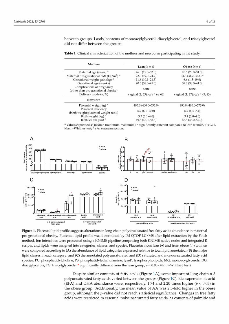
Annotated species were separated into the lipid categories glycerophospholipids, sph-ingolipids, fatty acyls, glycerolipids, and sterols [44]. In both groups, glycerophospholipidswas the most abundant category corresponding to 70% of the annotated signals, followed bysphingolipids (17%), fatty acyls (8%), glycerolipids (5%), and sterols (< 1%) (Figure 1A). Nodifferences were observed in any lipid categories between the lean and obese women. Glyc-erophospholipids, sphingolipids, and glycerolipids were divided into classes. The mostabundant classes with biological significance are shown in Figure 1B. Total glycerophospho-lipid was similar between the groups. However, the content of lysophospholipids tendedto a marginal (p = 0.056) 1.25-fold increase in placentas from obese women. Ceramidesand sphingomyelins were the most abundant sphingolipids that showed similar contents
Nutrients 2021, 13, 2768 6 of 18
between groups. Lastly, contents of monoacylglycerol, diacylglycerol, and triacylglyceroldid not differ between the groups.
Table 1. Clinical characterization of the mothers and newborns participating in the study.
MothersLean (n = 6) Obese (n = 6)
Maternal age (years) A 26.0 (19.0–32.0) 26.5 (20.0–31.0)Maternal pre-gestational BMI (kg/m2) A 22.0 (19.0–24.2) 34.3 (31.2–37.6) *
Gestational weight gain (kg) A 11.6 (10.1–21.3) 6.6 (1.5–19.0)Gestational age (weeks) 40.5 (38.0–41.0) 39.0 (38.0–41.0)
Complications of pregnancy(other than pre-gestational obesity) none none
Delivery mode (n; %) vaginal (2; 33); c/s B (4; 66) vaginal (1; 17); c/s B (5; 83)
Newborn
Placental weight (g) A 485.0 (400.0–555.0) 480.0 (480.0–575.0)Placental efficiency
(birth weight:placental weight ratio) 6.9 (6.1–10.0) 6.9 (6.4–7.4)
Birth weight (kg) A 3.3 (3.1–4.0) 3.4 (3.0–4.0)Birth length (cm) A 49.5 (46.0–53.5) 48.5 (45.0–52.0)
A values expressed as median (minimum-maximum); * significantly different compared to lean women, p < 0.01,Mann–Whitney test; B c/s, cesarean section.
Figure 1. Placental lipid profile suggests alterations in long-chain polyunsaturated free fatty acids abundance in maternalpre-gestational obesity. Placental lipid profile was determined by IM-QTOF LC/MS after lipid extraction by the Folchmethod. Ion intensities were processed using a KNIME pipeline comprising both KNIME native nodes and integrated Rscripts, and lipids were assigned into categories, classes, and species. Placentas from lean (•) and from obese (#) womenwere compared according to (A) the abundance of lipid categories expressed relative to total lipid annotated; (B) the majorlipid classes in each category; and (C) the annotated polyunsaturated and (D) saturated and monounsaturated fatty acidspecies. PC: phosphatidylcholine; PS: phosphatidylethanolamine; lysoP: lysophospholipids; MG: monoacyglycerols; DG:diacyglycerols; TG: triacylglycerols. * Significantly different from the lean group; p < 0.05 (Mann–Whitney test).
Despite similar contents of fatty acyls (Figure 1A), some important long-chain n-3polyunsaturated fatty acids varied between the groups (Figure 1C). Eicosapentaenoic acid(EPA) and DHA abundance were, respectively, 1.74 and 2.20 times higher (p < 0.05) inthe obese group. Additionally, the mean value of AA was 2.5-fold higher in the obesegroup, although the p-value did not reach statistical significance. Changes in free fattyacids were restricted to essential polyunsaturated fatty acids, as contents of palmitic and
Nutrients 2021, 13, 2768 7 of 18
stearic (saturated) and of palmitoleic and oleic (monounsaturated) acids were similar inthe lean and obese women (Figure 1D).
Maternal obesity is often associated with an inflammatory milieu and dyslipidemia.Hence, the levels of IL-1 β, IL-6, IL-10, and TNF-α were measured in maternal blood,placental tissue, and umbilical cord blood (Supplementary Figure S1). No differenceswere found in any of these inflammatory markers, except for a lower content of IL-1β in placentas from women with pre-gestational obesity. Likewise, the content of totalcholesterol, triacylglycerols, and HDL, LDL, and VLDL lipoproteins in maternal plasmawere similar between lean and obese women (Supplementary Figure S2).
These results suggest that placentas from women with pre-gestational obesity didnot present signs of overall lipid accumulation and no apparent signs of inflammationnor maternal dyslipaemia were observed. However, placental polyunsaturated long-chainfatty acids were increased.
3.3. Fatty Acid Transporter Protein FABP1 Is Decreased and Negatively Associated withPolyunsaturated Fatty Acids in Placentas from Women with Pre-Gestational Obesity
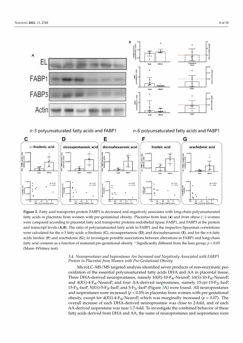
We next analysed the fatty acid transporter proteins that handle unsaturated fatty acidsby the placenta: endothelial lipase (EL), fatty acid binding protein-1 (FABP1), and fatty acidbinding protein-3 (FABP3), at the protein and mRNA levels (Figure 2A,B). Placentas fromthe obese group presented a significant 70% decrease in FABP1 protein content comparedwith placentas from the lean group (p < 0.05) and a non-significant 30% decrease in mRNAlevels. Endothelial lipase and FABP3 protein and transcript levels were similar betweengroups (Figure 2A,C).
The associations between the changes in FABP1 and polyunsaturated fatty acidswere assessed by Spearman rank correlations, and the ratios of fatty acid (FA) to FABP1were also assessed (Figure 2C–G). For all n-3 fatty acids (Figure 2C–E) and n-6 fattyacids (Figure 2F,G), the ratio FA:FABP1 was significantly higher in placentas from theobese group. The ratios EPA:FABP1 (Figure 2E), DHA:FABP1 (Figure 2F), and AA:FABP1(Figure 2G) were, respectively, 5.5-, 7-, and 6-fold higher in placentas from the obesecompared to the lean group. Spearman correlation analyses also showed that coefficientswere negative for all FA with statistically significant results for EPA (r = −0.82; p = 0.02;Figure 2E) and marginally significant for DHA (r = −0.57; p = 0.09; Figure 2F) and AA(r = −0.60; p = 0.054; Figure 2G). Taken together, these results suggest that FABP1 mightplay a role in placental handling of these fatty acids and that the decrease in its contentmight have an impact in the availability and signalling properties of EPA, DHA, and AA,in particular.
Nutrients 2021, 13, 2768 8 of 18
Figure 2. Fatty acid transporter protein FABP1 is decreased and negatively associates with long-chain polyunsaturatedfatty acids in placentas from women with pre-gestational obesity. Placentas from lean (•) and from obese (#) womenwere compared according to placental fatty acid transporter proteins endothelial lipase, FABP1, and FABP3 at the proteinand transcript levels (A,B). The ratio of polyunsaturated fatty acids to FABP1 and the respective Spearman correlationswere calculated for the n-3 fatty acids α-linolenic (C), eicosapentaenoic (D), and docosahexaenoic (E), and for the n-6 fattyacids linoleic (F) and arachidonic (G), to investigate possible associations between alterations in FABP1 and long-chainfatty acid contents as a function of maternal pre-gestational obesity. * Significantly different from the lean group; p < 0.05(Mann–Whitney test).
3.4. Neuroprostanes and Isoprostanes Are Increased and Negatively Associated with FABP1Protein in Placentas from Women with Pre-Gestational Obesity
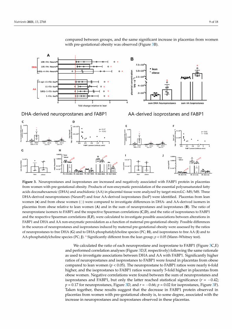
MicroLC–MS/MS targeted analysis identified seven products of non-enzymatic per-oxidation of the essential polyunsaturated fatty acids DHA and AA in placental tissue.Three DHA-derived neuroprostanes, namely 10(R)-10-F4t-NeuroP, 10(S)-10-F4t-NeuroP,and 4(RS)-4-F4t-NeuroP, and four AA-derived isoprostanes, namely 15-epi-15-F2t-IsoP,15-F2t-IsoP, 5(RS)-5-F2t-IsoP, and 5-F2c-IsoP (Figure 3A) were found. All neuroprostanesand isoprostanes were increased (p < 0.05) in placentas from women with pre-gestationalobesity, except for 4(RS)-4-F4t-NeuroP, which was marginally increased (p = 0.07). Theoverall increase of each DHA-derived neuroprostane was close to 2-fold, and of eachAA-derived isoprostane was near 1.7-fold. To investigate the combined behavior of thesefatty acids derived from DHA and AA, the sums of neuroprostanes and isoprostanes were
Nutrients 2021, 13, 2768 9 of 18
compared between groups, and the same significant increase in placentas from womenwith pre-gestational obesity was observed (Figure 3B).
Figure 3. Neuroprostanes and isoprostanes are increased and negatively associated with FABP1 protein in placentasfrom women with pre-gestational obesity. Products of non-enzymatic peroxidation of the essential polyunsaturated fattyacids docosahexaenoic (DHA) and arachidonic (AA) in placental tissue were analyzed by target microLC–MS/MS. ThreeDHA-derived neuroprostanes (NeuroP) and four AA-derived isoprostanes (IsoP) were identified. Placentas from leanwomen (•) and from obese women (#) were compared to investigate differences in DHA- and AA-derived isomers inplacentas from obese relative to lean women (A) and in the sum of neuroprostanes and isoprostanes (B). The ratio ofneuroprostane isomers to FABP1 and the respective Spearman correlations (C,D), and the ratio of isoprostanes to FABP1and the respective Spearman correlations (E,F), were calculated to investigate possible associations between alterations inFABP1 and DHA and AA non-enzymatic peroxidation as a function of maternal pre-gestational obesity. Possible differencesin the sources of neuroprostanes and isoprostanes induced by maternal pre-gestational obesity were assessed by the ratiosof neuroprostanes to free DHA (G) and to DHA-phosphatidylcholine species (PC; H), and isoprostanes to free AA (I) and toAA-phosphatidylcholine species (PC; J). * Significantly different from the lean group; p < 0.05 (Mann–Whitney test).
We calculated the ratio of each neuroprostane and isoprostane to FABP1 (Figure 3C,E)and performed correlation analyses (Figure 3D,F, respectively) following the same rationaleas used to investigate associations between DHA and AA with FABP1. Significantly higherratios of neuroprostanes and isoprostanes to FABP1 were found in placentas from obesecompared to lean women (p < 0.05). The neuroprostane to FABP1 ratios were nearly 6-foldhigher, and the isoprostanes to FABP1 ratios were nearly 5-fold higher in placentas fromobese women. Negative correlations were found between the sum of neuroprostanes andisoprostanes and FABP1, but only the latter reached statistical significance (r = −0.42;p = 0.17 for neuroprostanes, Figure 3D, and r = −0.66; p = 0.02 for isoprostanes, Figure 3F).Taken together, these results suggest that the decrease in FABP1 protein observed inplacentas from women with pre-gestational obesity is, to some degree, associated with theincrease in neuroprostanes and isoprostanes observed in these placentas.
Nutrients 2021, 13, 2768 10 of 18
The metabolic sources of DHA-derived neuroprostanes, in particular, and AA-derivedisoprostanes are not fully known; likely candidates are free DHA and AA, or phospho-lipids, mainly phosphatidylcholine, enriched in these fatty acids [45]. To address thisissue, we calculated the ratios of neuroprostanes to free DHA (Figure 3G) and DHA-enriched phosphatidylcholine species (Figure 3G,H), and of isoprostanes to free AA andAA-enriched phosphatidylcholine species (Figure 3I,J). No significant differences wereobserved in the ratios of neuroprostanes and isoprostanes to free FA between the leanand obese groups. On the other hand, marginally lower ratios were observed for DHA-enriched phosphatidylcholine species and AA-enriched phosphatidylcholine in placentasfrom the obese compared to the lean group. These results suggest that DHA and AA inphosphatidylcholine might be less susceptible to non-enzymatic peroxidation in placentasfrom women with pre-gestational obesity. Non-esterified DHA and AA seem to similarlycontribute to the synthesis of neuroprostanes and isoprostanes, respectively, in placentasfrom lean and obese women.
3.5. DHA-Derived Neuroprostanes and AA-Derived Isoprostanes Are Positively Associated withMaternal Pre-Gestational BMI and Endothelial Lipase Protein; and DHA-Derived NeuroprostanesOnly Are Negatively Associated with Inflammation and Birth Weight
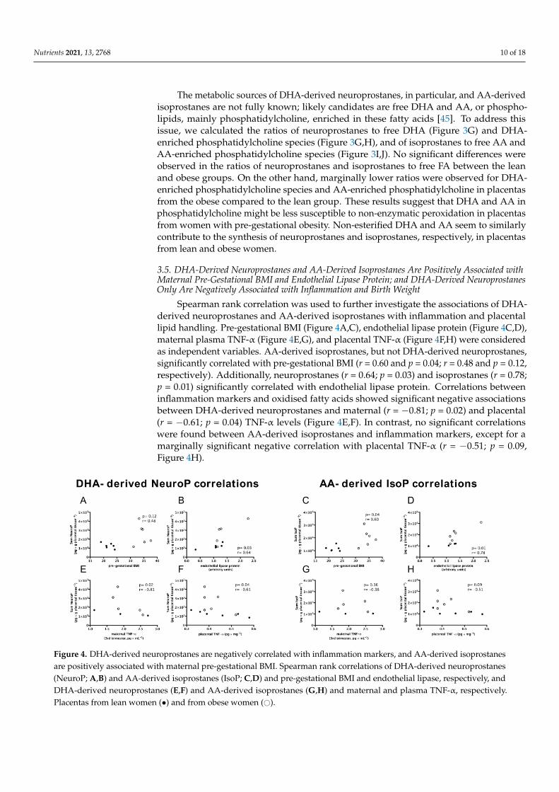
Spearman rank correlation was used to further investigate the associations of DHA-derived neuroprostanes and AA-derived isoprostanes with inflammation and placentallipid handling. Pre-gestational BMI (Figure 4A,C), endothelial lipase protein (Figure 4C,D),maternal plasma TNF-α (Figure 4E,G), and placental TNF-α (Figure 4F,H) were consideredas independent variables. AA-derived isoprostanes, but not DHA-derived neuroprostanes,significantly correlated with pre-gestational BMI (r = 0.60 and p = 0.04; r = 0.48 and p = 0.12,respectively). Additionally, neuroprostanes (r = 0.64; p = 0.03) and isoprostanes (r = 0.78;p = 0.01) significantly correlated with endothelial lipase protein. Correlations betweeninflammation markers and oxidised fatty acids showed significant negative associationsbetween DHA-derived neuroprostanes and maternal (r = −0.81; p = 0.02) and placental(r = −0.61; p = 0.04) TNF-α levels (Figure 4E,F). In contrast, no significant correlationswere found between AA-derived isoprostanes and inflammation markers, except for amarginally significant negative correlation with placental TNF-α (r = −0.51; p = 0.09,Figure 4H).
Figure 4. DHA-derived neuroprostanes are negatively correlated with inflammation markers, and AA-derived isoprostanesare positively associated with maternal pre-gestational BMI. Spearman rank correlations of DHA-derived neuroprostanes(NeuroP; A,B) and AA-derived isoprostanes (IsoP; C,D) and pre-gestational BMI and endothelial lipase, respectively, andDHA-derived neuroprostanes (E,F) and AA-derived isoprostanes (G,H) and maternal and plasma TNF-α, respectively.Placentas from lean women (•) and from obese women (#).

Nutrients 2021, 13, 2768 11 of 18
Multiple regression analysis was used to investigate the tentative predictors of pla-cental neuroprostanes and isoprostanes and their association with birth weight (Table 2).The sum of placental DHA-derived neuroprostanes was predicted by pre-gestationalBMI (β = 1.64 × 10²; positive association) and by placental TNF-α content, with negativeassociation (β = −5.56 × 10³) (Table 2; model 1). The sole predictor of the sum of pla-cental AA-derived isoprostanes was placental endothelial lipase protein (β = 1.21 × 10⁴;model 2). Most importantly, birth weight was significantly determined by pre-gestationalBMI (β = 1.26 × 10²), gestational weight gain (β = 7.72 × 101), and by the sum of placentalneuroprostanes, with a negative association (β = −4.06 × 10⁻¹) (Table 2, model 3).
Table 2. Multiple regression models for the assessment of predictors of placental Σ neuroprostanes and Σ isoprostanes andpredictors of birth weight.
DependentVariables
Independent Variablesβ Coefficients
Adj. R2 EstimatedError (%) 1 p2
Value SE p-Value
Σ Neuroprostanesin placenta
Model 1
Pre-gestational BMI 1.64 × 10² 2.61 × 101 0.000493.88 20.1 0.0000TNF-α, placenta −5.56 × 10³ 1.59 × 10³ 0.0101
Gestational weight gain — — ns — — nsIL-6, placenta — — ns — — ns
Endothelial lipase, protein — — ns — — nsFABP-1, protein — — ns — — ns
Σ Isoprostanesin placenta
Model 2
Endothelial lipase, protein 1.21 × 10⁴ 1.12 × 10³ 0.0000 93.65 22.9 0.0000Pre-gestational BMI — — ns — — ns
Gestational weight gain — — ns — — nsTNF-α, placenta — — ns — — ns
IL-6, placenta — — ns — — ns
Birth weightModel 3
Pre-gestational BMI 1.26 × 10² 1.38 × 101 0.000199.07 6.83 0.0000Gestational weight gain 7.72 × 101 1.71 × 101 0.0040
Σ Neuroprostanes, placenta −4.06 × 10⁻¹ 1.37 × 10⁻¹ 0.0253Σ Isoprostanes, placenta — — ns — — ns
IL-6, placenta — — ns — — nsTNF-α, placenta — — ns — — ns
Endothelial lipase, protein — — ns — — ns1 Estimated relative error of estimate = (estimated absolute error × 100%)/average value of the dependent variable; 2 Model significance.Significant associations were independent of the following variables, denoted as non-significant (ns) p-values: model 1, gestational weightgain, IL-6 (placenta), endothelial lipase protein (placenta), and FABP1 (placenta); model 2, pre-gestational BMI, gestational weight gain,TNF-α (placenta), and IL-6 (placenta); model 3, Σ of isoprostanes (placenta), interleukin-6 (placenta), TNF-α (placenta), and endotheliallipase protein (placenta);—blank cell; IL-6: Interleukin-6; TNF-α: Tumour Necrosis Factor-α.
Collectively, DHA-derived neuroprostanes might mediate a less inflammatory re-sponse and potentially prevent excessive neonatal weight gain, as opposed to AA-derivedisoprostanes, which appeared related to increased maternal adiposity.
4. Discussion
In this study, we show that despite no overall features of lipid accumulation, placentasfrom women with pre-gestational obesity contain an increased content of polyunsaturatedfree fatty acids. This increase is associated with a decreased content of FABP1, a cytoplas-mic protein that handles unsaturated fatty acids and is important for determining theirbiological fate. Additionally, higher contents of neuroprostanes and isoprostanes, whichare products of non-enzymatic oxidation of DHA and AA, respectively, were observed inplacentas from obese women. Correlation and multivariate analyses suggested that theseoxidised fatty acids are associated with maternal and placental inflammation, and alsowith birth weight. To our knowledge, this is the first report to characterize DHA-derivedneuroprostanes and AA-derived isoprostanes in the human placenta, and in particular toexplore their role in maternal and neonatal outcomes in the context of obesity.
Obesity in general is associated with chronic low-grade inflammation and insulinresistance. Therefore, obese women are at higher risk of developing metabolic disturbancesduring pregnancy [1]. In this study, samples from women who developed gestational
Nutrients 2021, 13, 2768 12 of 18
diabetes mellitus, pre-eclampsia, or other complications were excluded from the analysis.Therefore, one of the strengths of our study is the well characterized patient cohort andthe homogeneity of placental samples which reduced possible confounders of obesity-associated comorbidities. The median BMI value of the obese women was 34.3, indicatingthey present obesity class 1, and only one out of six women presented with a BMI of37.6 (obesity class II). Additionally, a non-significant 40% decrease in gestational weightgain was observed in obese women. This result is in agreement with previous reportsshowing that obese women gain less weight than lean women during pregnancy [30]. Themechanisms involved are not well established, but it might be a physiological response tocompensate for the excess stored energy. In our study, we found similar energy and nutrientintake between lean and obese women, possibly as a result of the nutritional counseling theyreceived during the pre-natal care. The fact that the obese women did not present with othermetabolic complications might explain the similar plasma concentrations of inflammatorymarkers, and lipoproteins and triacylglycerols as in lean women. We speculate, therefore,that despite being obese, they were metabolically healthy, and this may explain the lackof differences in placental weight and efficiency. Although several studies have shownthat maternal obesity is associated with an inflammatory and pro-oxidant milieu [46,47],there are reports of healthy obese mothers with no signs of dyslipidaemia [13,48]. Indeed,a lower content of IL-6 and reduced infiltration of macrophages in placentas from obesewomen was recently shown [49]. Conversely, there are consistent data showing that severalplacental responses appear to be affected by maternal pre-gestational obesity, irrespectiveof its severity [16,50].
The placenta is the highly specialized organ that interfaces between the mother andher baby. It integrates signals of maternal availability and fetal demand of nutrients andoxygen, and has a central role in determining the life-long health of the offspring [51].The importance of placental lipid metabolism in regulating maternal-fetal transfer offatty acids, in particular essential polyunsaturated fatty acids, has been recognized inhealthy pregnancies [14,52] and in pre-gestational obesity [17,18], although the mechanismsinvolved are not fully known. Indeed, in this study, we showed that placentas fromwomen with pre-gestational obesity contain a significant increase in DHA and EPA contentcompared to placentas from lean women. Additionally, the median value of placental AAfrom obese women was nearly 2.5-fold higher than the lean group, despite these differencesbeing non-significant statistically, which may reflect the small sample size.
Alterations in placental fatty acid transporter proteins are plausible mechanismsassociated with altered placental lipid handling in maternal pre-gestational obesity [53].Indeed, we show that FABP1 is decreased in placentas from obese women, corroborating aprevious study [25]. The decrease in FABP1 in placentas from the obese group was reflectedin significantly higher ratios of n-3 and n-6 polyunsaturated fatty acids to FABP1.
FABP1 is the liver isoform of FABP. Unlike other members in the FABP family, it hastwo ligand-binding sites for fatty acids and higher affinity for long-chain unsaturatedfatty acids. Besides fatty acids, FABP1 binds a range of hydrophobic molecules, suchas peroxisome proliferator-activated receptors (PPARs), prostaglandins, hydroxyl andhydroperoxyl metabolites of AA, lysophopholipids, and pro-oxidants such as heme [54].FABP1 has been found in the cytosol, nucleus, and mitochondria [55,56], and so may havemultiple roles. Hence, we speculate that the decreased content of FABP1 found in placentasfrom women with pre-gestational obesity might have affected placental function beyondjust lipid transport.
Due to the role of FABP1 in trafficking fatty acids to the nucleus and also its abilityto bind PPARs, the decrease in placentas from women with pre-gestational obesity likelyaltered pathways related to placental lipid handling and fatty acid metabolism. There isevidence that PPARs are involved in these processes in the human placenta [57,58]. Similarcontents of PPARs in placentas from obese and lean women [25] were described in parallelwith increased PPAR-γ (related to fatty acid synthesis) and decreased PPAR-α (related tofatty acid oxidation) [20]. Yang et al. have proposed that regulation of PPARs is important
Nutrients 2021, 13, 2768 13 of 18
at the maternal–fetal interface and this fact might explain the apparent discrepancies inPPARs content in maternal pre-gestational obesity [59]. It remains to be determined if andhow PPARs participate in the cross-talk between long chain polyunsaturated fatty acidsand FABP1 protein seen in the present study.
As FABP1 binds to pro-oxidant molecules and is considered an antioxidant, at least inthe liver [54], its decrease might render the placenta more susceptible to oxidative stressin obese women. A decreased total antioxidant capacity and increased activation of thepro-oxidant NFkB pathway has been observed in placentas from women with obesity [60].The significant increase in DHA-derived neuroprostanes and AA-derived isoprostanes weobserved supports a pro-oxidant milieu in placentas from the obese group. In addition toincreased concentrations of neuroprostanes and isoprostanes, we found that their ratiosto FAPB1 were also significantly altered, and that FABP1 negatively correlated with thetotal isoprostanes. These results not only support the concept of a pro-oxidant milieu, butalso suggest that FABP1 binds to isoprostanoids, in particular AA-derived isoprostanes, asobserved for enzymatically derived fatty acid mediators.
Neuroprostanes and isoprostanes are isoprostanoids (isomers of prostaglandins),formed by the non-enzymatic peroxidation of DHA and AA, respectively. They havebeen described as important signalling molecules, and AA-derived isoprostanes act invascular smooth muscle through tyrosine kinase and Rho kinase in human cells [61] andthrough prostanoid receptors in rats [62]. AA-derived isoprostanes are implicated incardiovascular diseases acting as vasoconstrictors and considered markers of oxidativestress [45]. Conversely, recent evidence suggests that DHA-derived neuroprostanes haveanti-inflammatory properties [63,64], despite the fact that they have been implicated inoxidative stress in neurodegenerative diseases [65]. Their role in placental function inpregnancies complicated by maternal obesity has not yet been described.
Oxidised 9-HODE, 13-HODE, and 15-HETE, products of AA produced by cycloxyge-nase activity, did not affect expression of syncytin, cyclin E, and p27, which are markers oftrophoblast differentiation, indicating that they are not implicated in trophoblast dysfunc-tion, at least in healthy trophoblasts [66]. On the other hand, in placentas from pregnanciescomplicated with pre-eclampsia, it has been shown that F2 class isoprostanes derived fromAA contribute to oxidative stress and have vasoconstrictive properties [67].
The formation of isoprostanoids is regulated to some extent, and are predominantlyformed in situ from oxidation of DHA or AA esterified to phosphatidylcholine in cell mem-branes. Therefore, their signalling properties depend upon phospholipase A2 activity [45],and a strict correlation between placental phospholipase A2 mRNA and free F2-isoprostanelevels has been found in pre-eclampsia [67].
Our marginally lower ratios of neuroprostanes to DHA-enriched and of isoprostanesto AA-enriched PC in placentas from obese women suggest that the proportion of thesemediators formed in situ is decreased in obesity. The significance of this result may be re-lated to compensatory mechanisms in placentas from obese women, controlling membranefunction and avoiding excessive release of mediators and possibly controlling neonatalbirth weight. Indeed, increased placental phospholipase A2 activity has been correlatedwith increased placental accumulation of lipids and adiposity in the newborn [68].
In the present study, AA-derived isoprostanes correlated with maternal pre-gestationalBMI. Additionally, in the multivariate model, the sole predictor of isoprostanoids in theplacenta was placental EL protein. These results could be interpreted in the light of thepro-inflammatory effect of AA-derived isoprostanes. A higher content of EL has beendescribed in placentas from pregnancies complicated with gestational diabetes mellitus, inaddition to obesity [69]. Consistent with these findings, we observed a non-statistical 25%increase in EL in placentas from women with pre-gestational obesity.
In the case of neuroprostanes, multivariate models indicated that both maternal pre-gestational BMI (positive β coefficient) and placental TNF-alpha (negative β coefficient)were tentative predictors of their content in the placenta, suggesting their possible anti-inflammatory role. This association was also observed in the correlation analysis, where
Nutrients 2021, 13, 2768 14 of 18
the sum of neuroprostanes was negatively correlated with maternal and placental TNF-αcontents. Importantly, when we evaluated the possible involvement of neuroprostanesand isoprostanes with birth weight, total neuroprostanes presented a negative contri-bution and, as expected, pre-gestational BMI and gestational weight gain had positiveassociations. However, total isoprostanes did not fit the same experimental model. Theseresults suggest that neuroprostanes present anti-inflammatory roles in placentas fromwomen with pre-gestational obesity, and by negatively affecting birth weight might at-tenuate the intergenerational cycles of detrimental effects on metabolic health as the riskof overweight/obesity in childhood increases gradually over the full range of maternalpre-gestational BMI [70]. Indeed, neonatal birth weight was similar between groups in thepresent study.
A word of caution is necessary when investigating birth weight in the context ofgestational obesity as it has been suggested that neonatal adiposity, and not total weight, isindependently associated with childhood adiposity [71]. The association between placentaloxidised fatty acids and neonatal outcomes, including adiposity, has never been addressed.An additional possible mechanism by which neuroprostanes exert anti-inflammatory effectsis the observation that 4-(RS)-4-F4t-NeuroP increased mRNA levels of the enzyme heme-oxygenase, which degrades heme, in human neuroblastoma cells and in primary culture ofneurons [65]. Given the fact that FABP1 is able to bind heme, a molecule with pro-oxidantproperties, the increase in neuroprostane isomers might counteract the decrease in FABP1protein in the obese group, providing a means of anti-oxidant defense. It remains to bedetermined if this is the case in the term human placenta in maternal pre-gestationalobesity.
The main limitation of the present study is the sample size, which impacts on thestatistical power. Consequently, results from the multiple regression analyses should beconsidered as an approach to screen for predictors in each model. In addition, we werenot able to address sexually dimorphic responses in placental fatty acid metabolism. Thisaspect is important as a recent study showed that placentas from females appear moreprone to store and esterify fatty acids, while placentas from males have a decreased capacityto transfer DHA to cord blood [72]. Nonetheless, our data are biochemically sound andbring new knowledge to the field, identifying isoprostanoids as potential novel mediatorsof placental function.
As maternal diet is the ultimate source of DHA and AA, our results add to the bodyof evidence that there must be a balance between intake of n-3 and n-6 fatty acids, andpossibly a higher intake of DHA is necessary during pregnancy [73]. A recent pilot trialdemonstrated that DHA supplementation starting early in pregnancy promoted higherlean mass accrual at birth and improved fetal growth [74]. The next step is to evaluate theintake of individual fatty acids and also to address the quality of the diet of these mothersby assessing other factors, e.g., dietary antioxidants, that contribute to placental lipidhandling in a larger cohort. Intervention studies are also needed to identify the molecularmechanisms involved. Such data would form the basis of efficient dietary strategies todecrease the burden of maternal pre-gestational obesity.
5. Conclusions
The decreased content of FABP1 and increased content of DHA, EPA, and AA inplacentas from women with pre-gestational obesity indicate important alterations in pla-cental lipid metabolism and fatty acid handling even in obese women that did not presentmajor signs of inflammation. Additionally, the decrease in FABP1 content was associatedwith increased contents of DHA-derived neuroprostanes and AA-derived isoprostanes inplacentas from obese women. Distinct mechanisms seemed to contribute to their increasedcontent, as total DHA-derived neuroprostanes was negatively associated with placentalTNF-α, and AA-isoprostanes associated with pre-gestational BMI. Importantly, placen-tal neuroprostanes negatively contributed to birth weight. Taken together, these resultsconfirm previous observations showing that placental lipid metabolism is disrupted in
Nutrients 2021, 13, 2768 15 of 18
maternal obesity. The possibility that these oxidised fatty acids act as mediators of placental,and possibly maternal, inflammation opens several possibilities for the investigation of themolecular mechanisms associated with alterations in placental fatty acid metabolism inmaternal pre-gestational obesity and their possible role in the health of the offspring.
Supplementary Materials: The following are available online at https://www.mdpi.com/article/10.3390/nu13082768/s1, Supplementary File S1: Supplementary methods; Supplementary Figure S1:Cytokines in maternal plasma, in the placenta and in cord blood plasma from lean (black bars) andobese (gray bars) women. Supplementary Figure S2: Total cholesterol, lipoproteins and triglyceridesin plasma of lean (black bars) and obese (gray bars) women; Supplementary Table S1: Internalstandards used in the Ion Mobility QTOF LC/MS lipid profile analysis. Supplementary Table S2:Energy and Nutrient intake of women participating in the study.
Author Contributions: Conceptualization, F.L.C.S., T.E.-B. and G.J.B.; methodology, A.M., C.L., C.V.,C.O., J.-M.G., J.L.G. and T.D.; formal analysis, L.B., C.S.F., M.A.S., D.B.M., G.D.A.P., A.M., C.L., C.V.,A.G.T. and T.E.-B.; investigation, L.B., C.S.F., M.A.S., D.B.M., A.M., C.L., G.D.A.P., C.V. and T.E.-B.;resources, T.E.-B., F.L.C.S., A.G.T., G.J.B., J.L.G., C.O., J.-M.G., C.V. and T.D.; data curation, A.M., C.L.and J.L.G.; writing—original draft preparation, T.E.-B. and L.B.; writing—review and editing, allauthors; visualization, all authors; supervision, T.E.-B., F.L.C.S. and G.J.B.; project administration,T.E.-B. and F.L.C.S.; funding acquisition, T.E.-B., F.L.C.S., A.G.T., G.J.B., J.L.G., C.O., J.-M.G., C.V. andT.D. All authors contributed to data interpretation and performed final editing checks and approvedthe final manuscript. All authors have read and agreed to the published version of the manuscript.
Funding: This research was funded by Fundação Carlos Chagas Filho de Amparo à Pesquisa do Es-tado do Rio de Janeiro (FAPERJ; E-26/110.111/2014 for F.L.C.S. and T.E.; E-026/203.254/2017 for T.E.),Isaac Newton Trust research grant for T.E., Global Challenge Research Fund (ref. 102642/A19819)for T.E., A.G.T., and G.J.B., and Coordenação de Aperfeiçoamento de Pessoal de Nível Superior–CAPES-Brasil (Finance code 001), Brazil. L.B. and D.B.M. were recipients of CAPES scholarships andC.S.F. was recipient of the CNPq scholarship (grant GM). The APC was covered partially by FAPERJ,PROEX funding via the post-graduate course in Nutrition, and the University of Cambridge.
Institutional Review Board Statement: The study was conducted according to the guidelines of theDeclaration of Helsinki, and approved by the Ethics Committee of the Maternidade Escola approvalnumber CEP: 34611513.0.0000.5257 on 14 October 2014.
Informed Consent Statement: Informed consent was obtained from all subjects involved in the study.
Acknowledgments: The authors acknowledge the contribution of the mothers that participated inthis study, without any direct compensation. The assistance of conticom.com.br in designing thegraphical abstract is greatly acknowledged.
Conflicts of Interest: The authors declare no conflict of interest. The funders, public funding agenciesfrom Brazil, had no role in the design of the study; in the collection, analyses, or interpretation ofdata; in the writing of the manuscript; or in the decision to publish the results.
References1. Catalano, P.M.; Shankar, K. Obesity and pregnancy: Mechanisms of short term and long term adverse consequences for mother
and child. BMJ 2017, 356, j1. [CrossRef]2. Brazil. Vigilancia de Fatores de Risco e Proteção Para Doenças Crônicas por Inquérito Telefônico. In Brasilia; 2018. Available
online: http://bvsms.saude.gov.br/bvs/publicacoes/vigitel_brasil_2018_vigilancia_fatores_risco.pdf (accessed on 4 April 2021).3. Dai, H.; Alsalhe, T.A.; Chalghaf, N.; Riccò, M.; Bragazzi, N.L.; Wu, J. The global burden of disease attributable to high body
mass index in 195 countries and territories, 1990–2017: An analysis of the Global Burden of Disease Study. PLoS Med. 2020, 17,e1003198. [CrossRef]
4. Schneider, B.C.; Menezes, A.M.B.; Wehrmeister, F.C.; Gonçalves, H. Gestational weight gain and childhood body mass indexacross three generations: Results from the 1993 Pelotas (Brazil) Birth Cohort. Pediatr. Obes. 2021, 16, e12760. [CrossRef] [PubMed]
5. Shen, Y.; Zhang, H.; Jiang, Y.; Mzayek, F.; Arshad, H.; Karmaus, W. Maternal Birth Weight and BMI Mediate the TransgenerationalEffect of Grandmaternal BMI on Grandchild’s Birth Weight. Obesity 2020, 28, 647–654. [CrossRef]
6. Lahti-Pulkkinen, M.; Bhattacharya, S.; Räikkönen, K.; Osmond, C.; Norman, J.; Reynolds, R.M. Intergenerational Transmission ofBirth Weight Across 3 Generations. Am. J. Epidemiol. 2018, 187, 1165–1173. [CrossRef]
7. Poston, L.; Caleyachetty, R.; Cnattingius, S.; Corvalan, C.; Uauy, R.; Herring, S.; Gillman, M.W. Preconceptional and maternalobesity: Epidemiology and health consequences. Lancet Diabetes Endocrinol. 2016, 4, 1025–1036. [CrossRef]

Nutrients 2021, 13, 2768 16 of 18
8. Bazinet, R.P.; Layé, S. Polyunsaturated fatty acids and their metabolites in brain function and disease. Nat. Rev. Neurosci. 2014, 15,771–785. [CrossRef] [PubMed]
9. Kuipers, R.S.; Luxwolda, M.F.; Offringa, P.J.; Boersma, E.R.; Dijck-Brouwer, D.J.; Muskiet, F.A. Fetal intrauterine whole bodylinoleic, arachidonic and docosahexaenoic acid contents and accretion rates. Prostaglandins Leukot. Essent. Fat. Acids 2012, 86,13–20. [CrossRef]
10. Uauy, R.; Hoffman, D.R.; Peirano, P.; Birch, D.; Birch, E. Essential fatty acids in visual and brain development. Lipids 2001, 36,885–895. [CrossRef]
11. Innis, S.M. Fatty acids and early human development. Early Hum. Dev. 2007, 83, 761–766. [CrossRef] [PubMed]12. Perazzolo, S.; Hirschmugl, B.; Wadsack, C.; Desoye, G.; Lewis, R.; Sengers, B.G. The influence of placental metabolism on fatty
acid transfer to the fetus. J. Lipid Res. 2017, 58, 443–454. [CrossRef]13. Gázquez, A.; Prieto-Sánchez, M.T.; Blanco-Carnero, J.E.; van Harskamp, D.; Perazzolo, S.; Oosterink, J.E.; Demmelmair, H.;
Schierbeek, H.; Sengers, B.G.; Lewis, R.; et al. In vivokinetic study of materno-fetal fatty acid transfer in obese and normal weightpregnant women. J. Physiol. 2019, 597, 4959–4973. [CrossRef]
14. Lewis, R.M.; Childs, C.E.; Calder, P.C. New perspectives on placental fatty acid transfer. Prostaglandins Leukot. Essent. Fat. Acids2018, 138, 24–29. [CrossRef]
15. Thornburg, K.L.; Kolahi, K.S.; Valent, A.M. What’s so special about lipid transport in the human placenta? J. Physiol. 2019, 597,4863–4864. [CrossRef] [PubMed]
16. Kelly, A.C.; Powell, T.L.; Jansson, T. Placental function in maternal obesity. Clin. Sci. 2020, 134, 961–984. [CrossRef]17. Gázquez, A.; Prieto-Sánchez, M.T.; Blanco-Carnero, J.E.; Ruíz-Palacios, M.; Nieto, A.; van Harskamp, D.; Oosterink, J.E.;
Schierbeek, H.; van Goudoever, J.B.; Demmelmair, H.; et al. Altered materno-fetal transfer of 13C-polyunsaturated fatty acids inobese pregnant women. Clin. Nutr. 2020, 39, 1101–1107. [CrossRef]
18. Hirschmugl, B.; Perazzolo, S.; Sengers, B.G.; Lewis, R.M.; Gruber, M.; Desoye, G.; Wadsack, C. Placental mobilization of free fattyacids contributes to altered materno-fetal transfer in obesity. Int. J. Obes. 2021, 45, 1114–1123. [CrossRef] [PubMed]
19. Hirschmugl, B.; Desoye, G.; Catalano, P.; Klymiuk, I.; Scharnagl, H.; Payr, S.; Kitzinger, E.; Schliefsteiner, C.; Lang, U.; Wadsack,C.; et al. Maternal obesity modulates intracellular lipid turnover in the human term placenta. Int. J. Obes. 2017, 41, 317–323.[CrossRef]
20. Calabuig-Navarro, V.; Haghiac, M.; Minium, J.; Glazebrook, P.; Ranasinghe, G.C.; Hoppel, C.; De-Mouzon, S.H.; Catalano, P.;O’Tierney-Ginn, P. Effect of Maternal Obesity on Placental Lipid Metabolism. Endocrinology 2017, 158, 2543–2555. [CrossRef][PubMed]
21. Segura, M.T.; Demmelmair, H.; Krauss-Etschmann, S.; Nathan, P.; Dehmel, S.; Padilla, M.C.; Rueda, R.; Koletzko, B.; Campoy, C.Maternal BMI and gestational diabetes alter placental lipid transporters and fatty acid composition. Placenta 2017, 57, 144–151.[CrossRef]
22. Duttaroy, A.K.; Basak, S. Maternal dietary fatty acids and their roles in human placental development. Prostaglandins Leukot.Essent. Fat. Acids 2020, 155, 102080. [CrossRef]
23. Hotamisligil, G.S.; Bernlohr, D.A. Metabolic functions of FABPs—Mechanisms and therapeutic implications. Nat. Rev. Endocrinol.2015, 11, 592–605. [CrossRef] [PubMed]
24. Lager, S.; Ramirez, V.I.; Gaccioli, F.; Jang, B.; Jansson, T.; Powell, T.L. Protein expression of fatty acid transporter 2 is polarized tothe trophoblast basal plasma membrane and increased in placentas from overweight/obese women. Placenta 2016, 40, 60–66.[CrossRef] [PubMed]
25. Dubé, E.; Gravel, A.; Martin, C.; Desparois, G.; Moussa, I.; Ethier-Chiasson, M.; Forest, J.-C.; Giguère, Y.; Masse, A.; Lafond, J.Modulation of Fatty Acid Transport and Metabolism by Maternal Obesity in the Human Full-Term Placenta1. Biol. Reprod. 2012,87, 14. [CrossRef]
26. Scifres, C.M.; Chen, B.; Nelson, D.M.; Sadovsky, Y. Fatty Acid Binding Protein 4 Regulates Intracellular Lipid Accumulation inHuman Trophoblasts. J. Clin. Endocrinol. Metab. 2011, 96, E1083–E1091. [CrossRef] [PubMed]
27. Johnson, R.D.; Polakoski, K.L.; Huang, X.; Sadovsky, Y.; Nelson, D. The release of 15-hydroxyeicosatetraenoic acid by humanplacental trophoblast is increased in preeclampsia. Am. J. Obstet. Gynecol. 1998, 178, 54–58. [CrossRef]
28. Bilodeau, J.-F. Review: Maternal and placental antioxidant response to preeclampsia–Impact on vasoactive eicosanoids. Placenta2014, 35, S32–S38. [CrossRef]
29. WHO. Consultation on Obesity (1999: Geneva, Switzerland) & World Health Organization. (2000). Obesity: Preventingand Managing the Global Epidemic: Report of a WHO Consultation, World Health Organization. Available online: https://apps.who.int/iris/handle/10665/42330 (accessed on 1 July 2021).
30. Institute of Medicine and National Research Council. Weight Gain during Pregnancy: Reexamining the Guidelines; Rasmussen, K.M.,Yaktine, A.L., Eds.; National Academies Press: Washington, DC, USA, 2009; pp. 73–83. ISBN 9780309149150.
31. Villar, J.; Ismail, L.C.; Victora, C.G.; Ohuma, E.O.; Bertino, E.; Altman, D.G.; Lambert, A.; Papageorghiou, A.T.; Carvalho, M.;Jaffer, Y.A.; et al. International standards for newborn weight, length, and head circumference by gestational age and sex: TheNewborn Cross–Sectional Study of the INTERGROWTH–21st Project. Lancet 2014, 384, 857–868. [CrossRef]
32. Burton, G.; Sebire, N.; Myatt, L.; Tannetta, D.; Wang, Y.-L.; Sadovsky, Y.; Staff, A.; Redman, C. Optimising sample collection forplacental research. Placenta 2014, 35, 9–22. [CrossRef] [PubMed]
Nutrients 2021, 13, 2768 17 of 18
33. Durand, T.; Cracowski, J.-L.; Guy, A.; Rossi, J.-C. Syntheses and preliminary pharmacological evaluation of the two epimers ofthe 5-F2t-isoprostane. Bioorg. Med. Chem. Lett. 2001, 11, 2495–2498. [CrossRef]
34. Durand, T.; Guy, A.; Vidal, J.-P.; Rossi, J.-C. Total Synthesis of (15R)- and (15S)-F2t-Isoprostanes by a Biomimetic Process Usingthe Cyclization of Acyclic Dihydroxylated Octa-5,7-dienyl Radicals. J. Org. Chem. 2002, 67, 3615–3624. [CrossRef] [PubMed]
35. Oger, C.; Brinkmann, Y.; Bouazzaoui, S.; Durand, T.; Galano, J.-M. Stereocontrolled Access to Isoprostanes via a Bicy-clo[3.3.0]octene Framework. Org. Lett. 2008, 10, 5087–5090. [CrossRef] [PubMed]
36. Oger, C.; Bultel-Poncé, V.; Guy, A.; Balas, L.; Durand, T.; Rossi, J.-C.; Galano, J.-M. The Handy Use of Brown’s P2-Ni Catalystfor a Skipped Diyne Deuteration: Application to the Synthesis of a [D4]-Labeled F4t-Neuroprostane. Chem. A. Eur. J. 2010, 16,13976–13980. [CrossRef]
37. Guy, A.; Oger, C.; Heppekausen, J.; Signorini, C.; De Felice, C.; Fürstner, A.; Durand, T.; Galano, J.-M. Oxygenated Metabolitesofn-3 Polyunsaturated Fatty Acids as Potential Oxidative Stress Biomarkers: Total Synthesis of 8-F3t-IsoP, 10-F4t-NeuroP and[D4]-10-F4t-NeuroP. Chem. A Eur. J. 2014, 20, 6374–6380. [CrossRef]
38. Livak, K.J.; Schmittgen, T.D. Analysis of relative gene expression data using real-time quantitative PCR and the 2(-Delta DeltaC(T)) Method. Methods 2001, 25, 402–408. [CrossRef]
39. Cook, D.B.; McLucas, B.C.; Montoya, L.A.; Brotski, C.M.; Das, S.; Miholits, M.; Sebata, T.H. Multiplexing protein and gene levelmeasurements on a single Luminex platform. Methods 2019, 158, 27–32. [CrossRef]
40. Friedewald, W.T.; Levy, R.I.; Fredrickson, D.S. Estimation of the concentration of low-density lipoprotein cholesterol in plasma,without use of the preparative ultracentrifuge. Clin. Chem. 1972, 18, 499–502. [CrossRef]
41. Folch, J.; Lees, M.; Stanley, G.H.S. A simple method for the isolation and purification of total lipides from animal tissues. J. Biol.Chem. 1957, 226, 497–509. [CrossRef]
42. Liggi, S.; Hinz, C.; Hall, Z.; Santoru, M.L.; Poddighe, S.; Fjeldsted, J.; Atzori, L.; Griffin, J.L. KniMet: A pipeline for the processingof chromatography–mass spectrometry metabolomics data. Metabolomics 2018, 14, 1–4. [CrossRef] [PubMed]
43. Dupuy, A.; Le Faouder, P.; Vigor, C.; Oger, C.; Galano, J.-M.; Dray, C.; Lee, J.C.-Y.; Valet, P.; Gladine, C.; Durand, T.; et al.Simultaneous quantitative profiling of 20 isoprostanoids from omega-3 and omega-6 polyunsaturated fatty acids by LC–MS/MSin various biological samples. Anal. Chim. Acta 2016, 921, 46–58. [CrossRef] [PubMed]
44. Liebisch, G.; Fahy, E.; Aoki, J.; Dennis, E.A.; Durand, T.; Ejsing, C.S.; Fedorova, M.; Feussner, I.; Griffiths, W.J.; Köfeler, H.; et al.Update on LIPID MAPS classification, nomenclature, and shorthand notation for MS-derived lipid structures. J. Lipid Res. 2020,61, 1539–1555. [CrossRef]
45. Ahmed, O.S.; Galano, J.-M.; Pavlickova, T.; Revol-Cavalier, J.; Vigor, C.; Lee, J.C.-Y.; Oger, C.; Durand, T. Moving forward withisoprostanes, neuroprostanes and phytoprostanes: Where are we now? Essays Biochem. 2020, 64, 463–484. [CrossRef]
46. Wallace, M.K.; Shivappa, N.; Wirth, M.D.; Hébert, J.R.; Huston-Gordesky, L.; Alvarado, F.; Mouzon, S.H.-D.; Catalano, P.M.Longitudinal Assessment of Relationships Between Health Behaviors and IL-6 in Overweight and Obese Pregnancy. Biol. Res.Nurs. 2021, 23, 481–487. [CrossRef] [PubMed]
47. Aye, I.L.; Lager, S.; Ramirez, V.I.; Gaccioli, F.; Dudley, D.J.; Jansson, T.; Powell, T. Increasing Maternal Body Mass Index IsAssociated with Systemic Inflammation in the Mother and the Activation of Distinct Placental Inflammatory Pathways1. Biol.Reprod. 2014, 90, 129. [CrossRef] [PubMed]
48. Martino, J.; Sebert, S.; Segura, M.T.; García-Valdés, L.; Florido, J.; Padilla, M.C.; Marcos, A.; Rueda, R.; McArdle, H.J.; Budge,H.; et al. Maternal Body Weight and Gestational Diabetes Differentially Influence Placental and Pregnancy Outcomes. J. Clin.Endocrinol. Metab. 2016, 101, 59–68. [CrossRef]
49. Nogues, P.; Dos Santos, E.; Couturier-Tarrade, A.; Berveiller, P.; Arnould, L.; Lamy, E.; Grassin-Delyle, S.; Vialard, F.; Dieudonne,M.-N. Maternal Obesity Influences Placental Nutrient Transport, Inflammatory Status, and Morphology in Human Term Placenta.J. Clin. Endocrinol. Metab. 2021, 106, e1880–e1896. [CrossRef]
50. Fowden, A.L.; Camm, E.; Sferruzzi-Perri, A. Effects of Maternal Obesity On Placental Phenotype. Curr. Vasc. Pharmacol. 2020, 19,113–131. [CrossRef] [PubMed]
51. Burton, G.J.; Fowden, A.L.; Thornburg, K.L. Placental Origins of Chronic Disease. Physiol. Rev. 2016, 96, 1509–1565. [CrossRef]52. Lewis, R.; Desoye, G. Placental Lipid and Fatty Acid Transfer in Maternal Overnutrition. Ann. Nutr. Metab. 2017, 70, 228–231.
[CrossRef] [PubMed]53. Easton, Z.J.W.; Regnault, T.R.H. The Impact of Maternal Body Composition and Dietary Fat Consumption upon Placental Lipid
Processing and Offspring Metabolic Health. Nutrients 2020, 12, 3031. [CrossRef]54. Wang, G.; Bonkovsky, H.L.; de Lemos, A.; Burczynski, F.J. Recent insights into the biological functions of liver fatty acid binding
protein 1. J. Lipid Res. 2015, 56, 2238–2247. [CrossRef] [PubMed]55. Schachtrup, C.; Emmler, T.; Bleck, B.; Sandqvist, A.; Spener, F. Functional analysis of peroxisome-proliferator-responsive element
motifs in genes of fatty acid-binding proteins. Biochem. J. 2004, 382, 239–245. [CrossRef] [PubMed]56. Wolfrum, C.; Borrmann, C.M.; Börchers, T.; Spener, F. Fatty acids and hypolipidemic drugs regulate peroxisome proliferator-
activated receptors-and-mediated gene expression via liver fatty acid binding protein: A signaling path to the nucleus. Proc. Natl.Acad. Sci. USA 2001, 98, 2323–2328. [CrossRef]
57. Peng, L.; Yang, H.; Ye, Y.; Ma, Z.; Kuhn, C.; Rahmeh, M.; Mahner, S.; Makrigiannakis, A.; Jeschke, U.; Von Schönfeldt, V. Role ofPeroxisome Proliferator-Activated Receptors (PPARs) in Trophoblast Functions. Int. J. Mol. Sci. 2021, 22, 433. [CrossRef]
Nutrients 2021, 13, 2768 18 of 18
58. Schaiff, W.T.; Carlson, M.G.; Smith, S.D.; Levy, R.; Nelson, D.M.; Sadovsky, Y. Peroxisome Proliferator-Activated Receptor?Modulates Differentiation of Human Trophoblast in a Ligand-Specific Manner 1. J. Clin. Endocrinol. Metab. 2000, 85, 3874–3881.[CrossRef]
59. Yang, X.; Glazebrook, P.; Ranasinghe, G.C.; Haghiac, M.; Calabuig-Navarro, V.; Minium, J.; O’Tierney-Ginn, P. Fatty acidtransporter expression and regulation is impaired in placental macrovascular endothelial cells in obese women. J. Matern.Neonatal Med. 2017, 32, 971–978. [CrossRef] [PubMed]
60. Saben, J.; Lindsey, F.; Zhong, Y.; Thakali, K.; Badger, T.; Andres, A.; Gomez-Acevedo, H.; Shankar, K. Maternal obesity is associatedwith a lipotoxic placental environment. Placenta 2014, 35, 171–177. [CrossRef] [PubMed]
61. Janssen, L.J.; Premji, M.; Netherton, S.; Coruzzi, J.; Lu-Chao, H.; Cox, P.G. Vasoconstrictor actions of isoprostanes via tyrosinekinase and Rho kinase in human and canine pulmonary vascular smooth muscles. Br. J. Pharmacol. 2001, 132, 127–134. [CrossRef][PubMed]
62. Fukunaga, M.; Makita, N.; Roberts, L.J.; Morrow, J.D.; Takahashi, K.; Badr, K.F. Evidence for the existence of F2-isoprostanereceptors on rat vascular smooth muscle cells. Am. J. Physiol. Physiol. 1993, 264, C1619–C1624. [CrossRef]
63. Musiek, E.; Brooks, J.D.; Joo, M.; Brunoldi, E.; Porta, A.; Zanoni, G.; Vidari, G.; Blackwell, T.S.; Montine, T.J.; Milne, G.; et al.Electrophilic Cyclopentenone Neuroprostanes Are Anti-inflammatory Mediators Formed from the Peroxidation of the ω-3Polyunsaturated Fatty Acid Docosahexaenoic Acid. J. Biol. Chem. 2008, 283, 19927–19935. [CrossRef]
64. Bosviel, R.; Joumard-Cubizolles, L.; Chinetti-Gbaguidi, G.; Bayle, D.; Copin, C.; Hennuyer, N.; Duplan, I.; Staels, B.; Zanoni,G.; Porta, A.; et al. DHA-derived oxylipins, neuroprostanes and protectins, differentially and dose-dependently modulate theinflammatory response in human macrophages: Putative mechanisms through PPAR activation. Free. Radic. Biol. Med. 2017, 103,146–154. [CrossRef] [PubMed]
65. Lee, Y.Y.; Galano, J.; Leung, H.H.; Balas, L.; Oger, C.; Durand, T.; Lee, J.C. Nonenzymatic oxygenated metabolite of docosahex-aenoic acid, 4(RS)-4-F4t-neuroprostane, acts as a bioactive lipid molecule in neuronal cells. FEBS Lett. 2020, 594, 1797–1808.[CrossRef] [PubMed]
66. Schild, R.L.; Schaiff, W.T.; Carlson, M.G.; Cronbach, E.J.; Nelson, D.M.; Sadovsky, Y. The Activity of PPARγ in Primary HumanTrophoblasts Is Enhanced by Oxidized Lipids. J. Clin. Endocrinol. Metab. 2002, 87, 1105–1110. [CrossRef]
67. Brien, M.; LaRose, J.; Greffard, K.; Julien, P.; Bilodeau, J. Increased placental phospholipase A 2 gene expression and free F2-isoprostane levels in response to oxidative stress in preeclampsia. Placenta 2017, 55, 54–62. [CrossRef] [PubMed]
68. Varastehpour, A.; Radaelli, T.; Minium, J.; Ortega-Senovilla, H.; Herrera, E.; Catalano, P.; Mouzon, S.H.-D. Activation ofPhospholipase A2 Is Associated with Generation of Placental Lipid Signals and Fetal Obesity. J. Clin. Endocrinol. Metab. 2006, 91,248–255. [CrossRef] [PubMed]
69. Gauster, M.; Hiden, U.; Van Poppel, M.; Frank, S.; Wadsack, C.; Mouzon, S.H.-D.; Desoye, G. Dysregulation of PlacentalEndothelial Lipase in Obese Women With Gestational Diabetes Mellitus. Diabetes 2011, 60, 2457–2464. [CrossRef]
70. Voerman, E.; Santos, S.; Golab, B.P.; Amiano, P.; Ballester, F.; Barros, H.; Bergström, A.; Charles, M.-A.; Chatzi, L.; Chevrier, C.;et al. Maternal body mass index, gestational weight gain, and the risk of overweight and obesity across childhood: An individualparticipant data meta-analysis. PLoS Med. 2019, 16, e1002744. [CrossRef] [PubMed]
71. Josefson, J.L.; Scholtens, D.M.; Kuang, A.; Catalano, P.M.; Lowe, L.P.; Dyer, A.R.; Petito, L.C.; Lowe, W.L.; Metzger, B.E. NewbornAdiposity and Cord Blood C-Peptide as Mediators of the Maternal Metabolic Environment and Childhood Adiposity. DiabetesCare 2021, 44, 1194–1202. [CrossRef] [PubMed]
72. Powell, T.L.; Barner, K.; Madi, L.; Armstrong, M.; Manke, J.; Uhlson, C.; Jansson, T.; Ferchaud-Roucher, V. Sex-specific responsesin placental fatty acid oxidation, esterification and transfer capacity to maternal obesity. Biochim. Biophys. Acta (BBA) Mol. CellBiol. Lipids 2021, 1866, 158861. [CrossRef]
73. Alvarado, F.L.; Calabuig-Navarro, V.; Haghiac, M.; Puchowicz, M.; Tsai, P.-J.S.; O’Tierney-Ginn, P. Maternal obesity is notassociated with placental lipid accumulation in women with high omega-3 fatty acid levels. Placenta 2018, 69, 96–101. [CrossRef]
74. Monthé-Drèze, C.; Sen, S.; Mouzon, S.H.-D.; Catalano, P.M. Effect of Omega-3 Supplementation in Pregnant Women with Obesityon Newborn Body Composition, Growth and Length of Gestation: A Randomized Controlled Pilot Study. Nutrients 2021, 13, 578.[CrossRef] [PubMed]