deep sequencing data and infectivity assays indicate that
TRANSCRIPT

viruses
Article
Deep Sequencing Data and Infectivity AssaysIndicate that Chickpea Chlorotic Dwarf Virus is theEtiological Agent of the “Hard Fruit Syndrome”of Watermelon
Takoua Zaagueri 1,2 ID , Laura Miozzi 2 ID , Monia Mnari-Hattab 1 ID , Emanuela Noris 2 ID ,Gian Paolo Accotto 2 ID and Anna Maria Vaira 2,* ID
1 Laboratoire de Biotechnologie Appliquée à l’Agriculture, Institut National de la Recherche Agronomique deTunisie (INRAT), Université de Carthage, El Rue Hedi Karray Menzah, 1004 Tunis, Tunisia;[email protected] (T.Z.); [email protected] (M.M.-H.)
2 Institute for Sustainable Plant Protection (IPSP), CNR, 10135 Turin, Italy; [email protected] (L.M.);[email protected] (E.N.); [email protected] (G.P.A.)
* Correspondence: [email protected]; Tel.: +39-011-3977-942
Received: 13 September 2017; Accepted: 21 October 2017; Published: 25 October 2017
Abstract: Chickpea chlorotic dwarf virus (CpCDV), a polyphagous mastrevirus, family Geminiviridae,has been recently linked to the onset of the “hard fruit syndrome” of watermelon, first describedin Tunisia, that makes fruits unmarketable due to the presence of white hard portions in the flesh,chlorotic mottling on the rind, and an unpleasant taste. To investigate the etiological agent of thisdisease, total RNA extracted from symptomatic watermelon fruits was subjected to small RNAsequencing through next generation sequencing (NGS) techniques. Data obtained showed thepresence of CpCDV and two other viral species. However, following validation through polymerasechain reaction (PCR), CpCDV was the only viral species consistently detected in all samples.Watermelon seedlings were then challenged by an agroinfectious CpCDV clone; several plantsproved to be CpCDV-infected, and were able to produce fruits. CpCDV infected and replicated inwatermelon fruits and leaves, leading to abnormality in fruits and in seed production, similar tothose described in field. These results indicate that CpCDV is the etiological agent of the “hard fruitsyndrome” of watermelon.
Keywords: watermelon; CpCDV infectious clone; next generation sequencing; mastrevirus;Geminiviridae; hard fruit syndrome; Amalgaviridae
1. Introduction
Watermelon (Citrullus lanatus) production in the world accounted to about 111 million tons in2014 (Food and Agriculture Organization Corporate Statistical Database), with China being the topproducer. This crop is largely cultivated in tropical and subtropical areas, and is also very popular inthe Mediterranean countries, where it is one of the most common and popular summer fruit crops.Watermelon is widely cultivated in the Central and Southern parts of Tunisia, with a yearly productionof 510,000 tons in 2014. A hard-fruit syndrome of watermelon (WHFS) has been reported in Tunisiasince 1994, with up to 70% incidence in some areas; diseased plants produced unmarketable fruitsexhibiting chlorotic mottling on the rind, white hard portions inside the flesh, and an altered taste.In recent years, the incidence has ranged between 10 and 40%, leading growers to switch to alternativecrops. WHFS etiology is uncertain up to now, but two begomoviruses (Tomato yellow leaf curl virus(TYLCV) and Tomato yellow leaf curl Sardinia virus, (TYLCSV)) were recently found associated withthis syndrome [1], suggesting a viral etiology for a disease initially attributed to physiological and/or
Viruses 2017, 9, 311; doi:10.3390/v9110311 www.mdpi.com/journal/viruses
Viruses 2017, 9, 311 2 of 13
nutritional disorders. Recently, a rolling-circle amplification assay, able to amplify the circular genomeof single-stranded (ss) DNA viruses (such as geminiviruses) was performed on fruit samples showingtypical symptoms, collected through years in different areas of Tunisia. This led to the unexpectedidentification of another ssDNA virus, Chickpea chlorotic dwarf virus, (CpCDV, genus Mastrevirus, familyGeminiviridae) [2]. This virus was also detected in a high percentage of diseased plants. CpCDV isa polyphagous dicot-infecting mastrevirus, inducing the chickpea stunt disease across North Africa,the Middle East and the Indian subcontinent [3]. Beside chickpea (Cicer aretinum), where it wasoriginally described [4], several pulse crops, staple food for millions of people, have been foundinfected by CpCDV, such as faba bean (Vicia faba), lentil (Lens culinaris), bean (Phaseolus vulgaris)and wild legume species (Accasia spp. Cajanus cajan, Dolichus lablab, Rinchosia minima) in Sudan [5];CpCDV was also detected in Beta vulgaris in Iran [6], in papaya (Carica papaya) in Burkina Faso [7],in some economically important Solanaceae such as pepper (Capsicum annuum) in Oman [8], and tomato(Lycopersicon esculentum) in Pakistan and Burkina Faso [7,9], in Malvaceae as okra (Abelmoschus esculentus)in Pakistan [10], and cotton (Gossypium hirsutum) in India [11]. CpCDV infects also other cucurbitsbeside watermelon [2], including squash (Cucurbita pepo) in Egypt [12] and cucumber (Cucumis sativus)in Pakistan [13]. CpCDV, as with other geminiviruses, is considered to be a dangerous emergingvirus, with a high potential of recombination and spread in new areas infecting new crops [14,15]. It istherefore crucial to evaluate its presence and its pathogenicity effects, in order to adopt preventionprocedures and limit crop losses. We recently suggested that CpCDV is the etiological agent of WHFS;therefore we investigated this issue in depth on diseased fruits using an NGS approach.
2. Materials and Methods
2.1. Plant Material
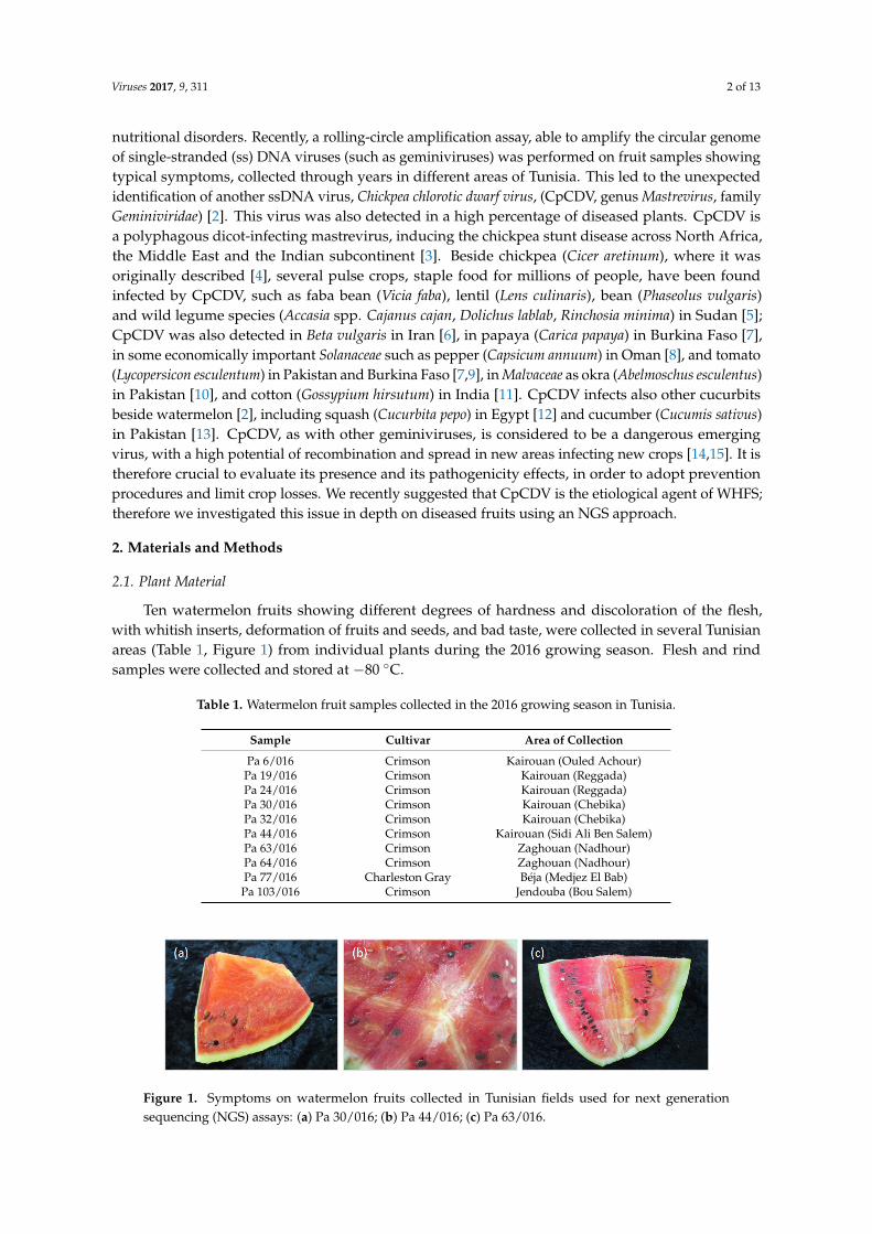
Ten watermelon fruits showing different degrees of hardness and discoloration of the flesh,with whitish inserts, deformation of fruits and seeds, and bad taste, were collected in several Tunisianareas (Table 1, Figure 1) from individual plants during the 2016 growing season. Flesh and rindsamples were collected and stored at −80 ◦C.
Table 1. Watermelon fruit samples collected in the 2016 growing season in Tunisia.
Sample Cultivar Area of Collection
Pa 6/016 Crimson Kairouan (Ouled Achour)Pa 19/016 Crimson Kairouan (Reggada)Pa 24/016 Crimson Kairouan (Reggada)Pa 30/016 Crimson Kairouan (Chebika)Pa 32/016 Crimson Kairouan (Chebika)Pa 44/016 Crimson Kairouan (Sidi Ali Ben Salem)Pa 63/016 Crimson Zaghouan (Nadhour)Pa 64/016 Crimson Zaghouan (Nadhour)Pa 77/016 Charleston Gray Béja (Medjez El Bab)
Pa 103/016 Crimson Jendouba (Bou Salem)
Viruses 2017, 9, 311 2 of 13
attributed to physiological and/or nutritional disorders. Recently, a rolling-circle amplification assay, able to amplify the circular genome of single-stranded (ss) DNA viruses (such as geminiviruses) was performed on fruit samples showing typical symptoms, collected through years in different areas of Tunisia. This led to the unexpected identification of another ssDNA virus, Chickpea chlorotic dwarf virus, (CpCDV, genus Mastrevirus, family Geminiviridae) [2]. This virus was also detected in a high percentage of diseased plants. CpCDV is a polyphagous dicot-infecting mastrevirus, inducing the chickpea stunt disease across North Africa, the Middle East and the Indian subcontinent [3]. Beside chickpea (Cicer aretinum), where it was originally described [4], several pulse crops, staple food for millions of people, have been found infected by CpCDV, such as faba bean (Vicia faba), lentil (Lens culinaris), bean (Phaseolus vulgaris) and wild legume species (Accasia spp. Cajanus cajan, Dolichus lablab, Rinchosia minima) in Sudan [5]; CpCDV was also detected in Beta vulgaris in Iran [6], in papaya (Carica papaya) in Burkina Faso [7], in some economically important Solanaceae such as pepper (Capsicum annuum) in Oman [8], and tomato (Lycopersicon esculentum) in Pakistan and Burkina Faso [7,9], in Malvaceae as okra (Abelmoschus esculentus) in Pakistan [10], and cotton (Gossypium hirsutum) in India [11]. CpCDV infects also other cucurbits beside watermelon [2], including squash (Cucurbita pepo) in Egypt [12] and cucumber (Cucumis sativus) in Pakistan [13]. CpCDV, as with other geminiviruses, is considered to be a dangerous emerging virus, with a high potential of recombination and spread in new areas infecting new crops [14,15]. It is therefore crucial to evaluate its presence and its pathogenicity effects, in order to adopt prevention procedures and limit crop losses. We recently suggested that CpCDV is the etiological agent of WHFS; therefore we investigated this issue in depth on diseased fruits using an NGS approach.
2. Materials and Methods
2.1 Plant Material
Ten watermelon fruits showing different degrees of hardness and discoloration of the flesh, with whitish inserts, deformation of fruits and seeds, and bad taste, were collected in several Tunisian areas (Table 1, Figure 1) from individual plants during the 2016 growing season. Flesh and rind samples were collected and stored at −80 °C.
Table 1. Watermelon fruit samples collected in the 2016 growing season in Tunisia.
Sample Cultivar Area of CollectionPa 6/016 Crimson Kairouan (Ouled Achour)
Pa 19/016 Crimson Kairouan (Reggada) Pa 24/016 Crimson Kairouan (Reggada) Pa 30/016 Crimson Kairouan (Chebika) Pa 32/016 Crimson Kairouan (Chebika) Pa 44/016 Crimson Kairouan (Sidi Ali Ben Salem) Pa 63/016 Crimson Zaghouan (Nadhour) Pa 64/016 Crimson Zaghouan (Nadhour) Pa 77/016 Charleston Gray Béja (Medjez El Bab) Pa 103/016 Crimson Jendouba (Bou Salem)
Figure 1. Symptoms on watermelon fruits collected in Tunisian fields used for next generation sequencing (NGS) assays: (a) Pa 30/016; (b) Pa 44/016; (c) Pa 63/016.
Figure 1. Symptoms on watermelon fruits collected in Tunisian fields used for next generationsequencing (NGS) assays: (a) Pa 30/016; (b) Pa 44/016; (c) Pa 63/016.

Viruses 2017, 9, 311 3 of 13
2.2. Nucleic Acid Extraction and Sequencing
For small RNA (sRNA) sequencing, total RNA was extracted from 10 symptomatic fruits usingTRIzol Reagent (Thermo Fisher Scientific, Waltham, MA, USA), according to the manufacturer’sinstructions. The RNA extracts were individually evaluated using NanoDrop spectrophotometerND-1000 (Thermo Scientific, Wilmington, DE, USA) and pooled into one sample; three microgramsof the pooled RNA were sent to Human Genetic Foundation sequencing service [16] for librarypreparation with the TruSeq RNA library Prep Kit v2 (Illumina, San Diego, CA, USA) and sequencingwith Illumina NextSeq 500.
Total RNA was also used for complementary DNA (cDNA) synthesis by High-Capacity cDNAReverse Transcription Kit (Thermo Fisher Scientific, Waltham, MA, USA); total nucleic acid extractionswere performed from the same samples, by silica gel-mediated extraction [17].
2.3. Small RNA Bioinformatic Analysis
Raw data (Sequence Read Archive database acc. num. SRP119446) were checked for qualityreads with FastQC software [18] and Fastx-toolkit [19] was used for removing adapter sequences,low-quality reads and artefacts; sequences shorter than 19 nt and longer than 27 nt were discarded.Reads were then analyzed by the software package VirusDetect [20], using its plant virus databaseas reference and default parameters. According to this pipeline, reads were assembled in contigswith both a reference-guided and a de novo assembly approach. De novo-assembled contigs werepooled together with those generated from reference-guided assemblies, and then processed to removeredundant sequences. An homology-dependent strategy to identify known and novel virus sequencesfrom the assembled contigs was then employed. Contigs were first compared against reference virusnucleotide sequences using BLASTn and then against the reference virus protein sequences usingBLASTx. Contigs matching the same reference sequence were merged to form the final VirusDetectoutput, and used to derive the coverage of the reference by virus contigs. Based on contigs lengthand nucleotide identity with the reference viral genome, viral sequences were selected as candidatesfor validation.
2.4. Validation of Candidate Viruses
To confirm the presence of the viral sequences identified in the sRNA library, PCR and reversetranscription PCR (RT-PCR) assays were conducted separately on each of the 10 plants. Specificprimers described in literature were used for CpCDV [2], TYLCV and TYLCSV [21], while new primerswere designed on de novo-assembled contigs showing similarities to Watermelon mosaic virus (WMV)and to amalgaviruses (see Table 2). RNA was used as templates for cDNA synthesis to validateRNA virus infections; total nucleic acids were directly used in PCR to validate DNA virus infections.Four positive controls were used, one for each known virus, and no positive control was obviouslyavailable for the new putative amalgavirus, here provisionally named Watermelon amalgavirus1 (WmAV1); the CpCDV positive control was a DNA extraction from an experimentally-inoculatedN. benthamiana [2]; for WMV, cDNA synthetized from total RNA extracted from a WMV-infected N.benthamiana (isolate 157C, from the IPSP collection) was used; for TYLCV and TYLCSV, DNA extractsfrom tomato plants experimentally-inoculated with TYLCV (Genbank Acc. No. DQ144621) andTYLCSV (Genbank Acc. No. X61153) agroclones [22] were used. The PCR negative control originatedfrom a non-agroinoculated watermelon fruit obtained from a seed-borne plant (cv. ‘Bontà’). Amplifiedfragments were analyzed on 1.5% agarose gel in 0.5 X Tris buffer EDTA (TBE).

Viruses 2017, 9, 311 4 of 13
Table 2. Primers used for validation assays, described in literature or designed on sequences of thede novo-assembled contigs. Chickpea chlorotic dwarf virus (CpCDV); Watermelon mosaic virus(WMV); Watermelon amalgavirus 1 (WmAV1), putative novel amalgavirus; Tomato yellow leaf curlvirus/Tomato yellow leaf curl Sardinia virus (TYLCV/TYLCSV).
Primers Sequences Targeted Virus-Amplicon Size Targeted Contigs
CpCDV-CP-F/R 1 GCAGAATCAAGGGCGAAGAGCGGACCGGGACCATAGTAAG
CpCDV501 bp CONTIG494
WMV-CP-F/R TGATGAGCAGATGGGTGTGAGCTGTTAATTCCCGCGAGAG
WMV379 bp CONTIG1352
WmAV1-F/R TTGCCTGGTCGTGTCTTGATGCTCAACGATGACAGATGCT
WmAV1333 bp CONTIG917
TY1(+)/TY2(-) 2 GCCCATGTA(T/C)CG(A/G)AAGCCGG(A/G)TTAGA(A/G)GCATG(A/C)GTAC
TYLCV/TYLCSV580 bp -
1 [2]; 2 [21].
2.5. Agroinfection of Watermelon Seedlings
For the infectivity assays, the agroinfectious clone of the CpCDV Tunisian isolate(TN-Zaghouan-TB2-Watermelon-2015, GenBank accession No. KX580024) [2] was used. This clone wasobtained by deleting 455 bp using NcoI/PstI from the full length CpCDV genome previously clonedwith SpeI into pBluescriptKS+, followed by the insertion of a full-length genome in the remainingSpeI site. The obtained fragment, corresponding to a ca. 1.8mer of the viral genome, was recoveredwith HindIII/SacI restriction and transferred into the binary vector pBin19, linearized accordingly.The 1.8mer clone was transformed into Rhizobium radiobacter (common name Agrobacterium tumefaciens,strain LBA4404). Bacteria were grown in liquid YEB/kanamycin/rifampicin medium for 48 h at 28 ◦C,with shaking, pelleted and resuspended in sterile water. About 30–40 µL of the suspension wereinoculated in the stems of watermelon seedlings at first true leaf stage or into the leaf axils of the modelplants Nicotiana benthamiana, according to described protocols [23].
Several watermelon cultivars for a total of 19 ‘Sugar Baby’, 3 ‘Crimson’, 32 ‘Bontà’, and 61‘Sentinel’ plants were tested for infection 28 days post agroinoculation (dpa) by tissue print orPCR. Tissue print positive reactions were always confirmed by dot blotting. Infected watermelonplants were maintained in confined environment in 20cm-diameter pots until fruit delivery. Healthy,non-inoculated watermelon plants were grown as controls in the same conditions. Fruits of about10 cm in diameter were collected once ripening occurred, and samples from a mixture of flesh and rindwere individually taken for DNA extraction.
2.6. CpCDV Detection and Replication
Leaf/stem prints (tissue print assay) or total nucleic acids extracts (dot blot assay), fixed onnylon membranes, were used to detect CpCDV. To visualize virus genomic forms, Southern blotassays were performed using total nucleic acids extracted from watermelon leaves and fruits withthe TLES buffer-based method (50 mM Tris–HCl, pH 9, 150 mM LiCl, 5 mM EDTA, and 5% SDS),according to [23]. Analyses were conducted on non-infiltrated young stems or leaves, to avoid falsepositive results owing to A. tumefaciens residual presence in the tissue. When PCR was performedfor CpCDV detection in experimentally infected watermelon seedlings, primers amplifying a 1300 bpfragment of CpCDV genome (CpCDV-seq1 5′-GTTGCCACCTGCAACGATT-3′ and CpCDV-seq25′-CGACACATAAGGTTCAGGTTG-3′) were used. A digoxigenin-labelled probe was synthesizedby PCR DIG probe Synthesis kit (Sigma-Aldrich, Darmstadt, Germany) according to manufacturer’sinstructions, using the primer pair CpCDV-CP-F/R targeting the coat protein open reading frame (ORF)of the CpCDV Tunisian isolate [2], which amplified a diagnostic 501 bp DNA fragment. The probe wasthen purified according to instructions and used at 8.4 ng/mL hybridization buffer. The assays wereessentially performed as previously described [24].
Viruses 2017, 9, 311 5 of 13
3. Results
3.1. Identification of Viral Sequences in the NGS Data
Sequencing resulted in 33,429,123 reads. Once removed low-quality reads and artefacts, 32,765,804reads were obtained; reads shorter than 19 nt or longer than 27 nt were then discarded, for a totalof 20,943,962 remaining reads. Contigs assembly and BLASTn analysis led to the identification ofsequences ascribable to CpCDV and Watermelon mosaic virus (genus Potyvirus, family Potyviridae)(Table 3 and Table S1).
Table 3. Viruses identified in the small RNA (sRNA) dataset by VirusDetect analysis; RPKM: reads perkilo base per million mapped reads.
Viral Species Genus GenomeLength (nt)
No. ofContigs
Contigs Length(Min–Max) (nt)
No. ofReads RPKM Type of
Analysis
Chickpea chlorotic dwarf virus(complete genome) Mastrevirus 2573 9 46–2702 3,197,315 59,331 BLASTn
Watermelon mosaic virus(complete genome) Potyvirus 10,051 80 41–10051 402,821 1913 BLASTn
Blueberry latent virus/Rhododendron virus A
(fusion protein)Amalgavirus 3162/3231 10 53–423 29,626 447/437 BLASTx
Ambrosia asymptomatic virus 2(polyprotein) Badnavirus 624 1 134 708 54 BLASTx
Cassava vein mosaic virus(ORF3 protein) Cavemovirus 1956 2 103–244 3506 85 BLASTx
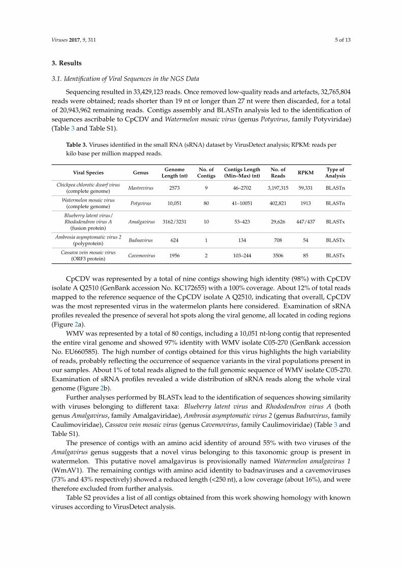
CpCDV was represented by a total of nine contigs showing high identity (98%) with CpCDVisolate A Q2510 (GenBank accession No. KC172655) with a 100% coverage. About 12% of total readsmapped to the reference sequence of the CpCDV isolate A Q2510, indicating that overall, CpCDVwas the most represented virus in the watermelon plants here considered. Examination of sRNAprofiles revealed the presence of several hot spots along the viral genome, all located in coding regions(Figure 2a).
WMV was represented by a total of 80 contigs, including a 10,051 nt-long contig that representedthe entire viral genome and showed 97% identity with WMV isolate C05-270 (GenBank accessionNo. EU660585). The high number of contigs obtained for this virus highlights the high variabilityof reads, probably reflecting the occurrence of sequence variants in the viral populations present inour samples. About 1% of total reads aligned to the full genomic sequence of WMV isolate C05-270.Examination of sRNA profiles revealed a wide distribution of sRNA reads along the whole viralgenome (Figure 2b).
Further analyses performed by BLASTx lead to the identification of sequences showing similaritywith viruses belonging to different taxa: Blueberry latent virus and Rhododendron virus A (bothgenus Amalgavirus, family Amalgaviridae), Ambrosia asymptomatic virus 2 (genus Badnavirus, familyCaulimoviridae), Cassava vein mosaic virus (genus Cavemovirus, family Caulimoviridae) (Table 3 andTable S1).
The presence of contigs with an amino acid identity of around 55% with two viruses of theAmalgavirus genus suggests that a novel virus belonging to this taxonomic group is present inwatermelon. This putative novel amalgavirus is provisionally named Watermelon amalgavirus 1(WmAV1). The remaining contigs with amino acid identity to badnaviruses and a cavemoviruses(73% and 43% respectively) showed a reduced length (<250 nt), a low coverage (about 16%), and weretherefore excluded from further analysis.
Table S2 provides a list of all contigs obtained from this work showing homology with knownviruses according to VirusDetect analysis.
Viruses 2017, 9, 311 6 of 13Viruses 2017, 9, 311 6 of 13
Figure 2. Distribution of sRNAs along (a) Chickpea chlorotic dwarf virus isolate A Q2510 (GenBank accession No. KC172655) and (b) Watermelon mosaic virus isolate C05-270 (GenBank accession No. EU660585). Viral genome organization is shown below each graphic; MP: movement protein, CP: capsid protein, Rep: replication protein, RepA: replication protein A.
3.2. Validation of Viral Sequences by RT-PCR/PCR
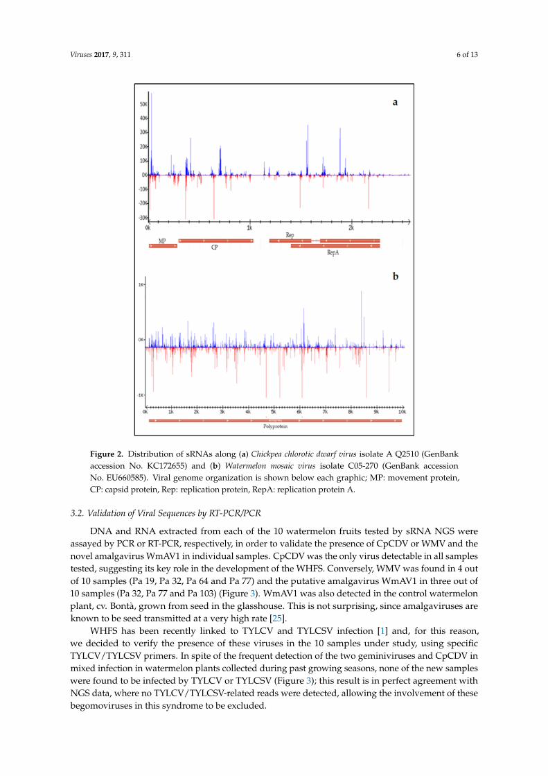
DNA and RNA extracted from each of the 10 watermelon fruits tested by sRNA NGS were assayed by PCR or RT-PCR, respectively, in order to validate the presence of CpCDV or WMV and the novel amalgavirus WmAV1 in individual samples. CpCDV was the only virus detectable in all samples tested, suggesting its key role in the development of the WHFS. Conversely, WMV was found in 4 out of 10 samples (Pa 19, Pa 32, Pa 64 and Pa 77) and the putative amalgavirus WmAV1 in three out of 10 samples (Pa 32, Pa 77 and Pa 103) (Figure 3). WmAV1 was also detected in the control watermelon plant, cv. Bontà, grown from seed in the glasshouse. This is not surprising, since amalgaviruses are known to be seed transmitted at a very high rate [25].
WHFS has been recently linked to TYLCV and TYLCSV infection [1] and, for this reason, we decided to verify the presence of these viruses in the 10 samples under study, using specific TYLCV/TYLCSV primers. In spite of the frequent detection of the two geminiviruses and CpCDV in mixed infection in watermelon plants collected during past growing seasons, none of the new samples were found to be infected by TYLCV or TYLCSV (Figure 3); this result is in perfect
Figure 2. Distribution of sRNAs along (a) Chickpea chlorotic dwarf virus isolate A Q2510 (GenBankaccession No. KC172655) and (b) Watermelon mosaic virus isolate C05-270 (GenBank accessionNo. EU660585). Viral genome organization is shown below each graphic; MP: movement protein,CP: capsid protein, Rep: replication protein, RepA: replication protein A.
3.2. Validation of Viral Sequences by RT-PCR/PCR
DNA and RNA extracted from each of the 10 watermelon fruits tested by sRNA NGS wereassayed by PCR or RT-PCR, respectively, in order to validate the presence of CpCDV or WMV and thenovel amalgavirus WmAV1 in individual samples. CpCDV was the only virus detectable in all samplestested, suggesting its key role in the development of the WHFS. Conversely, WMV was found in 4 outof 10 samples (Pa 19, Pa 32, Pa 64 and Pa 77) and the putative amalgavirus WmAV1 in three out of10 samples (Pa 32, Pa 77 and Pa 103) (Figure 3). WmAV1 was also detected in the control watermelonplant, cv. Bontà, grown from seed in the glasshouse. This is not surprising, since amalgaviruses areknown to be seed transmitted at a very high rate [25].
WHFS has been recently linked to TYLCV and TYLCSV infection [1] and, for this reason,we decided to verify the presence of these viruses in the 10 samples under study, using specificTYLCV/TYLCSV primers. In spite of the frequent detection of the two geminiviruses and CpCDV inmixed infection in watermelon plants collected during past growing seasons, none of the new sampleswere found to be infected by TYLCV or TYLCSV (Figure 3); this result is in perfect agreement withNGS data, where no TYLCV/TYLCSV-related reads were detected, allowing the involvement of thesebegomoviruses in this syndrome to be excluded.

Viruses 2017, 9, 311 7 of 13
Viruses 2017, 9, 311 7 of 13
agreement with NGS data, where no TYLCV/TYLCSV-related reads were detected, allowing the involvement of these begomoviruses in this syndrome to be excluded.
Figure 3. Validation of virus infection in individual watermelon fruit sample used for NGS analysis; polymerase chain reaction (PCR) (a,d) and real time PCR (RT-PCR) (b,c); amplified fragments were separated by electrophoresis in 1.5% agarose gels in 0.5× TBE: (a) CpCDV, 501 bp amplified fragment; (b) WMV, 379 bp amplified fragment; (c) WmAV1, putative novel amalgavirus, 333 bp amplified fragment (no positive control is available); (d) TYLCV/TYLCSV, 580 bp amplified fragment, positive controls: TYLCV (+left) and TYLCSV (+right). H, seed-borne watermelon fruit; nt, no template; +, positive controls; M, 100 bp DNA Ladder, (*) indicates the 500 bp fragment.
3.3. CpCDV Sequence Analysis
The BLASTn analysis of the contig representing the full-length CpCDV genome (CONTIG494), showed maximum similarity with the CpCDV KX580024, obtained from watermelon collected in Tunisia in 2015, one year before the collection of the samples used in the present study. The two sequences are 2571 nt long and share a nucleotide identity of 99.1% (2549/2571 nt). The next-closest sequence is a CpCDV isolate from Syria (FR687959) [26], typed as strain A [27,28]. The genome organization is typical of mastreviruses [26].
CONTIG494 contains an unusual nonanucleotide TAATGTTAC in the stem-loop region, different from the canonical geminiviral nonanucleotide TAATATTAC. This non-canonical sequence is also present in CpCDV KX580024, and was confirmed by amplification and sequencing of additional watermelon samples collected in Tunisia in 2015 (2 samples) and 2016 (3 samples). However, all other sequences of CpCDV available in the GenBank repository (more than 200) have a TAATATTAC sequence. To verify whether the non-canonical sequence is correlated with the host plant species infected by CpCDV, we analyzed the two other CpCDV sequences isolated from cucurbits (KF692356 from squash and KT719388 from cucumber) and found that both contained the canonical geminiviral nonanucleotide.
The non-canonical nonanucleotide is uncommon in geminiviruses, but can be found in some chickpea-infecting mastreviruses from Australia, e.g., Chickpea chlorosis virus (National Center for Biotechnology Information (NCBI) Reference Sequence NC_014740), Chickpea chlorosis Australia virus (NC_022131.1), and Chickpea redleaf virus (NC_014739.1) [29,30], as well as in Dragonfly-associated mastrevirus (two records), and in one isolate of Maize streak virus.
3.4. Infectivity Assays
A CpCDV infectious agroclone was used to inoculate watermelon plants in controlled conditions using a tissue print assay to identify infected plants, to be grown for fruit delivery. The infection rate was about 7% (6 out of 83 tested plants), while it reached almost 99% with model plant
Figure 3. Validation of virus infection in individual watermelon fruit sample used for NGS analysis;polymerase chain reaction (PCR) (a,d) and reverse transcription PCR (RT-PCR) (b,c); amplifiedfragments were separated by electrophoresis in 1.5% agarose gels in 0.5× TBE: (a) CpCDV, 501 bpamplified fragment; (b) WMV, 379 bp amplified fragment; (c) WmAV1, putative novel amalgavirus,333 bp amplified fragment (no positive control is available); (d) TYLCV/TYLCSV, 580 bp amplifiedfragment, positive controls: TYLCV (+left) and TYLCSV (+right). H, seed-borne watermelon fruit;nt, no template; +, positive controls; M, 100 bp DNA Ladder, (*) indicates the 500 bp fragment.
3.3. CpCDV Sequence Analysis
The BLASTn analysis of the contig representing the full-length CpCDV genome (CONTIG494),showed maximum similarity with the CpCDV KX580024, obtained from watermelon collected inTunisia in 2015, one year before the collection of the samples used in the present study. The twosequences are 2571 nt long and share a nucleotide identity of 99.1% (2549/2571 nt). The next-closestsequence is a CpCDV isolate from Syria (FR687959) [26], typed as strain A [27,28]. The genomeorganization is typical of mastreviruses [26].
CONTIG494 contains an unusual nonanucleotide TAATGTTAC in the stem-loop region, differentfrom the canonical geminiviral nonanucleotide TAATATTAC. This non-canonical sequence is alsopresent in CpCDV KX580024, and was confirmed by amplification and sequencing of additionalwatermelon samples collected in Tunisia in 2015 (2 samples) and 2016 (3 samples). However, all othersequences of CpCDV available in the GenBank repository (more than 200) have a TAATATTACsequence. To verify whether the non-canonical sequence is correlated with the host plantspecies infected by CpCDV, we analyzed the two other CpCDV sequences isolated from cucurbits(KF692356 from squash and KT719388 from cucumber) and found that both contained the canonicalgeminiviral nonanucleotide.
The non-canonical nonanucleotide is uncommon in geminiviruses, but can be found in somechickpea-infecting mastreviruses from Australia, e.g., Chickpea chlorosis virus (National Center forBiotechnology Information (NCBI) Reference Sequence NC_014740), Chickpea chlorosis Australia virus(NC_022131.1), and Chickpea redleaf virus (NC_014739.1) [29,30], as well as in Dragonfly-associatedmastrevirus (two records), and in one isolate of Maize streak virus.
3.4. Infectivity Assays
A CpCDV infectious agroclone was used to inoculate watermelon plants in controlled conditionsusing a tissue print assay to identify infected plants, to be grown for fruit delivery. The infection ratewas about 7% (6 out of 83 tested plants), while it reached almost 99% with model plant N. benthamianaused as infection controls (222 out of 225 agroinoculated plants). A further experiment was performed,with the hypothesis that the virus concentration could be below the detection limit of the tissue printassay. In this new experiment PCR was used as detection tool, and 18 infected watermelons out of 32

Viruses 2017, 9, 311 8 of 13
were detected, resulting in a 56% infection rate. A possible explanation is that experimental watermeloninfection may in some cases be below tissue print detection threshold, limiting the possibility of routineusage of this widely-used technique for mass diagnosis in these circumstances. Infected watermelonplants showed reduced growth and chlorosis, with smaller and deformed leaves compared to noninoculated plants.
Four CpCDV-infected watermelon plants (Figure 4a) were kept until fruit delivery. All fruitsobtained from CpCDV-infected or healthy watermelons were small in size (10 cm maximum diameter)possibly due to limiting experimental growth conditions: nevertheless they all reached the ripeningstage. All the eight fruits collected from the infected plants were positive for virus infection in dotblot assays (see Table 4 and Figure 4b), and showed several degrees of symptoms compared to fruitsfrom healthy plants (Figure 4c), including yellowish/whitish areas or stripes in the flesh, that wasdiscolored (orange instead of red) and, in some cases, displayed a clearly deformed shape. In addition,they all produced few seeds, mainly aborted or immature, especially in the symptomatic parts of theflesh (Figure 4d,e). In five cases, the small fruits showed growth arrest by precocious pod necrosis.
Viruses 2017, 9, 311 8 of 13
N. benthamiana used as infection controls (222 out of 225 agroinoculated plants). A further experiment was performed, with the hypothesis that the virus concentration could be below the detection limit of the tissue print assay. In this new experiment PCR was used as detection tool, and 18 infected watermelons out of 32 were detected, resulting in a 56% infection rate. A possible explanation is that experimental watermelon infection may in some cases be below tissue print detection threshold, limiting the possibility of routine usage of this widely-used technique for mass diagnosis in these circumstances. Infected watermelon plants showed reduced growth and chlorosis, with smaller and deformed leaves compared to non inoculated plants.
Four CpCDV-infected watermelon plants (Figure 4a) were kept until fruit delivery. All fruits obtained from CpCDV-infected or healthy watermelons were small in size (10 cm maximum diameter) possibly due to limiting experimental growth conditions: nevertheless they all reached the ripening stage. All the eight fruits collected from the infected plants were positive for virus infection in dot blot assays (see Table 4 and Figure 4b), and showed several degrees of symptoms compared to fruits from healthy plants (Figure 4c), including yellowish/whitish areas or stripes in the flesh, that was discolored (orange instead of red) and, in some cases, displayed a clearly deformed shape. In addition, they all produced few seeds, mainly aborted or immature, especially in the symptomatic parts of the flesh (Figure 4d,e). In five cases, the small fruits showed growth arrest by precocious pod necrosis.
Figure 4. (a) Tissue print assay of selected agro-infected watermelon seedlings. Plant and leaf stems are tested, four prints for each plant are performed; H, plant 3/12; 1, plant 4/27; 2, plant 6/14; 3, plant 6/11; 4, plant 6/21. CpCDV CP-specific digoxigenin-labelled probe is used and chemiluminescent detection is performed. (b) Dot blot assay of watermelon fruits. Total DNA extracts are prepared using both rind and flesh tissues, 400 ng of DNA/drop, 5 μL volume are spotted on membrane. 1, 2, 3, 4, the four fruits produced by plant 4/27; 5, 6, the two fruits produced by plant 6/11; 7, sole fruit produced by plant 6/14; 8, sole fruit produced by plant 6/21; 9, one of the fruits produced by plant 8/27, agroinoculated but not CpCDV-infected; H, not agro-inoculated watermelon fruit, grown in the
Figure 4. (a) Tissue print assay of selected agro-infected watermelon seedlings. Plant and leaf stems aretested, four prints for each plant are performed; H, plant 3/12; 1, plant 4/27; 2, plant 6/14; 3, plant 6/11;4, plant 6/21. CpCDV CP-specific digoxigenin-labelled probe is used and chemiluminescent detectionis performed. (b) Dot blot assay of watermelon fruits. Total DNA extracts are prepared using both rindand flesh tissues, 400 ng of DNA/drop, 5 µL volume are spotted on membrane. 1, 2, 3, 4, the four fruitsproduced by plant 4/27; 5, 6, the two fruits produced by plant 6/11; 7, sole fruit produced by plant6/14; 8, sole fruit produced by plant 6/21; 9, one of the fruits produced by plant 8/27, agroinoculatedbut not CpCDV-infected; H, not agro-inoculated watermelon fruit, grown in the same conditions;+, CpCDV-infected N. benthamiana leaf tissue; −, healthy N. benthamiana leaf tissue; TE, 1x Tris/EDTAbuffer, diluent of DNA. (c) Not agro-inoculated watermelon fruit cv. ‘Bontà’; (d) Symptomatology inwatermelon fruit 1 produced by experimentally-infected plant 6/11, cv. ‘Bontà’; (e) Symptomatologyin watermelon fruit produced by experimentally-infected plant 6/21, cv. ‘Sentinel’; infected fruits showvarious degree of abnormality in flesh structure with whitish stripes inside the red/orange area, and inseed production.

Viruses 2017, 9, 311 9 of 13
Table 4. Summary of watermelon fruits assays. Visual interpretation of chemiluminescent reactionsdata: “+”, slightly positive reaction; “++”, moderately positive reaction; “+++” strongly positivereaction; “−“ negative reaction.
Plant Cultivar CpCDV Infection in Plants 1 Fruits Tested 2 CpCDV Infection in Fruits 3
4/27 Sugar Baby +
1234
+++++
6/11 Bontà +++ 1 4
2+++
6/14 Bontà +++ 1 ++
6/21 Sentinel +++ 1 5 ++
8/27 Sentinel − 1 −Healthy Bontà − 1 −
1 According to tissue print results. 2 All fruits obtained from CpCDV-infected plants were tested; a selection ofnon-infected plant fruit is presented. 3 According to dot blot results. 4 Shown in Figure 4d. 5 Shown in Figure 4e.
3.5. CpCDV Replication
To evaluate the ability of CpCDV to replicate in the aerial parts and in fruits ofexperimentally inoculated watermelon plants, DNA samples were analyzed by Southern blottingusing a CpCDV-specific probe. As it can be seen in Figure 5, both ss and replicative dsDNA forms(in supercoiled and open circular conformations) were detected in watermelon leaves, and also insymptomatic portions of fruits, though at a relatively lower concentration. These viral forms weresimilar to those detected in the model plant N. benthamiana. Conversely, no signal was observed inhealthy plants.
This confirms that the CpCDV agroclone obtained from a symptomatic watermelon plant inTunisia can efficiently replicate in watermelon plants and their fruits, that exhibit symptoms similar tothose induced by WHFS.

Viruses 2017, 9, 311 10 of 13Viruses 2017, 9, 311 10 of 13
Figure 5. Southern blot assay of DNA extracts of: 1, CpCDV-infected N. benthamiana; 2, healthy N. benthamiana; 3, CpCDV-infected watermelon plant 6/14 (leaf extract); 4, CpCDV-infected watermelon plant 6/23 (leaf extract); 5, CpCDV-infected watermelon plant 4/27 (fruit extract); 6, healthy watermelon (leaf plus fruit); P, 6 ng of CpCDV genome full length fragment; M, 1 Kb Ladder Plus DNA ladder: the pale 1650 bp band is indicated (Thermo Fisher Scientific, Waltham, MA, USA). On the right, the four CpCDV genome forms found in watermelon leaf and fruit extracts are shown by black arrows; the forms of the double-stranded DNA are indicated: open circular (OC), linear (Lin) and covalently closed circular (CCC), single-stranded DNA is marked (ssDNA) (a) ethidium bromide-stained agarose gel showing the high molecular weight genomic DNA bands (white arrow); (b) autoradiography film resulting from chemiluminescent detection using CpCDV DIG-labelled probe.
4. Discussion
Over the past years, deep sequencing technologies have opened novel doors to reconstruct viral populations in a high-throughput and cost-effective manner. Up to now, an increasing number of studies have used NGS to either analyze known viruses by means of a reference-guided approach or discover novel viruses using a de novo-based strategy [31]. In this study, the sRNA sequencing of a pool of 10 symptomatic samples was a powerful mean for the identification of CpCDV as the etiological agent of the WHFS and the assembling of its full-length sequence. Indeed, CpCDV was the only virus present in all tested samples, and its agroclone was able to infect watermelon seedlings, actively replicating in the tissues and inducing, in controlled conditions, fruits abnormalities similar to those described for the diseased watermelon plants occurring in open field.
CpCDV, with its wide host range, the seriousness of symptoms induced and its expanding geographical distribution, is an emerging virus and has the potential to become a serious pest for several crops in tropical and Mediterranean countries and worldwide. The two known vectors of CpCDV are Orosius albicinctus (Distant) and Orosius orientalis (Matsumura), both originally found in Asia. O. orientalis was also reported in Tunisia [32] and it is likely that its vector/vectors are already
Figure 5. Southern blot assay of DNA extracts of: 1, CpCDV-infected N. benthamiana; 2, healthyN. benthamiana; 3, CpCDV-infected watermelon plant 6/14 (leaf extract); 4, CpCDV-infected watermelonplant 6/23 (leaf extract); 5, CpCDV-infected watermelon plant 4/27 (fruit extract); 6, healthy watermelon(leaf plus fruit); P, 6 ng of CpCDV genome full length fragment; M, 1 Kb Ladder Plus DNA ladder:the pale 1650 bp band is indicated (Thermo Fisher Scientific, Waltham, MA, USA). On the right,the four CpCDV genome forms found in watermelon leaf and fruit extracts are shown by black arrows;the forms of the double-stranded DNA are indicated: open circular (OC), linear (Lin) and covalentlyclosed circular (CCC), single-stranded DNA is marked (ssDNA) (a) ethidium bromide-stained agarosegel showing the high molecular weight genomic DNA bands (white arrow); (b) autoradiography filmresulting from chemiluminescent detection using CpCDV DIG-labelled probe.
4. Discussion
Over the past years, deep sequencing technologies have opened novel doors to reconstruct viralpopulations in a high-throughput and cost-effective manner. Up to now, an increasing number ofstudies have used NGS to either analyze known viruses by means of a reference-guided approachor discover novel viruses using a de novo-based strategy [31]. In this study, the sRNA sequencingof a pool of 10 symptomatic samples was a powerful mean for the identification of CpCDV as theetiological agent of the WHFS and the assembling of its full-length sequence. Indeed, CpCDV was theonly virus present in all tested samples, and its agroclone was able to infect watermelon seedlings,actively replicating in the tissues and inducing, in controlled conditions, fruits abnormalities similar tothose described for the diseased watermelon plants occurring in open field.
CpCDV, with its wide host range, the seriousness of symptoms induced and its expandinggeographical distribution, is an emerging virus and has the potential to become a serious pest forseveral crops in tropical and Mediterranean countries and worldwide. The two known vectors ofCpCDV are Orosius albicinctus (Distant) and Orosius orientalis (Matsumura), both originally found inAsia. O. orientalis was also reported in Tunisia [32] and it is likely that its vector/vectors are already

Viruses 2017, 9, 311 11 of 13
present in other North African countries, and possibly in Southern Europe, posing a new real threatfor cucurbits cultivation.
Beside the full-length sequence of the DNA virus CpCDV, the sRNA sequencing has allowedthe assembly of the full-length sequence of the RNA virus WMV. WMV infection is widespread inTunisia on all cucurbit species, at least since the late eighties [33,34]. The virus induces severe green leafmottling and plant stunting, mainly on melon and squash, both in open field and protected crops, in allmajor production regions. The virus is less prevalent on watermelon, but when present, it inducesmainly leaf symptoms. As a matter of fact, the PCR experiments showed its infection occurred only ina few samples, thus excluding a correlation between this virus and WHFS.
The sRNA sequencing also allowed to identify a previously undescribed virus infectingwatermelon, here provisionally named WmAV1, belonging to the Amalgavirus genus: as reported forother viral species belonging to the same genus, its infection is apparently symptomless and transmittedthrough seeds. Amalgaviruses have small dsRNA genomes (about 3.4 kbp) and their virions havenot yet been detected or identified; moreover, very little is known about their transmission, apartfrom seed involvement. The family Amalgaviridae is a recently recognized taxon, currently comprisingfour species of plant-infecting viruses (Blueberry latent virus, Rhododendron virus A, Southern tomatovirus, and Vicia cryptic virus M) [25,35–38]. Recently, several new species ascribable to this genus havebeen described in diverse plant species, following mining transcriptomic data available in publicrepositories [39]. It is not surprising that amalgavirus-like sequences have been found in watermelonin the present study by analyzing sRNA populations. Considering all these aspects, it is likely that theWmAV1 has a more widespread distribution than expected, but no role in the WHFS.
In summary, this work confirmed that high throughput sequencing analysis of sRNAs isa powerful tool for the identification of both known and new viral sequences and for the definitionof new etiological agents. The data obtained by this technique, supported by PCR validation andexperimental infection in controlled conditions, allowed the causal link between CpCDV and WHFS tobe highlighted for the first time.
Supplementary Materials: The following are available online at www.mdpi.com/1999-4915/9/11/311/s1,Table S1: Viral species identified by (a) BLASTn and (b) BLASTx analyses, Table S2: List of all contigs obtainedshowing homology with known viruses according BLASTn and BLASTx analysis.
Acknowledgments: Authors wish to thank Daniele Marian for technical support, Elena Zocca, Caterina Perrone,Luca Bordone and Aziza Ghazouani for plant management. Takoua Zaagueri was financially supported by grantsfrom the Tunisian Ministry of Higher Education and Scientific Research (MERST).
Author Contributions: Takoua Zaagueri and Anna Maria Vaira performed the experiments; Laura Miozzicontributed NGS costs and analyzed data; Anna Maria Vaira and Laura Miozzi conceived and designedthe experiments; Emanuela Noris performed Southern blotting experiments and analyzed replication data;Takoua Zaagueri and Monia Mnari-Hattab provided plant extracts for NGS analysis; Anna Maria Vaira,Laura Miozzi and Gian Paolo Accotto wrote the paper.
Conflicts of Interest: The authors declare no conflict of interest. The funding sponsors had no role in the designof the study; in the collection, analyses, or interpretation of data; in the writing of the manuscript, and in thedecision to publish the results.
References
1. Mnari-Hattab, M.; Zammouri, S.; Hajlaoui, M. First report of hard watermelon syndrome in Tunisiaassociated with Tomato yellow leaf curl virus infection. New Dis. Rep. 2014, 30. [CrossRef]
2. Zaagueri, T.; Mnari-Hattab, M.; Zammouri, S.; Hajlaoui, M.; Accotto, G.P.; Vaira, A.M. First Report ofChickpea chlorotic dwarf virus in Watermelon (Citrullus lanatus) in Tunisia. Plant Dis. 2017, 101, 392–393.[CrossRef]
3. Kanakala, S.; Sakhare, A.; Verma, H.; Malathi, V. Infectivity and the phylogenetic relationship of a mastreviruscausing chickpea stunt disease in India. Eur. J. Plant Pathol. 2013, 135, 429–438. [CrossRef]
4. Horn, N.; Reddy, S.; Roberts, I.; Reddy, D. Chickpea chlorotic dwarf virus, a new leafhopper-transmittedgeminivirus of chickpea in India. Ann. Appl. Biol. 1993, 122, 467–479. [CrossRef]

Viruses 2017, 9, 311 12 of 13
5. Ali, M.; Kumari, S.; Makkouk, K.; Hassan, M. Chickpea chlorotic dwarf virus (CpCDV) naturally infectsPhaseolus bean and other wild species in the Gezira region of Sudan. Arab J. Plant Prot. 2004, 22, 96.
6. Farzadfar, S.; Pourrahim, R.; Golnaraghi, A.; Ahoonmanesh, A. PCR detection and partial molecularcharacterization of Chickpea chlorotic dwarf virus in naturally infected sugar beet plants in Iran. J. Plant Pathol.2008, 90, 247–251.
7. Ouattara, A.; Tiendrebeogo, F.; Lefeuvre, P.; Hoareau, M.; Claverie, S.; Traore, E.V.; Barro, N.; Traoré, O.;Varsani, A.; Lett, J.M. New strains of chickpea chlorotic dwarf virus discovered on diseased papaya andtomato plants in Burkina Faso. Arch. Virol. 2017, 162, 1791–1794. [CrossRef] [PubMed]
8. Akhtar, S.; Khan, A.; Briddon, R. A distinct strain of Chickpea chlorotic dwarf virus infecting pepper in Oman.Plant Dis. 2014, 98, 286. [CrossRef]
9. Zia-Ur-Rehman, M.; Hameed, U.; Herrmann, H.W.; Iqbal, M.; Haider, M.; Brown, J.K. First report of Chickpeachlorotic dwarf virus infecting tomato crops in Pakistan. Plant Dis. 2015. [CrossRef]
10. Zia-Ur-Rehman, M.; Hameed, U.; Ali, C.; Haider, M.; Brown, J. First Report of Chickpea chlorotic dwarf virusInfecting Okra in Pakistan. Plant Dis. 2017. [CrossRef]
11. Manzoor, M.; Ilyas, M.; Shafiq, M.; Haider, M.; Shahid, A.; Briddon, R. A distinct strain of Chickpea chloroticdwarf virus (genus Mastrevirus, family Geminiviridae) identified in cotton plants affected by leaf curl disease.Arch. Virol. 2014, 159, 1217–1221. [CrossRef] [PubMed]
12. Fahmy, I.F.; Taha, O.; El-Ashry, A.N. First genome analysis and molecular characterization of Chickpeachlorotic dwarf virus Egyptian isolate infecting squash. Virus Dis. 2015, 26, 33–41. [CrossRef] [PubMed]
13. Hameed, U.; Zia-Ur-Rehman, M.; Ali, S.; Haider, M.; Brown, J. First Report of Chickpea chlorotic dwarf virusInfecting Cucumber in Pakistan. Plant Dis. 2017, 101, 848. [CrossRef]
14. Kraberger, S.; Kumari, S.G.; Hamed, A.A.; Gronenborn, B.; Thomas, J.E.; Sharman, M.; Harkins, G.W.;Muhire, B.M.; Martin, D.P.; Varsani, A. Molecular diversity of Chickpea chlorotic dwarf virus in Sudan: Highrates of intra-species recombination—A driving force in the emergence of new strains. Infect. Genet. Evol.2015, 29, 203–215. [CrossRef] [PubMed]
15. Kraberger, S.; Harkins, G.; Kumari, S.; Thomas, J.; Schwinghamer, M.; Sharman, M.; Collings, D.A.;Briddon, R.W.; Martin, D.P.; Varsani, A. Evidence that dicot-infecting mastreviruses are particularly prone tointer-species recombination and have likely been circulating in Australia for longer than in Africa and theMiddle East. Virology 2013, 444, 282–291. [CrossRef] [PubMed]
16. Human Genetic Foundation Sequencing Service. Available online: http://www.hugef-torino.org/site/index.php (accessed on 28 October 2016).
17. Foissac, X.; Svanella-Dumas, L.; Dulucq, M.; Candresse, T.; Gentit, P. Polyvalent detection of fruit treetricho, capillo and foveaviruses by nested RT-PCR using degenerated and inosine containing primers (PDORT-PCR). In XVIII International Symposium on Virus and Virus-like Diseases of Temperate Fruit Crops-TopFruit Diseases. Acta Hortic. 2001, 550, 37–44. [CrossRef]
18. FastQC Software. Available online: http://www.bioinformatics.babraham.ac.uk/projects/fastqc (accessedon 6 November 2016).
19. Fastx-Toolkit Software. Available online: http://hannonlab.cshl.edu/fastx_toolkit/ (accessed on8 November 2016).
20. Zheng, Y.; Gao, S.; Padmanabhan, C.; Li, R.; Galvez, M.; Gutierrez, D.; Fuentes, S.; Ling, K.S.; Kreuze, J.;Fei, Z. VirusDetect: An automated pipeline for efficient virus discovery using deep sequencing of smallRNAs. Virology 2017, 500, 130–138. [CrossRef] [PubMed]
21. Accotto, G.P.; Navas-Castillo, J.; Noris, E.; Moriones, E.; Louro, D. Typing of Tomato Yellow Leaf Curl Virusesin Europe. Eur. J. Plant Pathol. 2000, 106, 179–186. [CrossRef]
22. Kheyr-Pour, A.; Bendahmane, M.; Matzeit, V.; Accotto, G.P.; Crespi, S.; Gronenborn, B. Tomato yellow leafcurl virus from sardinia is a whitefly-transmitted monoparatite geminivirus. Nucleic Acids Res. 1991, 19,6763–6769. [CrossRef] [PubMed]
23. Noris, E.; Accotto, G.P.; Tavazza, R.; Brunetti, A.; Crespi, S.; Tavazza, M. Resistance to tomato yellow leafcurl Geminivirus in Nicotiana benthamiana plants transformed with a truncated viral C1 gene. Virology 1996,224, 130–138. [CrossRef] [PubMed]
24. Accotto, G.P.; Vaira, A.M.; Noris, E.; Vecchiati, M. Using non-radioactive probes on plants: A few examples.J. Biolumin. Chemilumin. 1998, 13, 295–301. [CrossRef]

Viruses 2017, 9, 311 13 of 13
25. Martin, R.R.; Zhou, J.; Tzanetakis, I.E. Blueberry latent virus: An amalgam of the Partitiviridae andTotiviridae. Virus Res. 2011, 155, 175–180. [CrossRef] [PubMed]
26. Mumtaz, H.; Kumari, S.; Mansoor, S.; Martin, D.; Briddon, R. Analysis of the sequence of a dicot-infectingmastrevirus (family Geminiviridae) originating from Syria. Virus Genes 2011, 42, 422–428. [CrossRef] [PubMed]
27. Muhire, B.; Martin, D.P.; Brown, J.K.; Navas-Castillo, J.; Moriones, E.; Zerbini, F.M.; Rivera-Bustamante, R.;Malathi, V.G.; Briddon, R.W.; Varsani, A. A genome-wide pairwise-identity-based proposal for theclassification of viruses in the genus Mastrevirus (family Geminiviridae). Arch. Virol. 2013, 158, 1411–1424.[CrossRef] [PubMed]
28. Muhire, B.M.; Varsani, A.; Martin, D.P. SDT: A virus classification tool based on pairwise sequence alignmentand identity calculation. PLoS ONE 2014, 9, e108277. [CrossRef] [PubMed]
29. Hadfield, J.; Thomas, J.E.; Schwinghamer, M.W.; Kraberger, S.; Stainton, D.; Dayaram, A.; Parry, J.N.;Pande, D.; Martin, D.P.; Varsani, A. Molecular characterisation of dicot-infecting mastreviruses from Australia.Virus Res. 2012, 166, 13–22. [CrossRef] [PubMed]
30. Thomas, J.; Parry, J.; Schwinghamer, M.; Dann, E. Two novel mastreviruses from chickpea (Cicer arietinum)in Australia. Arch. Virol. 2010, 155, 1777–1788. [CrossRef] [PubMed]
31. Hadidi, A.; Flores, R.; Candresse, T.; Barba, M. Next-Generation Sequencing and Genome Editing in PlantVirology. Front. Microbiol. 2016, 7. [CrossRef] [PubMed]
32. Boukhris-Bouhachem, S.; Chabbouh, N.; Harbi, M.; Danet, J.L. (Eds.) Les Cicadiaires Vecteurs Potentielsde Phytopathogènes en Vignoble Tunisien (Hemiptera: Cicadomorpha: Fulgoromorpha); Annales de la SociétéEntomologique de France; Taylor & Francis Group: Abingdon, UK, 2007.
33. Mnari, M.; Cherif, C.; Jebari, H. Importance des virus des Cucurbitacées en Tunisie et étude des souches duvirus de la mosaïque du concombre. Ann. Inst. Natl. Rech. Agron. Tunis. 1990, 63, 15.
34. Mnari-Hattab, M.; Jebari, H.; Zouba, A. Identification et distribution des virus responsables de mosaiqueschez les cucurbitacées en Tunisie. Eur. Public Prosec. Off. Bull. 2008, 38, 497–506. [CrossRef]
35. Adams, M.J.; Lefkowitz, E.J.; King, A.M.Q.; Carstens, E.B. Ratification vote on taxonomic proposals to theInternational Committee on Taxonomy of Viruses (2014). Arch. Virol. 2014, 159, 2831–2841. [CrossRef][PubMed]
36. Liu, W.; Chen, J. A double-stranded RNA as the genome of a potential virus infecting. Virus Genes 2009, 39,126–131. [CrossRef] [PubMed]
37. Sabanadzovic, S.; Valverde, R.A.; Brown, J.K.; Martin, R.R.; Tzanetakis, I.E. Southern tomato virus: The linkbetween the families Totiviridae and Partitiviridae. Virus Res. 2009, 140, 130–137. [CrossRef] [PubMed]
38. Sabanadzovic, S.; Ghanem-Sabanadzovic, N.A.; Valverde, R. A novel monopartite dsRNA virus fromrhododendron. Arch. Virol. 2010, 155, 1859–1863. [CrossRef] [PubMed]
39. Nibert, M.L.; Pyle, J.D.; Firth, A.E. A+1 ribosomal frameshifting motif prevalent among plant amalgaviruses.Virology 2016, 498, 201–208. [CrossRef] [PubMed]
© 2017 by the authors. Licensee MDPI, Basel, Switzerland. This article is an open accessarticle distributed under the terms and conditions of the Creative Commons Attribution(CC BY) license (http://creativecommons.org/licenses/by/4.0/).