defects of mutant dnmt1 are linked to a spectrum of...
TRANSCRIPT

Defects of mutant DNMT1 are linked to aspectrum of neurological disorders
Jonathan Baets,1,2,3,* Xiaohui Duan,4,5,* Yanhong Wu,6 Gordon Smith,7 William W. Seeley,8
Ines Mademan,1,2 Nicole M. McGrath,9 Noah C. Beadell,10 Julie Khoury,10
Maria-Victoria Botuyan,11 Georges Mer,11 Gregory A. Worrell,12 Kaori Hojo,13
Jessica DeLeon,14 Matilde Laura,15,16 Yo-Tsen Liu,15,16,17,18 Jan Senderek,19 Joachim Weis,20
Peter Van den Bergh,21 Shana L. Merrill,22 Mary M. Reilly,15,16 Henry Houlden,15,16
Murray Grossman,22 Steven S. Scherer,22 Peter De Jonghe,1,2,3 Peter J. Dyck4 andChristopher J. Klein4,6,23
*These authors contributed equally to this work.
We report a broader than previously appreciated clinical spectrum for hereditary sensory and autonomic neuropathy type 1E
(HSAN1E) and a potential pathogenic mechanism for DNA methyltransferase (DNMT1) mutations. The clinical presentations and
genetic characteristics of nine newly identified HSAN1E kinships (45 affected subjects) were investigated. Five novel mutations of
DNMT1 were discovered; p.C353F, p.T481P, p.P491L, p.Y524D and p.I531N, all within the target-sequence domain, and two
mutations (p.T481P, p.P491L) arising de novo. Recently, HSAN1E has been suggested as an allelic disorder of autosomal dom-
inant cerebellar ataxia, deafness and narcolepsy. Our results indicate that all the mutations causal for HSAN1E are located in the
middle part or N-terminus end of the TS domain, whereas all the mutations causal for autosomal dominant cerebellar ataxia,
deafness and narcolepsy are located in the C-terminus end of the TS domain. The impact of the seven causal mutations in this
cohort was studied by cellular localization experiments. The binding efficiency of the mutant DNMT proteins at the replication
foci and heterochromatin were evaluated. Phenotypic characterizations included electromyography, brain magnetic resonance and
nuclear imaging, electroencephalography, sural nerve biopsies, sleep evaluation and neuropsychometric testing. The average sur-
vival of HSAN1E was 53.6 years. [standard deviation = 7.7, range 43–75 years], and mean onset age was 37.7 years. (standard
deviation = 8.6, range 18–51 years). Expanded phenotypes include myoclonic seizures, auditory or visual hallucinations, and renal
failure. Hypersomnia, rapid eye movement sleep disorder and/or narcolepsy were identified in 11 subjects. Global brain atrophy
was found in 12 of 14 who had brain MRI. EEGs showed low frequency (delta waves) frontal-predominant abnormality in five of
six patients. Marked variability in cognitive deficits was observed, but the majority of patients (89%) developed significant cog-
nitive deficit by the age of 45 years. Cognitive function decline often started with personality changes and psychiatric manifest-
ations. A triad of hearing loss, sensory neuropathy and cognitive decline remains as the stereotypic presentation of HSAN1E.
Moreover, we show that mutant DNMT1 proteins translocate to the cytoplasm and are prone to form aggresomes while losing
their binding ability to heterochromatin during the G2 cell cycle. Our results suggest mutations in DNMT1 result in imbalanced
protein homeostasis through aggresome-induced autophagy. This mechanism may explain why mutations in the sole DNA main-
tenance methyltransferase lead to selective central and peripheral neurodegeneration.
1 Neurogenetics Group, VIB-Department of Molecular Genetics, University of Antwerp, Antwerpen, Belgium2 Laboratory of Neurogenetics, Institute Born-Bunge, University of Antwerp, Antwerpen, Belgium3 Department of Neurology, Antwerp University Hospital, Antwerpen, Belgium4 Peripheral Neuropathy Research Laboratory, Mayo Clinic, Rochester, MN, USA5 Department of Neurology, China-Japan Friendship Hospital, Beijing China
doi:10.1093/brain/awv010 BRAIN 2015: 138; 845–861 | 845
Received October 7, 2014. Revised November 22, 2014. Accepted December 5, 2014. Advance Access publication February 12, 2015
� The Author (2015). Published by Oxford University Press on behalf of the Guarantors of Brain. All rights reserved.
For Permissions, please email: [email protected]

6 Department of Laboratory Medicine and Pathology, Mayo Clinic Rochester MN, USA7 Department of Neurology, University of Utah, UT, USA8 Departments of Neurology and Pathology, University of California San Franciso, California, USA9 Department of Medicine, Whangarei Hospital, Whangarei, New Zealand
10 Department of Neurology, Oregon Health and Science University, Oregon, WA, USA11 Department of Biochemistry and Molecular Biology, Mayo Clinic Rochester MN, USA12 Epilepsy Research Laboratory, Department of Neurology, Mayo Clinic Rochester MN, USA13 Harima Sanatorium, Division of Neuropsychiatry, Hyogo, Japan14 Department of Neurology, University of California, San Francisco, California, USA15 MRC Centre for Neuromuscular Diseases, UCL Institute of Neurology and National Hospital for Neurology and Neurosurgery,
London, UK16 Department of Molecular Neuroscience, UCL Institute of Neurology and National Hospital for Neurology and Neurosurgery,
London, UK17 Department of Neurology, Neurological Institute, Taipei Veterans General Hospital, Taipei, Taiwan18 National Yang-Ming University School of Medicine, Taipei, Taiwan19 Friedrich-Baur Institute, Department of Neurology, Ludwig-Maximilians University Munich, Munich, Germany20 Institute of Neuropathology, RWTH Aachen University Hospital, Aachen, Germany21 Centre de Reference Neuromusculaire, Cliniques universitaires St-Luc, Universite de Louvain, Brussels, Belgium22 Department of Neurology, University of Pennsylvania, Philadelphia, PA, USA23 Department of Medical Genetics, Mayo Clinic Rochester MN, USA
Correspondence to: Christopher J. Klein, MD,
Mayo Clinic, 200 First Street SW,
Rochester,
MN 55905, USA
E-mail: [email protected]
Keywords: protein aggregation; sensory neuropathy; narcolepsy; REM sleep behaviour disorder; neurodegeneration
Abbreviations: ADCA-DN = dominant negative cerebellar ataxia, deafness, and narcolepsy; HSAN1E = hereditary sensoryautonomic neuropathy with dementia and hearing loss
IntroductionThe pathogeneses of adult-onset neurodegenerative dis-
orders have been associated with misfolded protein aggre-
gation (Choi et al., 2013; Takalo et al., 2013) and
epigenetic dysregulation (Marques et al., 2011). Defects in
DNA methylation related genes have been linked with two
neurodevelopmental disorders: immunodeficiency centro-
meric instability-facial anomalies (ICF; DNMT3B) and
Rett syndrome (MECP2; Jin and Robertson, 2013; Klein
and Benarroch, 2014). The mutations in DNMT3B and
MECP2 are either homozygous (ICF) or X-linked (Rett),
leading to childhood onset and mortality. In contrast, auto-
somal dominantly inherited heterozygous mutations of
DNMT1 are recently identified as causal for two neurode-
generative diseases with characteristics of adult-onset and
age-dependent progression: (i) hereditary sensory autonomic
neuropathy with dementia and hearing loss (HSAN1E;
OMIM#614116); and (ii) cerebellar ataxia, deafness, and
narcolepsy (ADCA-DN; OMIM#604121). A recent study
has suggested that narcolepsy may be a common feature
of HSAN1E (Moghadam et al., 2014). HSAN1E is a sub-
type of the HSAN, a group of genetic disorders predomin-
antly affecting the sensory and autonomic neurons of the
peripheral nervous system. Genetic causes of HSAN are
diverse, with 15 causal genes discovered to date (Rotthier
et al., 2012; Rossor et al., 2013). However, each causal
gene only accounts for a small percentage of HSAN patients
(�1–12%), and the genetic causes for the majority of pa-
tients with HSAN remains to be resolved (Klein et al.,
2005; Rotthier et al., 2009; Davidson et al., 2012, Rossor
et al., 2013).
DNMT1 is the sole maintenance methyltransferase and
an essential component of cellular epigenetic regulation. It
is indispensable in embryonic development and performs
crucial functions in chromatin structure, neuronal survival
and cell cycle regulation. DNMT1 is comprised of a large
regulatory N-terminal region and a smaller catalytic
C-terminal region. The proper allosteric interaction of
N-terminal regulatory region with the catalytic C-terminal
region is required for enzymatic function and controls its
preference for hemimethylated DNA (Margot et al., 2000).
Specifically, the TS domain in the N-terminal regulatory
region, where all the causal mutations reside, regulates
DNMT1 binding to hemimethylated DNA during S phase
and is essential for its persistent association to heterochro-
matin during G2 phases. DNMT1 has high preference for
hemimethylated cytosines on the newly synthesized DNAs,
and TS domain serves as a controller to precisely direct
DNMT1 to hemimethylated sites (Frauer and Leonhardt,
2011; Song et al., 2011). The TS domain positions itself
in the DNMT1 catalytic pocket when the enzyme is in-
active, and the activation of DNMT1 is achieved by the
intricate interactions of multiple factors that induce the
846 | BRAIN 2015: 138; 845–861 J. Baets et al.

TS domain to exit the enzymatic active pocket and allow
hemimethylated CpG sites to enter (Song et al., 2011).
Thus, the proper folding of the TS domain is especially
crucial for faithful DNA methylation maintenance. The
DNMT1 misfolding due to TS domain mutations not
only has deleterious impact on its DNA binding and critical
interactions with other cellular regulators, but also impairs
the faithful maintenance of DNA methylation of the newly
synthesized DNA strand, as a significant portion of main-
tenance methylation occurs during the G2 phase within
heterochromatin (Spada et al., 2007).
Herein, we report nine newly recognized kindreds (Fig. 1)
with HSAN1E from the USA, Belgium, England, New
Zealand and Germany. Five novel mutations in the TS
domain are identified and the phenotype–genotype correl-
ations of HSAN1E are expanded. We review their common
and unique clinical presentations, and summarize their
overlap symptoms with frontotemporal dementia, sleep dis-
orders (including narcolepsy) and myoclonic seizures. Our
functional studies indicate that in addition to the aberrant
global DNA methylation we previously reported
(Sun et al., 2014), DNMT1 mutations also lead to protein
homeostasis imbalance due to DNMT1 mutant protein
misfolding, implicating aggresome-induced autophagy in
the pathogenesis of HSAN1E.
Materials and methods
Kindreds
The Institutional Review Boards of the participating centresapproved the study, written informed consent was obtainedfrom all patients and relatives or from their legal representativesprior to enrollment. Nine kindreds from five countries (USA,England, New Zealand, Germany and Belgium) are identified.
Mutation identification
Whole-genome paired-end sequencing was performed inKindred 6 as described previously (Drmanac et al., 2010;Zimon et al., 2012). Whole-exome sequencing was performedin Kindreds 5 and 7. Library preparation was conducted
Figure 1 Pedigrees of nine recently identified HSAN1E kindreds. Kindreds 1–4 and 6 have novel mutations in the TS domain of DNMT1,
and four kindreds have mutations at Y495. Black symbols represent affected subjects. The age of death is marked under the deceased patients.
A spectrum of disorders caused by mutant DNMT1 BRAIN 2015: 138; 845–861 | 847

according to the TruSeq (Illumina) sample-preparation proto-col and sequencing (100 bp paired-end reads) was run onHiSeq 2000 (Illumina). The coverage was 490% for at leasttwo reads, encompassing 85% of the targeted regions withcoverage 410� sequencing depth. The identified variants inKindreds 5–7 were validated using Sanger sequencing. TheDNMT1 mutations in other kindreds/cases were identifiedthrough Sanger sequencing of DNMT1 coding exons.Nucleotide numbering was based on the DNMT1 cDNA se-quence (NM_001379).
Expression vectors andmutant constructs
The expression constructs EGFP-human DNMT1 and mRFP-human PCNA were kindly provided by Dr H. Leonhardt(Ludwig Maximilians University Munich, Germany) as previ-ously described (Klein et al., 2011). The pEGFP-hDNMT1contains a CMV promoter and enhanced green fluorescentprotein (GFP) fused into the N-terminal of full length humanDNMT1 (NM_001379). Similarly, pmRFP-human PCNAcontains a CMV promoter and mRFP cDNA fused into theN-terminal of human PCNA. All mutant constructs were gen-erated using QuikChange0� mutagenesis kit (Stratagene).Primer sequences are available upon request. The full lengthopen reading frames of all DNMT1 constructs were confirmedby Sanger sequencing.
HEK293 transfection and live-cellconfocal microscopy
HEK293 cells were seeded on Lab-TekTM II chambered coverglass 24 h before transfection to reach 50–60% of confluencyat the time of transfection. Cells were co-transfected withEGFP-DNMT1 and mRFP-PCNA plasmids using FuGENE�
HD (Promega). Thymidine was added to synchronize cells to Sphase after 24 h of transfection and released after 12 h. Forty-eight hours after transfection, digital images of live cell micros-copy were captured and analysed with Carl Zeiss LSM 780META laser scanning confocal microscopes. EGFP and mRFP1were excited sequentially at 488 nm, 547 nm to minimize thecrosstalk.
Aggresome fluorescence markerstaining
Red fluorescent staining of cellular aggresome was performedusing a ProteoStat Aggresome Detection Kit (Enzo LifeScience), according to the manufacturer’s protocol. Briefly,HEK293 cells were transfected with wt-EGFP-DNMT1 andvarious constructs of mutant EGFP-DNMT1. Transfectedcells were fixed in 4% formaldehyde and permeabilized by0.1% TritonTM X-100. Cells were stained with ProteostatAggresome red-fluorescent dye Detection Reagent (1:4000)and Hoechst 33342 (1:2000), then washed extensively withphosphate-buffered saline. Images of cellular immunofluores-cence were acquired using Carl Zeiss LSM 780 META laserscanning confocal microscopes.
GFP-tagged proteinquantification assay
The amount of GFP-DNMT1 protein was quantified usingGFP Quantitation Kit (BioVision). HEK293 cells were trans-fected with mutant and wild-type DNMT1 constructs. Fortyhours after transfection, cells were treated with cycloheximideto inhibit the protein synthesis and cells were collected andlysed after 8 h of treatment. The quantities of GFP-tagged pro-tein are determined by comparing the fluorescence with that ofGFP standards and converted to the percentage of wild-typeGFP-DNMT1 after normalizing with total protein concentra-tion. The concentration of total proteins was checked usingBCA protein assay (Pierce). The fluorescence value for eachwell was measured at 510 nm using a Microplate Reader(SpectraMAX Gemini XPS, Molecular Devices). The calcula-tions are in triplicates based on three independent experiments.
Results
Clinical findings and case descriptions
A detailed overview of clinical findings of total 45 patients
(21 female, 24 male) from nine Caucasian kindreds is sum-
marized in Table 1, and the pedigrees are shown in Fig. 1.
Representative findings on EEG, brain MRI and neuropath-
ology of sural nerve biopsy are shown in Fig. 2.
Kindred 1 with de novo mutationp.T481P
A 34-year-old female (Patient II-2) had normal develop-
ment and childhood. In her late teens she started to develop
progressive hearing loss and behavioural changes (anger
outbursts and social withdrawal). Audiometry revealed
moderate to severe bilateral sensorineural hearing loss at
age 20. In her early 20s, she developed paraesthesia in
the hands and feet with reduced sensation and recurrent
foot ulcers. She began hearing negative authoritative
voices and schizophrenia was initially considered. At age
31, she was diagnosed with sensory ataxia after being
admitted to a hospital with septicaemia from a foot ulcer,
and nerve conductions studies confirmed a pure pansensory
neuropathy with areflexia.
Her short term memory declined significantly in her 30s
and behavioural problems progressed including mood fluc-
tuations and loss of empathy. She also started showing
abnormal sleep patterns: sleeping 12–20 h per day and
falling asleep while eating. She often acted out her
dreams during sleep, with recollection of the dreams on
awakening. During wakefulness she has arm and leg myo-
clonic jerks. On the Epworth Sleepiness Scale she scored 16
points (0 = normal and 24 worst sleepiness) without cata-
plexy, hypnagogic hallucinations, or sleep paralysis.
Electroencephalography (EEG) showed abnormal diffuse
delta background with more marked slowing in
bifrontal regions, correlating with a mild to moderate
848 | BRAIN 2015: 138; 845–861 J. Baets et al.

Tab
le1
Clin
ical
featu
res
of
45
pati
en
tsfr
om
nin
ekin
dre
ds
wit
hD
NM
T1
mu
tati
on
s
Kin
dre
d,
mu
tati
on
ID (gen
der)
Age
Pre
sen
tin
g
sym
pto
m
Heari
ng
loss
Ulc
ers
/
am
pu
tati
on
Gait
defe
ct
NC
S
ab
no
rmal
Co
gn
itiv
e
declin
e
Bra
in
MR
I
ch
an
ges
Sle
ep
dis
ord
er
Myo
clo
nu
sN
ote
s
Kin
dre
d-1
p.T
481`
P
II-2
(F)
34
Hear
ing
and
beha-
viour
chan
ge
(19)
YY
/NY
YY,au
ditory
hal
lu-
cinat
ions,
be-
hav
iour
chan
ge
(20s)
Atr
ophy
(G/C
e/F
)
Hyp
ers
om
nia
,
RB
Dw
/o
nar
cole
psy
YV
isual
/auditory
hal
luci
nat
ions
dia
gnose
d
schiz
ophre
nia
initia
llyK
ind
red
-2
p.P
491`
L
II-1
(M)
†(4
8)
Hear
ing
loss
(18)
YY
/YY
YY,au
ditory
hal
lu-
cinat
ion,behav
-
iour
chan
ge(3
9)
Atr
ophy
(G,C
e)
Hyp
ers
om
nia
,
SOR
EM
P
YSu
ralbio
psy
:
axonal
neur-
opat
hy;EEG
:
diff
use
slow
ing
Kin
dre
d-3
p.Y
524`
D
I-1
(M)
†(4
3)
-Y
Y/-
Y-
--
--
†R
enal
failu
re,
amyl
oid
due
chro
nic
wound
infe
ctio
nII-1
(M)
46
Senso
rylo
ss(2
6)
YY
/NY
YN
-N
-Su
ralbio
psy
:
axonal
neuro
pat
hyK
ind
red
-4
p.I
531`
N
I-1
(M)
†(4
5)
Hear
ing
and
sen-
sory
loss
(25)
Y-
Y,at
axic
YY
-O
SAS
w/o
nar
cole
psy
YSp
ells
of
pos-
tura
ltone
loss
+sy
nco
pe
II-1
(F)
39
Gai
tin
stab
ility
(32)
YN
/NY,at
axic
YY
NO
SAS
w/o
nar
cole
psy
N
Kin
dre
d-5
p.Y
495`
C
II-1
(M)
45
Senso
rylo
ss(3
0)
YY
/NY
YY,behav
iour
chan
ge
Atr
ophy
(G)
PLM
D,R
SWA
,
MSL
Ts
dis
turb
ed
NPai
nle
ss
frac
ture
s
Kin
dre
d-6
p.C
353`
F
I-1
(F)
†(7
5)
-Y
Y/-
Y-
--
--
II-1
(F)
†(6
5)
-Y
-Y
--
--
-
II-2
(M)
†(6
8)
-Y
Y/Y
Y-
--
--
III-
2(M
)73
Hear
ing
loss
(42)
YY
/YY
YY,hal
luci
nat
ion
(52)
Atr
ophy
(G)
Hyp
ers
om
nia
-R
enal
failu
re
(dia
lysi
s),
lym
-
phoedem
ale
ft
leg
III-
3(M
)†
(65)
Hear
ing
loss
(39)
YY
/NY
YN
--
-B
rain
hae
mor-
rhag
e(6
3)
IV-1
(F)
53
Gai
tin
stab
ility
(51)
NN
/NY
NN
--
-
IV-4
(M)
48
Hear
ing
loss
(48)
YN
/NN
NN
--
-
Kin
dre
d-7
p.Y
495`
C
II-3
(F)
†(5
4)
Cogn
itiv
edecl
ine
(40)
YY
/-Y
-Y
-N
NFT
D
II-6
(F)
†(5
5)
Tro
phic
ulc
er
(43)
YY
/NY
YY
-N
NSe
izure
s
II-8
(F)
†(5
5)
Hear
ing
loss
(40’s)
YN
/NY
-Y
-N
N
II-9
(F)
†(5
0s)
Hear
ing
loss
(40’s)
YN
/NY
-Y
-N
N
III-
1(F
)†
(57)
Hear
ing
loss
(40’s)
YN
/NY
YY
Atr
ophy
(G)
NN
III-
2(M
)53
Impai
red
bal
ance
(36)
YY
/NY
YY
Atr
ophy
(G),T
2
lesi
ons
NN
EEG
:diff
use
slow
ing
(continued)
A spectrum of disorders caused by mutant DNMT1 BRAIN 2015: 138; 845–861 | 849

Tab
le1
Co
nti
nu
ed
Kin
dre
d,
mu
tati
on
ID (gen
der)
Age
Pre
sen
tin
g
sym
pto
m
Heari
ng
loss
Ulc
ers
/
am
pu
tati
on
Gait
defe
ct
NC
S
ab
no
rmal
Co
gn
itiv
e
declin
e
Bra
in
MR
I
ch
an
ges
Sle
ep
dis
ord
er
Myo
clo
nu
sN
ote
s
III-
3(F
)†
(58)
Senso
rylo
ss(4
3)
YY
/YY
YY
-N
N
III-
5(F
)†
(51)
Senso
rylo
ss(4
2)
YN
/NY
YY
Atr
ophy
(G),T
2
Lesi
ons
NN
MS?
IV-3
(F)
43
Hear
ing
loss
(41)
YN
/NY
YN
Atr
ophy
,T2
Lesi
ons
NY
Dys
arth
ria
IV-4
(F)
48
Foot
arth
ropat
hy
(38)
YY
/YY
YY
Atr
ophy
(G)
NY
EEG
:post
eri
or
slow
ing
Kin
dre
d-8
p.Y
495`
H
I-2
(M)
†(-
)N
euro
pat
hyY
--
--
--
-
I-3
(M)
†(-
)H
ear
ing
loss
Y-
Y-
--
--
I-4
(M)
†(6
1)
Hear
ing
loss
(40s)
YY
/NY
-Y
-Y,(R
LS)
-
II-2
(M)
50s
Neuro
pat
hy(4
0s)
--
YY
--
--
II-3
(M)
†(5
6)
Neuro
pat
hy(4
6)
YY
/NY
YY,behav
iour
chan
ge
Atr
ophy
(G)
Y,(R
LS)
NExtr
em
ean
xie
ty,
FTD
,
lym
phoedem
aII-4
(F)
56
Neuro
pat
hy(4
5)
YN
/NY,fa
llsY
Y,dem
entia
N-
N
II-5
(M)
†(5
2)
Neuro
pat
hyan
d
dem
entia
YY
/YY
YY
--
NFT
D
II-6
(F)
†(5
1)
Neuro
pat
hyan
d
infe
ctio
ns
YY
/YY
YY
--
NPro
nounce
d
lym
phoedem
aII-7
(M)
52
Hear
ing
loss
(30s)
Y-
YY
YA
trophy
(G)
-N
FTD
II-8
(M)
50
Neuro
pat
hy-
--
--
--
-
Kin
dre
d-9
p.Y
495`
C
I-1
(M)
†(4
3)
-Y
Y/Y
YY
Y-
--
II-3
(M)
†(4
9)
Behav
iour
chan
ges
(30s)
YY
/YY
YY
-N
arco
lepsy
-
II-5
(F)
†(4
8)
Hear
ing
loss
(30s)
YN
/NY
YY
--
-
III-
1(F
)†
(46)
Hear
ing
loss
YY
/-Y
YY
--
-N
orm
alC
Tan
d
EEG
III-
3(F
)†
(50)
Senso
rylo
ss(2
6)
YY
/-Y
YY
--
-EEG
:post
eri
or
slow
ing
III-
4(F
)†
(47)
-Y
N/-
YY
Y-
Nar
cole
psy
-
III-
6(M
)†
(53)
-Y
Y/-
YY
Y-
--
III-
7(M
)†
(53)
-Y
-Y
YY
--
-
III-
9(M
)†
(50)
-Y
Y/-
YY
Y-
--
IV-4
(M)
47
Hear
ing
loss
(30)
YY
/NY
YY
Atr
ophy
(G,F)
Nar
cole
psy
N
IV-5
(F)
46
Hear
ing
loss
(30s)
YY
/-Y
YY,hal
luci
nat
ion
(46)
--
-
ID=
indiv
idual
;N
CS
=nerv
eco
nduct
ion
studie
s;F
=fe
mal
e;M
=m
ale;Y
=ye
s;N
=no;‘-’=
info
rmat
ion
not
avai
lable
;R
=ri
ght;
L=
left
;w
/o=
without;
†=
dece
ased
(age
ofdeat
his
indic
ated
betw
een
bra
ckets
ifknow
n);
for
atro
phy
on
bra
in
MR
I:G
=gl
obal
;C
e=
cere
bella
r;F
=fr
onta
l;R
BD
=R
EM
-sle
ep
behav
iour
dis
ord
er;
SOR
EM
P=
sleep-o
nse
tR
EM
sleep
peri
ods;
OSA
S=
obst
ruct
ive
sleep
apnoea
syndro
me;PLM
D=
peri
odic
limb
move
ment
dis
ord
er;
RSW
A=
REM
sleep
without
atonia
;M
SLT
=m
ultip
lesl
eep
late
ncy
test
;R
LS
=re
stle
ssle
gssy
ndro
me;M
S=
multip
lesc
lero
sis;
FTD
=fr
onto
tem
pora
ldem
entia.
For
allad
ditio
nal
clin
ical
feat
ure
sth
eag
eat
onse
tis
indic
ated
betw
een
bra
ckets
ifth
isin
form
atio
nw
as
avai
lable
.
850 | BRAIN 2015: 138; 845–861 J. Baets et al.
diffuse encephalopathy. Twenty-four hour continuous
video recordings demonstrated frequent myoclonic extrem-
ity movements associated with runs of higher voltage and
irregular theta activity potentially epileptic (Fig. 2E and F).
No epileptiform activity correlated with dream enactment
during recorded REM sleep. She was empirically initiated
on lamotrigine and the extremity myoclonic movements
abated, supporting myoclonic seizures. Brain MRI showed
global atrophy most prominently affecting the frontal lobes,
also the cerebellum. Detailed neuropsychiatric testing indi-
cated global difficulties with slowed digit span, processing
speed and memory (1 percentile for her age and education).
Genetic testing revealed a novel, de novo p.T481P mutation
in DNMT1.
Kindred 2 with de novo mutationp.P491L
Patient II-1 presented with progressive perceptive hearing
loss since the age of 18 years requiring hearing aids at age
23. Around that same time he developed progressive sen-
sory loss with ulcerations in the feet, later complicated by
osteomyelitis requiring amputation of several toes, increas-
ing postural instability caused further gait impairment.
Nerve conduction studies showed a severe pure sensory
axonal neuropathy, and a sural nerve biopsy revealed loss
of myelinated and unmyelinated fibres. He had myoclonic
seizures from the age of 24 and mental decline started at
the age of 39 with behavioural changes (aggression towards
family), disturbances of executive functions and auditory
hallucinations. MRI showed global cerebral and cerebellar
atrophy and the EEG was diffusely slowed. Late in the
disease course, he developed daytime hypersomnolence
with abnormal polysomnography identifying multiple
sleep onset REM periods. He was transferred to a residen-
tial care facility at age 40 and died around the age of 48.
A de novo p.P491L mutation in DNMT1 was found using
Sanger sequencing. Previously a P491Y mutation affecting
the same residues was reported (Klein et al., 2011).
Kindred 3 with mutation p.Y524D
The 46-year-old proband (Patient II-1) developed numbness
and pain in his legs with gait unsteadiness at age 26. Due
to a pronounced inability to feel pain or temperature, he
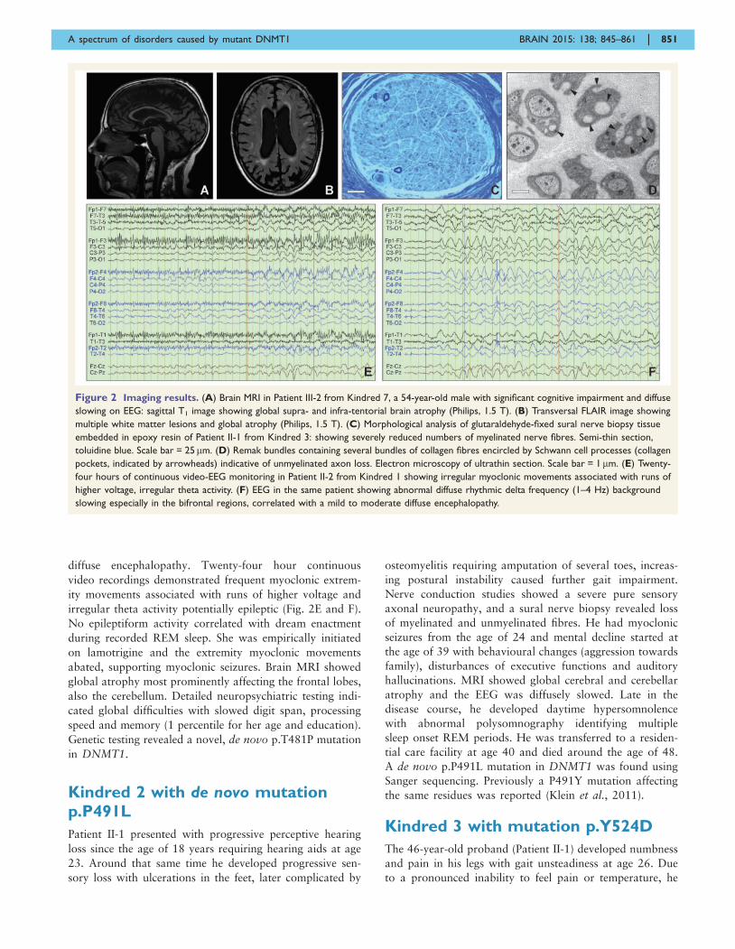
Figure 2 Imaging results. (A) Brain MRI in Patient III-2 from Kindred 7, a 54-year-old male with significant cognitive impairment and diffuse
slowing on EEG: sagittal T1 image showing global supra- and infra-tentorial brain atrophy (Philips, 1.5 T). (B) Transversal FLAIR image showing
multiple white matter lesions and global atrophy (Philips, 1.5 T). (C) Morphological analysis of glutaraldehyde-fixed sural nerve biopsy tissue
embedded in epoxy resin of Patient II-1 from Kindred 3: showing severely reduced numbers of myelinated nerve fibres. Semi-thin section,
toluidine blue. Scale bar = 25mm. (D) Remak bundles containing several bundles of collagen fibres encircled by Schwann cell processes (collagen
pockets, indicated by arrowheads) indicative of unmyelinated axon loss. Electron microscopy of ultrathin section. Scale bar = 1mm. (E) Twenty-
four hours of continuous video-EEG monitoring in Patient II-2 from Kindred 1 showing irregular myoclonic movements associated with runs of
higher voltage, irregular theta activity. (F) EEG in the same patient showing abnormal diffuse rhythmic delta frequency (1–4 Hz) background
slowing especially in the bifrontal regions, correlated with a mild to moderate diffuse encephalopathy.
A spectrum of disorders caused by mutant DNMT1 BRAIN 2015: 138; 845–861 | 851

developed multiple injuries in the feet and hands followed
by ulcerations. In addition he developed bilateral hearing
loss starting at the age of 33. At the time of last examin-
ation (35 years) there was no evidence of cognitive decline.
Nerve conduction studies confirmed a sensory neuropathy
and a sural nerve biopsy (Fig. 2C and D) was compatible
with a chronic axonal neuropathy affecting myelinated and
unmyelinated fibres with limited axonal regeneration. The
patient’s father also had a history of severe sensory neur-
opathy with ulcerations and hearing loss. He died at the
age of 43 due to chronic renal failure attributed to amyl-
oidosis secondary to chronic wound infection. A novel
p.Y524D mutation in DNMT1 was found using Sanger
sequencing.
Kindred 4 with mutation p.I531N
The proband (Patient II-1) presented at age 37 with a 5-year
history of gait instability that first became apparent with a
painless fracture of her left foot with neurogenic arthropa-
thy. Three years earlier, she noted bilateral hearing loss that
required hearing aids. Audiometry confirmed sensorineural
hearing loss. She had significant and progressive sensory loss
resulting in the need to use a walking aid and the loss of the
ability to drive. Nerve conduction studies revealed pure sen-
sory axonal neuropathy. She graduated college and worked
as an accountant for 10 years. However, at the age of 36 she
noted her thinking and communication increasingly became
difficult. Clinical examinations revealed normal alertness,
comprehension and orientation with moderate impairment
in repetition and reduction in fluency. She also exhibited
excessive daytime somnolence with sleep attacks. Formal
sleep evaluation diagnosed obstructive sleep apnoea with
severe dry eyes, but REM onset sleep and narcolepsy were
not diagnosed.
Her father passed away at age 45 following 20 years of
similar illness that started with distal loss of sensation and
hearing loss. He had blacking out spells and episodes of loss
of postural tone with generalized atonic seizures. EEG
showed frontal slowing but no epileptiform activity. He
was diagnosed with chorea and myoclonus and was noted
to have gait ataxia. Nerve biopsy revealed a severe sensory
axonal neuropathy. A novel DNMT1 mutation p.I531N
was identified in the patient, and not found in her
mother. The DNA of the deceased father was not available.
Kindred 5 with mutation p.Y495C
The 43-years-old patient (Patient II-1) developed sensory
loss and ulcerations in his feet at age 30, followed by pro-
gressive hearing loss at 36. Neurological examination
showed reduced pinprick and vibration sense and decreased
reflexes in the lower limbs. He reported painless left tibia
and fibula fractures at the age of 38, also complained of
excessive daytime sleepiness. Nerve conduction studies were
consistent with a pure sensory axonal neuropathy. Brain
MRI showed generalized cerebral and cerebellar atrophy.
Detailed neuropsychometric testing showed mild under-
functioning in the verbal domain and a severe
under-functioning in the performance domain. On focal
cognitive tests speed of information processing was ex-
tremely low. Polysomnography showed mild periodic limb
movement disorder, loss of REM atonia, disturbance of
multiple sleep latency tests but not a typical pattern for
narcolepsy. He had no family history of neurological dis-
eases, suggesting de novo origin, but the parents’ DNA
samples were not available for testing. Whole-exome
sequencing identified a previously reported, heterozygous
mutation p.Y495C in DNMT1 (Klein et al., 2011).
Kindred 6 with mutation p.C353F
The index patient (Patient III-3) developed progressive per-
ceptive hearing loss at 39 years of age. At age 45 she de-
veloped progressive gait difficulties due to balance
problems, and poorly healing ulcerations in the feet. She
remained cognitively intact until she suffered a haemor-
rhagic stroke at 63 years. Her elder brother had a similar
disease course with onset of hearing loss at 42 years and
gait instability 5 years later in combination with foot ulcer-
ations necessitating toe amputations. From the age of 52
onward there was mental decline with disturbance of ex-
ecutive functions in combination with visual and auditory
hallucinations. He is currently 73-years-old and undergoes
dialysis since renal failure 5 years ago. At that time he was
found to have a bilateral renal atrophy on ultrasound, but
kidney biopsy was not performed. There was no uncon-
trolled hypertension or other obvious causes of nephropa-
thy. Lymphoedema affecting the left leg was noted. Nerve
conduction studies in both siblings showed a pronounced
pure sensory axonal neuropathy. Their father, paternal
aunt and paternal grandmother all had a similar disease
course. Whole-genome sequencing in Patient III-3 and her
elder brother revealed a novel p.C353F mutation. Among
the younger generation with the mutation, Patient IV-1 has
gait instability at the age of 51. Audiometry showed mild
to moderate perceptive hearing loss. Patient IV-4 is cur-
rently 48-years-old and has asymmetric perceptive hearing
loss. Both showed normal nerve conduction studies.
Patients IV-2 and IV-3 do not carry the mutation, and
they have no symptoms or signs of the disease and have
normal nerve conduction studies and audiometry.
Kindred 7 with mutation p.Y495C
The 48-year-old proband (Patient IV-4) had the first nerve
conduction studies at age 33 when she was asymptomatic
because her mother had a severe sensory neuropathy, and
showed reduced sensory nerve amplitudes. At age 38, she
presented with a Charcot neuroarthropathy of the left
ankle and subsequently had numerous neuropathic ulcers
on both feet. Repeat nerve conduction studies showed
absent sensory nerve responses. She required a right
below knee amputation for an infected, necrotic foot
852 | BRAIN 2015: 138; 845–861 J. Baets et al.

ulcer at age 42. At age 43, she complained of progressive
hearing loss. Audiology revealed moderate to severe sen-
sorineural hearing loss and hearing aids were advised. At
age 47, her family raised concerns about her cognitive func-
tion and behaviour. She had obvious short-term memory
impairment, difficulties caring for her 8-year-old daughter
and maintaining the household. On the Addenbrooke’s
Cognitive Exam ACE-R, she scored 66/100 (normal486/
100). She had poor recall, verbal fluency and visuospatial
abilities. Her EEG showed paroxysmal predominantly pos-
terior delta wave activity. Her MRI showed severe general-
ized cerebral and cerebellar atrophy, but no white matter
lesions. Many of her relatives also had slowly progressive
sensory neuropathy, hearing loss, gait ataxia, and cognitive
problems, often with death in their 50s. Her cousin, Patient
IV-3 has dysarthria and three subcortical white matter T2
hyperintense lesions in her brain MRI (Fig. 2A and B). Her
uncle (Patient III-2) had global atrophy and widespread
white matter changes on MRI. Patient III-5 also had an
MRI consistent with demyelination. The maternal grand-
mother was diagnosed with early onset frontal temporal
dementia at age 40, along with sensory neuropathy, hear-
ing loss and gait ataxia, and she died at age 54. DNA
testing of the index, her affected cousin and uncle revealed
the previously reported DNMT1 mutation p.Y495C
(Klein et al., 2011).
Kindred 8 with mutation p.Y495H
The index patient (Patient II-4) was first seen at the age of
47 and diagnosed with peripheral sensory neuropathy after
experiencing a non-healing cuneiform fracture and loss of
sensation in her foot for 2 years, and confirmed by nerve
conduction studies. At current age of 56, she has pro-
gressed to a severe hearing loss, as documented by audiom-
etry, and showed cognitive decline in addition to her
progressive neuropathy. In retrospect, her hearing loss
and cognitive decline were likely present at the time of ini-
tial evaluation, but not appreciated and reported. Her af-
fected brother Patient II-7 was also evaluated and noted to
have severe peripheral neuropathy with foot ulcers, psychi-
atric complaints, cognitive decline, hearing loss, and lym-
phoedema. In addition, her other siblings suffered a similar
disease course consisting of severe peripheral neuropathy
with foot ulcers, psychiatric complaints, cognitive decline,
hearing loss, and lymphoedema. The patient’s father, two
uncles and three siblings all died in their early 50’s due to
various complications of the disease. A previously reported
p.Y495H mutation in DNMT1 was found using conven-
tional Sanger sequencing in the index patient and confirmed
in one definitely affected sibling (Klein et al., 2013).
Kindred 9 with mutation p.Y495C
The 47-year-old male proband (Patient IV-4) was first
noticed with hearing decline around age 30. By 38, he
was diagnosed with sensorineural hearing loss and had to
wear hearing aids. Around the same time, he was also
diagnosed with sleep apnoea and started to use a sleep
apnoea machine. Between 39 and 43, his family noticed
that he started to change jobs frequently, and his employers
mentioned his behaviour became increasingly irrational.
His family also noticed that he would spend a lot more
time on simple things and became obsessed with small de-
tails. His balance declined progressively resulting in a grad-
ual need for walking aids. He had ulcers on his feet and
nerve conduction studies confirmed prominent sensory
neuropathy. His long-term memory has not been affected,
but he had an increasingly difficult time having normal
conversations as he cannot stay focused on a topic. FTD
was considered as he had predominance of frontal-execu-
tive dysfunction and behavioural symptoms (disinhibition,
agitation, anger outbursts, hyper-religiosity), but not for-
mally diagnosed. Brain MRI revealed frontally predomin-
ant but distributed cerebral cortical and cerebellar atrophy.
His sister who started to experience visual hallucinations
recently, and many other affected family members, had
similar disease course. Death typically occurred in their
40’s. DNA testing found a previously reported DNMT1
mutation p.Y495C in the patient and his affected sister.
Summary
Among these nine kindreds, seven different mutations
(C353F, T481P, P491L, Y524D, I531N, Y495C, and
Y495H) were found, of which five (C353F, T481P,
P491L, Y524D, I531N) are novel. T481P (Kindred 1),
and P491L (Kindred 2) arose de novo in the index patients,
Y495C in Kindred 5 was also likely de novo as neither
parents had any phenotypes. In the other kindreds, the
transmitted mutations segregated with the disease. All
seven mutations reside in the TS domain of DNMT1, six
of them are very close to the previously described hotspot
mutant site Y495 (Klein et al., 2013), except C353F which
resides at the very edge of the domain (Fig. 3). The other
mutations are all positioned at the middle part of TS
domain where a highly-conserved region has been identified
essential for the binding of heterochromatin (Easwaran
et al., 2004). All seven mutations are predicted as ‘deleteri-
ous’ by SIFT (score: 0) ‘probably damaging’ by PolyPhen-2
(P = 1), also ‘disease-causing’ by MutationTaster (P = 1).
Using this cohort of 45 affected patients, we estimated
the average age of onset is 37.7 years (SD = 8.6, range
18–51), and recognized hearing loss as the most common
initial symptom (36%), followed by sensory loss, ulcer-
ations and/or arthropathy (33%), cognitive decline (7%)
and gait imbalance (7%). As the disease progresses, hearing
loss and ulceromutilating pure sensory neuropathy become
obligatory features. Gait difficulties are often present, but
are typically attributed to sensory ataxia and/or wound
infections rather than muscle weakness. Cerebellar involve-
ment, as demonstrated by cerebellar atrophy, may contrib-
ute to the ataxia. The degree of cerebral involvement is also
variable; the majority of patients display cognitive decline
A spectrum of disorders caused by mutant DNMT1 BRAIN 2015: 138; 845–861 | 853

(89%), ranging from overt (frontal and temporally predom-
inant) dementia to milder cognitive deficit features. In our
cohort of 45 patients, only 11% remain free of significant
cognitive decline after age 45. Dementia seems to be the
main factor driving the reduced life expectancy: the average
age of death at 53.6 years (SD = 7.7, range: 43–75). Brain
atrophy, was observed in 12 of 14 patients who underwent
MRI exam. Most often global atrophy, in some cases se-
lective frontal or cerebellar atrophy, was observed.
Additionally, new phenotypic features were identified
including myoclonus (in three kinships), seizures (in three
kinships), and auditory or visual hallucinations (in four
kinships) that lead to consideration of schizophrenia
(Table 1).
DNMT1 mutants are mislocalized tothe cytosol and form aggregates
The maintenance of methylation is a continuous process
extending to G2 phase, and DNMT1 binding to hetero-
chromatin is essential for the maintenance of DNA methy-
lation (Easwaran et al., 2004; Schermelleh et al., 2007;
Spada et al., 2007). During S phase, multiple proteins,
such as PCNA and UHRF1, recruit DNMT1 to the repli-
cation foci, where DNMT1 functions as an essential com-
ponent of the DNA replication machinery (Leonhardt et al.,
1992). DNMT1 is also continuously loaded onto hetero-
chromatin during G2 phase, and partial deletion of the TS
domain abolishes DNMT1 association with heterochroma-
tin (Easwaran et al., 2004). In our previous study using
HeLa cells, we demonstrated the heterochromatin binding
abilities of Y495C-DNMT1 and P491Y-DNMT1 are abol-
ished during G2 phase, even though these mutants
co-localize with PCNA at replication foci during S phase
(Klein et al., 2011). To investigate the characteristics of
newly identified novel mutants, we used confocal
microscopy to analyse HEK293 cells that had been co-
transfected to express various mutation constructs of
GFP-DNMT1 and S-phase marker RFP-PCNA. Similar to
our previous results, we found that C353F, T481P, P491L,
Y524D, I531N, Y495C, and Y495H mutants were co-loca-
lized with PCNA to replication foci during S phase. After S
phase, however, when RFP-PCNA diffuses and is no longer
presents in toroid structures within nucleus, these mutants
were mislocalized to cytosol, often in aggregates (Fig. 4).
We also noticed that RFP-PCNA proteins exist in the ag-
gregates structures, suggesting they are being
co-translocated to cytoplasm while still bound to DNMT1.
DNMT1 mutant proteins formaggresomes and are prone todegradation
Aggresomes are juxtanuclear inclusion bodies that have
been proposed to function as intermediate structures for
the disposal of protein aggregates. They are often induced
by protein homeostasis imbalance when cells sequester
toxic misfolded proteins from the cytoplasm and facilitate
their degradation through the autophagy pathway.
Aggresomes formed by pathological proteins have been
discovered in multiple neurodegenerative diseases, such as
synphilin-1 (SNCAIP), HTT and copper-zinc superoxide
dismutase (SOD1) in Parkinson’s disease, Huntington’s dis-
ease and amyotrophic lateral sclerosis (ALS), respectively
(Ross and Poirier, 2004). To determine whether the
observed mutant DNMT1 aggregates in cytosol are actual
aggresomes, we stained the cells transfected with various
constructs of EGFP-DNMT1 with red fluorescent molecular
marker (ProteoStat�, Enzo Life Science) that specifically
labels aggresome. The results showed that the red-fluores-
cent aggresomal marker overlaid with the mutant GFP-
DNMT1 protein aggregates in the cytoplasm, whereas
wild-type DNMT1 stayed within the nucleus. These results
demonstrate the DNMT1 mutants form aggresomes
(Fig. 5A), suggesting that lysosome-autophagy pathway is
likely involved in removing misfolded mutant DNMT1. It
has been suggested that the misfolded nucleus proteins are
translocated and sequestered in the cytoplasm for degrad-
ation to reduce its toxicity (Yamamoto and Simonsen,
2011). DNMT1 mutation-induced translocation of
DNMT1 proteins from the nucleus to the cytosol could
impose extracellular toxic stress. Previously we reported
that Y495C-DNMT1 and P491Y-DNMT1 mutant proteins
are prone to early degradation (Klein et al., 2011). To in-
vestigate whether the newly identified DNMT1 mutant pro-
teins are also subject to early degradation in cytosol, we
treated HEK293 cells with cycloheximide and quantified
the DNMT1-GFP fusion protein with a GFP protein quan-
tification assay. The amount of GFP tagged DNMT1 mu-
tants was significantly reduced compared to wild-type
DNMT1 (Fig. 5B), ranging from 40–80% compared to
wild-type.
Figure 3 Targeting sequence (TS) domain structure with
mutations of DNMT1 shown. The TS domain (grey) is the site
for all known causal mutations for HSAN1E (red) and ADCA-DN
(blue). Images were created using PyMol (www.pymol.org) and
crystal structures of the human DNMT1 TS domain.
854 | BRAIN 2015: 138; 845–861 J. Baets et al.

Figure 4 Representative images from confocal microscopy in HEK293 cells co-transfected with RFP-PCNA and GFP-wild-type
DNMT1, or various mutant GFP-DNMT1. Wild-type and mutant DNMT1 appear in green in the left panels, PCNA appears in red in the
middle panels and merged images are shown in the right panels. Scale bars = 5 mm. In the S phase, PCNA locates at the toroidal structures of the
replication foci; in the G2 phase, PCNA shows a diffused pattern in the nucleus. Both wild-type DNMT1 and mutant DNMT1 proteins co-localize
with PCNA at replication foci during the S phase. However, during G2 phase (when PCNA diffuses) wild-type DNMT1 stays in the nucleus and
binds to heterochromatin while mutant DNMT1 proteins mislocated into cytoplasm and formed aggregates.
A spectrum of disorders caused by mutant DNMT1 BRAIN 2015: 138; 845–861 | 855

DiscussionThe current study expands the clinical phenotypes of
HSAN1E, the number of causal mutations, and provides
evidence of a higher than previously expected HSAN1E
occurrence in clinical practice. We also identified an add-
itional potential pathogenic mechanism of mutant DNMT1
beyond its impact on genome methylation. The occurrence
of HSAN1E was initially thought to be quite low as only
four kindreds were found during the initial genetic
Figure 5 DNMT1 mutants. (A) Representative images from fluorescence aggresomes marker staining of HEK293 cells transfected with
either enhanced GFP-wild type DNMT1 or various mutant enhanced GFP-DNMT1. Wild-type DNMT1 stays in the nucleus and binds to het-
erochromatin whereas mutant DNMT1 aggregates in cytosol overlaid with red fluorescence aggresome markers. Scale bars = 5 mm. (B)
Quantification of GFP-DNMT1 proteins in HEK293 cells transfected with eGFP-wild type DNMT1, or various mutant enhanced GFP-DNMT1. All
mutant DNMT1 proteins showed degradation comparing to wild-type DNMT protein. Data are derived from three independent experiments,
asterisk represents there is statistically significant change between wild-type and mutant proteins.
856 | BRAIN 2015: 138; 845–861 J. Baets et al.

discovery (Klein et al., 2011). However, after reporting the
causal mutations in DNMT1, we quickly identified two
more kindreds with HSAN1E, both caused by a mutation
at position Y495, suggesting Y495 as a hotspot for muta-
tions (Klein et al., 2013). Subsequently, additional DNMT1
mutations within the TS domain were reported in patients
with narcolepsy, expanding DNMT1 mutation-associated
phenotypes (Winkelmann et al., 2012). Herein, we report
nine newly identified HSAN1E kindreds, totalling 45 pa-
tients, all with pathogenic mutations within the TS domain.
In addition, five novel mutations in DNMT1 are dis-
covered, further expanding the phenotype–genotype correl-
ation of DNMT1 to HSAN1E.
Kindreds 2, 3, 6 and 8 are part of a previously reported
cohort of 100 unrelated HSAN index patients who were
systematically screened for the known HSAN genes at that
time (Rotthier et al., 2009). This clinically heterogeneous
cohort also included patients with early disease onset and
recessive inheritance. When only taking those patients with
an adult disease onset into account, either in isolated cases
or in dominantly transmitting pedigrees, these four kin-
dreds represent 5.9% of the overall cohort. Among all
HSAN causal genes with autosomal dominant inheritance,
only RAB7A and SPTLC1 were reported with higher fre-
quencies (7% and 12%, respectively), although both are in
part overestimated due to founder effects in the studied
population. Among the recessively inherited HSAN genes,
NTRK1 accounts for a specific recognizable recessive syn-
drome and is the only one with frequency above 5% in the
screened cohort (Rotthier et al., 2009; Davidson et al.,
2012). Thus, among all 15 genes causal for HSAN,
DNMT1 seems to have a higher occurrence rate.
The marked morbidity and reduced life expectancy
underscore the importance of diagnosing HSAN1E, and
the unique combination of clinical features should help to
make the diagnosis. Previous reports described that patients
with HSAN1E have pan-sensory peripheral neuropathy
(myelinated and unmyelinated axons), hearing loss and de-
mentia as stereotypic symptoms, and present with different
initial symptoms at varied onset ages. In this largest
HSAN1E cohort study to date, we recognize that the
most frequent presenting symptom is hearing loss, followed
by sensory loss and its complications (ulcerations and
arthropathy). A minority of patients (7%) presented with
changes in mental status, but upon disease progression
nearly all patients developed clear cognitive decline that
often started with personality change. This study further
expanded the clinical spectrum of HSAN1E including audi-
tory and/or visual hallucinations, myoclonic seizures, renal
failure and frontotemporal dementia. In addition, diverse
parasomnias such as REM sleep disorder were appreciated.
Because of the variability and severity of presenting symp-
toms, HSAN1E patients were first seen in different subspe-
cialty clinics, where the focus was frequently given to the
most severe symptom, and the initial diagnosis emphasized
the most severe symptoms only. As hearing loss is the
common presenting symptom, and the course of sensory
neuropathy and cognitive decline is slowly progressive,
many patients did not visit a neurologist until they pro-
gressed to difficulties in walking, severe ulcerations, or no-
ticeable memory decline, or evident personality change.
Subtle changes in behaviour and personality in combination
with disturbances of short-term memory usually did not
prompt a specific diagnosis in the early stages of the dis-
ease. For some patients there were gaps of several years
between the appearances of each triad disease element.
The diverse clinical presentations are not surprising because
methylation maintenance is a fundamental aspect of DNA
replication and cell survival, although the predominance of
neuronal dysfunction is not explained.
Mutations in the TS domain of DNMT1 have also been
linked to ADCA-DN. A recent study reported the occur-
rence of narcolepsy in two HSAN1E families (Moghadam
et al., 2014) suggesting that narcolepsy is a common
phenotype for both ADCA-DN and HSAN1E. In our
cohort, 11 subjects displayed sleep disorders, most com-
monly hypersomnolence. The subsequent extensive clinical
testing of these 11 patients showed that only three had the
clinical feature sufficient to diagnose narcolepsy, and all
three had dementia. Establishing a diagnosis of narcolepsy
and a progressive encephalopathy, however, is not straight-
forward. Detailed questioning of patients and their families
often did not reveal symptoms of sleep disorders. In add-
ition, excessive daytime sleepiness might be part of the
global CNS phenotype, given the high rate of EEG delta
slowing in our cohort. In patients with ADCA-DN, a pri-
mary diagnosis of narcolepsy might have been easier to
establish as many patients were not reported to have sig-
nificant cognitive decline. It is possible, however, that a
more generalized encephalopathy was overlooked, as
some patients with ADCA-DN displayed marked atrophy
affecting multiple regions of the brain on MRI studies and
abnormalities on SPECT imaging. The results of EEGs have
not been reported for ADCA-DN patients (Moghadam
et al., 2014).
Except for the prominence of the narcolepsy, HSAN1E
and ADCA-DN patients have many similar phenotypes. All
patients with ADCA-DN had bilateral sensorineural hear-
ing loss that presented around the same time of the narco-
lepsy diagnosis, and mild to severe neuropathy typically
developed later. Both have similar disease onset age and
life-expectancy. Cognitive decline is seen in majority of
both HSAN1E and ADCA-DN patients by their late 40’s
(Table 2). Although severe cognitive decline seems to be the
main cause of early death among patients with HSAN1E,
the causes for premature death in ADCA-DN patients were
not reported. Taken together, these findings suggest that
the HSAN1E and ADCA-DN share much more overlapping
clinical features than their acronyms suggest, and we pro-
pose that a unified disease terminology such as ‘DNMT1-
complex disorder’ to better encompass both diseases.
Our functional study in cultured cells demonstrates that
the mutations in TS domain affect the localization of
DNMT1 protein. These DNMT1 mutants translocate out
A spectrum of disorders caused by mutant DNMT1 BRAIN 2015: 138; 845–861 | 857

Tab
le2
Co
mp
ari
son
of
the
clin
ical
featu
res
of
all
rep
ort
ed
fam
ilie
s/p
ati
en
tsw
ith
DN
MT
1m
uta
tio
ns
HS
AN
1E
Rep
ort
sA
DC
A-D
NR
ep
ort
s
Baets
et
al.
(cu
rren
tre
po
rt)
Kle
inet
al.,
2011
Kle
inet
al.,
2013
Yu
an
et
al.,
2013
Mo
gh
ad
am
et
al.,
2014
Win
kelm
an
net
al.,
2012
Ped
roso
et
al.,
2013
Focu
sof
the
study
Lar
geH
SAN
1E
cohort
study
with
in-d
epth
clin
ical
de-
scri
ption
of
nin
e
new
HSA
N1E
fam
ilies
and
pro
tein
funct
ional
assa
ysin
cell
culture
.
Initia
lge
netic
dis
cov-
ery
of
HSA
N1E,
limited
desc
ription
of
clin
ical
feat
ure
s
of
four
HSA
N1E
fam
ilies.
Genetic
and
clin
ical
studie
sof
two
new
HSA
N1E
fam
ilies
Singl
enew
HSA
N1E
case
In-d
epth
clin
ical
de-
scri
ption
of
two
AD
CA
-DN
fam
ilies
(als
oin
Win
kelm
ann
etal
.
report
)an
dtw
o
new
HSA
N1E
fam
ilies
Initia
lge
netic
dis
cov-
ery
for
AD
CA
-DN
.
Lim
ited
clin
ical
de-
scri
ption
of
four
AD
CA
-DN
fam
ilies.
Singl
enew
AD
CA
-DN
case
Num
ber
ofkin
dre
ds
(pat
ients
)
inve
stig
ated
9(4
5)
4(2
1)
2(6
)1
(1)
4(5
)4
(9)
1(1
)
Num
ber
of
muta
-
tions
(nove
l)
7(5
)2
(2)
2(1
)1
(1)
4(1
)3
(3)
1(1
)
Ave
rage
age
of
onse
t
Lat
e30’s
Ear
ly40’s
Ear
ly40’s
Lat
ete
en
Ear
ly30’s
Mid
30’s
toear
ly40’s
21
Com
mon
pre
senting
sym
pto
m
Hear
ing
loss
Hear
ing
loss
Hear
ing
loss
Senso
ryneuro
pat
hyN
arc
ole
psy
(AD
CA
-DN
)
Hea
rin
glo
ss
(HSA
N1
E)
Nar
cole
psy
Nar
cole
psy
Hear
ing
loss
Yes,
all
Yes,
all
Yes,
all
Yes
Yes,
all
Yes,
all
Yes
Senso
ryneuro
pat
hyYes,
all
Yes,
all
Yes,
all
Yes,
Yes,
all
Yes,
all
No
(age
32)
Nar
cole
psy
/
cata
ple
xy
3dia
gnose
dan
d11
report
ed
sleep
dis
ord
ers
.
Sym
pto
ms
not
re-
port
ed
by
pat
ients
,
but
were
not
the
focu
sof
inve
stig
atio
n.
No
Not
report
ed
Yes,
all
Yes,
all
Yes
Cogn
itiv
edecl
ine
or
dem
entia
Yes,
32/4
5fo
ur
hav
e
FTD
Yes,
13/2
1Yes,
all,
one
has
FTD
Mild
menta
l
reta
rdat
ion
Yes,
1/3
Yes,
4/9
no
Ata
xia
Yes,
5/4
5Yes,
4/2
1N
oN
oYes,
4/5
Yes,
all
Yes
Bra
inA
trophy
by
MR
I
Yes,
12/1
4perf
orm
ed
Yes,
5/5
perf
orm
ed
Yes,
4/4
perf
orm
ed
Yes
Yes,
all
Yes,
report
ed
intw
o
fam
ilies
Yes
Optic
atro
phy
Not
check
ed
Not
check
ed
Not
check
ed
Not
report
ed
Yes,
all
Yes,
7/9
no
Ave
rage
age
of
deat
h
Mid
50’s
Ear
ly60’s
Ear
ly60’s
Aliv
eat
42
Mid
50’s
Mid
50’s
Aliv
eat
32
EEG
/low
freq
uency
Yes,
5of
6perf
orm
ed
Not
check
ed
Yes,
1/1
perf
orm
ed
Not
report
ed
Not
report
ed
Not
report
ed
Not
report
ed
Hyp
ocr
etin
1N
ot
check
ed
Not
check
ed
Not
check
ed
Not
check
ed
No
rmal
in
HSA
N1
E
Lo
win
AD
CA
-DN
low
Slig
htly
low
Lym
phoedem
aYes,
3/4
5N
oN
oN
ot
report
ed
No
Yes,
2/9
Not
report
ed
858 | BRAIN 2015: 138; 845–861 J. Baets et al.

of the nucleus into the cytoplasm during G2 phase, and
form aggresomes in the cytoplasm. It has been suggested
that binding to replication machinery and hemimethylated
DNA require conformation change of the TS domain
(Frauer and Leonhardt, 2011). We speculate that
DNMT1 binding to replication foci during S phase may
have exacerbated the misfolding of mutant DNMT1, caus-
ing mutant proteins being recognized by the chaperone fac-
tors that are involved in transporting misfolded protein out
of nucleus. These findings suggest that mutants have dom-
inant effects by failing to interact with DNA in neurons
and by forming cytosolic aggregates. Of interest, although
DNMT1 mutations have been shown to cause global DNA
hypomethylation and site-specific hypermethylation (Klein
et al., 2011; Sun et al., 2014), a phenomenon often seen in
cancer, cancer has never been reported in any patients with
HSAN1E.
The misfolding and aggregation of DNMT1 mutant pro-
teins could contribute to the pathogenesis of HSAN1E
through a toxic gain-of-function, as has been shown for
other neurological diseases (Knaevelsrud and Simonsen,
2010). Although the issue of aggresome formation is still
under debate pertaining to whether protein misfolding is
the mechanism of toxic gain-of-function, and whether
aggresomes are part of compensatory cellular process.
The nuclear-to-cytoplasm transport of other nuclear pro-
teins, including HTT, ATXN1 and TARDBP mutants, has
been proposed as a pathogenic mechanism in their asso-
ciated neurodegenerative disorders (Levine and Kroemer,
2008). Cytoplasmic accumulated aggresomes often impose
toxic cellular stress (Saifi et al., 2003). The post-mitotic
neurons and the long length of peripheral nerves could be
particularly vulnerable to mutated protein misfolding and
the cellular stress arisen from it, and worsening with age
(Klein et al., 2013). Our study indicates that the mutations
of DNMT1 have deleterious impact on the central and per-
ipheral nervous system, emphasizing the importance of the
TS domain for maintaining proper DNMT1 function and
its associate epigenetic regulatory mechanism.
All mutations reported to date that cause either HSAN1E
or ADCA-DN are missense mutations in the TS domain.
Our results better differentiated the location distinction be-
tween mutations causal for ADCA-DN (located in the C-
terminus end of the TS domain) and for HSAN1E (located
in the middle part or N-terminus of TS domain). This large
cohort study suggests that the location and nature of the
mutations may influence the disease presentations and se-
verity. Two HSAN1E mutations, p.T481P (Kindred 1) and
p.P491L (Kindred 2), both from patients with de novo mu-
tations, had young onset age in their late teens, and the
disease course progressed rather quickly. Whereas one
HSAN1E mutation, p.C353F mutation (Kindred 6), may
be associated with a relatively slow disease progression
rate: the proband had no apparent cognitive problem
until she suffered a stroke at age 63, and the younger gen-
eration had later onset and milder symptoms. However, the
link between the location of mutation and phenotype
diversity is likely complex. The proband’s brother in
Kindred 6 had cognitive decline starting at age 52 along
with earlier presented hearing loss and sensory neuropathy,
illustrating that the phenotypic expressions vary consider-
ably even between siblings.
Our study recognized several new clinical features asso-
ciated with HSAN1E, including myoclonic seizures, and
auditory or visual hallucinations. In addition, two patients
had end-stage renal failure. Patient III-2 of Kindred 6
(C353F) is undergoing hemodialysis; the renal failure in
Patient I-1 of Kindred 3 (Y524D) was attributed to amyl-
oidosis due to chronic osteomyelitis, but this was not docu-
mented. A patient from the previously reported kindred
(Klein et al., 2011) with Y495C also died of renal failure
after chronic pyelonephritis; the kidneys showed an atro-
phied cortex and large numbers of nodules. At this point it
remains unclear if the renal involvement is an accidental
feature or in fact causally linked with DNMT1 mutations.
Moreover, lymphoedema was noted in several individuals
from Kindred 8 and one individual from Kindred 6. Of
interest, several individuals with ADCA-DN were previ-
ously reported to have lymphoedema (Winkelmann et al.,
2012; Moghadam et al., 2014). The direct causal link of
lymphoedema with the DNMT1 mutation remains to be
discovered. The diversity of the DNMT1-associated pheno-
types certainly suggests the identification of additional cases
and warrants further mechanistic investigations. In sum-
mary, our study suggests that DNMT1-related disorders
appeared to be more common than previously estimated,
and the extent of the phenotypic spectrum much broader
than currently appreciated, contributing to delayed diagno-
sis. We hope that this report will lead to more cases of
DNMT1-related disorders being recognized and diagnosed
by candidate gene testing rather than relatively expensive
whole-exome sequencing. At the same time, it will be inter-
esting to see if a greater range of neurodegenerative dis-
orders may also be linked with DNMT1 function as
whole-exome sequencing is increasingly being used for gen-
etic screening in clinical setting. Given the complexity of
DNMT1 function in mammalian cells, a substantial
amount of research is required to unravel its pathogenic
mechanisms. DNMT1 mutants seem to exert their dama-
ging impact by two complex pathways. First, epigenetic
pathways are impaired, leading to abnormal global methy-
lation in HSAN1E patients. Indeed, the disrupted binding
of heterochromatin and the early degradation of mutant
Y495C DNMT1 have been shown impairing global
genome methylation maintenance (Klein et al., 2011; Sun
et al., 2014). Second, misfolded mutant DNMT1 imposes
cellular stress by putting protein homeostasis network out
of balance, which may eventually lead to cell death. The
fact that DNMT1 mutations underlie such a unique yet
variable clinical spectrum underscores the super-stringent
requirement for the highly conserved DNMT1 protein.
This is not entirely surprising as DNMT1 is the sole essen-
tial enzyme that keeps the fidelity of methylation inherit-
ance starting from embryogenesis to the very end of life.
A spectrum of disorders caused by mutant DNMT1 BRAIN 2015: 138; 845–861 | 859

AcknowledgementsWe thank all patients and their relatives for their willing-
ness to participate in this study.
FundingThis study was supported by the National Institute of
Health (NIH) (NS065007, DP3DK104394,
R01DK064814, U10NS077305), and American Diabetes
Association (ADA) 7-11-AEC-23 of USA, the University
of Antwerp (UA), the Association Belge contre les
Maladies Neuromusculaires (ABMM), the Medical
Foundation Queen Elisabeth (GSKE), the agency for
Innovation by Science and Technology (IWT) and the EU
FP7/2007-2013 under grant agreement number 2012-
305121 (NEUROMICS) and Judy Seltzer Levenson
Memorial Fund for CMT Research. MMR and ML are
grateful to the Medical Research Council (MRC), MRC
Centre grant (G0601943), and the National Institutes of
Neurological Diseases and Stroke and office of Rare
Diseases (U54NS065712) for their support. Part of this
work was undertaken at University College London
Hospitals/University College London, which received a pro-
portion of funding from the Department of Health’s
National Institute for Health Research Biomedical
Research Centres funding scheme. J.W. is supported by
the German Research Council (DFG WE1406/13-1). Dr
Baets receives research grant from Association Belge
contre les Maladies Neuro-Musculaires, Medical
Foundation Queen Elisabeth and EU 7th Framework
Programme (FP7, ‘NEUROMICS’); Dr Grossman sits on
the board of International FTD Society; Dr Dyck receives
an honorarium for serving as an Associate Editor of
Diabetes, and receives support for teaching of neuropathy
examinations from Alnylam and ISIS Pharmaceutical com-
panies; Dr Houlden receives research grant from MRC UK,
The Wellcome Trust and NIHR UCL/UCLH BRC; Dr
Seeley receives research grant from National Institute of
Aging, John D. French Alzheimer’s Disease Foundation,
Consortium for Frontotemporal Dementia Research,
James S. McDonnell Foundation, Alzheimer’s Drug
Discovery Foundation and serves as a consultant for
Bristol-Myers Squibb. Dr Klein, Dr Smith and Dr Scherer
receive research grants from National Institute of
Neurological Disorders and Stroke. Other co-authors
have nothing to disclose.
ReferencesChoi AM, Ryter SW, Levine B. Autophagy in human health and dis-
ease. N Engl J Med 2013; 368: 1845–6.
Davidson G, Murphy S, Polke J, Laura M, Salih M, Muntoni F, et al.
Frequency of mutations in the genes associated with hereditary sen-
sory and autonomic neuropathy in a UK cohort. J Neurol 2012;
259: 1673–85.
Drmanac R, Sparks AB, Callow MJ, Halpern AL, Burns NL,
Kermani BG, et al. Human genome sequencing using unchained
base reads on self-assembling DNA nanoarrays. Science 2010;
327: 78–81.Easwaran HP, Schermelleh L, Leonhardt H, Cardoso MC.
Replication-independent chromatin loading of Dnmt1 during G2
and M phases. EMBO Rep 2004; 5: 1181–6.Frauer C, Leonhardt H. Twists and turns of DNA methylation. Proc
Natl Acad Sci USA 2011; 108: 8919–20.
Jin B, Robertson KD. DNA methyltransferases, DNA damage repair,
and cancer. Adv Exp Med Biol 2013; 754: 3–29.
Klein CJ, Benarroch EE. Epigenetic regulation: basic concepts and
relevance to neurologic disease. Neurology 2014; 82: 1833–40.Klein CJ, Bird T, Ertekin-Taner N, Lincoln S, Hjorth R, Wu Y,
et al. DNMT1 mutation hot spot causes varied phenotypes of
HSAN1 with dementia and hearing loss. Neurology 2013; 80:
824–8.
Klein CJ, Botuyan MV, Wu Y, Ward CJ, Nicholson GA, Hammans S,
et al. Mutations in DNMT1 cause hereditary sensory neuropathy
with dementia and hearing loss. Nat Genet 2011; 43: 595–600.
Klein CJ, Duan X, Shy ME. Inherited neuropathies: clinical overview
and update. Muscle Nerve 2013; 48: 604–22.Klein CJ, Wu Y, Kruckeberg KE, Hebbring SJ, Anderson SA,
Cunningham JM, et al. SPTLC1 and RAB7 mutation analysis in
dominantly inherited and idiopathic sensory neuropathies. J
Neurol Neurosurg Psychiatry 2005; 76: 1022–4.
Knaevelsrud H, Simonsen A. Fighting disease by selective autophagy of
aggregate-prone proteins. FEBS Lett 2010; 584: 2635–45.Leonhardt H, Page AW, Weier HU, Bestor TH. A targeting sequence
directs DNA methyltransferase to sites of DNA replication in mam-
malian nuclei. Cell 1992; 71: 865–73.Levine B, Kroemer G. Autophagy in the pathogenesis of disease. Cell
2008; 132: 27–42.
Margot JB, Aguirre-Arteta AM, Di Giacco BV, Pradhan S, Roberts RJ,
Cardoso MC, et al. Structure and function of the mouse DNA
methyltransferase gene: Dnmt1 shows a tripartite structure. J Mol
Biol 2000; 297: 293–300.Marques SC, Oliveira CR, Pereira CM, Outeiro TF. Epigenetics in
neurodegeneration: a new layer of complexity. Prog
Neuropsychopharmacol Biol Psychiatry 2011; 35: 348–55.Moghadam KK, Pizza F, La Morgia C, Franceschini C, Tonon C,
Lodi R, et al. Narcolepsy is a common phenotype in HSAN IE
and ADCA-DN. Brain 2014; 137(Pt 6): 1643–55.Pedroso JL, Povoas Barsottini OG, Lin L, Melberg A, Oliveira AS,
Mignot E. A novel de novo exon 21 DNMT1 mutation causes
cerebellar ataxia, deafness, and narcolepsy in a Brazilian patient.
Sleep 2013; 36: 1257–9, 9A.
Ross CA, Poirier MA. Protein aggregation and neurodegenerative dis-
ease. Nat Med 2004; ; 10(Suppl): S10–7.Rossor AM, Polke JM, Houlden H, Reilly MM. Clinical implications
of genetic advances in Charcot-Marie-Tooth disease. Nat Rev
Neurol 2013; 9: 562–71.Rotthier A, Baets J, De Vriendt E, Jacobs A, Auer-Grumbach M,
Levy N, et al. Genes for hereditary sensory and autonomic neuro-
pathies: a genotype-phenotype correlation. Brain 2009; 132(Pt 10):
2699–711.
Rotthier A, Baets J, Timmerman V, Janssens K. Mechanisms of disease
in hereditary sensory and autonomic neuropathies. Nat Rev Neurol
2012; 8: 73–85.
Saifi GM, Szigeti K, Snipes GJ, Garcia CA, Lupski JR. Molecular
mechanisms, diagnosis, and rational approaches to management of
and therapy for Charcot-Marie-Tooth disease and related peripheral
neuropathies. J Investig Med 2003; 51: 261–83.
Schermelleh L, Haemmer A, Spada F, Rosing N, Meilinger D,
Rothbauer U, et al. Dynamics of Dnmt1 interaction with the repli-
cation machinery and its role in postreplicative maintenance of DNA
methylation. Nucleic Acids Res 2007; 35: 4301–12.
860 | BRAIN 2015: 138; 845–861 J. Baets et al.

Song J, Rechkoblit O, Bestor TH, Patel DJ. Structure of DNMT1-DNA complex reveals a role for autoinhibition in maintenance
DNA methylation. Science 2011; 331: 1036–40.
Spada F, Haemmer A, Kuch D, Rothbauer U, Schermelleh L,
Kremmer E, et al. DNMT1 but not its interaction with the replica-tion machinery is required for maintenance of DNA methylation in
human cells. J Cell Biol 2007; 176: 565–71.
Sun Z, Wu Y, Ordog T, Baheti S, Nie J, Duan X, et al. Aberrant
Signature Methylome by DNMT1 hot spot mutation in hereditarysensory and autonomic neuropathy 1E. Epigenetics 2014; 9:
1184–93.
Takalo M, Salminen A, Soininen H, Hiltunen M, Haapasalo A.Protein aggregation and degradation mechanisms in neurodegenera-
tive diseases. Am J Neurodegener Dis 2013; 2: 1–14.
Winkelmann J, Lin L, Schormair B, Kornum BR, Faraco J, Plazzi G,et al. Mutations in DNMT1 cause autosomal dominant cerebel-
lar ataxia, deafness and narcolepsy. Hum Mol Genet 2012; 21:
2205–10.
Yamamoto A, Simonsen A. The elimination of accumulated and aggre-gated proteins: a role for aggrephagy in neurodegeneration.
Neurobiol Dis 2011; 43: 17–28.
Yuan J, Higuchi Y, Nagado T, Nozuma S, Nakamura T, Matsuura E,
et al. Novel mutation in the replication focus targeting sequencedomain of DNMT1 causes hereditary sensory and autonomic neu-
ropathy IE. J Peripher Nerv Syst 2013; 18: 89–93.
Zimon M, Baets J, Almeida-Souza L, De Vriendt E, Nikodinovic J,Parman Y, et al. Loss-of-function mutations in HINT1 cause axonal
neuropathy with neuromyotonia. Nat Genet 2012; 44: 1080–3.
A spectrum of disorders caused by mutant DNMT1 BRAIN 2015: 138; 845–861 | 861