definition: primary malignant tumors of lymphoid tissue, they represent 5% of all malignant tumors....
TRANSCRIPT

Definition: Definition:
Primary malignant tumors of lymphoid Primary malignant tumors of lymphoid tissue, they represent 5% of all malignant tissue, they represent 5% of all malignant tumors.tumors.
Incidence:Incidence:
Male: Female HD. Male: Female HD. 2:12:1 N.H.L. 1.4 : 1N.H.L. 1.4 : 1

According to the U.S. National Institutes of Health, lymphomas account for about five percent of all cases of cancer in the United States, and Hodgkin's lymphoma in particular accounts for less than one percent of all cases of cancer in the United States.

Because the whole system is part of the body's immune system, patients with a weakened immune system, such as from HIV infection or from certain drugs or medication, also have a higher incidence of lymphoma.

1- Hodgkin1- Hodgkin’’s disease :s disease :
a-a-Infectious aetiology (may be initiated by an infection but Infectious aetiology (may be initiated by an infection but may not be transmitted)may not be transmitted)
**may be hypersensitive immune response to a virus.may be hypersensitive immune response to a virus. **relation to Ebstein Barr virus as evidenced relation to Ebstein Barr virus as evidenced by:by:
-- +ve antibodies +ve antibodies-- Increase incidence in-patient with inf. Increase incidence in-patient with inf. OnonucleosisOnonucleosis
b-b-Abnormal immune responseAbnormal immune response- - depression of cellular immunity should be depression of cellular immunity should be considered an inherent character of the patient considered an inherent character of the patient who ultimatIy develop H.Dwho ultimatIy develop H.D
2-Non Hodgkin2-Non Hodgkin’’s lymphoma:s lymphoma: ** Neoplasia of the immune system Neoplasia of the immune system
** Can be considered is a disorder of lymphoid Can be considered is a disorder of lymphoid differentiation.differentiation.
Aetiology of Predisposing factors:Aetiology of Predisposing factors:

Possible Aetiologic factors of NHL:Possible Aetiologic factors of NHL:I-Host Factors:-I-Host Factors:-
a-a- Primary Primary Immune deficiencyImmune deficiency b-b- Immuno suppressive ttt. Immuno suppressive ttt.
2-Invironmental Factors:-2-Invironmental Factors:-
a-a- I Ionizing radiations.onizing radiations. b-b-DrugsDrugs: : (Hydantion, Anaesthesia)(Hydantion, Anaesthesia) c-c- Chemicals: Chemicals: (Petrol. Viny1 Chloride)(Petrol. Viny1 Chloride) d-d- E.B.V E.B.V

Age IncidenceAge Incidence-:-:

1- H.D.:1- H.D.:
Disease of Lymphatic tissue characterized by the Disease of Lymphatic tissue characterized by the presence of Reed Sternberg cells and variable proliferations presence of Reed Sternberg cells and variable proliferations of lymphocytes.of lymphocytes.
*Rye Classification*Rye ClassificationSub Type Average %Sub Type Average %
Lymphatic predominance (L.P.) Lymphatic predominance (L.P.) 10 – 12% 10 – 12%Nodular sclerosis (N.S.)Nodular sclerosis (N.S.) 45 - 55% 45 - 55%Mixed Cellularity (MC.)Mixed Cellularity (MC.) 30 - 35% 30 - 35%Lymphocytic Deplition (L.D.)Lymphocytic Deplition (L.D.) 8- 10% 8- 10%
L.P., or, N.S. are more favourable than M.C and L.D.L.P., or, N.S. are more favourable than M.C and L.D.
Histopathology:-Histopathology:-

Classifications of
Non Hodgkin’s Lymphoma

a-Rappaport classification:- depends on:a-Rappaport classification:- depends on:
1-1-Pattern of growth:-Pattern of growth:- -- Nodular (N.) [ Follicular, Indolent] Nodular (N.) [ Follicular, Indolent] -- Diffuse (D.) [Aggressive] Diffuse (D.) [Aggressive]
2-2- Cell population: Cell population: - - Lymphocytic (L.)Lymphocytic (L.)- - Histiocytic (H.)Histiocytic (H.)- - Mixed (L & H.)Mixed (L & H.)
TypesTypes : :** Favorable histology: N.L.,N. Mixed, DL.W.D(well differentiated) Favorable histology: N.L.,N. Mixed, DL.W.D(well differentiated)**Unfavorable histology: N.H. + all D., except DL. W.DUnfavorable histology: N.H. + all D., except DL. W.D** Histioocytic of several varities: Histioocytic of several varities:
-- majority (large cell L. immunoblastic L.) majority (large cell L. immunoblastic L.) -- true H is very rare true H is very rare
Many classifications according toHistopathology:-Many classifications according toHistopathology:-

B-A number of different classification systems exist for lymphoma.
: Kiel Karl Lennert of Kiel, Germany, proposed
a new system of classifying lymphomas based on cellular morphology and their relationship to cells of the normal peripheral lymphoid system
:REALIn the mid 1990s, the Revised European-American Lymphoma (REAL) Classification attempted to apply immunophenotypic and genetic features in identifying distinct clinicopathologic NHL entities
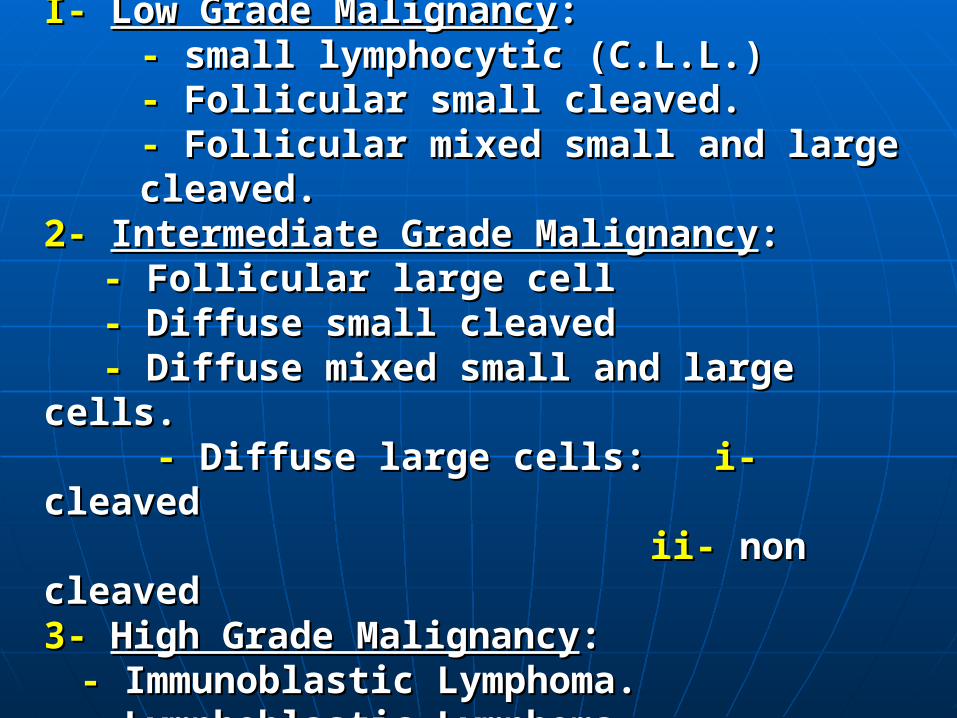
C-C-Working FormulationWorking Formulation::I- I- Low Grade MalignancyLow Grade Malignancy::
-- small lymphocytic (C.L.L.) small lymphocytic (C.L.L.)-- Follicular small cleaved. Follicular small cleaved.-- Follicular mixed small and large cleaved. Follicular mixed small and large cleaved.
2- 2- Intermediate Grade MalignancyIntermediate Grade Malignancy:: -- Follicular large cell Follicular large cell
-- Diffuse small cleaved Diffuse small cleaved -- Diffuse mixed small and large cells. Diffuse mixed small and large cells.
-- Diffuse large cells: Diffuse large cells: i-i- cleaved cleaved ii-ii- non cleaved non cleaved
3-3- High Grade MalignancyHigh Grade Malignancy::-- Immunoblastic Lymphoma. Immunoblastic Lymphoma.-- Lymphoblastic Lymphoma Lymphoblastic Lymphoma
- - Small non cleaved (Burkitt’s)Small non cleaved (Burkitt’s)

*In NHL:*In NHL: The presence of a The presence of a nodular pattern (follicular) nodular pattern (follicular)
remains an important remains an important prognostic feature in addition prognostic feature in addition
to the cell typeto the cell type

Classification: D-WHOThe WHO Classification, published in 2001 and updated in 2008, is the latest classification of lymphoma . This system attempts to group lymphomas by cell type (i.e. the normal cell type that most resembles the tumour) and defining phenotypic, molecular or cytogenetic characteristics .

There are three large groups: the B cell, T cell, and natural killer cell tumours. Other less common groups, are also recognized. Hodgkin's lymphoma, although considered separately within the WHO classifications, is now recognized as being a tumour of, albeit markedly abnormal, lymphocytes of mature B cell lineage

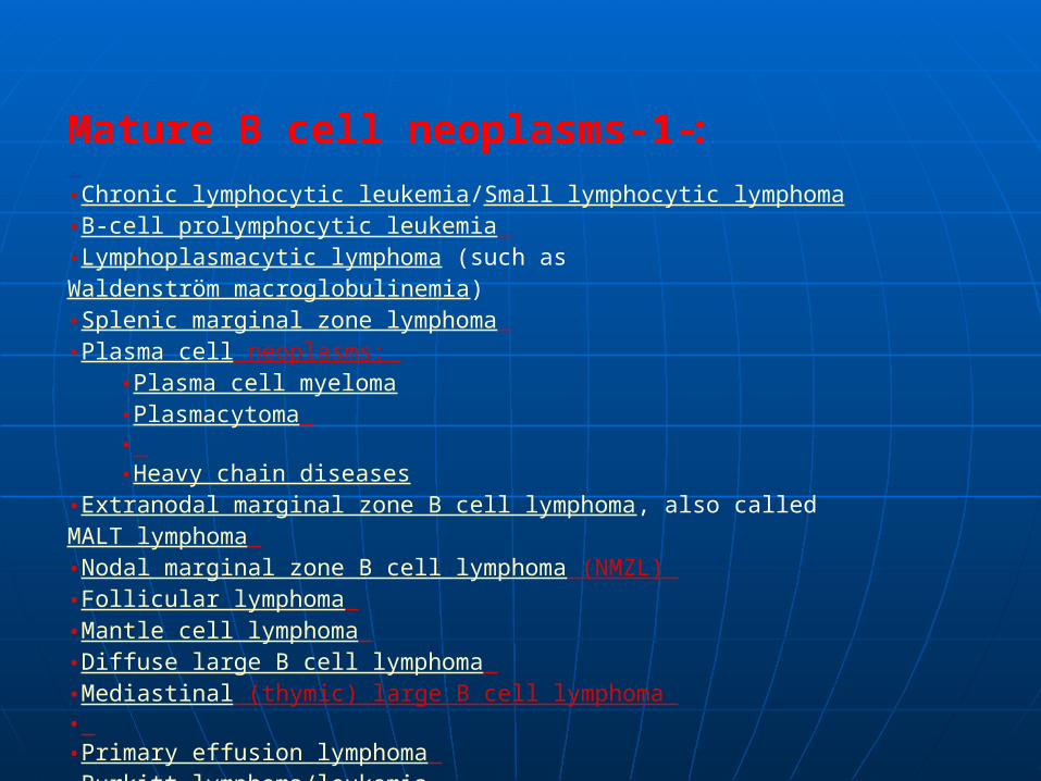
:-1-Mature B cell neoplasms.•Chronic lymphocytic leukemia/Small lymphocytic lymphoma •B-cell prolymphocytic leukemia •Lymphoplasmacytic lymphoma (such as Waldenström macroglobulinemia) •Splenic marginal zone lymphoma •Plasma cell neoplasms:
•Plasma cell myeloma •Plasmacytoma • •Heavy chain diseases
•Extranodal marginal zone B cell lymphoma, also called MALT lymphoma •Nodal marginal zone B cell lymphoma (NMZL) •Follicular lymphoma •Mantle cell lymphoma •Diffuse large B cell lymphoma •Mediastinal (thymic) large B cell lymphoma • •Primary effusion lymphoma •Burkitt lymphoma/leukemia

-2-Mature T cell and natural killer (NK) cell :neoplasmsT cell prolymphocytic leukemia T cell large granular lymphocytic leukemia Aggressive NK cell leukemia
Adult T cell leukemia/lymphoma Extranodal NK/T cell lymphoma, nasal type Enteropathy-type T cell lymphoma Hepatosplenic T cell lymphoma Blastic NK cell lymphoma Mycosis fungoides / Sezary syndrome Primary cutaneous CD30-positive T cell lymphoproliferative disorders Primary cutaneous anaplastic large cell lymphoma Lymphomatoid papulosis Angioimmunoblastic T cell lymphoma Peripheral T cell lymphoma, unspecified Anaplastic large cell lymphoma

-3-Hodgkin lymphomaClassical Hodgkin lymphomas :
*Nodular sclerosis Mixed cellularity
*Lymphocyte-rich *Lymphocyte depleted or not
depleted *Nodular lymphocyte-predominant
Hodgkin lymphoma

-4-Immunodeficiency-associated :lymphoproliferative disorders
*Associated with a primary immune disorder *Associated with the Human Immunodeficiency
Virus (HIV) *Post-transplant
*Associated with methotrexate therapy

General ConsiderationsGeneral Considerations::
I-H-D.:I-H-D.: ** usually have a unicentric origin & usually usually have a unicentric origin & usually starts starts by by involvement involvement L.N. inL.N. in one or more one or more a adjacent areas djacent areas esp. in cervical region.esp. in cervical region. N.H.L. :N.H.L. : ** usually multicentric from the start. usually multicentric from the start. ** in 1/3 onset occur in extranodal sites. in 1/3 onset occur in extranodal sites.2- Mode of spread:2- Mode of spread:
H. D.H. D. ** in the great majority via lymphatics to in the great majority via lymphatics to contagious L.N. and other lymphatic structurescontagious L.N. and other lymphatic structures
N.H.L. : *N.H.L. : * more often rapidly spread to distant nodal more often rapidly spread to distant nodal and extranodal sites via blood streamand extranodal sites via blood stream

1- Asymptomatic Lymphadenopathy:-1- Asymptomatic Lymphadenopathy:-
** the majority of patients with H.D. (extranodal the majority of patients with H.D. (extranodal only in 10%) .only in 10%) .** 2/3 of patients with N.H.L. (1/3 extranodal). 2/3 of patients with N.H.L. (1/3 extranodal).Usually in cervical L.N.(cervical onset 65- 80%, Usually in cervical L.N.(cervical onset 65- 80%, in H.D. ,while in N.H.L. only in 30 - 40%).in H.D. ,while in N.H.L. only in 30 - 40%).
a-H.D.:a-H.D.:
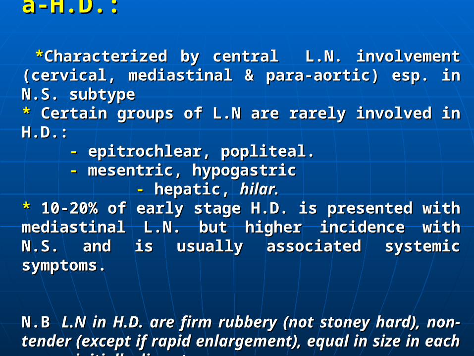
**Characterized by central L.N. involvement (cervical, Characterized by central L.N. involvement (cervical, mediastinal & para-aortic) esp. in N.S. subtypemediastinal & para-aortic) esp. in N.S. subtype** Certain groups of L.N are rarely involved in H.D.: Certain groups of L.N are rarely involved in H.D.:
-- epitrochlear, popliteal. epitrochlear, popliteal. -- mesentric, hypogastric mesentric, hypogastric -- hepatic, hepatic, hilar.hilar.** 10-20% of early stage H.D. is presented with mediastinal L.N. 10-20% of early stage H.D. is presented with mediastinal L.N. but higher incidence with N.S. and is usually associated systemic but higher incidence with N.S. and is usually associated systemic symptoms.symptoms.
N.B N.B L.N in H.D. are firm rubbery (not stoney hard), non-tender L.N in H.D. are firm rubbery (not stoney hard), non-tender (except if rapid enlargement), equal in size in each group, initially (except if rapid enlargement), equal in size in each group, initially discrete.discrete.
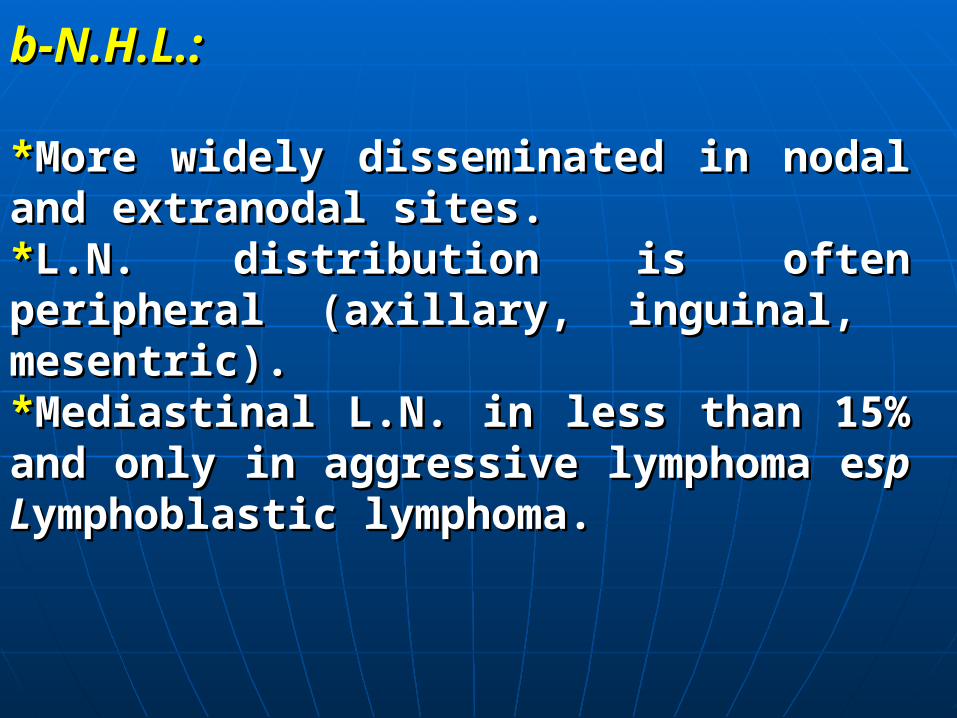
b-N.H.L.:b-N.H.L.:
**More widely disseminated in nodal and More widely disseminated in nodal and extranodal sites.extranodal sites.**L.N. distribution is often peripheral (axillary, L.N. distribution is often peripheral (axillary, inguinal, mesentric).inguinal, mesentric).**Mediastinal L.N. in less than 15% and only in Mediastinal L.N. in less than 15% and only in aggressive lymphoma eaggressive lymphoma esp Lsp Lymphoblastic ymphoblastic lymphoma.lymphoma.

2-Extra Nodal Lesions:2-Extra Nodal Lesions:
*In N.H.L.:*In N.H.L.: the most commonly involved sites are the the most commonly involved sites are the head & neck areas (Waldeyer’s ring,head & neck areas (Waldeyer’s ring, nas nasal cavities, al cavities, sinuses & orbit) followed by G.I.T.sinuses & orbit) followed by G.I.T.
*In H.D.:*In H.D.: splenic & hepatic involvement esp. in splenic & hepatic involvement esp. in presence of :-L.N enlargement above and below the presence of :-L.N enlargement above and below the diaphragm. diaphragm. -systemic symptoms-systemic symptoms - and in M.C and L.D. subtypes.- and in M.C and L.D. subtypes.
N.B Hepatic N.B Hepatic involvement usually in association with involvement usually in association with concomitant splenic involvement.concomitant splenic involvement.

a-G.I.T.:a-G.I.T.:** diarrhoea , malabsorption syndrom ( G.I.T. infiltration) diarrhoea , malabsorption syndrom ( G.I.T. infiltration)** ulceration & bleeding. ulceration & bleeding.** retroperitoneal m retroperitoneal masses esp. in N.H.L. asses esp. in N.H.L. resulting in I.resulting in I.V.C V.C obstruction, leading to ascitis and oedema.obstruction, leading to ascitis and oedema.
Primary GI N.H.L:Primary GI N.H.L:-- Adults: stomach Adults: stomach >> intestine. intestine.-- Children: intestine Children: intestine >> stomach esp. for Burkitt’s stomach esp. for Burkitt’s (iliocaecal valve)(iliocaecal valve)--There is association between affection of Waldeyer's ring There is association between affection of Waldeyer's ring & G.I. lymphoma esp. the stomach.& G.I. lymphoma esp. the stomach.
Jaundice in H.D: may be due to:Jaundice in H.D: may be due to: -- intrahepatic cholestasis: involvement of portal tracts. intrahepatic cholestasis: involvement of portal tracts. - - extrahepatic cholestasis due to glands in porta hepatis.extrahepatic cholestasis due to glands in porta hepatis. -- auto immune haemolytic anaemia. auto immune haemolytic anaemia.

b- Chestb- Chest::
** involvement of lung parenchyma (nodules, involvement of lung parenchyma (nodules, cavitations).cavitations). ** usually due to infiltration by contiguity from usually due to infiltration by contiguity from adenopathy .adenopathy . ** Pleural and / or pericardial involvement (usually with Pleural and / or pericardial involvement (usually with massive mediastinal L.N. enlargement.)massive mediastinal L.N. enlargement.) ** massive mediastinal L.N. enlargement leading to massive mediastinal L.N. enlargement leading to mediastinal syndrome esp. with :mediastinal syndrome esp. with :
- - N.S. (H.D.)N.S. (H.D.)-- Lymphoblastic (N.H.L.) Lymphoblastic (N.H.L.)

C-Bone Pain & Tenderness:C-Bone Pain & Tenderness:
** may be oesteolytic or oesteoblastic and may be may be oesteolytic or oesteoblastic and may be pathological fractures.pathological fractures.
d- Neurological Manifestation;d- Neurological Manifestation;
** Herpes Zoster. Herpes Zoster.** Spinal cord compression (H.D.) . Spinal cord compression (H.D.) .** Peripheral nerve palsies. [Horner's, brachial plexus, Peripheral nerve palsies. [Horner's, brachial plexus, phrenic nerve, vocal cords].phrenic nerve, vocal cords].** Cranial Cranial nerve palsies.nerve palsies.** Cerebral signs and symptoms Cerebral signs and symptoms

e-Cutaneous Manifestations:-
* Pruritis * Skin nodules.* Herpes zoster * Alopecia and icthyosis * Hyperpigmentation.

3- Systemic or constitutional symptoms:3- Systemic or constitutional symptoms:
** May May occur with L.N. enlargement or may precedes it.occur with L.N. enlargement or may precedes it.** Common with H.D. (30-40%) than N.H.L. (10%). Common with H.D. (30-40%) than N.H.L. (10%). * * Fever > 38 ,night sweats, wt . loss, pruritis, other systemic Fever > 38 ,night sweats, wt . loss, pruritis, other systemic
symptoms.symptoms.
** Alcohol intolerance (H.D.): Alcohol intolerance (H.D.):- - localized localized acute acute pain at one or more sites of Hodgkin's pain at one or more sites of Hodgkin's
involvement involvement - - anorexia, malaise. fatigueanorexia, malaise. fatigue** Systemic symptoms means unfavorable prognosis. Systemic symptoms means unfavorable prognosis.
Diagnostic Evaluation and Staging:Diagnostic Evaluation and Staging: Proper ttt depends on:Proper ttt depends on:
-- Hisopathologic Type. Hisopathologic Type. -- Anatomic extent of Anatomic extent of disease (staging)disease (staging)

**Diagnosis:Diagnosis:
-- The precise unequivocal diagnosis requires The precise unequivocal diagnosis requires histopathologic confirmation by the examination of histopathologic confirmation by the examination of suitable biopsy material.suitable biopsy material.
-- Most satisfactory nodes: (lower cervical & Most satisfactory nodes: (lower cervical & axillary).axillary).
-- If there is a group of L.N. take the central ones. If there is a group of L.N. take the central ones.
- - If there is only mediastinal L.N.:(mediastinascopy If there is only mediastinal L.N.:(mediastinascopy or thoracotomy).or thoracotomy).
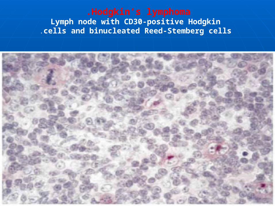
Hodgkin’s lymphoma .Lymph node with CD30-positive Hodgkin
cells and binucleated Reed-Stemberg cells.
Immunophenotyping is an important diagnostic modality and is crucial for the classifi cation of non-Hodgkin’s lymphoma. Certain markers, such as the CD-20 receptor, have become crucial for treatment as newer biologic agents have utilized this receptor therapeutically .

Characteristic cellSurface antigens
EBV
Reed-Sternberg cell
CD15+,CD30+, CD20+/–, CD45–,
EMA–
+EBV in 50%
Lymphocytic and histiocytic cell “popcorn cells”
CD15–, CD30–, CD20+, CD45+,
EMA+EBV–
Classical HLNLPHL
NLPHL is the most favorable HL with an indolent course.

Clinical StaginClinical Stagingg::
I-I- Detailed history esp. systemic symptoms Detailed history esp. systemic symptoms..22-- Clinical exam. including Waldeyer's ring, areas of Clinical exam. including Waldeyer's ring, areas of
bone tendernessbone tenderness..33-- Adequate Adequate surgical biopsysurgical biopsy
44-- Routine laboratory tests(CBC,ESR. liver function test, Routine laboratory tests(CBC,ESR. liver function test, serum uric acid)serum uric acid)
55-- Plain chest X ray & bilateral lower extremity Plain chest X ray & bilateral lower extremity lymphangiographylymphangiography..
66-- Radiologic examination of GIT & gastroscopy. If Radiologic examination of GIT & gastroscopy. If +ve +ve Waldeyer's ringWaldeyer's ring..

Imaging studiesLaboratory tests
–CT scan of chest, abdomen, –Complete blood
and pelviscount
–FDG-PET scan or PET/CT –Serum chemistry
–Gallium-67 scan (if PET –Lactate dehydro
unavailable(genase (LDH)
–MRI or CT of the brain –Liver function tests
)if symptomatically indicated( –Renal function tests
–Bone scan –Erythrocyte sedi-
)if symptomatically indicated(mentation rate (ESR)
Imaging and laboratory work-ups for non-Hodgkin’s lymphoma
■

Positron emission tomography (PET) using fluorodeoxyglucose has been found useful for the staging and follow-up of patients with HL. PET is sensitive and, in most instances, specific enough to detect involvement by HL. In untreated patients, a higher stage is found in at least 20% of cases using PET imaging as compared with conventional imaging. PET may also be used in patients with residual tumor masses to discriminate

Pathologic staging:Pathologic staging:1-1- Bone marrow biopsy Bone marrow biopsy2-2- Staging Staging laparotomy, laparotomy, after -ve bone after -ve bone narrow narrow biopsy in Clinical stage I & II H.D.biopsy in Clinical stage I & II H.D.3-3- Lumbar puncture with cytologic exam. Of Lumbar puncture with cytologic exam. Of C.S.F in all N.H.L with bone marrow C.S.F in all N.H.L with bone marrow involvement.involvement.4- 4- Cytologic exam. of Cytologic exam. of any any effusioneffusion
The The Ann Arbor staging Ann Arbor staging classificationclassification :mentioned before. :mentioned before.

Bone marrow biopsy (bilateral) is recommended for all cases of NHL because of the high probability of bone marrow involvement for certain types of NHL in : 70% in small lymphocytic lymphoma, 50% in follicular lymphoma, and >10% in diffuse large-cell lym-phoma or marginal zone lymphoma. Cytological examination of cranial spinal fluid (CSF) is indicated for stage IV disease with bone marrow, testis, and parameningeal involvement.

Stage I
Stage II
Stage III
Stage IV
Involvement of single lymph node (I) or extralymphatic site (IE)
Involvement of two or more involved lymph node sites on the same side of the diaphragm (II) or localized involvement of one extra-lymphatic organ or site plus one or more lymph node regions on the same side of diaphragm (IIE)
Involvement of lymph node regions on both sides of diaphragm (III), which can also include involvement of the spleen (IIIS) or localized extralymphatic site or organ extension (IIIE) or both (IIISE)
Diffuse (multifocal) involvement of one or more extralymphatic organs or sitesA=No “B” symptoms B=Unexplained fever >38°C, weight loss >10% in previous 6 months, drenching night sweats X=Bulky disease
Ann Arbor staging system
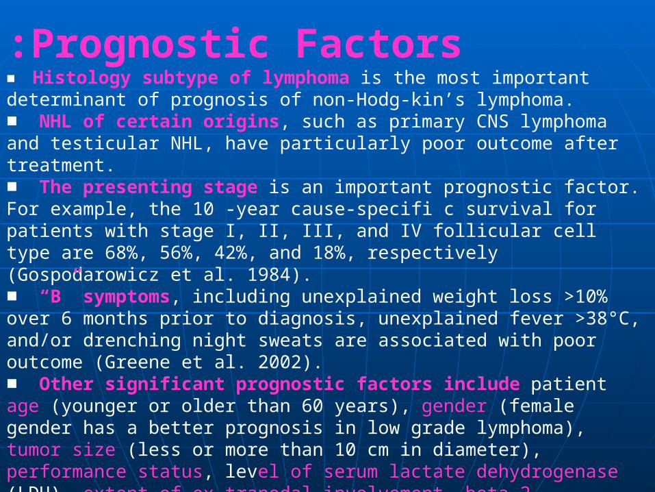
Prognostic Factors:■ Histology subtype of lymphoma is the most important determinant of prognosis of non-Hodg-kin’s lymphoma.■ NHL of certain origins, such as primary CNS lymphoma and testicular NHL, have particularly poor outcome after treatment.■ The presenting stage is an important prognostic factor. For example, the 10 -year cause-specifi c survival for patients with stage I, II, III, and IV follicular cell type are 68%, 56%, 42%, and 18%, respectively (Gospodarowicz et al. 1984).■ “B” symptoms, including unexplained weight loss >10% over 6 months prior to diagnosis, unexplained fever >38°C, and/or drenching night sweats are associated with poor outcome (Greene et al. 2002).■ Other significant prognostic factors include patient age (younger or older than 60 years), gender (female gender has a better prognosis in low grade lymphoma), tumor size (less or more than 10 cm in diameter), performance status, level of serum lactate dehydrogenase (LDH), extent of ex-tranodal involvement, beta-2 microglobulin, and S-phase fraction.

The International Prognostic Index (IPI) for aggressive NHL includes five of the above-mentioned significant risk factors to predict overall survival : 1-stage (I or II vs. III or IV),2- serum LDH (normal vs. abnormal), 3-extranodal site involvement (0 or 1 vs. >1),4- age of the patient (younger than 60 vs. older than 60),5- and performance status (ECOG 0 or 1 vs. 2-4).

The IPI risk groups are determined by the numerical summation of the number of adverse risk factors (0 to 5), and a higher number of adverse risk factors are associated with poor prognosis

Risk groupIPI score
5-Year survival(%)
Low-risk Low-intermediate
High-intermediate
High-risk
0–1 2
3
4–5
73% 51%
43%
26%
International prognostic index for aggressive
non-Hodgkin’s lymphoma

The frequency subtypes of HD differs in different parts of the world. At present, with effective treatments for HL, the subtypes are no longer prognostically relevant. However, some of these types have particular clinical features: nodular sclerosis is more frequent in young women with a large mediastinal mass. The lymphocyte-predominant HL resembles a low-grade, B-cell lymphoma, and can be treated with limited irradiation at least in early stages.
Prognostic Factors for HD

Prognostic factors in HL are age, sex, stage, and some serum markers such as sedimentation rate and soluble CD25. Recently, a prognostic score was established for advanced HL. This score, into which seven unfavorable clinical and laboratory parameters , predicts treatment failure (low serum albumin, anemia, male sex, age > 45 yr, stage IV, leukocytosis, and lymphocytopenia).

Treatment Strategy:Treatment Strategy:
A Role of SurgerA Role of Surgery :y : Limited to five clinical situations: Limited to five clinical situations:
1-1- Initial diagnosis Initial diagnosis P.S. P.S.2-2- ttt of concurrent unrelated diseases ttt of concurrent unrelated diseases3-3- Complications of lymphomas as hypersplenism. Complications of lymphomas as hypersplenism.4-4- Extirpaition of disease involving L.N. as pressure Extirpaition of disease involving L.N. as pressure symptoms related to a localized L.N. enlargement not symptoms related to a localized L.N. enlargement not responding to local ttt.responding to local ttt.5-5- Management of G.I.T lymphoma (to decrease Management of G.I.T lymphoma (to decrease incidence of perforation or Hge due to rapid tumour incidence of perforation or Hge due to rapid tumour necrosis after necrosis after effective chemotherapy.)effective chemotherapy.)

B-Role of Radiotherapy:B-Role of Radiotherapy:
I- H.I- H.D.:D.: Now mainly in the form of involved field radiotherapy to localized Now mainly in the form of involved field radiotherapy to localized disease or in bulky lymphadenopathy in more advanced stagesdisease or in bulky lymphadenopathy in more advanced stages
High High incidence of recurrence after radiothincidence of recurrence after radiotherapy in:erapy in:
I- I- IIA with bulky mediastinal diseaseIIA with bulky mediastinal disease2- 2- Extra nodal involvement lEA , IIEA.Extra nodal involvement lEA , IIEA.3- 3- Mixed cellularity & lymphocytic depletion subtypes.Mixed cellularity & lymphocytic depletion subtypes.II- II- N. H. L.:N. H. L.:
* * compared to H.D. less firm guid lines for routine Iry radiotherapy compared to H.D. less firm guid lines for routine Iry radiotherapy can be provided in PS, I, II N.H.L.can be provided in PS, I, II N.H.L.** For localised NHL :PSI, II. For localised NHL :PSI, II.
-- low grade low grade regional extended radiotherapy (4500 R) regional extended radiotherapy (4500 R)-- aggressive aggressive radiotherapy + chemotherapyradiotherapy + chemotherapy
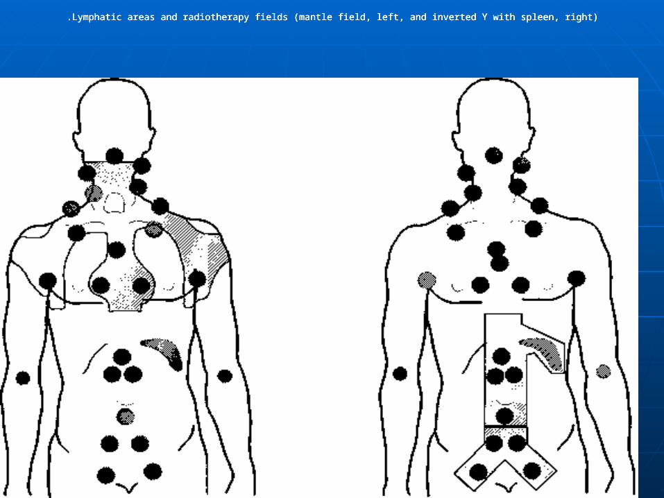
Lymphatic areas and radiotherapy fields (mantle field, left, and inverted Y with spleen, right).Lymphatic areas and radiotherapy fields (mantle field, left, and inverted Y with spleen, right).Lymphatic areas and radiotherapy fields (mantle field, left, and inverted Y with spleen, right).


C-ChemotherapyC-Chemotherapy::
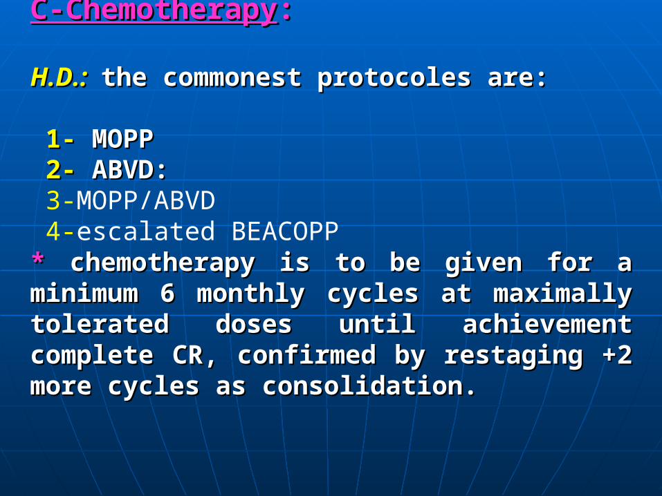
H.D.:H.D.: the commonest protocoles arethe commonest protocoles are::
1-1- MOPP MOPP 2-2- ABVD: ABVD: 3-MOPP/ABVD 4-escalated BEACOPP ** chemotherapy is to be given for a minimum 6 chemotherapy is to be given for a minimum 6 monthly cycles at maximally tolerated doses until monthly cycles at maximally tolerated doses until achievement complete CR, confirmed by restaging +2 achievement complete CR, confirmed by restaging +2 more cycles as consolidation.more cycles as consolidation.
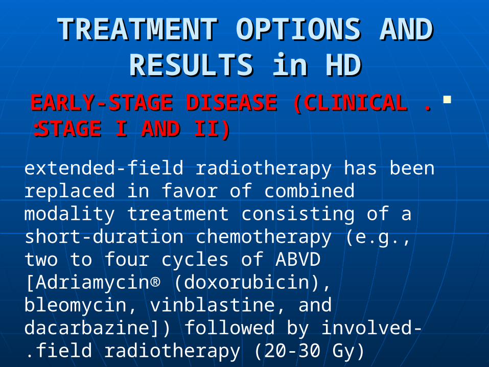
TREATMENT OPTIONS AND TREATMENT OPTIONS AND RESULTS in HDRESULTS in HD
. .EARLY-STAGE DISEASE EARLY-STAGE DISEASE (CLINICAL (CLINICAL ::STAGE I AND II)STAGE I AND II)
extended-field radiotherapy has been replaced in favor of combined modality treatment consisting of a short-duration chemotherapy (e.g., two to four cycles of ABVD [Adriamycin® (doxorubicin), bleomycin, vinblastine, and dacarbazine]) followed by involved-field radiotherapy (20-30 Gy).

.Advanced Stage HD(Clinical Stages III and IV) ABVD , The main advantage of ABVD alone is the relatively low incidence of long-term toxic effects as compared with alkylating agents-based regimens.
escalated BEACOPP regimen :
achieved an 87% freedom from progression and a 91% overall survival after 5 yr. Both schemes are toxic and therefore should only be administered in larger centers with much experience and within clinical trials

Non-Hodgkin’s Lymphoma Treatment of Stage I and II Indolent NHL:Radiation Therapy■ Radiation therapy is the mainstay treatment of stage I and II grade I and II follicular lymphoma, marginal zone lymphoma (non-gastric), and small lymphocytic lymphoma .
■IFRT delivers treatment to the clinically involved region
TREATMENT OPTIONS AND RESULTS
Treatment of Stage III & IV Indolent Lymphoma■ Asymptomatic patients with more advanced stage III or stage IV low-grade NHL can be closely monitored (watchful waiting) .
■ Rituximab is a “humanized” anti-CD20 monoclonal antibody that can be recommended for the treatment of indolent non-Hodgkin’s lymphoma (CD20 positive), and its efficacy in the treatment of relapsed or refractory indolent NHL has been repeatedly demonstrated .■ Combined rituximab and chemotherapy should be recommended for indolent NHL patients who have indication for treatment .
.

Indications for treatment include:1 -active symptoms
2-cytopenias3-progression of disease
4 -potential organ compromises .Results from trials comparing R-CHOP to CHOP and R-CVP to CVP revealed that overall chemoimmunotherapy appears to be superior to chemotherapy alone
Treatment of Stage III & IV Indolent Lymphoma
Treatment of Gastric Mucosa-Associated :(Lymphoid Tumors (MALT)
General Principles:
■ For H. Pylori-positive stage IE gastric MALT, antibiotic treatment of H. Pylori should be used as the initial treatment . Radiation therapy is an effective modality for definitive treatment of localized (stage IE or II) gastric MALT and is recommended for H. Pylori-negative cases, as well as for patients with deep invasion, active symptoms, or disease progression after antibiotic treatment .
■ For stage III or IV gastric MALT, chemotherapy and/or rituximab should be considered . Radiation therapy is indicated for local symptomatic control.■ Treatment strategy of the more commonly diagnosed large B-cell lymphoma of the stomach (comprises approximately 60% of all gastric lymphoma cases) is identical to that of the intermediate-grade NHL.
Treatment of Intermediate-Grade (Aggressive) Non-Hodgkin’s Lymphoma General Principles:■ Treatment strategies of the more commonly diagnosed aggressive NHL, including diffuse large B-cell lymphoma, grade III follicular lymphoma,peripheral T-cell lymphoma, and mantle-cell lymphoma, follow similar recommendations:■ For stage I and II aggressive NHL, CHOP-based chemotherapy followed by adjuvant IFRT is the standard treatment. Rituximab is indicated for CD20-positive large-cell non-Hodgkin’s lymphoma .■ For stage III and IV aggressive NHL, CHOP-based chemotherapy is the mainstay treatment. Rituximab is indicated for CD20 positive large-cell non-Hodgkin’s lymphoma.
COURSE AND PROGNOSIS:COURSE AND PROGNOSIS:
H.D.:H.D.:
-- 10 years actuarial survival has progressively increased from 10 years actuarial survival has progressively increased from 1% with no therapy to 70% with modern ttt.1% with no therapy to 70% with modern ttt.
N.H.L.:N.H.L.:
-- Modern chemotherapy progress have revolutionized the Modern chemotherapy progress have revolutionized the prognosis for many aggressive lymphomas esp., large cell type prognosis for many aggressive lymphomas esp., large cell type and Burkitt’s lymphoma.and Burkitt’s lymphoma.

Thank youThank you