dehydrated human amniotic tissue improves healing time, cost of care · 2020-03-13 · tional wound...
TRANSCRIPT

businessbriefsDon Fetterolf, MD & Rachel Savage
Human amniotic membrane al-lografts have been used in surgical procedures for more than 100 years.
Known to decrease inflammation, re-duce scar tissue formation, and support soft tissue regeneration, dehydrated hu-man amniotic membrane allografts also have been proven to reduce wound-clo-sure time, overall cost to treat wounds, and scarring. Clinicians who have gone through basic training for chronic wound management are all taught that lower extremity compression is the “gold stan-dard” for management of ulcers caused by chronic venous insufficiency, as long as there is no co-existing arterial disease significant enough to prevent the use of compression therapy.
ROLE OF AMNIOTIC MEMBRANEHuman amniotic membrane is non-
immunogenic, non-vascular tissue com-prising the innermost layer of the pla-centa (the amnion and the chorion). Composed of a single layer of epithelial cells, a basement membrane, and an avas-cular connective tissue matrix, amniotic membrane contains extracellular matrix (including collagen and laminins), cell-signaling proteins (such as cytokines), and growth factors that are essential to the healing process. Amniotic mem-brane layers also consist of epithelium cells (lining of hollow organs and glands that protect or enclose); a thick, compact layer (composed of reticular fibers); and a fibroblast layer. The membrane also con-tains cell-anchoring collagen types IV, V, and VII — structural proteins that are es-sential for wound healing.
In vitro testing confirms presence of essential soft tissue growth factors and cytokines in human amniotic membrane
allografts. Growth factors bind to the ex-tracellular matrix and are released into surrounding tissue, providing a continual release of growth factors during the tis-sue regeneration process.
Unlike many of the xenografts and composite dermal substitutes on the market today, dehydrated human amni-otic membrane allografts may be used in a wide variety of applications that can reduce the need to carry products in in-ventory. Wound healing applications for dehydrated amniotic membrane include acute and chronic full- and partial-thick-ness wounds such as diabetic foot ulcers, venous leg ulcers, arterial ulcers, pressure ulcers, post-surgical or post traumatic wounds, wound dehiscence, burn inju-ries, acute suture line repairs, and sub-cutaneous wound tunnel repair. With a variety of sizes available, the waste typi-cally realized with other grafts is reduced significantly. Many diabetic foot ulcers may be less than 4 sq cm, so sizing op-tions are important.
IMPROVING HEALING TIME In 2007, Surgical Biologics (Kennesaw,
GA) developed the PURION® process for the use of dehydrated amniotic mem-brane as an allograft. First utilized in oph-thalmic surgery (there have been more than 45,000 implants to date without any adverse events associated with dehydrat-ed amniotic membrane), amniotic mem-brane has been utilized more recently as a potent facilitator of wound healing in various fields, including lower extremity ulcers, ophthalmological surgery, burns, gynecologic surgery, orthopedics, and a variety of other applications.1-7
The process works by safely and gently separating the placental tissues. The vari-
ous layers of the amniotic membrane are cleaned and reassembled with minimal manipulation of the tissue to maintain the structure. The tissue is then dehy-drated to preserve the elements that are key to healing, with no chemicals being utilized. Dehydrated human amniotic membrane has a shelf life of five years and may be micronized to create a pow-der configuration to be used as topical powder or injectable solution. The final product may be stored at room tempera-ture and is regulated by the FDA under section 361 of the Public Health Service Act as Human Cells, Tissues, & Cellu-lar and Tissue Products. Placentas are recovered only by scheduled Caesarean section procedures, and each donation is subject to FDA compliant screening cri-teria and blood testing.
Among other benefits, the PURION process allows tissue to be dehydrated and sterilized, producing an easy-to-use graft. To date, 100,000 allografts have been distributed for human implanta-tion in various surgical applications, and a number of recent studies have demon-strated the clinical cost effectiveness of using dehydrated human amniotic mem-brane allografts. One prospective, strati-fied, randomized, comparative, parallel group, single-center clinical trial com-pared the proportion of diabetic foot ulcers completely healed by use of de-hydrated amniotic membrane graft (Epi-Fix,® MiMedx Group Inc., Kennesaw, GA) every other week, plus standard of care (SOC) versus a SOC protocol of ad-vanced wound care dressings in patients living with a nonhealing diabetic foot ulcer with adequate arterial perfusion. Following surgical debridement, all pa-tients underwent weekly dressing chang-
DEHYDRATED HUMAN AMNIOTIC TISSUE IMPROVES HEALING TIME, COST OF CARE
www.todayswoundclinic.com Today’s Wound Clinic® January/February 2013 19 Leased and used with permission from HMP Communications, LLC from October 2013 through October 2014.
amnioticallografts
20 January/February 2013 Today’s Wound Clinic® www.todayswoundclinic.com
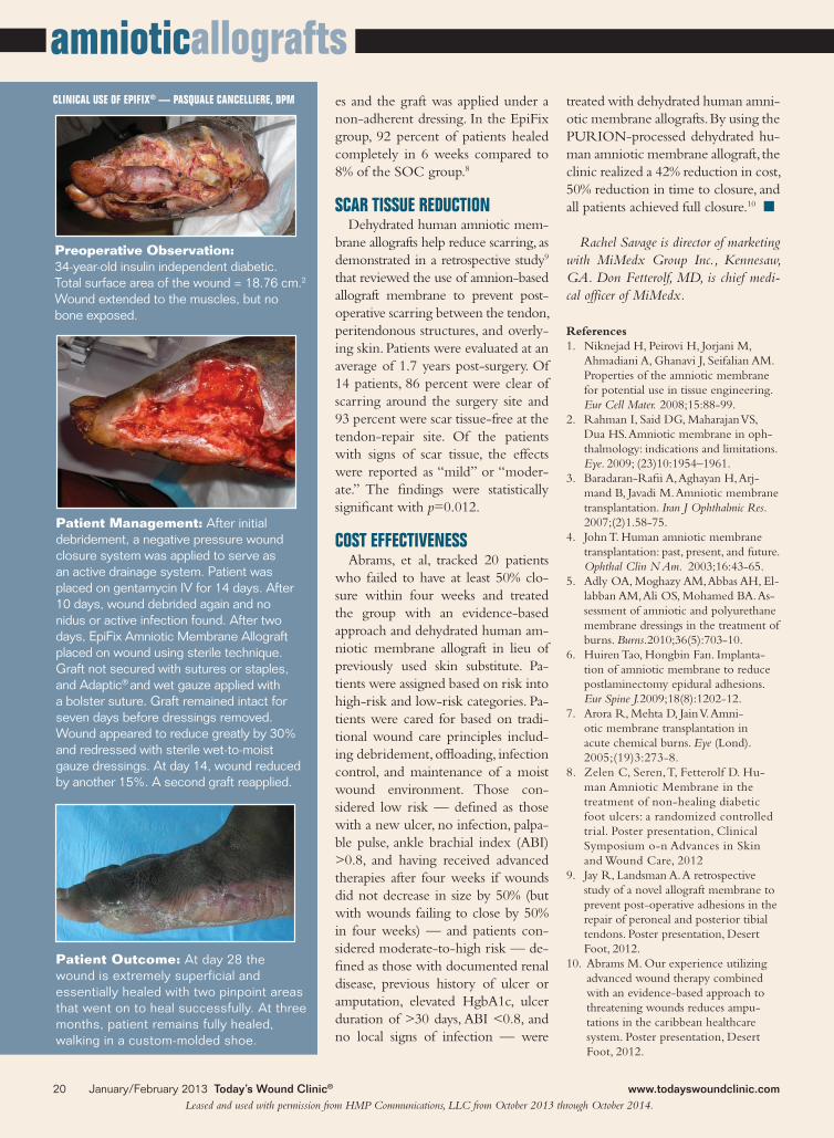
Preoperative Observation: 34-year-old insulin independent diabetic. Total surface area of the wound = 18.76 cm.2 Wound extended to the muscles, but no bone exposed.
Patient Management: After initial debridement, a negative pressure wound closure system was applied to serve as an active drainage system. Patient was placed on gentamycin IV for 14 days. After 10 days, wound debrided again and no nidus or active infection found. After two days, EpiFix Amniotic Membrane Allograft placed on wound using sterile technique. Graft not secured with sutures or staples, and Adaptic® and wet gauze applied with a bolster suture. Graft remained intact for seven days before dressings removed. Wound appeared to reduce greatly by 30% and redressed with sterile wet-to-moist gauze dressings. At day 14, wound reduced by another 15%. A second graft reapplied.
es and the graft was applied under a non-adherent dressing. In the EpiFix group, 92 percent of patients healed completely in 6 weeks compared to 8% of the SOC group.8
SCAR TISSUE REDUCTIONDehydrated human amniotic mem-
brane allografts help reduce scarring, as demonstrated in a retrospective study9
that reviewed the use of amnion-based allograft membrane to prevent post-operative scarring between the tendon, peritendonous structures, and overly-ing skin. Patients were evaluated at an average of 1.7 years post-surgery. Of 14 patients, 86 percent were clear of scarring around the surgery site and 93 percent were scar tissue-free at the tendon-repair site. Of the patients with signs of scar tissue, the effects were reported as “mild” or “moder-ate.” The findings were statistically significant with p=0.012.
COST EFFECTIVENESSAbrams, et al, tracked 20 patients
who failed to have at least 50% clo-sure within four weeks and treated the group with an evidence-based approach and dehydrated human am-niotic membrane allograft in lieu of previously used skin substitute. Pa-tients were assigned based on risk into high-risk and low-risk categories. Pa-tients were cared for based on tradi-tional wound care principles includ-ing debridement, offloading, infection control, and maintenance of a moist wound environment. Those con-sidered low risk — defined as those with a new ulcer, no infection, palpa-ble pulse, ankle brachial index (ABI) >0.8, and having received advanced therapies after four weeks if wounds did not decrease in size by 50% (but with wounds failing to close by 50% in four weeks) — and patients con-sidered moderate-to-high risk — de-fined as those with documented renal disease, previous history of ulcer or amputation, elevated HgbA1c, ulcer duration of >30 days, ABI <0.8, and no local signs of infection — were
treated with dehydrated human amni-otic membrane allografts. By using the PURION-processed dehydrated hu-man amniotic membrane allograft, the clinic realized a 42% reduction in cost, 50% reduction in time to closure, and all patients achieved full closure.10 n
Rachel Savage is director of marketing with MiMedx Group Inc., Kennesaw, GA. Don Fetterolf, MD, is chief medi-cal officer of MiMedx.
References1. Niknejad H, Peirovi H, Jorjani M,
Ahmadiani A, Ghanavi J, Seifalian AM. Properties of the amniotic membrane for potential use in tissue engineering. Eur Cell Mater. 2008;15:88-99.
2. Rahman I, Said DG, Maharajan VS, Dua HS. Amniotic membrane in oph-thalmology: indications and limitations. Eye. 2009; (23)10:1954–1961.
3. Baradaran-Rafii A, Aghayan H, Arj-mand B, Javadi M. Amniotic membrane transplantation. Iran J Ophthalmic Res. 2007;(2)1.58-75.
4. John T. Human amniotic membrane transplantation: past, present, and future. Ophthal Clin N Am. 2003;16:43-65.
5. Adly OA, Moghazy AM, Abbas AH, El-labban AM, Ali OS, Mohamed BA. As-sessment of amniotic and polyurethane membrane dressings in the treatment of burns. Burns.2010;36(5):703-10.
6. Huiren Tao, Hongbin Fan. Implanta-tion of amniotic membrane to reduce postlaminectomy epidural adhesions. Eur Spine J.2009;18(8):1202-12.
7. Arora R, Mehta D, Jain V. Amni-otic membrane transplantation in acute chemical burns. Eye (Lond). 2005;(19)3:273-8.
8. Zelen C, Seren, T, Fetterolf D. Hu-man Amniotic Membrane in the treatment of non-healing diabetic foot ulcers: a randomized controlled trial. Poster presentation, Clinical Symposium o-n Advances in Skin and Wound Care, 2012
9. Jay R, Landsman A. A retrospective study of a novel allograft membrane to prevent post-operative adhesions in the repair of peroneal and posterior tibial tendons. Poster presentation, Desert Foot, 2012.
10. Abrams M. Our experience utilizing advanced wound therapy combined with an evidence-based approach to threatening wounds reduces ampu-tations in the caribbean healthcare system. Poster presentation, Desert Foot, 2012.
Patient Outcome: At day 28 the wound is extremely superficial and essentially healed with two pinpoint areas that went on to heal successfully. At three months, patient remains fully healed, walking in a custom-molded shoe.
CLINICAL USE OF EPIFIX® — PASQUALE CANCELLIERE, DPM
Leased and used with permission from HMP Communications, LLC from October 2013 through October 2014.