delivery herpesvirusand adenovirus to nuderat intracerebral … · 9830 medical sciences: nilaveret...
TRANSCRIPT
Proc. Natl. Acad. Sci. USAVol. 92, pp. 9829-9833, October 1995Medical Sciences
Delivery of herpesvirus and adenovirus to nude rat intracerebraltumors after osmotic blood-brain barrier disruptionGmAANAN NILAVER*t, LESLIE L. MULDOONt, ROBERT A. KROLL*, MICHAEL A. PAGEL*4, XANDRA 0. BREAKEFIELDS,BEVERLY L. DAVIDSONII, AND EDWARD A. NEUWELT*t§**Departments of *Neurology, tCell Biology and Anatomy, and §Biochemistry and Molecular Biology, Division of Neurosurgery, Oregon Health SciencesUniversity, Portland, OR 97201; tVeterans Administration Medical Center, Portland, OR 97201; 1Department of Neurology, Neuroscience Program,and Cancer Center, Harvard Medical School and Massachusetts General Hospital-East, Charlestown, MA 02129; and bDepartment ofInternal Medicine, University of Iowa College of Medicine, Iowa City, IA 52242
Communicated by Ronald W Estabrook, University of Texas Southwestern Medical Center, Dallas, 7X, June 15, 1995 (received for reviewApril 10, 1995)
ABSTRACT The delivery of viral vectors to the brain fortreatment of intracerebral tumors is most commonly accom-plished by stereotaxic inoculation directly into the tumor.However, the small volume of distribution by inoculation maylimit the efficacy of viral therapy of large or disseminatedtumors. We have investigated mechanisms to increase vectordelivery to intracerebral xenografts of human LX-1 small-celllung carcinoma tumors in the nude rat. The distribution ofEscherichia coli lacZ transgene expression from primary viralinfection was assessed after delivery of recombinant virus byintratumor inoculation or intracarotid infusion with or with-out osmotic disruption of the blood-brain barrier (BBB).These studies used replication-compromised herpes simplexvirus type 1 (HSV; vector RH105) and replication-defectiveadenovirus (AdRSVlacZ), which represent two of the mostcommonly proposed viral vectors for tumor therapy. Trans-vascular delivery of both viruses to intracerebral tumor wasdemonstrated when administered intraarterially (i.a.) afterosmotic BBB disruption (n = 9 for adenovirus; n = 7 for HSV),while no virus infection was apparent after i.a. administrationwithout BBB modification (n = 8 for adenovirus; n = 4 forHSV). The thymidine kinase-negative HSV vector infectedclumps of tumor cells as a result of its ability to replicateselectively in dividing cells. Osmotic BBB disruption in com-bination with i.a. administration of viral vectors may offer amethod of global delivery to treat disseminated brain tumors.
Gene therapy, using recombinant viral vectors, has recentlygained prominence as a potential therapeutic modality for ma-lignant gliomas and central nervous system (CNS) neoplasms (1,2). One strategy is to target the herpes simplex virus thymidinekinase (HSV-tk) gene to tumor cells, rendering them sensitive tothe antiviral agent ganciclovir (1-5). In most studies evaluatinggene therapy of experimental brain tumors, the delivery of therecombinant viral vectors was accomplished by direct stereotaxicinoculation of the intracerebral tumor. However, brain tumorssuch as malignant gliomas and metastases are rarely localized toone brain region and often spread to other areas. The scheme ofusing retroviral vector packaging cell lines to transfer HSV-tk tobrain tumor cells has recently been approved for use in humansubjects following the encouraging results of preliminary in vitroand animal experiments. Antitumor activity has been observed,but gene delivery was observed only 5-25 cell diameters from theinjection site.ttOur laboratory has been studying ways of facilitating greater
access of therapeutic agents to brain and intracerebral neoplasmsby temporary osmotic opening of the blood-brain barrier (BBB)(6). The BBB is a single layer of brain capillary endothelial cellsthat are bound together by tight junctions, which excludes entry
of blood-borne molecules >180 Da and particularly large andcomplex biological molecules such as viruses from the brain (6,7).Even the leaky barrier at the brain-tumor interface is relativelyimpermeable to such particles. Intraarterial (i.a.) administrationof hypertonic mannitol leads to a reversible and transient openingof the BBB (6). We have previously demonstrated the feasibilityof global delivery of a variety of agents including antibodies,chemotherapeutic drugs, and imaging agents throughout thecerebral hemisphere after osmotic modification of the BBB (6, 8).This technique may also be applicable for global delivery of viralparticles (7, 9, 10).
In the present study, we have compared the ability ofreplication-compromised HSV (HSV-tk- RH105) (11, 12) andreplication-defective adenovirus (AdRSVlacZ) (9, 13) totransfer the Escherichia coli lacZ gene into intracerebraltumors consisting of human small-cell lung carcinoma (LX-1)xenografted in nude rats. We tested the feasibility of usingosmotic BBB disruption (BBBD) to increase vector delivery tobrain tumors by comparing the effects of direct inoculation ofthe recombinant viruses to their intracarotid infusion with andwithout osmotic disruption of the BBB.
MATERIALS AND METHODSViral Vectors. The HSV mutant RH105 bears a mutation in
the HSV-tk gene with the lacZ gene under control of the HSVIE3 promoter inserted into the HSV-tk locus (12). RH105 isreplication-compromised in that it can replicate only in divid-ing cells (11). The titer of the HSV vector was determined tobe 1 x 10¹⁰ plaque-forming units (pfu)/ml by plaque-formingassays on confluent monolayers ofVero African green monkeykidney cells (1).The adenovirus mutant AdRSVlacZ contains a deletion in
IE1 and a partial deletion in IE3 rendering it replicationdefective (9, 13). The lacZ gene has been inserted in IE1 undercontrol of the Rous sarcoma virus promoter (9, 13). Freshlyprepared virus stock, concentrated by gradient centrifugationin CsCl, was desalted by G50 Sephadex chromatography. Atiter of 1.5 x 10¹² particles per ml was determined by mea-suring absorbance at 260 nm. This has previously been shownto represent 1-5 x 10¹⁰ pfu/ml by plaque assay (13).
Abbreviations: CNS, central nervous system; HSV, herpes simplexvirus; tk, thymidine kinase; BBB, blood-brain barrier; BBBD, BBBdisruption; pfu, plaque-forming unit; i.a., intraarterial(ly).**To whom reprint requests should be addressed at: Blood-Brain
Barrier Program, L603, Oregon Health Sciences University, 3181S.W. Sam Jackson Park Road, Portland, OR 97201.
ttRam, Z., Culver, K W., Oshiro, E. M., Viola, J. J., DeVroom, H. L.,Otto, E., Long, Z., Chiang, Y., McGarrity, G. J., Muul, L. M., Katz,D., Blase, R. M. & Oldfield, E. H., Oral Presentation, AnnualMeeting of the American Association of Neurological Surgeons,April 25-27, 1995, Orlando, FL.
The publication costs of this article were defrayed in part by page chargepayment. This article must therefore be hereby marked "advertisement" inaccordance with 18 U.S.C. §1734 solely to indicate this fact.
9829

9830 Medical Sciences: Nilaver et al.
Table 1. Delivery of adenovirus and HSV to nude rat intracerebral tumors
No. of Time of % tumor Normal brainanimals Titer sacrifice labeling labeling
Adenovirus by direct inoculation4 1 x 10⁹ 2 days None* None*2 1.2 x 10⁹ 4 days 8-10 1-2+
HSV by direct inoculation3 2.4 x 10⁷ 2 days 10-30* 1-2+ (necrosis)1 2.4 x 10⁸ 4 days 5-10* 1-2+ (necrosis)3 2.4 x 10⁸ 4 days 30-50 2-5+ (necrosis)1 2.4 x 10⁸ 1 day 60 3-5+ (necrosis)
Adenovirus by BBBD1 2 x 10¹⁰ 4 days 1-2 3-5 glial cells2 5 x 10¹⁰ 4 days 1-3 None3 2 x 10¹¹ 3-5 days 1-2 0-2 glial cells1 5 x 10"1 1 day (died) 0 None4 5 x 101" 4 days 1-3 0-20 glial cells
HSV by BBBD1 1 X 10⁸ 3 days 10-15 None1 4 x 10⁸ 3 days 5-10 None1 5 x 10⁸ 3 days 0* None*3 5 x 10⁸ 2-3 days 1-30 0-2 cells labeled1 5 x 10⁹ 1 day (died) 10-15 None
In direct inoculation studies, staining was graded as follows: 1+, -10%; 2+, "20%; 3+, -30%; 4+,-40%; 5+, -50% of cells labeled. Titers are expressed as particle numbers for adenovirus and pfu forHSV.*Animals analyzed by 5-bromo-3-chloro-3-indolyl 3-D-galactoside histochemistry.
LX-1 Cells and Tumors. Lung carcinoma cell line LX-1 wassuspended at a concentration of 9 x 10⁴ cells per ,ul. Adultfemale nude rats (mu/mu) from the National Institutes ofHealth breeding colony (weighing 220-250 g) were anesthe-tized (ketamine, 50 mg/kg; xylazine, 2 mg/kg; i.p.) and inoc-ulated stereotaxically with 10 ,ul of the cell suspension in theright caudate/putamen as described (14).Virus Delivery by Direct Inoculation. Recombinant viral
vectors were administered to nude rats, 6 days after tumor-cellinoculation by stereotaxic injection into the intracerebraltumor, using the same anesthesia and coordinates as described(14). Viral vectors (RH105, 2.4 x 10⁸ pfu, n = 8; AdRSVlacZ,1 x 109-1011 particles, n = 6) suspended in 24 ,ul of 0.9%(wt/vol) saline were delivered over a 20-min period.Virus Delivery by BBBD. Six days after tumor implantation,
nude rats were anesthetized with isoflurane inhalant (5%induction, 2% maintenance) in an air atmosphere, and man-nitol (25% in H20) warmed to 37°C was infused cephalad viaa catheter in the right external carotid artery, at a rate of 0.12ml/sec using a constant flow pump (Harvard Apparatus) asdescribed (14). Rats received either RH105 (n = 7) orAdRSVlacZ (n = 9) administered as an i.a. bolus at the doseindicated in Table 1 (1 ml over 1 min) immediately after themannitol infusion. Control animals (sham modification) re-ceived normal saline (i.a.) instead of mannitol, followed by asimilar amount of virus (n = 4 for RH105; n = 8 forAdRSVlacZ).Animals that received adenovirus were sacrificed 2-5 days
after virus administration with the majority sacrificed at 4 days(Table 1). Animals receiving HSV were sacrificed at 2 daysafter vector administration, corresponding to periods of peaktransgene expression, as determined by preliminary studies(Table 1) and published data (13, 15).Immunohistochemistry. After sacrifice, the rats were per-
fusion-fixed prior to serial sectioning at 100 pum (in the coronalplane) with a Vibratome (Oxford). Histochemical detection off3-galactosidase was initially used as described (14), but im-munocytochemical detection gave more consistent results.Tissue sections were immunocytochemically labeled with pu-rified polyclonal (rabbit) antibodies to E. coli 3-galactosidase(Cappel) as described (16). The extent of transgene expression
in tumor was determined by the ratio of the number ofimmunoreactive cells to the total number of tumor cells inrandomly chosen, thionin counterstained coronal sections oftumor-bearing brain. The extent of gene transfer to normalbrain and brain around tumor was estimated by a 1-5 gradingsystem (1+, -10%; 2+, =20%; 3+, =30%, 4+, =40%; 5+,=50% of cells labeled).
RESULTSlacZ Transgene Expression After Delivery of Virus to Rat
Brain by Direct Inoculation. Both AdRSVlacZ and RH105infected the tumor and normal brain around tumor after directintratumoral inoculation (Figs. 1 and 2A) The extent ofadenovirus-mediated gene expression was considerably less
.Cc. .
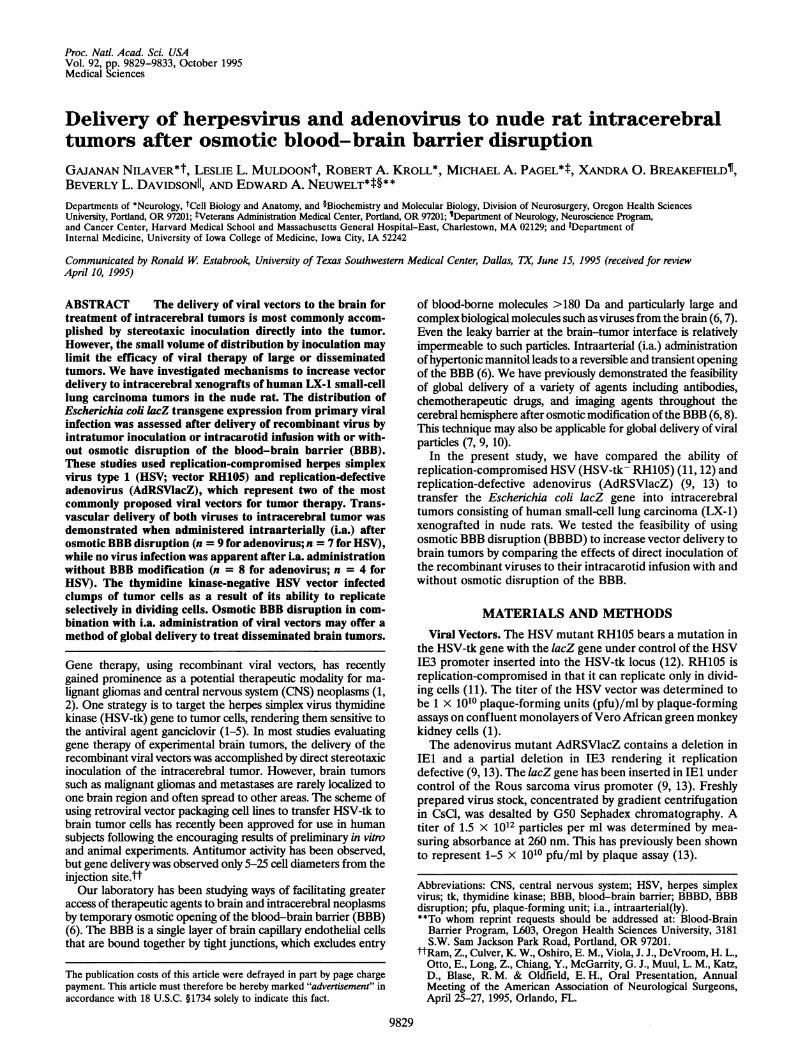
FIG. 1. Coronal section through tumor-bearing region of rat brainlabeled for (-galactosidase 4 days after inoculation of AdRSVLacZ (1.2x 10⁹ particles). About 10% of tumor cells are labeled for the transgeneproduct, which is dispersed as clumps in the tumor xenograft. T, tumor;BG, basal ganglia; CC, corpus callosum. (x17.)
Proc. Natl. Acad. Sci. USA 92 (1995)

Proc. Natl. Acad. Sci. USA 92 (1995) 9831
than that seen with the HSV vector RH105, despite the use of100-fold higher adenoviral titers (Table 1). After adenovirusinoculation, the extent of lacZ gene expression within thetumor varied between 5 and 10% of the total tumor cell mass,with the stained cells being in dispersed clumps scattered overa wide area of the tumor xenograft (Fig. 1). Parenchymal cellsexpressing f3-galactosidase reactivity could be detected in cellswith the morphological appearance of astrocytes extendingalong the corpus callosum (Fig. 1). No cytopathic effects werenoted. The tumor cells expressing the lacZ gene product(Table 1) in the xenograft did not show evidence of oncolysis,and there was no evidence of inflammation, cell death, orcavitation in brain parenchyma adjacent to tumor or along thecorpus callosum.Tumors that received tk- HSV by direct inoculation, in
contrast, frequently demonstrated necrotic centers after 4 days(Fig. 24). Intense f3-galactosidase reactivity was observed intumor cells that were immediately adjacent to these necroticregions (Fig. 24). Approximately 50-60% of the tumor xe-nograft was immunoreactive for the 13-galactosidase transgeneproduct. The parenchymal cells adjacent to the tumor werealso infected, with immunoreactivity detected primarily withinneurons (Fig. 2B). Infection could also be traced in neuronsalong the corpus callosum (Fig. 2A4), with no astrocytic profilesbeing apparent as noted with the adenoviral vector (Fig. 1).Tissue damage was evident in normal brain with necrosisevident along the corpus callosum (Fig. 24).LacZ Transgene Expression Following Viral Delivery by
BBBD. Transvascular delivery of both viruses to intracerebraltumor was achieved when the recombinant vectors were ad-ministered i.a. after osmotic BBBD (Figs. 3A and 4). No viralinfection or f3-galactosidase transgene product could be de-tected in tumor or normal brain around tumor of sham-disrupted animals (where saline was used in lieu of mannitol).These control experiments indicate that the brain capillaryendothelium is not sufficiently compromised, even at the"leaky" brain-tumor interface, to allow transvascular entry ofeither viral vector after i.a. delivery.
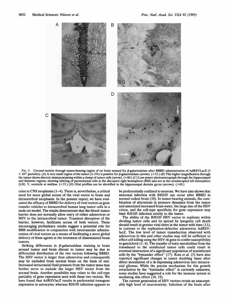
Adenoviral infection could be detected in the tumor xe-nograft (Table 1) after transvascular delivery with osmoticBBBD, as evidenced by B3-galactosidase staining (Fig. 3). Only2-3% of tumor cells were immunoreactive 4 days after BBBD(Fig. 3A), with the infected cells organized as isolated clusterswithin the tumor (Fig. 3B). Although no labeling of paren-chymal cells could be detected at the tumor-brain interface,immunoreactive cells were found scattered throughout theosmotically disrupted cerebral hemisphere. A few brain re-gions, such as the hippocampal dentate gyrus (Fig. 3 C and D),the thalamus, and the perisagittal cortex, showed the highestdensity of stained cells. The immunolabeled parenchymal cellsin the disrupted cerebral hemisphere appeared to be primarilyastrocytes, and the neurons did not appear to be labeled. Therewas no evidence of necrosis in immunostained regions of thetumor, at the brain-tumor interface, or along the corpuscallosum (Fig. 14).HSV infection following BBBD, in contrast, was confined
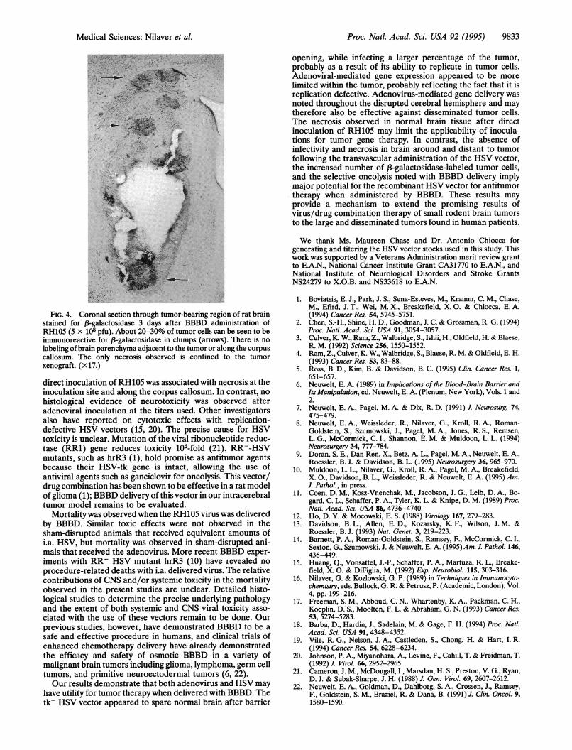
essentially to the tumor xenograft (Fig. 4). The immunolabeledcells in the tumor accounted for 20-30% of the tumor cellmass. The 13-galactosidase staining was organized in clumps,and areas of necrosis were often evident at the centers of thelabeled tumor cell clusters. No labeling could be detected innormal brain parenchyma of the disrupted cerebral hemi-sphere distant from the tumor, although in a few animals someneurons at the tumor-brain interface were stained for ,-ga-lactosidase. In contrast to results of the direct 'inoculationstudies, there was no necrosis of the brain parenchyma alongthe corpus callosum or any other region of the disruptedcerebral hemisphere. The only necrosis observed was confinedselectively to the tumor xenograft.
BG
tS
.. 4*:::
.- -. ^ ..... . .-, t,e ..
A-,|.t., S.'
9-
I.0
*p S
q
'4,' ~,I.- ;P W,e
a, 1
I * X,I
4.
I,.
W.
- 1
'V0
FIG. 2. Coronal section through tumor-bearing region of rat brainlabeled for j3-galactosidase 4 days after inoculation with HSV RH105(2.4 x 10⁸ pfu). (A) Labeling can be seen within tumor cells in thexenograft (-50% stained), parenchymal cells in the brain aroundtumor (BAT), and within neurons along the corpus callosum. Somenecrosis is apparent along the corpus callosum (arrow). BG, basalganglia. (x15.) (B) Immunolabeling within tumor cells and primarilywithin neurons in brain parenchyma (arrows) at the brain-tumorinterface. t, Tumor. (X60.)
Mortality Studies. No mortality was observed in the animalsthat received the recombinant viral vectors (RH105, n = 8;AdRSVlacZ, n = 6) by direct stereotaxic inoculation. Histo-logical analysis of brain was performed on all injected animals(Table 1).Of the 13 animals in the control group (sham modification)
that received AdRSVlacZ i.a. after saline administration, 8survived (38.5% mortality) and were used for histologicalanalysis. All four controls (sham modification with saline)administered RH105 survived (0% mortality). Of a total of 16tumor-bearing nude rats that received AdRSVlacZ by BBBD,7 died after this procedure (mortality rate, 44%). The brainsof 11 survivors were processed for histology as described. Nineanimals were administered RH105 by BBBD, 3 of which died(mortality rate, 33%). Histological analysis was performed onthe 6 survivors and on 1 animal that died 24 hr after virusadministration.
DISCUSSIONGene therapy using recombinant viral vectors holds consider-able promise as an approach for delivery of oncolytic mole-
Medical Sciences: Nilaver et al.
3
9832 Medical Sciences: Nilaver et al.
A B._ss .; .:
X >,,4.: i $,4
s.. : 3...:. . . ... ,. ,l. j
E:z o< *.%K ,.t Y d e <.
.k
-> X
:7. 4' * ,. ,, , * J.E b ..
.. n
.w '#
bws #
SHiS;,,, ,
6e _;5$6 wS >\
FIG. 3. Coronal section through tumor-bearing region of rat brain stained for 13-galactosidase after BBBD administration of AdRSVLacZ (5X 10¹¹ particles). (A) A very small region of the tumor (2-3%) is positive for f-galactosidase (arrow). (xi.) (B) This higher magnification throughthe tumor shows discrete immunostaining within a clump of tumor cells (arrow). (x 60.) (C) Low-power photomicrograph through the hippocampaland thalamic regions, showing labeling of parenchymal cells in the disrupted right hemisphere (RH) and not in the nondisrupted left hemisphere(LH). V, ventricle at midline. (Xij.) (D) Glial profiles can be identified in the hippocampal dentate gyrus (arrows). (x60.)
cules to CNS neoplasms (1-4). There is, nevertheless, a criticalneed for more global access of the viral vector to brain andintracerebral neoplasms. In the present report, we have eval-uated the efficacy of BBBD for delivery ofviral vectors as genetransfer vehicles to intracerebral human lung tumor cells in anude rat model. The results demonstrate that the blood-tumorbarrier does not normally allow entry of either adenovirus orHSV to the intracerebral tumor. Transient disruption of thebarrier, however, facilitates access of both vectors. Theseencouraging preliminary results suggest a potential role forBBB modification in conjunction with intravascular adminis-tration of viral vectors as a means of facilitating a more globaldelivery of these agents in the treatment of disseminated braintumors.
Striking differences in 13-galactosidase staining in brainaround tumor and brain distant to tumor may be due todifferential distribution of the two vectors following BBBD.The HSV vector is larger than adenovirus and consequentlymay be excluded from normal brain on the basis of size.Increased intracranial fluid pressure from the tumor mass mayfurther serve to exclude the larger HSV vector from thenormal brain. Another possibility may relate to the cell-typepartiality of gene expression seen with these two vectors. Wehave found that AdRSVlacZ results in preferential transgeneexpression in astrocytes, whereas RH105 infection appears to
be preferentially confined to neurons. We have also shown thatneuronal infection with RH105 can occur after BBBD innormal rodent brain (10). In tumor-bearing animals, the com-bination of alterations in pressure dynamics from the tumorand associated increased brain water, the large size of the HSVvirion, and the cell-type specificity for gene expression maylimit RH105 infection strictly to the tumor.The ability of the RH105 HSV vector to replicate within
dividing tumor cells and its spread by lysogenic cell deathshould result in greater viral titers in the tumor with time (11),in contrast to the replication-defective adenovirus AdRSV-lacZ. The low level of tumor transduction observed withadenovirus in this and other studies may still be sufficient toeffect cell killing using the HSV-tk gene to confer susceptibilityto ganciclovir (1-4). The transfer of toxic metabolites from thetransduced to the uninfected tumor cells could result ineventual destruction of a significant population of noninfectedcells by the "bystander effect" (17). Ross et al. (5) have alsoreported significant changes in tumor doubling times afterdirect inoculation of a tk-expressing adenovirus into intracra-nial gliomas. While the precise mechanism for this growthretardation by the "bystander effect" is currently unknown,some studies have suggested a role for the immune system inmediating this effect (18, 19).The current generation of HSV vectors retain an unaccept-
ably high level of neurotoxicity. Infection of the brain after
Proc. Natl. Acad. Sci. USA 92 (1995)
Proc. Natl. Acad. Sci. USA 92 (1995) 9833
FIG. 4. Coronal section through tumor-bearing region of rat brainstained for 3-galactosidase 3 days after BBBD administration ofRH105 (5 x 10⁸ pfu). About 20-30% of tumor cells can be seen to beimmunoreactive for fB-galactosidase in clumps (arrows). There is no
labeling of brain parenchyma adjacent to the tumor or along the corpuscallosum. The only necrosis observed is confined to the tumorxenograft. (x 17.)
direct inoculation of RH105 was associated with necrosis at theinoculation site and along the corpus callosum. In contrast, nohistological evidence of neurotoxicity was observed afteradenoviral inoculation at the titers used. Other investigatorsalso have reported on cytotoxic effects with replication-defective HSV vectors (15, 20). The precise cause for HSVtoxicity is unclear. Mutation of the viral ribonucleotide reduc-tase (RR1) gene reduces toxicity 106-fold (21). RR--HSVmutants, such as hrR3 (1), hold promise as antitumor agentsbecause their HSV-tk gene is intact, allowing the use ofantiviral agents such as ganciclovir for oncolysis. This vector/drug combination has been shown to be effective in a rat modelof glioma (1); BBBD delivery of this vector in our intracerebraltumor model remains to be evaluated.
Mortality was observed when the RH105 virus was deliveredby BBBD. Similar toxic effects were not observed in thesham-disrupted animals that received equivalent amounts ofi.a. HSV, but mortality was observed in sham-disrupted ani-mals that received the adenovirus. More recent BBBD exper-iments with RR- HSV mutant hrR3 (10) have revealed no
procedure-related deaths with i.a. delivered virus. The relativecontributions of CNS and/or systemic toxicity in the mortalityobserved in the present studies are unclear. Detailed histo-logical studies to determine the precise underlying pathologyand the extent of both systemic and CNS viral toxicity asso-
ciated with the use of these vectors remain to be done. Ourprevious studies, however, have demonstrated BBBD to be a
safe and effective procedure in humans, and clinical trials ofenhanced chemotherapy delivery have already demonstratedthe efficacy and safety of osmotic BBBD in a variety ofmalignant brain tumors including glioma, lymphoma, germ celltumors, and primitive neuroectodermal tumors (6, 22).Our results demonstrate that both adenovirus and HSV may
have utility for tumor therapy when delivered with BBBD. Thetk- HSV vector appeared to spare normal brain after barrier
opening, while infecting a larger percentage of the tumor,probably as a result of its ability to replicate in tumor cells.Adenoviral-mediated gene expression appeared to be morelimited within the tumor, probably reflecting the fact that it isreplication defective. Adenovirus-mediated gene delivery wasnoted throughout the disrupted cerebral hemisphere and maytherefore also be effective against disseminated tumor cells.The necrosis observed in normal brain tissue after directinoculation of RH105 may limit the applicability of inocula-tions for tumor gene therapy. In contrast, the absence ofinfectivity and necrosis in brain around and distant to tumorfollowing the transvascular administration of the HSV vector,the increased number of 13-galactosidase-labeled tumor cells,and the selective oncolysis noted with BBBD delivery implymajor potential for the recombinant HSV vector for antitumortherapy when administered by BBBD. These results mayprovide a mechanism to extend the promising results ofvirus/drug combination therapy of small rodent brain tumorsto the large and disseminated tumors found in human patients.
We thank Ms. Maureen Chase and Dr. Antonio Chiocca forgenerating and titering the HSV vector stocks used in this study. Thiswork was supported by a Veterans Administration merit review grantto E.A.N., National Cancer Institute Grant CA31770 to E.A.N., andNational Institute of Neurological Disorders and Stroke GrantsNS24279 to X.O.B. and NS33618 to E.A.N.
1. Boviatsis, E. J., Park, J. S., Sena-Esteves, M., Kramm, C. M., Chase,M., Efird, J. T., Wei, M. X., Breakefield, X. 0. & Chiocca, E. A.(1994) Cancer Res. 54, 5745-5751.
2. Chen, S.-H., Shine, H. D., Goodman, J. C. & Grossman, R. G. (1994)Proc. Natl. Acad. Sci. USA 91, 3054-3057.
3. Culver,K W., Ram, Z., Walbridge, S., Ishii, H., Oldfield, H. & Blaese,R. M. (1992) Science 256, 1550-1552.
4. Ram, Z., Culver, K. W., Walbridge, S., Blaese, R. M. & Oldfield, E. H.(1993) Cancer Res. 53, 83-88.
5. Ross, B. D., Kim, B. & Davidson, B. C. (1995) Clin. Cancer Res. 1,651-657.
6. Neuwelt, E. A. (1989) in Implications of the Blood-Brain Barrier andIts Manipulation, ed. Neuwelt, E. A. (Plenum, New York), Vols. 1 and2.
7. Neuwelt, E. A., Pagel, M. A. & Dix, R. D. (1991) J. Neurosurg. 74,475-479.
8. Neuwelt, E. A., Weissleder, R., Nilaver, G., Kroll, R. A., Roman-Goldstein, S., Szumowski, J., Pagel, M. A., Jones, R. S., Remsen,L. G., McCormick, C. I., Shannon, E. M. & Muldoon, L. L. (1994)Neurosurgery 34, 777-784.
9. Doran, S. E., Dan Ren, X., Betz, A. L., Pagel, M. A., Neuwelt, E. A.,Roessler, B. J. & Davidson, B. L. (1995) Neurosurgery 36, 965-970.
10. Muldoon, L. L., Nilaver, G., Kroll, R. A., Pagel, M. A., Breakefield,X. O., Davidson, B. L., Weissleder, R. & Neuwelt, E. A. (1995) Am.J. Pathol., in press.
11. Coen, D. M., Kosz-Vnenchak, M., Jacobson, J. G., Leib, D. A., Bo-gard, C. L., Schaffer, P. A., Tyler, K. L. & Knipe, D. M. (1989) Proc.Natl. Acad. Sci. USA 86, 4736-4740.
12. Ho, D. Y. & Mocowski, E. S. (1988) Virology 167, 279-283.13. Davidson, B. L., Allen, E. D., Kozarsky, K. F., Wilson, J. M. &
Roessler, B. J. (1993) Nat. Genet. 3, 219-223.14. Barnett, P. A., Roman-Goldstein, S., Ramsey, F., McCormick, C. I.,
Sexton, G., Szumowski, J. & Neuwelt, E. A. (1995)Am. J. Pathol. 146,436-449.
15. Huang, Q., Vonsattel, J.-P., Schaffer, P. A., Martuza, R. L., Breake-field, X. 0. & DiFiglia, M. (1992) Exp. Neurobiol. 115, 303-316.
16. Nilaver, G. & Kozlowski, G. P. (1989) in Techniques in Immunocyto-chemistry, eds. Bullock, G. R. & Petrusz, P. (Academic, London), Vol.4, pp. 199-216.
17. Freeman, S. M., Abboud, C. N., Whartenby, K. A., Packman, C. H.,Koeplin, D.-S., Moolten, F. L. & Abraham, G. N. (1993) Cancer Res.53, 5274-5283.
18. Barba, D., Hardin, J., Sadelain, M. & Gage, F. H. (1994) Proc. Natl.Acad. Sci. USA 91, 4348-4352.
19. Vile, R. G., Nelson, J. A., Castleden, S., Chong, H. & Hart, I. R.(1994) Cancer Res. 54, 6228-6234.
20. Johnson, P. A., Miyanohara, A., Levine, F., Cahill, T. & Freidman, T.(1992) J. Virol. 66, 2952-2965.
21. Cameron, J. M., McDougall, I., Marsdan, H. S., Preston, V. G., Ryan,D. J. & Subak-Sharpe, J. H. (1988) J. Gen. Virol. 69, 2607-2612.
22. Neuwelt, E. A., Goldman, D., Dahlborg, S. A., Crossen, J., Ramsey,F., Goldstein, S. M., Braziel, R. & Dana, B. (1991) J. Clin. Oncol. 9,1580-1590.
Medical Sciences: Nilaver et al.