delivery of protein kinase a by crisprmax and its effects
TRANSCRIPT

This is an electronic reprint of the original article. This reprint may differ from the original in pagination and typographic detail.
Delivery of Protein Kinase A by CRISPRMAX and Its Effects on Breast Cancer Stem-Like PropertiesZhou, Jun-Nian; Rautio, Tzu-Chen; Liu, Chang; Xu, Xiao-Yu; Wang, Dong-Qing; Guo, Yong;Eriksson, John; Zhang, HongboPublished in:Pharmaceutics
DOI:10.3390/pharmaceutics13010011
Published: 01/12/2020
Document VersionPublisher's PDF, also known as Version of record
Document LicenseCC BY
Link to publication
Please cite the original version:Zhou, J-N., Rautio, T-C., Liu, C., Xu, X-Y., Wang, D-Q., Guo, Y., Eriksson, J., & Zhang, H. (2020). Delivery ofProtein Kinase A by CRISPRMAX and Its Effects on Breast Cancer Stem-Like Properties. Pharmaceutics, 13(1).https://doi.org/10.3390/pharmaceutics13010011
General rightsCopyright and moral rights for the publications made accessible in the public portal are retained by the authors and/or other copyright ownersand it is a condition of accessing publications that users recognise and abide by the legal requirements associated with these rights.
Take down policyIf you believe that this document breaches copyright please contact us providing details, and we will remove access to the work immediatelyand investigate your claim.
This document is downloaded from the Research Information Portal of ÅAU: 17. Jan. 2022

pharmaceutics
Article
Delivery of Protein Kinase A by CRISPRMAX and Its Effects onBreast Cancer Stem-Like Properties
Jun-Nian Zhou 1,2,3,4,† , Tzu-Chen Rautio 3,5,† , Chang Liu 3,4, Xiao-Yu Xu 3,4, Dong-Qing Wang 6,Yong Guo 3,4,7,*, John Eriksson 4,* and Hongbo Zhang 3,4,*
�����������������
Citation: Zhou, J.; Rautio, T.; Liu, C.;
Xu, X.; Wang, D.; Guo, Y.; Eriksson, J.;
Zhang, H. Delivery of Protein Kinase
A by CRISPRMAX and Its Effects on
Breast Cancer Stem-Like Properties.
Pharmaceutics 2021, 13, 11. https://
dx.doi.org/10.3390/pharmaceutics
13010011
Received: 6 November 2020
Accepted: 18 December 2020
Published: 23 December 2020
Publisher’s Note: MDPI stays neu-
tral with regard to jurisdictional claims
in published maps and institutional
affiliations.
Copyright: © 2020 by the authors. Li-
censee MDPI, Basel, Switzerland. This
article is an open access article distributed
under the terms and conditions of the
Creative Commons Attribution (CC BY)
license (https://creativecommons.org/
licenses/by/4.0/).
1 Experimental Hematology and Biochemistry Lab, Beijing Institute of Radiation Medicine,Beijing 100850, China; [email protected]
2 Stem Cell and Regenerative Medicine Lab, Institute of Health Service and Transfusion Medicine,Beijing 100850, China
3 Pharmaceutical Sciences Laboratory, Åbo Akademi University, 20520 Turku, Finland;[email protected] (T.-C.R.); [email protected] (C.L.); [email protected] (X.-Y.X.)
4 Turku Bioscience Center, University of Turku and Åbo Akademi University, 20520 Turku, Finland5 Biomedical Imaging, Faculty of Medicine, University of Turku, 20520 Turku, Finland6 Department of Radiology, Affiliated Hospital of Jiangsu University, Jiangsu University,
Zhenjiang 212001, China; [email protected] Department of Endocrinology, Key Laboratory of National Health and Family Planning Commission for Male
Reproductive Health, National Research Institute for Family Planning, Beijing 100081, China* Correspondence: [email protected] (Y.G.); [email protected] (J.E.); [email protected] (H.Z.);
Tel.: +86-10-62179175 (Y.G.); +358-22153313 (J.E.); +358-22154141 (H.Z.)† These authors contributed equally to this paper.
Abstract: Protein kinase A (PKA) activation has recently been reported to inhibit epithelial-mesenchymaltransition (EMT) and cancer stem cell (CSC) ability, which is considered to be responsible for chemore-sistance and tumor recurrence in patients. While current studies mainly focus on gene manipulationof the EMT process, the direct delivery of PKA enzymes to cancer cells has never been investigated.Here, we utilize the commercial Lipofectamine CRISPRMAX reagent to directly deliver PKAs tobreast cancer cells and evaluate its effects on EMT regulation. We optimized the delivery parameterswith fluorescent-labeled bovine serum albumin, and successfully delivered fluorescent PKAs throughCRISPRMAX into breast cancer cells. Then, we evaluated the biological effects by immunofluo-rescence, flow cytometry, mammosphere assay, and chemoresistance assay. Our data showed theexpression of EMT-related markers, α-smooth muscle actin and N-cadherin, was downregulated afterCRISPRMAX-PKA treatment. Although the CD44+/CD24− population did not change considerably,the size of mammospheres significantly decreased. In paclitaxel and doxorubicin chemoresistanceassays, we noticed PKA delivery significantly inhibited paclitaxel resistance rather than doxorubicinresistance. Taken together, these results suggest our direct enzyme delivery can be a potential strat-egy for inhibiting EMT/CSC-associated traits, providing a safer approach and having more clinicaltranslational efficacy than gene manipulation. This strategy will also facilitate the direct testing ofother target enzymes/proteins on their biological functions.
Keywords: CRISPRMAX; drug delivery; protein kinase A; chemoresistance; cancer stem cells;epithelial-mesenchymal transition
1. Introduction
The cancer stem cell (CSC) theory has been proposed as one of the viable explanationsfor the resistance of cancer cells to conventional cancer therapies, including chemother-apy and radiotherapy in the clinic [1]. Emerging studies demonstrate that epithelial-to-mesenchymal transition (EMT) plays a crucial role in the dedifferentiation of differentiatedcancer cells into CSCs [2,3]. Many key EMT regulators, including transcription factorSnail family transcriptional repressor 1 (SNAIL1), Twist family BHLH transcription factor
Pharmaceutics 2021, 13, 11. https://dx.doi.org/10.3390/pharmaceutics13010011 https://www.mdpi.com/journal/pharmaceutics
Pharmaceutics 2021, 13, 11 2 of 25
1 (TWIST), zinc finger E-Box binding homeobox 1 (ZEB1), and microRNAs (miRNAs),such as miR-200 family, let-7, have been revealed and have shown promise in inhibition ofthe EMT process in studies [4]. Therefore, targeting EMT-induced CSCs or reversing EMThas become an attractive strategy for inhibiting CSCs and CSC-associated chemoresistance.
As a kind of biological macromolecule, enzymes are one of the main targets in thedevelopment of many disease-associated drugs in cancers. Many small-molecule com-pounds have been developed as inhibitors or agonists of enzyme activity and are widelyused in antitumor research and treatment. On the other hand, enzymes can also be useddirectly as medicine. For example, therapeutic enzymes used in replacement therapyfor genetic diseases. Adagen1 (pegadamase bovine) was the first therapeutic enzymeapproved by FDA under the Orphan Drug Act in 1990. Compared with other therapeutics,enzymes have two important properties as drugs: they specifically bind to the targets,have catalytic properties, and can catalyze many target molecules into target products [5].Based on previous studies mentioned above, we hypothesized that introducing a func-tional enzyme that inhibits EMT/CSCs as a candidate drug into cancer cells could inhibitEMT/CSC-associated properties, leading to a catalytic specificity and lower cytotoxicity.Most enzymes have been reported to be overexpressed in the EMT process [6–8], but wecannot intervene in the EMT process by directly delivering enzymes into cells. Recently,Pattabiraman et al. reported that induction of protein kinase A (PKA) can evoke mesenchy-mal human mammary epithelial cells that undergo mesenchymal-to-epithelial transition(MET) [9], an inverse process of EMT, which promotes the differentiation of CSCs, causingthe loss of tumor-initiating capacity. This indicates PKA may be an ideal target for ourdirect enzyme transfer platform.
Because PKA enzyme needs to function inside the cell, we need a carrier to transferit into the cell while maintaining the activity of the enzyme. In recent times, rapid devel-opments in nanoparticle-based drug delivery systems have led to improvements in thetherapeutic efficacy of cancer treatment [10,11]. Among these particles, lipid nanoparti-cles (LNPs) are the most common and well-investigated nanocarriers for targeted drugdelivery. They have many advantages, such as flexible physicochemical and biophysicalproperties, easy operation for different delivery requirements, high efficiency of enteringcells and tissues, and reduced system toxicity [12], etc. In 1995, the first clinically approvednanomedicine for the treatment of cancer was liposomal doxorubicin (Doxil) [13], whichwas expected to reduce the cardiotoxicity of doxorubicin (DOX). The poorly water-solublechemotherapy drug paclitaxel has also been prepared as liposomal paclitaxel in order toimprove its efficiency in targeting tissues [14]. There are also other chemotherapy drugsencapsulated in LNPs [15], and some have been approved for clinical trials. On this basis,biomolecules, such as nucleic acid molecules, are also used for delivery by LNPs, such asmiRNA and siRNA that can regulate the expression of target proteins [16,17]. They arenot only widely used in life science-related laboratories to study the function of genes butalso in the field of tumor treatment research [18]. The focus for this study is the biologicalunderstanding of a direct delivery of PKA enzymes into the cancer cells. PKA has thefunction of inhibiting epithelial-to-mesenchymal transition and cancer stem cells, thusit has great potential against resistance to cancer treatment. In this work, we utilized acommercial ready-to-use product, CRISPRMAX (Lipofectamine CRISPRMAX Cas9 trans-fection reagent), which is the first optimized lipid nanoparticle transfection reagent forCRISPR-Cas9 protein delivery [19–21]. Since this commercial reagent has been well es-tablished, the results obtained by using this commercial kit will provide a cornerstone ofenzyme delivery strategy for broader biological community, and these results will be easilyrepeated by other laboratories, even they have no material science background. This willalso be a practically handled method for clinical doctors in future clinical applications.
In fact, there are many other functionalized nanosystems that can be used to deliverproteins/enzymes. For example, in our research group, we have developed differentplatforms for protein delivery, including: (a) liposomes [22], (b) polymer nanoparticles [23],(c) polymersomes [24], (d) mesoporous nanoparticles [25], (e) metal-organic framework

Pharmaceutics 2021, 13, 11 3 of 25
with biomineralization [26], (f) DNA nanoparticles [27,28], (g) emulsions [29], etc. However,in the end, we selected the commercial CRISPRMAX, because CRISPRMAX has the follow-ing advantages. First, this reagent has low toxicity to cells, allowing us to focus on eval-uating the biological effects of PKA enzymes on cells, including epithelial-mesenchymaltransition, cancer stem cells, and chemoresistance. Second, the experimental conditions forthis reagent have been optimized; there is a standard protocol for CRISPRMAX-enzymepreparation and cell transfection, which is involved in cell seeding density, reagent usage,the time of each step, and the precautions for each step. Third, such reagent is an idealdelivery solution in 96-well format plates for high-throughput analysis. These advantagesmake us easily focus on the biological function of a direct delivery of PKA enzymes into thecancer cells rather than nanocarriers themselves, and provide a good paradigm of potentialtranslational application in the clinic.
Therefore, Lipofectamine CRISPRMAX reagent was selected as a model nanocarrier forinvestigating the establishment of direct transfer of PKAs into breast cancer cells. We usedfluorescent-labeled bovine serum albumin (BSA) as a positive control to optimize relevanttransfection parameters, and proved that PKA enzymes through CRISPRMAX can beintroduced into breast cancer cells. The biological effects of CRISPRMAX-PKA complexeswere further examined through functional experiments, including chemoresistance assay,flow cytometry (FCM), and mammosphere assay, to test whether the CSC- and/or EMT-related properties were repressed through the biotherapeutic effects of the complexes. Weobserved that the expression of EMT-related markers, α-smooth muscle actin (α-SMA)and N-cadherin (N-cad), was downregulated. Although the CD44+/CD24− populationdid not change significantly, in another functional test of CSCs, we found that the sizeof mammospheres (MSs) was significantly downregulated. In the chemoresistance assay,we observed a drug-dependent result, that is, PKA delivery has a significant inhibitoryeffect to paclitaxel (PTX) resistance, but there is no effect against DOX. Interestingly, thisinhibitory effect is order dependent, that is, the inhibitory effect can be exerted only in thecells that were treated with PKA before PTX administration. This indicates that the role ofPKA enzymes in chemotherapy drug resistance may be drug dependent. Taken together,as a proof-of-concept, we confirmed the direct enzyme delivery as a potential strategyfor inhibiting EMT/CSC-associated chemoresistance. This strategy will also facilitate thescreening of target enzymes/proteins for the construction of a new drug delivery systemor complex nanoparticles.
2. Materials and Methods2.1. Cell Culture
Human breast cancer MDA-MB-231 cells and MCF-7 cells were cultured with Dul-becco’s Modified Eagle’s medium (DMEM, Lonza, Walkersville, MD, USA), which con-tained 10% fetal bovine serum (FBS, Gibco, Grand Island, NY, USA), 0.5% penicillin/streptomycin(Pen/Strep, Gibco), and 1% L-Glutamine (Gibco). Human breast cancer BT-549 cells andHs-578T cells were grown in ATCC-modified RPMI 1640 medium (Gibco), which contained10% FBS, 0.5% Pen/Strep, and 1% L-glutamine. Human normal breast cells MCF-10A werecultured in DMEM/F12 medium (Gibco) containing 5% FBS, D-(+)-Glucose (4.5 g/L, Sigma,St. Louis, MO, USA), human insulin (10 µg/mL, Sigma), cholera toxin (Sigma, 0.1 µg/mL),hydrocortisone (0.5 µg/Ml, Sigma), epidermal growth factor (EGF, 20 ng/mL, Peprotech,London, UK), and 1% L-glutamine. All the cells were cultured in a humidified incubatorwith 5% CO2 at 37 ◦C. MCF-7 cells, BT-549 cells, Hs-578T, and MCF-10A cells were kindlyprovided by Prof. Jukka Westermarck (Turku Bioscience, Turku, Finland).
2.2. Enzyme Delivery by Lipofectamine CRISPRMAX Reagent
Lipofectamine CRISPRMAX reagent (Invitrogen, Carlsbad, CA, USA) was used fordelivering BSA and PKA enzymes in this study. All procedures were followed accordingto the manufacturer’s protocol. Breast cancer cells and MCF-10A cells were seeded in96-well plates (5000 cells per well, except MDA-MB-231 cells were seeded at the density

Pharmaceutics 2021, 13, 11 4 of 25
of 2000 cells per well) one day before transfection. The seeded plates were incubatedovernight; the cells were grown at 50–70% confluence in 96-well plates for transfection.On the day of transfection, the culture medium of each well in the plates was replacedwith 100 µL of Opti-MEM (Invitrogen) before performing CRISPRMAX-based enzymedelivery. As for the preparation of enzymes-loaded CRISPRMAX, tube 1 contained 5 µL ofOpti-MEM medium, 0.5 µL of PKA enzyme solution (500 ng per well, 1 µg/µL of stocksolution), and 0.5 µL of Cas9 Plus reagent were added in sequence. The control cellswere treated with the same amount of purified BSAs, or Alexa Fluor 680-labeled BSAs(Invitrogen). Tube 2 contained 5 µL of Opti-MEM medium with 0.5 µL of CRISPRMAXreagent. Next, the solution of tube 1 was added to tube 2 within 3 min, and then mixedwell; the complex solution was incubated at room temperature. After 5–10 min, 10 µL/wellof CRISPRMAX complex solution were added into the cancer cells for 1 day of incubation.Then, the CRISPRMAX-containing medium was replaced with complete growth mediumin each well; the transfected cells continued to grow for 1 to 3 days before conductingfurther experiments.
2.3. Characterization of PKAs- and BSAs-Loaded CRISPRMAX Complexes
The morphology of the enzymes-loaded CRISPRMAX complexes was observed bymeans of transmission electron microscopy (TEM) and confocal laser scanning microscopy(CLSM). For TEM analysis, fresh CRISPRMAX-BSA complexes, CRISPRMAX-PKA com-plexes, and null CRISPRMAX complexes were prepared, respectively, according to theprotocol mentioned in Section 2.2. The grids used for the electron microscope sampleswere immersed in the complex solution for each setup for a few seconds, then taken out,and placed on an absorbent paper for drying overnight. The grids were observed underTEM (LVEM 5, Delong Instruments, Brno, Czech Republic). For CLSM analysis, fluorescentBSAs (Alexa Fluor 680-labeled BSAs) were used to prepare fresh CRISPRMAX-BSA-Alexa680 complexes; the same concentration of BSA-Alexa 680 solution without CRISPRMAXwas used as a control group. The solution of the complexes or the control group wasdripped onto the glass slides, and then these samples were covered with cover glasses;fluorescent particles were observed under CLSM (Zeiss, LSM880 with Airyscan, Carl ZeissAG, Oberkochen, Germany). The particle size of CRISPRMAX-PKAs and CRISPRMAX-BSAs was measured with dynamic light scattering using a Zetasizer Nano ZS (MalvernInstruments Ltd., Malvern, Worcs, UK). The surface charge of the complexes (zeta potential)was measured using a Zetasizer Nano ZS with disposable folded capillary cells (DTS1070,Malvern, Worcs, UK).
2.4. Encapsulation Efficiency
Fluorescent-labeled BSAs (BSA-Alexa 680) were used to calculate the encapsulationefficiency of CRISPRMAX. Briefly, fresh CRISPRMAX-BSA-Alexa 680 complexes wereprepared, according to the protocol mentioned in Section 2.2. In the meantime, the sameamount of BSA-Alexa 680 without CRISPRMAX was prepared in the same volume ofsolution as positive control groups, of which the fluorescence intensity represented thetotal amount of BSA-Alexa 680 (Vt). Then, the complex solution was added into theupper chamber of the centrifugal filter device (100K, Merck Millipore, Billerica, MA, USA),followed by centrifugation at 13,000 rpm for 5 min. The molecular weight of CRISPRMAXand CRISPRMAX-BSA-Alexa 680 complexes is greater than 100 kDa, and the complexes willbe trapped in the filter, while the molecular weight of unencapsulated free BSA-Alexa 680is less than 100 kDa, and will be passed through the filter. After centrifugation, the filtratewas collected. Through this method, the unencapsulated free BSAs in the preparationsystem were obtained. The fluorescence intensity of these complexes was analyzed witha Varioskan multimode reader, and the fluorescence intensity of BSA-Alexa 680 (Vx) wasobtained. The encapsulation efficiency was calculated based on the following formula:
Encapsulation efficiency (%) = (Vt − Vx)/Vt × 100% (1)
Pharmaceutics 2021, 13, 11 5 of 25
2.5. Release Efficiency
For in vitro release study of CRISPRMAX, fluorescent BSAs (BSA-Alexa 680) were usedto calculate the release efficiency of CRISPRMAX. The release properties were measured atthe following time points: 0, 1, 2, 4, 8, 12, and 24 h. First, fresh CRISPRMAX-BSA-Alexa680 complexes were prepared, according to the protocol mentioned in Section 2.2; 1.25 µgof BSA-Alexa 680 in 250 µL of complex solution per tube, each tube for one time point,respectively. Meanwhile, the same amount of BSA-Alexa 680 without CRISPRMAX (1.25 µgof BSA-Alexa 680 in 250 µL of solution without CRISPRMAX per tube, each tube for onetime point, respectively) was prepared as positive control groups, of which the fluorescenceintensity represented the total amount of BSA-Alexa 680 (Vt). All samples of the releasegroups and control groups were put into a shaking water bath at 37 ◦C, protected from thelight. For the experimental setup, the solution of CRISPRMAX-BSA-Alexa 680 complexeswas added into the upper chamber of the centrifugal filter device (100K, Merck Millipore),followed by centrifugation at 13,000 rpm for 5 min; the filtrate of the release groups persetup was collected. The fluorescence intensity values of the free BSA-Alexa 680 solution(V0, V1, V2, V4, V8, V12, and V24) in the release groups were measured at each time point.The fluorescence intensity values of the positive control groups (Vt0, Vt1, Vt2, Vt4, Vt8, Vt12,and Vt24) were also measured at each time point. Based on the fluorescence value of thepositive controls at each time point, the release percentage was normalized at each timepoint, and subtracted the proportion of the original unencapsulated free BSA-Alexa 680.For example, the percentage release at the first hour time point was calculated as follows:
Percentage release (%) = [(V1/Vt1) − (V0/Vt0)] × 100% (2)
2.6. Cellular Uptake Efficiency Analysis of CRISPRMAX-BSA-Alexa Fluor 680
According to the protocol mentioned in Section 2.2, 500 ng of BSA-Alexa Fluor 680 wascoated by CRISPRMAX reagent and added into BT-549 cells per well (n = 3). The cellularuptake was measured at indicated time points (10 min, 30 min, 1 h, 2 h, 3 h, 4 h, 6 h,and 23 h) after CRISPRMAX-BSA-Alexa 680 complex treatment in cells. In the meantime,the fluorescence intensity value of CRISPRMAX-BSA-Alexa 680 without cells (n = 3) wasmeasured at indicated time points. The fluorescence intensity value for each sample wasmeasured at 679 (excitation) and 702 nm (emission) with a Varioskan LUX multimodereader (Thermo Fisher Scientific, Waltham, MA, USA). Cellular uptake efficiency wascalculated based on the following formula: Ic = the fluorescence intensity value of BSA-Alexa 680 (500 ng) only in 110 µL of OPTI-MEM without CRISPRMAX complex andcells, and It = the fluorescence intensity value of BSA-Alexa 680 in CRISPRMAX-complex-containing cell transfection supernatant at the indicated time points:
Cellular uptake efficiency = (Ic − It)/Ic × 100% (3)
2.7. Immunofluorescence Analysis by Confocal Laser Scanning Microcopy
CRISPRMAX-PKAs- and CRISPRMAX-BSAs-delivered cancer cells were seeded inconfocal dishes (Thermo Fisher Scientific) and continued to grow for 3 to 5 days afterdelivery. Cell fixation was performed using 4% paraformaldehyde (PFA, Sigma) in the con-focal dishes for 30 min at room temperature. Then, 0.1% triton X-100 (Sigma) was used topermeabilize the cell membrane of the samples for 5–10 min, followed by blocking with 10%goat serum (Life Technologies, Auckland, New Zealand) for 30 min at room temperaturewithout PBS washing after discarding. The primary antibodies were directly incubatedwith the cells according to the manufacturer’s protocol. For EMT markers expressionanalysis, mouse anti-human E-cad antibody (1:50, Santa Cruz Biotechnology, Santa Cruz,CA, USA), mouse anti-human N-cadherin antibody (1:50, Santa Cruz Biotechnology), andmouse anti-human α-SMA antibody (1:100, Santa Cruz Biotechnology) were diluted in PBSand incubated with the cells overnight at 4 ◦C, followed by TRITC-labeled goat anti-mousesecondary antibodies incubation for 30 min at room temperature. For PKA expression
Pharmaceutics 2021, 13, 11 6 of 25
analysis, rabbit anti-human/bovine PKA antibodies (1:300, Invitrogen) were incubatedwith the cells overnight at 4 ◦C, followed by TRITC-labeled goat anti-rabbit secondaryantibodies incubation for 1 h at room temperature. All prepared samples were rinsedwith PBS three times before nuclei staining. To visualize the cell nuclei of these samples,the samples were treated with 5 µg/mL of 4′,6-diamidino-2-phenylindole (DAPI, LifeTechnologies) solution for 5 min at room temperature; the stained samples were observedusing confocal laser scanning microcopy (CLSM) (Zeiss, LSM880).
2.8. Flow Cytometry
For analysis of CD44 and CD24, the flow cytometric samples were prepared accordingto the manufacturer’s protocol (Invitrogen). Briefly, the trypsinized single cell suspensionwas washed with PBS twice and centrifuged for 8 min at 330 g. For each test (100 µL ofPBS), 0.15 µL of mouse anti-human APC-labeled CD44 monoclonal antibody (eBioscience,San Diego, CA, USA) and 5 µL of mouse anti-human PE-labeled CD24 monoclonal antibodywere incubated together in an Eppendorf tube for 15 min at 4 ◦C, which was packed withaluminum lamination foils to protect from light exposure. Similar to the preparation ofCD44 and CD24 markers, the amount of isotype controls (APC-labeled mouse IgG and PE-labeled mouse IgG) were prepared, that was, 0.3 µL of APC-IgG (eBioscience) and 1.25 µLof PE-IgG (eBioscience) in a total volume of 100 µL of PBS. After antibody incubation,the cells were washed with PBS 2–3 times and centrifuged for 8 min at 330 g. Then, thesamples were resuspended in 500 µL of PBS for flow cytometric analysis with a BD LSRFortessa analyzer.
2.9. Mammosphere Formation Assay
Mammosphere assay has been used for identification of tumor spherical colonies,CSCs, also known as mammospheres (MSs) [16,30,31]. The single cell suspensions wereobtained by trypsinizing the breast cancer cells that were transfected with CRISPRMAX-PKAs or CRISPRMAX-BSAs. The same number of the cancer cells from each group weregrown in ultra-low attachment plates (Sigma) containing serum-free DMEM/F12 (Lonza),B27 (1:50, Gibco), N2 (1:100, Gibco), 2 µg/mL of human insulin solution (Sigma), 4 µg/mLof heparin (Sigma), human epidermal growth factor (EGF, Peprotech), and human basicfibroblast growth factor (bFGF, Peprotech). The MSs were observed and imaged under abright-field microscope one week later. Diameter measurement of MSs was analyzed byImage J, and the size of MSs greater than 70 µm in diameter was counted.
2.10. Chemoresistance Assay
The effects of PKA-loaded CRISPRMAX complexes in breast cancer cells and normalbreast cells on the chemoresistance of PTX (Arisun ChemPharm, Xi’an, China) and DOX(Arisun ChemPharm) were determined by WST-1 cell viability assay [26]. Breast cancercells and MCF-10A cells were seeded in 96-well plates (5000 cells per well, except MDA-MB-231 cells were seeded at the density of 2000 cells per well, three parallel wells for eachconcentration) in complete growth medium containing 5% FBS, and cultured in the cellincubator overnight. Then, each well of the medium was replaced with fresh Opti-MEMmedium containing CRISPRMAX-PKAs or CRISPRMAX-BSAs complexes. After deliveringthe complexes, PTX or DOX at the indicated concentrations was added under differenttransfection time points (at 0 and 12 h). Transfected cells were treated with DOX at 0, 0.1,0.5, 1, and 1.5 µg/mL or with PTX at 0, 0.05, 0.1, 0.2, and 0.5 µg/mL. The duration of PTXand DOX treatment was 48 h. For WST-1 assay, according to the manufacturer’s protocol(Roche, Mannheim, BW, Germany), the drug-containing medium was replaced with 10 µLof WST-1 reagent that were dissolved in 100 µL of complete growth medium for each well.After 2 h of incubation in the cell incubator, the absorbance was measured at 440 nm with aVarioskan LUX multimode reader (Thermo Fisher Scientific).

Pharmaceutics 2021, 13, 11 7 of 25
2.11. Statistical Analyses
All data were presented as means with standard deviations (SD); n = 3 replicates fortransfection with PKA and BSA (control) group. Statistical graphs were generated usingGraphPad Prism 8 software (GraphPad Software Inc., San Diego, CA, USA) or Origin6.1 software (OriginLab, Northampton, MA, USA). Quantitative analysis of the mean offluorescence intensity of PKA was performed by ImageJ. The data of chemoresistanceassays were further performed with AUC (area under curve) to compare the effectivenessof drug effect between transfection with PKA and BSA enzymes. The statistical significanceof differences between two groups was determined by unpaired two-tailed t tests or two-way ANOVA analysis, according to the number of parameters of the data. p < 0.05 wasconsidered statistically significant (* p < 0.05, ** p < 0.01, and *** p < 0.001); p > 0.05 wasconsidered not significant.
3. Results3.1. Characterization of PKAs- and BSA-Loaded CRISPRMAX Complexes3.1.1. Morphology
The morphology data of the enzymes-loaded CRISPRMAX complexes were collectedby means of TEM and CLSM. Under TEM, the different appearances of CRISPRMAX andthe encapsulated proteins were clearly shown. CRISPRMAX was shown as a light grayouter outline of the complexes, while the encapsulated BSA or PKA proteins were presentedin black (Figure 1a). For null CRISPRMAX, there was only a light gray outline. These resultsshowed that the proteins were successfully coated by CRISPRMAX. For CLSM analysis,fluorescent BSAs were used to prepare fresh CRISPRMAX-BSA-Alexa 680 complexes;the same concentration of BSA-Alexa 680 solution without CRISPRMAX was used as acontrol group. Fluorescent particles were clearly observed under CLSM; the images in2-D and 3-D display showed that small red particles were distributed in the solution,while BSA-Alexa 680 in the control group had no significant red particles due to a lack ofCRISPRMAX encapsulation (Figure 1b,c), which further indicates BSA-Alexa 680 proteinswere successfully coated by CRISPRMAX.
3.1.2. Encapsulation Efficiency and Release Efficiency of CRISPRMAX
For evaluation of the encapsulation capability of CRISPRMAX, fluorescent-labeledBSAs (BSA-Alexa 680) were used as a model to mimic the encapsulation efficiency ofCRISPRMAX, since it is very difficult to directly quantify PKA. The same amount in thesame volume of BSA-Alexa 680 without CRISPRMAX as well as in CRISPRMAX-BSA-Alexa680 complexes was used as a positive control to indicate a total of 100% fluorescence value.Our results showed that the encapsulation efficiency using CRISPRMAX to encapsulatethe proteins was 76.11% ± 4.320% (Figure 1d). As for the analysis of the release propertyof CRISPRMAX, our results showed that there were no significant releases of the freeBSA-Alexa 680 from CRISPRMAX-BSA-Alexa 680 complexes within the first four hour.A small release (<10%) was detected at the eighth hour (Figure 1e), but no further releasewas detected thereafter, indicating CRISPRMAX-BSA-Alexa 680 complexes were relativelystable in in vitro.
3.1.3. Determination of Zeta Potential and Particle Size of the Prepared Lipid Nanoparticles
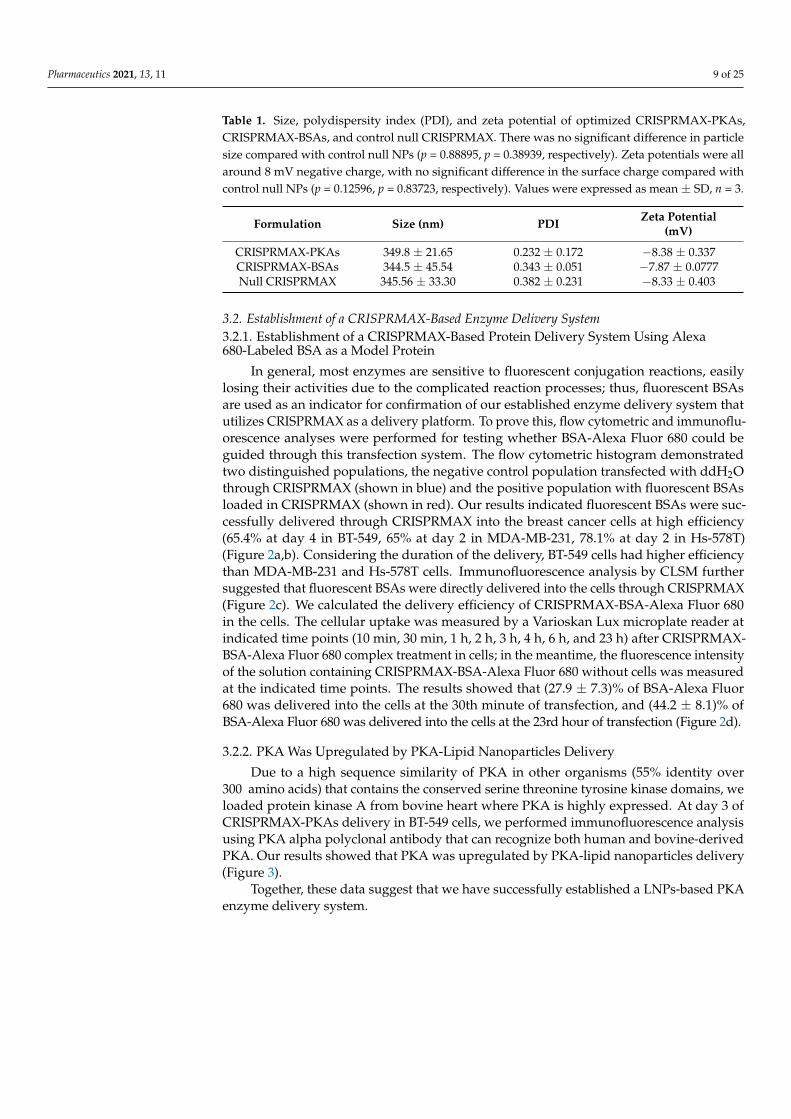
Two of the most important parameters of nanoparticles are particle size and zetapotential [32]. Zeta potential is the overall surface charge of a particle in a particularsolution. In this study, 10 µL of CRISPRMAX complexes was added into 100 µL of Opti-MEM, mixed well, followed by a 20-fold dilution in Opti-MEM. Both the size and zetapotential of these samples were measured using a Zetasizer Nano ZS. The mean of thesize of PKA, BSA-loaded, and control CRISPRMAX (null) was found to be 349.8 ± 21.65,344.5 ± 45.54, 345.56 ± 33.30 nm in Opti-MEM medium, respectively. The overall surfacecharge of PKA, BSA-loaded, and control CRISPRMAX (null) was found to be−8.38 ± 0.337,

Pharmaceutics 2021, 13, 11 8 of 25
−7.87± 0.0777,−8.33± 0.403 in Opti-MEM medium, respectively. There was no significantdifference between each group in terms of size and surface charge (Table 1).
Pharmaceutics 2021, 13, x FOR PEER REVIEW 8 of 25
same volume of BSA-Alexa 680 without CRISPRMAX as well as in CRIS-PRMAX-BSA-Alexa 680 complexes was used as a positive control to indicate a total of 100% fluorescence value. Our results showed that the encapsulation efficiency using CRISPRMAX to encapsulate the proteins was 76.11% ± 4.320% (Figure 1d). As for the analysis of the release property of CRISPRMAX, our results showed that there were no significant releases of the free BSA-Alexa 680 from CRISPRMAX-BSA-Alexa 680 com-plexes within the first four hour. A small release (<10%) was detected at the eighth hour (Figure 1e), but no further release was detected thereafter, indicating CRIS-PRMAX-BSA-Alexa 680 complexes were relatively stable in in vitro.
Figure 1. Characterization of PKAs- and BSAs-loaded CRISPRMAX complexes. (a) TEM images of CRISPRMAX-BSAs, CRISPRMAX-PKAs and null CRISPRMAX; the yellow circles indicate the location of the complexes (scale bars, 500 nm in BSAs and PKAs, and 1 μm in null complexes). (b) CLSM images of BSA-Alexa 680 and CRISPRMAX-BSA-Alexa 680 in a 2-D display under the same parameters; the right panel is a partial enlargement of CRISPRMAX-BSA-Alexa 680 in a 2-D display (scale bars, 20 μm in the left and middle panels; 10 μm in the right panel). (c) A CLSM image of CRIS-
Figure 1. Characterization of PKAs- and BSAs-loaded CRISPRMAX complexes. (a) TEM images of CRISPRMAX-BSAs,CRISPRMAX-PKAs and null CRISPRMAX; the yellow circles indicate the location of the complexes (scale bars, 500 nm inBSAs and PKAs, and 1 µm in null complexes). (b) CLSM images of BSA-Alexa 680 and CRISPRMAX-BSA-Alexa 680 in a 2-Ddisplay under the same parameters; the right panel is a partial enlargement of CRISPRMAX-BSA-Alexa 680 in a 2-D display(scale bars, 20 µm in the left and middle panels; 10 µm in the right panel). (c) A CLSM image of CRISPRMAX-BSA-Alexa680 in a 3-D display (scale bar, 50 µm). (d) The encapsulation efficiency of CRISPRMAX based on the calculation of loadedBSA-Alexa 680. (e) The release efficiency of CRISPRMAX based on the calculation of loaded BSA-Alexa 680 at the indicatedtime points.
Pharmaceutics 2021, 13, 11 9 of 25
Table 1. Size, polydispersity index (PDI), and zeta potential of optimized CRISPRMAX-PKAs,CRISPRMAX-BSAs, and control null CRISPRMAX. There was no significant difference in particlesize compared with control null NPs (p = 0.88895, p = 0.38939, respectively). Zeta potentials were allaround 8 mV negative charge, with no significant difference in the surface charge compared withcontrol null NPs (p = 0.12596, p = 0.83723, respectively). Values were expressed as mean ± SD, n = 3.
Formulation Size (nm) PDI Zeta Potential(mV)
CRISPRMAX-PKAs 349.8 ± 21.65 0.232 ± 0.172 −8.38 ± 0.337CRISPRMAX-BSAs 344.5 ± 45.54 0.343 ± 0.051 −7.87 ± 0.0777Null CRISPRMAX 345.56 ± 33.30 0.382 ± 0.231 −8.33 ± 0.403
3.2. Establishment of a CRISPRMAX-Based Enzyme Delivery System3.2.1. Establishment of a CRISPRMAX-Based Protein Delivery System Using Alexa680-Labeled BSA as a Model Protein
In general, most enzymes are sensitive to fluorescent conjugation reactions, easilylosing their activities due to the complicated reaction processes; thus, fluorescent BSAsare used as an indicator for confirmation of our established enzyme delivery system thatutilizes CRISPRMAX as a delivery platform. To prove this, flow cytometric and immunoflu-orescence analyses were performed for testing whether BSA-Alexa Fluor 680 could beguided through this transfection system. The flow cytometric histogram demonstratedtwo distinguished populations, the negative control population transfected with ddH2Othrough CRISPRMAX (shown in blue) and the positive population with fluorescent BSAsloaded in CRISPRMAX (shown in red). Our results indicated fluorescent BSAs were suc-cessfully delivered through CRISPRMAX into the breast cancer cells at high efficiency(65.4% at day 4 in BT-549, 65% at day 2 in MDA-MB-231, 78.1% at day 2 in Hs-578T)(Figure 2a,b). Considering the duration of the delivery, BT-549 cells had higher efficiencythan MDA-MB-231 and Hs-578T cells. Immunofluorescence analysis by CLSM furthersuggested that fluorescent BSAs were directly delivered into the cells through CRISPRMAX(Figure 2c). We calculated the delivery efficiency of CRISPRMAX-BSA-Alexa Fluor 680in the cells. The cellular uptake was measured by a Varioskan Lux microplate reader atindicated time points (10 min, 30 min, 1 h, 2 h, 3 h, 4 h, 6 h, and 23 h) after CRISPRMAX-BSA-Alexa Fluor 680 complex treatment in cells; in the meantime, the fluorescence intensityof the solution containing CRISPRMAX-BSA-Alexa Fluor 680 without cells was measuredat the indicated time points. The results showed that (27.9 ± 7.3)% of BSA-Alexa Fluor680 was delivered into the cells at the 30th minute of transfection, and (44.2 ± 8.1)% ofBSA-Alexa Fluor 680 was delivered into the cells at the 23rd hour of transfection (Figure 2d).
3.2.2. PKA Was Upregulated by PKA-Lipid Nanoparticles Delivery
Due to a high sequence similarity of PKA in other organisms (55% identity over300 amino acids) that contains the conserved serine threonine tyrosine kinase domains, weloaded protein kinase A from bovine heart where PKA is highly expressed. At day 3 ofCRISPRMAX-PKAs delivery in BT-549 cells, we performed immunofluorescence analysisusing PKA alpha polyclonal antibody that can recognize both human and bovine-derivedPKA. Our results showed that PKA was upregulated by PKA-lipid nanoparticles delivery(Figure 3).
Together, these data suggest that we have successfully established a LNPs-based PKAenzyme delivery system.

Pharmaceutics 2021, 13, 11 10 of 25
Pharmaceutics 2021, 13, x FOR PEER REVIEW 10 of 25
(44.2 ± 8.1)% of BSA-Alexa Fluor 680 was delivered into the cells at the 23rd hour of transfection (Figure 2d). 3.2.2. PKA Was Upregulated by PKA-Lipid Nanoparticles Delivery
Due to a high sequence similarity of PKA in other organisms (55% identity over 300 amino acids) that contains the conserved serine threonine tyrosine kinase domains, we loaded protein kinase A from bovine heart where PKA is highly expressed. At day 3 of CRISPRMAX-PKAs delivery in BT-549 cells, we performed immunofluorescence analysis using PKA alpha polyclonal antibody that can recognize both human and bovine-derived PKA. Our results showed that PKA was upregulated by PKA-lipid nanoparticles deliv-ery (Figure 3).
Together, these data suggest that we have successfully established a LNPs-based PKA enzyme delivery system.
Figure 2. Establishment of a CRISPRMAX-based protein delivery system using the Alexa 680-labeled BSA as a modelprotein. (a) Bright-field images of three different mesenchymal breast cancer cell lines were taken at 200×magnification(scale bars, 200 µm). (b) Single-parameter histograms for comparison of the negative control (cells transfected with ddH2O,blue) and the positive population (the cell of interest, red): (left) BT-549 cells were seeded at the density of 8000 cells/well,and FCM was conducted at day 4 after transfection with fluorescent BSAs; (middle) MDA-MB-231 cells were seeded atthe density of 4000 cells/well, and FCM was conducted at day 2 after transfection with fluorescent BSAs; (right) Hs-578Tcells were seeded at the density of 8000 cells/well, and FCM was conducted at day 2 after transfection with fluorescentBSAs. Each kind of cell was transfected with 500 ng of BSA-Alexa Fluor 680 per well through CRISPRMAX. (c) CLSManalysis of BT-549 cells transfected with fluorescent BSAs delivered by CRISPRMAX. Fluorescent excitation and emissionfor BSA-Alexa Flour 680 were 633 and 702 nm, respectively. Confocal images were taken at 200×magnification (scale bars,50 µm). (d) Cellular uptake efficiency of BSA-Alexa Fluor 680 delivered by CRISPRMAX within 24 h in BT-549 cells. CLSM,confocal laser scanning microscopy.

Pharmaceutics 2021, 13, 11 11 of 25
Pharmaceutics 2021, 13, x FOR PEER REVIEW 11 of 25
Figure 2. Establishment of a CRISPRMAX-based protein delivery system using the Alexa 680-labeled BSA as a model protein. (a) Bright-field images of three different mesenchymal breast cancer cell lines were taken at 200x magnification (scale bars, 200 μm). (b) Single-parameter histograms for comparison of the negative control (cells transfected with ddH2O, blue) and the positive population (the cell of interest, red): (left) BT-549 cells were seeded at the density of 8000 cells/well, and FCM was conducted at day 4 after transfection with fluorescent BSAs; (middle) MDA-MB-231 cells were seeded at the density of 4000 cells/well, and FCM was conducted at day 2 after transfection with fluorescent BSAs; (right) Hs-578T cells were seeded at the density of 8000 cells/well, and FCM was conducted at day 2 after transfection with flu-orescent BSAs. Each kind of cell was transfected with 500 ng of BSA-Alexa Fluor 680 per well through CRISPRMAX. (c) CLSM analysis of BT-549 cells transfected with fluorescent BSAs delivered by CRISPRMAX. Fluorescent excitation and emission for BSA-Alexa Flour 680 were 633 and 702 nm, respectively. Confocal images were taken at 200x magnification (scale bars, 50 μm). (d) Cellular uptake efficiency of BSA-Alexa Fluor 680 delivered by CRISPRMAX within 24 h in BT-549 cells. CLSM, confocal laser scanning microscopy.
Figure 3. Immunofluorescence analysis of PKA expression in BT-549 cells after CRISPRMAX-based PKA delivery. (a) PKA expression was examined by immunofluorescence analysis using CLSM; 3 days after CRISPRMAX-based PKA de-livery vs. CRISPRMAX-based BSA delivery (control) in BT-549 cells. Cell nuclei were visualized by DAPI. Confocal im-ages were taken at 200x magnification (scale bars, 50 μm). (b) Quantitative analysis of the mean of florescence intensity of PKA performed by ImageJ (n = 3), and the mean of fluorescence intensity of PKA was significantly higher compared with BSA group, p = 0.00459, ** p < 0.01. The quantitative data were presented as Mean ± SD.
Figure 3. Immunofluorescence analysis of PKA expression in BT-549 cells after CRISPRMAX-based PKA delivery. (a) PKAexpression was examined by immunofluorescence analysis using CLSM; 3 days after CRISPRMAX-based PKA delivery vs.CRISPRMAX-based BSA delivery (control) in BT-549 cells. Cell nuclei were visualized by DAPI. Confocal images weretaken at 200× magnification (scale bars, 50 µm). (b) Quantitative analysis of the mean of florescence intensity of PKAperformed by ImageJ (n = 3), and the mean of fluorescence intensity of PKA was significantly higher compared with BSAgroup, p = 0.00459, ** p < 0.01. The quantitative data were presented as Mean ± SD.
3.3. Analysis of the Effects of PKA-Lipid Nanoparticles Delivery on EMT-Associated MarkerExpression and CSCs3.3.1. Expression Analysis of E-Cadherin, N-Cadherin, and α-SMA after PKA DeliveryUsing CLSM
Since BT-549 cells have higher efficiency than MDA-MB-231 and Hs-578T cells, herein,we focused on the effects of PKA-lipid nanoparticles delivery on EMT-associated markerexpression and CSCs in BT-549 cells. Our results showed that the expression of α-SMAand N-cadherin was decreased in the cells transfected with CRISPRMAX-PKAs comparedwith the control (CRISPRMAX-BSAs); the expression of E-cadherin was not remarkablydifferent between the PKA and BSA group (Figure 4).

Pharmaceutics 2021, 13, 11 12 of 25
3.3.2. Analysis of the Effects of PKA Enzyme Delivery on CSCs
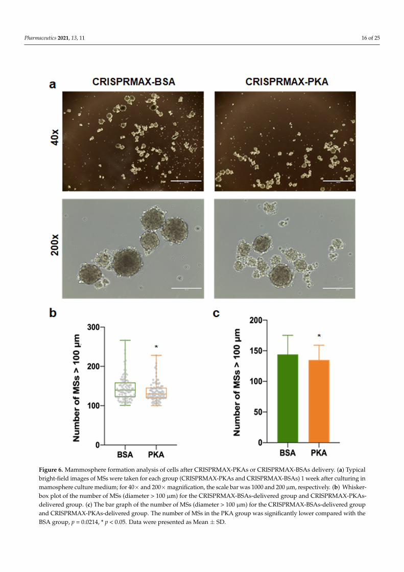
As mentioned earlier, our immunofluorescence results showed that the expressionof EMT-related mesenchymal markers, α-SMA and N-cadherin, was decreased in thebreast cancer cells treated with PKA, indicating the suppression of the EMT process atthe molecular level (Figure 4). Other functional experiments were also performed toexamine the impact on CSCs after introducing PKAs. Flow cytometry detection of changesin cell surface markers is a common method for rapid detection of changes in the CSCpopulation. It is believed that the CD44+/CD24− population can enrich the CSC populationin breast cancer cells [33]. Therefore, we performed flow cytometry analysis to examine theCD44/CD24 population in four different kinds of breast cancer cells (BT-549, MDA-MB-231,Hs-578T, and MCF-7 cells) 3 to 5 days after CRISPRMAX-PKAs delivery. Compared withthe CRISPRMAX-BSAs group, the CD44+/CD24− population of the CRISPRMAX-PKAsgroup did not significantly decrease (Figure 5). Next, we utilized the mammosphereassay, which is considered as one of the most effective evaluation methods of breast CSCsin vitro [34], to evaluate the biological effects of CRISPRMAX-PKAs delivery on breastcancer cells. The size and number of mammospheres are related to the mamosphere-formation ability that represents the stemness of the CSCs in the culture process. Here, wefocused on the relatively large size of MSs (diameter > 100 µm). Our results showed thatthe size of MSs grown in the mammosphere culture system for 7 days was significantlyreduced after CRISPRMAX-PKAs delivery (Figure 6). The diameter of CRISPRMAX-PKAswas 134.915 ± 24.123 µm compared with that of CRISPRMAX-BSAs (144.163 ± 31.121 µm)(p = 0.0214) (Figure 6b,c). Together, these data suggested that CRISPRMAX-based PKAdelivery decreased the expression of mesenchymal markers (α-SMA and N-cadherin) andreduced the mammosphere-forming capacity of breast cancer cells.
3.4. Analysis of the Synergy Effects of CRISPRMAX-Based PKA Delivery along withChemotherapy Drugs
To explore the effects of co-treatment of breast cancer cells with PKA along with thechemotherapy drug (PTX or DOX), and whether the transfection with PKA could increasethe chemosensitivity of breast cancer cells, the experimental setups were designed underdifferent transfection times: 0 and 12 h, then PTX or DOX were added after transfection.Data of cell viability were analyzed, and we also performed area under curve (AUC)analysis for measurements of the effectiveness of treatments between BSA (control) andPKA transfection (Figures 7 and 8).
3.4.1. Analysis of Synergy Effect of CRISPRMAX-Based PKA Delivery and DOX in BreastCancer Cells
The breast cancer cells (MCF-7, BT-549, and Hs-578T) were transfected with 500 ng ofPKA and treated with DOX at 0, 0.1, 0.5, 1, and 1.5 µg/mL at the same time for 48 h. Theresults of WST-1 analysis showed that the cell viability was significantly increased in MCF-7, BT-549, and Hs-578T (statically significant comparing with BSA, p = 0.0294, p = 0.0002,p = 0.0425, respectively) shown in Figure 7a,c,e. The cell viability greatly increased withthe treatment with DOX following 12 h of PKA transfection in MCF-7 and BT-549 cells(statically significant compared with BSA, p = 0.0067, p = 0.0043, respectively) shown inFigure 7b,d. These results demonstrated the treatment with CRISPRMAX-PKAs along withDOX (neither at 0 h nor at 12 h of CRISPRMAX-based PKA delivery) did not improve thesensitivity to DOX but had opposite effects that caused the growth of breast cancer cellscompared with controls.
3.4.2. Analysis of Synergy Effect of CRISPRMAX-PKAs Delivery and PTX in BreastCancer Cells
The breast cancer cells (MCF-7, BT-549, and Hs-578T) were transfected with 500 ngof PKA and treated with PTX at 0, 0.05, 0.1, 0.2, and 0.5 µg/mL at the same time for 48 h.When treating breast cancer cells with PTX and CRISPRMAX-PKAs simultaneously, theresults of cell viability and AUC showed that the PKA group was greatly higher compared

Pharmaceutics 2021, 13, 11 13 of 25
with the BSA group in BT-549 and Hs-578T cells (statically significant compared with theBSA group, p = 0.0004, p = 0.0088, respectively); as for MCF-7 cells, there was no differencebetween PKA and the control in these two cell lines (Figure 8a,c,e). The results showedthat guiding PKAs through CRISPRMAX along with PTX at the same time provokedan increase of cell growth in BT-549 and Hs-578T. Whereas, adding PTX following 12 hof transfection with CRISPRMAX-PKAs in breast cancer cells, the cell viability was allremarkably decreased in MCF-7, BT-549, and Hs-578T (statically significant compared withthe BSA group, p = 0.0018, p = 0.0290, p = 0.0031, respectively) shown in Figure 8b,d,f,which indicates that PKAs must be guided into breast cancer cells to trigger the repressionof EMT before adding PTX, resulting in an increased sensitivity to PTX.
3.4.3. Analysis of the Synergy Effect of CRISPRMAX-PKA Delivery and PTX in NormalBreast Cells
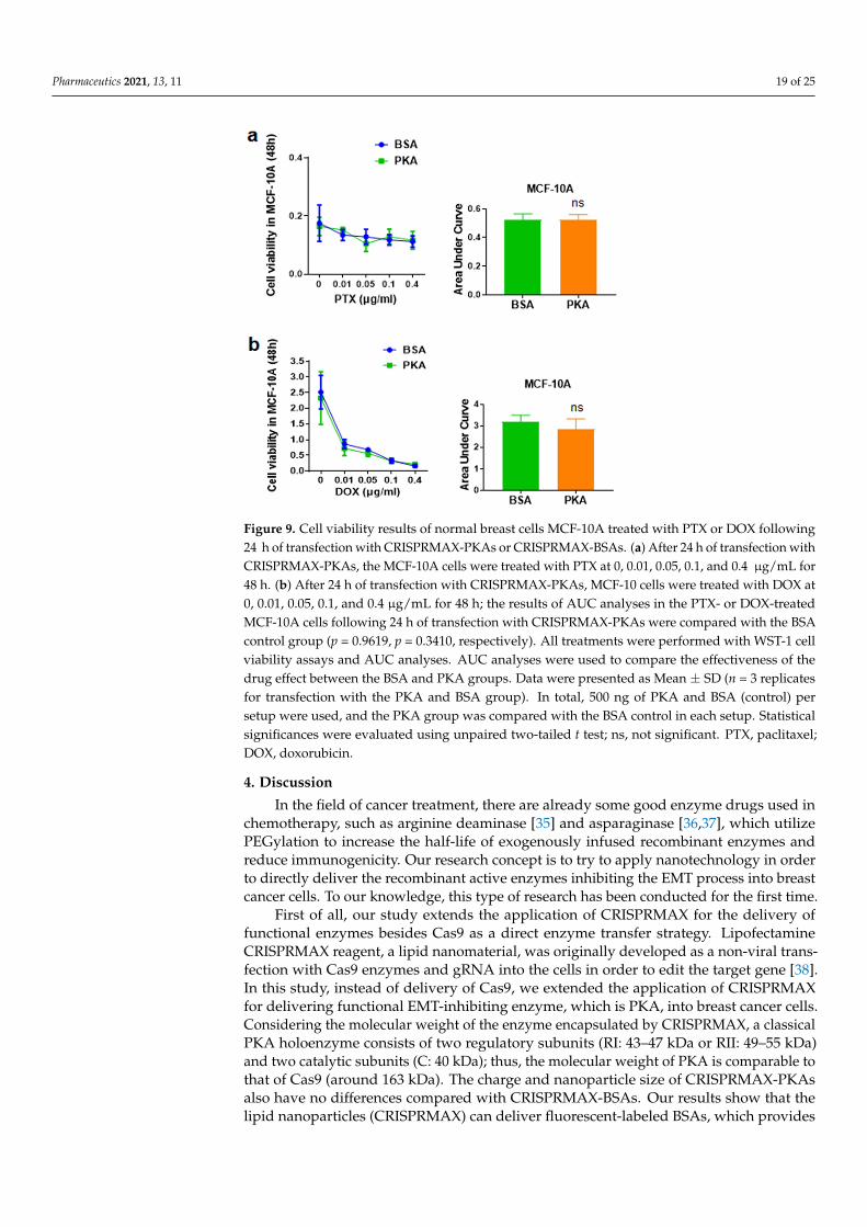
The side effect of chemotherapeutics lies in the non-selective killing of normal cells.In this study, we previously demonstrated that transfection with CRISPRMAX-PKAs caninitiate an inhibition process that represses EMT and CSCs in breast cancer cells, therebyfurther weakening chemoresistance in breast cancer cells. Does the same phenomenonalso occur in normal breast cells? To investigate whether PKA transfection can also affectsensitivity to chemotherapy drugs in normal cells, we delivered CRISPRMAX-PKAs alongwith PTX or DOX in normal breast epithelial cells MCF-10A. After 24 h of CRISPRMAX-PKAs delivery, different concentrations of PTX and DOX were added into the cells for a 48-hincubation. WST-1 was utilized for the detection of cell viability. The results demonstratedthat CRISPRMAX-PKAs delivery did not alter the effects of the 48-h treatment with PTXor DOX in MCF-10A cells (Figure 9), which further confirmed that CRISPRMAX-PKAsdelivery is specific to increase the sensitivity to PTX in mesenchymal breast cancer cellsrather than in normal breast cells.

Pharmaceutics 2021, 13, 11 14 of 25Pharmaceutics 2021, 13, x FOR PEER REVIEW 13 of 25
Figure 4. Expression analysis of E-cadherin, N-cadherin, and α-SMA after PKA delivery observed by CLSM. E-cadherin, N-cadherin, and α-SMA expression were examined by immunofluorescence analysis using confocal laser scanning mi-croscopy 5 days after CRISPRMAX-based delivery, with the control (BSA) (left panel) vs. with PKA (right panel). Cell nuclei were visualized by DAPI. Confocal images were taken at 200x magnification (scale bars, 50 μm).
Figure 4. Expression analysis of E-cadherin, N-cadherin, and α-SMA after PKA delivery observed by CLSM. E-cadherin, N-cadherin, and α-SMA expression were examined by immunofluorescence analysis using confocal laser scanning microscopy5 days after CRISPRMAX-based delivery, with the control (BSA) (left panel) vs. with PKA (right panel). Cell nuclei werevisualized by DAPI. Confocal images were taken at 200×magnification (scale bars, 50 µm).

Pharmaceutics 2021, 13, 11 15 of 25
Pharmaceutics 2021, 13, x FOR PEER REVIEW 14 of 25
Figure 5. FCM analysis of the CD44+/CD24- population in breast cancer cells after introducing CRISPRMAX-PKAs or CRISPRMAX-BSAs. (a) MCF-7 cells; (b) MDA-MB-231 cells; (c) BT-549 cells; (d) Hs-578T cells. The CD44+/CD24– antigenic phenotype was shown in the Q3 area (at the right lower corner of the individual image). For isotype group: cells trans-fected with ddH2O through CRISPRMAX as controls were incubated with the same amount of APC-isotype IgG and PE-isotype IgG with CD44-APC and CD24-PE antibodies; as for the BSA and PKA groups, the cells transfected with BSAs or PKAs were incubated with CD44-APC and CD24-PE antibodies. X-axis presents CD44-APC of the cells; Y-axis shows CD24-PE of the cells. All cells were delivered with PKAs or BSAs for 3 to 5 days. FCM, flow cytometry.
Figure 5. FCM analysis of the CD44+/CD24− population in breast cancer cells after introducing CRISPRMAX-PKAs orCRISPRMAX-BSAs. (a) MCF-7 cells; (b) MDA-MB-231 cells; (c) BT-549 cells; (d) Hs-578T cells. The CD44+/CD24− antigenicphenotype was shown in the Q3 area (at the right lower corner of the individual image). For isotype group: cells transfectedwith ddH2O through CRISPRMAX as controls were incubated with the same amount of APC-isotype IgG and PE-isotypeIgG with CD44-APC and CD24-PE antibodies; as for the BSA and PKA groups, the cells transfected with BSAs or PKAswere incubated with CD44-APC and CD24-PE antibodies. X-axis presents CD44-APC of the cells; Y-axis shows CD24-PE ofthe cells. All cells were delivered with PKAs or BSAs for 3 to 5 days. FCM, flow cytometry.
Pharmaceutics 2021, 13, 11 16 of 25
Pharmaceutics 2021, 13, x FOR PEER REVIEW 15 of 25
Figure 6. Mammosphere formation analysis of cells after CRISPRMAX-PKAs or CRISPRMAX-BSAs delivery. (a) Typical bright-field images of MSs were taken for each group (CRISPRMAX-PKAs and CRISPRMAX-BSAs) 1 week after cultur-ing in mamosphere culture medium; for 40x and 200x magnification, the scale bar was 1000 and 200 μm, respectively. (b) Whisker-box plot of the number of MSs (diameter > 100 μm) for the CRISPRMAX-BSAs-delivered group and CRIS-PRMAX-PKAs-delivered group. (c) The bar graph of the number of MSs (diameter > 100 μm) for the CRIS-PRMAX-BSAs-delivered group and CRISPRMAX-PKAs-delivered group. The number of MSs in the PKA group was significantly lower compared with the BSA group, p = 0.0214, * p < 0.05. Data were presented as Mean ± SD.
Figure 6. Mammosphere formation analysis of cells after CRISPRMAX-PKAs or CRISPRMAX-BSAs delivery. (a) Typicalbright-field images of MSs were taken for each group (CRISPRMAX-PKAs and CRISPRMAX-BSAs) 1 week after culturing inmamosphere culture medium; for 40× and 200×magnification, the scale bar was 1000 and 200 µm, respectively. (b) Whisker-box plot of the number of MSs (diameter > 100 µm) for the CRISPRMAX-BSAs-delivered group and CRISPRMAX-PKAs-delivered group. (c) The bar graph of the number of MSs (diameter > 100 µm) for the CRISPRMAX-BSAs-delivered groupand CRISPRMAX-PKAs-delivered group. The number of MSs in the PKA group was significantly lower compared with theBSA group, p = 0.0214, * p < 0.05. Data were presented as Mean ± SD.

Pharmaceutics 2021, 13, 11 17 of 25
Pharmaceutics 2021, 13, x FOR PEER REVIEW 16 of 25
Figure 7. Chemoresistance evaluation of DOX in the cells following 0 or 12 h transfection with CRISPRMAX-PKAs. (a,c,e) Adding DOX following 0 h of transfection with CRISPRMAX-PKAs: MCF-7, BT-549, and Hs-578T cells were transfected with PKA by CRISPRMAX and treated with DOX at 0, 0.1, 0.5, 1, and 1.5 μg/mL simultaneously for 48 h; the results of AUC analyses in MCF-7, BT-549, and Hs-578T were compared with the BSA control group (p = 0.02094, p = 0.0002, p = 0.0425, respectively). (b,d,f) Cells were treated with DOX for 48 h following 12 h of transfection with CRISPRMAX-PKAs; the results of AUC analyses in MCF-7, BT-549, and Hs-578T were compared with the BSA control group (p = 0.0067, p = 0.0043, p = 0.8200, respectively). AUC analyses were used to compare the effectiveness of the drug effect between the BSA and PKA group. All treatments were performed with WST-1 cell viability assays and AUC analyses. Data were presented as Mean ± SD (n = 3 replicates for transfection with the PKA and BSA group). In total, 500 ng of PKA and BSA (control) per setup were used, and the PKA group was compared with the BSA control in each setup. Statistical significances were evaluated using unpaired two-tailed t test; * p < 0.05, ** p < 0.01, *** p < 0.001, and ns, not significant. DOX, doxorubicin.
Figure 7. Chemoresistance evaluation of DOX in the cells following 0 or 12 h transfection with CRISPRMAX-PKAs. (a,c,e)Adding DOX following 0 h of transfection with CRISPRMAX-PKAs: MCF-7, BT-549, and Hs-578T cells were transfectedwith PKA by CRISPRMAX and treated with DOX at 0, 0.1, 0.5, 1, and 1.5 µg/mL simultaneously for 48 h; the results of AUCanalyses in MCF-7, BT-549, and Hs-578T were compared with the BSA control group (p = 0.02094, p = 0.0002, p = 0.0425,respectively). (b,d,f) Cells were treated with DOX for 48 h following 12 h of transfection with CRISPRMAX-PKAs; theresults of AUC analyses in MCF-7, BT-549, and Hs-578T were compared with the BSA control group (p = 0.0067, p = 0.0043,p = 0.8200, respectively). AUC analyses were used to compare the effectiveness of the drug effect between the BSA and PKAgroup. All treatments were performed with WST-1 cell viability assays and AUC analyses. Data were presented as Mean ±SD (n = 3 replicates for transfection with the PKA and BSA group). In total, 500 ng of PKA and BSA (control) per setup wereused, and the PKA group was compared with the BSA control in each setup. Statistical significances were evaluated usingunpaired two-tailed t test; * p < 0.05, ** p < 0.01, *** p < 0.001, and ns, not significant. DOX, doxorubicin.

Pharmaceutics 2021, 13, 11 18 of 25Pharmaceutics 2021, 13, x FOR PEER REVIEW 17 of 25
Figure 8. Chemoresistance assay of PTX in the cells following 0 or 12 h of transfection with CRISPRMAX-PKAs. (a,c,e) Adding PTX following 0 h of transfection with CRISPRMAX-PKAs: MCF-7, BT-549, and Hs-578T cells were transfected with PKA by CRISPRMAX and treated with PTX at 0, 0.05, 0.1, 0.2, and 0.5 μg/mL simultaneously for 48 h; the results of AUC analyses in MCF-7, BT-549, and Hs-578T were compared with the BSA control group (p = 0.1652, p = 0.0004, p = 0.0088, respectively). (b,d,f) Cells were treated with PTX for 48 h following 12 h of transfection with CRISPRMAX-PKAs; the results of AUC analyses in MCF-7, BT-549, and Hs-578T were compared with the BSA control group (p = 0.0018, p = 0.0290, p = 0.0031, respectively). AUC analyses were used to compare the effectiveness of drug effect between the BSA and PKA groups. All treatments were performed with WST-1 cell viability assays and AUC analyses. Data were presented as Mean ± SD (n = 3 replicates for transfection with the PKA and BSA group). In total, 500 ng of PKA and BSA (control) per setup were used, and the PKA group was compared with the BSA control in each setup. Statistical significances were evaluated using unpaired two-tailed t test; * p < 0.05, ** p < 0.01, *** p < 0.001, and ns, not significant. PTX, paclitaxel.
3.4.1. Analysis of Synergy Effect of CRISPRMAX-Based PKA Delivery and DOX in Breast Cancer Cells
The breast cancer cells (MCF-7, BT-549, and Hs-578T) were transfected with 500 ng of PKA and treated with DOX at 0, 0.1, 0.5, 1, and 1.5 μg/mL at the same time for 48 h. The results of WST-1 analysis showed that the cell viability was significantly increased in MCF-7, BT-549, and Hs-578T (statically significant comparing with BSA, p = 0.0294, p = 0.0002, p = 0.0425, respectively) shown in Figure 7a,c,e. The cell viability greatly increased with the treatment with DOX following 12 h of PKA transfection in MCF-7 and BT-549 cells (statically significant compared with BSA, p = 0.0067, p = 0.0043, respectively) shown in Figure 7b,d. These results demonstrated the treatment with CRISPRMAX-PKAs along with DOX (neither at 0 h nor at 12 h of CRISPRMAX-based PKA delivery) did not im-
Figure 8. Chemoresistance assay of PTX in the cells following 0 or 12 h of transfection with CRISPRMAX-PKAs. (a,c,e)Adding PTX following 0 h of transfection with CRISPRMAX-PKAs: MCF-7, BT-549, and Hs-578T cells were transfectedwith PKA by CRISPRMAX and treated with PTX at 0, 0.05, 0.1, 0.2, and 0.5 µg/mL simultaneously for 48 h; the results ofAUC analyses in MCF-7, BT-549, and Hs-578T were compared with the BSA control group (p = 0.1652, p = 0.0004, p = 0.0088,respectively). (b,d,f) Cells were treated with PTX for 48 h following 12 h of transfection with CRISPRMAX-PKAs; theresults of AUC analyses in MCF-7, BT-549, and Hs-578T were compared with the BSA control group (p = 0.0018, p = 0.0290,p = 0.0031, respectively). AUC analyses were used to compare the effectiveness of drug effect between the BSA and PKAgroups. All treatments were performed with WST-1 cell viability assays and AUC analyses. Data were presented as Mean ±SD (n = 3 replicates for transfection with the PKA and BSA group). In total, 500 ng of PKA and BSA (control) per setup wereused, and the PKA group was compared with the BSA control in each setup. Statistical significances were evaluated usingunpaired two-tailed t test; * p < 0.05, ** p < 0.01, *** p < 0.001, and ns, not significant. PTX, paclitaxel.
Pharmaceutics 2021, 13, 11 19 of 25
Pharmaceutics 2021, 13, x FOR PEER REVIEW 19 of 25
Figure 9. Cell viability results of normal breast cells MCF-10A treated with PTX or DOX following 24 h of transfection with CRISPRMAX-PKAs or CRISPRMAX-BSAs. (a) After 24 h of transfection with CRISPRMAX-PKAs, the MCF-10A cells were treated with PTX at 0, 0.01, 0.05, 0.1, and 0.4 μg/mL for 48 h. (b) After 24 h of transfection with CRISPRMAX-PKAs, MCF-10 cells were treated with DOX at 0, 0.01, 0.05, 0.1, and 0.4 μg/mL for 48 h; the results of AUC analyses in the PTX- or DOX-treated MCF-10A cells following 24 h of transfection with CRISPRMAX-PKAs were com-pared with the BSA control group (p = 0.9619, p = 0.3410, respectively). All treatments were per-formed with WST-1 cell viability assays and AUC analyses. AUC analyses were used to compare the effectiveness of the drug effect between the BSA and PKA groups. Data were presented as Mean ± SD (n = 3 replicates for transfection with the PKA and BSA group). In total, 500 ng of PKA and BSA (control) per setup were used, and the PKA group was compared with the BSA control in each setup. Statistical significances were evaluated using unpaired two-tailed t test; ns, not signif-icant. PTX, paclitaxel; DOX, doxorubicin.
4. Discussion In the field of cancer treatment, there are already some good enzyme drugs used in
chemotherapy, such as arginine deaminase [35] and asparaginase [36,37], which utilize PEGylation to increase the half-life of exogenously infused recombinant enzymes and reduce immunogenicity. Our research concept is to try to apply nanotechnology in order to directly deliver the recombinant active enzymes inhibiting the EMT process into breast cancer cells. To our knowledge, this type of research has been conducted for the first time.
First of all, our study extends the application of CRISPRMAX for the delivery of functional enzymes besides Cas9 as a direct enzyme transfer strategy. Lipofectamine CRISPRMAX reagent, a lipid nanomaterial, was originally developed as a non-viral transfection with Cas9 enzymes and gRNA into the cells in order to edit the target gene [38]. In this study, instead of delivery of Cas9, we extended the application of CRIS-PRMAX for delivering functional EMT-inhibiting enzyme, which is PKA, into breast cancer cells. Considering the molecular weight of the enzyme encapsulated by CRIS-PRMAX, a classical PKA holoenzyme consists of two regulatory subunits (RI: 43–47 kDa or RII: 49–55 kDa) and two catalytic subunits (C: 40 kDa); thus, the molecular weight of PKA is comparable to that of Cas9 (around 163 kDa). The charge and nanoparticle size of
Figure 9. Cell viability results of normal breast cells MCF-10A treated with PTX or DOX following24 h of transfection with CRISPRMAX-PKAs or CRISPRMAX-BSAs. (a) After 24 h of transfection withCRISPRMAX-PKAs, the MCF-10A cells were treated with PTX at 0, 0.01, 0.05, 0.1, and 0.4 µg/mL for48 h. (b) After 24 h of transfection with CRISPRMAX-PKAs, MCF-10 cells were treated with DOX at0, 0.01, 0.05, 0.1, and 0.4 µg/mL for 48 h; the results of AUC analyses in the PTX- or DOX-treatedMCF-10A cells following 24 h of transfection with CRISPRMAX-PKAs were compared with the BSAcontrol group (p = 0.9619, p = 0.3410, respectively). All treatments were performed with WST-1 cellviability assays and AUC analyses. AUC analyses were used to compare the effectiveness of thedrug effect between the BSA and PKA groups. Data were presented as Mean ± SD (n = 3 replicatesfor transfection with the PKA and BSA group). In total, 500 ng of PKA and BSA (control) persetup were used, and the PKA group was compared with the BSA control in each setup. Statisticalsignificances were evaluated using unpaired two-tailed t test; ns, not significant. PTX, paclitaxel;DOX, doxorubicin.
4. Discussion
In the field of cancer treatment, there are already some good enzyme drugs used inchemotherapy, such as arginine deaminase [35] and asparaginase [36,37], which utilizePEGylation to increase the half-life of exogenously infused recombinant enzymes andreduce immunogenicity. Our research concept is to try to apply nanotechnology in orderto directly deliver the recombinant active enzymes inhibiting the EMT process into breastcancer cells. To our knowledge, this type of research has been conducted for the first time.
First of all, our study extends the application of CRISPRMAX for the delivery offunctional enzymes besides Cas9 as a direct enzyme transfer strategy. LipofectamineCRISPRMAX reagent, a lipid nanomaterial, was originally developed as a non-viral trans-fection with Cas9 enzymes and gRNA into the cells in order to edit the target gene [38].In this study, instead of delivery of Cas9, we extended the application of CRISPRMAXfor delivering functional EMT-inhibiting enzyme, which is PKA, into breast cancer cells.Considering the molecular weight of the enzyme encapsulated by CRISPRMAX, a classicalPKA holoenzyme consists of two regulatory subunits (RI: 43–47 kDa or RII: 49–55 kDa)and two catalytic subunits (C: 40 kDa); thus, the molecular weight of PKA is comparable tothat of Cas9 (around 163 kDa). The charge and nanoparticle size of CRISPRMAX-PKAsalso have no differences compared with CRISPRMAX-BSAs. Our results show that thelipid nanoparticles (CRISPRMAX) can deliver fluorescent-labeled BSAs, which provides

Pharmaceutics 2021, 13, 11 20 of 25
indirect evidence to support the notion that CRISPRMAX is able to deliver PKAs. Ourimmunofluorescence analysis of PKA expression further supports the notion that PKAenzymes can be delivered into breast cancer cells through CRISPRMAX. The establishmentof this method helps to quickly introduce potential therapeutic proteins or enzymes intotumor cells in order to evaluate their therapeutic effects (around 3 to 7 days) instead oftranscription or translation of the constructed nucleotide fragments, leading to the removalof risk of genomic integration.
Second, our study suggests a strategy that, combining EMT-targeting and the conven-tional chemotherapy, can improve the therapeutic effects of breast cancer treatment butrequires careful design of the dosing regimen. EMT is a key biological process for promot-ing tumor cells to dedifferentiate to new CSCs after external stimulation, especially afterradiotherapy and chemotherapy [39]. Many regulatory factors are involved in this process,including non-coding RNAs, proteins, enzymes, etc. When it comes to enzymes, most ofthem are overexpressed in the EMT process, which should be knockdown, and thereforeare not suitable for the enzyme delivery treatment strategy. Recently, the cAMP/PKAsignaling pathway was revealed to play a role in MET induction in mesenchymal breastcancer cells, which elicits their differentiation toward epithelial phenotypes, resulting inthe loss of tumor-initiating capability [9]. Thus, we selected PKA as the model enzyme forinvestigating the biopharmaceutical effects of CRISPRMAX-encapsulation of functionalenzymes. A recent study reported that the EMT process is not required for the developmentof lung metastasis but contributes to chemoresistance [40]. Therefore, the clinically relevanteffect that we focused on in this study is chemoresistance. We selected two commonchemotherapy drugs (paclitaxel and doxorubicin) with different mechanisms of actionfor combination therapy with PKA delivery, respectively. Paclitaxel is one of several cy-toskeletal drugs that target tubulin. Paclitaxel-treated cells have defects in mitotic spindleassembly, chromosome segregation, and cell division [41,42]. Doxorubicin interacts withDNA by intercalation and inhibition of macromolecular biosynthesis [43]. Our study foundthat there is no synergistic effect of PKA and DOX, but a good synergistic effect of PKAand PTX on the inhibition of chemoresistance has been confirmed. Interestingly, in orderto have good synergy effect of PKA and PTX, the breast cancer cells must be transfectedwith PKA for 12 h before adding PTX. If PTX and PKA are carried out at the same time,it will not only fail to inhibit tumor cells but even promote their growth. This indicatesa beforehand process related to EMT inhibition or differentiation is necessary for suchsynergy (an overview diagram is shown in Figure 10).
When we use nanoparticles for combining EMT-targeting and conventional chemother-apy, we cannot always carry out the commonly used strategy that is the so-called “one-stopsolution”, which loads two drugs in one nanoparticle at the same time [26], but need toconsider taking a sequential solution strategy in terms of delivering combinational thera-peutics in a single nanoparticle. Therefore, using nanoparticles for combinational therapyremains challenging. The significance of this study is that we discovered that deliveringPKA enzymes first before administrating PTX can have a good synergy in EMT inhibitionas well as chemosensitivity for breast cancer cells.
Why is there no synergy effect of PKA delivery and DOX, whereas adding PKAalong with PTX has a good synergy? So far, the exact mechanism of such a phenomenonis not clear. Considering our results, PKA delivery inhibits EMT in breast cancer cells,resulting in a decrease in α-SMA expression. α-SMA is an actin protein that belongs to theactin cytoskeletons, and paclitaxel targets another type of cytoskeleton tubulin; based onthese two facts, we hypothesized that the synergy effects of PKA and PTX may be relatedto cytoskeletal remodeling [44], which is also involved in driving the EMT process [45].However, such a hypothesis still requires further investigation in the future.
Except for liposomes, there are also many other functionalized nanosystems that canbe used to deliver enzymes, such as polymer nanoparticles [23], polymersomes [24], meso-porous nanoparticles [25], metal-organic framework (MOF) with biomineralization [26],DNA nanoparticles [27,28], emulsions [29], etc. In detail, for mesoporous silica nanopar-
Pharmaceutics 2021, 13, 11 21 of 25
ticles (MSNs), it is reported that enzymes can be loaded in the cavity of the pores in asize-selective adsorption manner [46]. Polymers, including microspheres, polymer-coatedsubstrates as small capsules (known as microencapsulation), hydrogels, and nanoparticles,can protect the protein drugs from premature degradation. Among these materials, polylac-tic acid (PLA) and poly(lactic-co-glycolic acid) (PLGA) are the most common biodegradablematerials used in the development of protein microspheres [47]. Most importantly, a newkind of MOF, such as a zeolitic imidazolate framework, ZIF-8, can store biologically activeenzymes in vitro for a long time [48]. In principle, enzyme-ZIF8 nanocomposition can bedelivered into cells by endocytosis and release the delivered enzymes in a pH-dependentmanner in the endosome/lysosome [49].
For characterization of PKAs- and BSAs-loaded CRISPRMAX complexes, dynamiclight scattering (DLS) measurements were performed for analyzing the particle size, poly-dispersity index (PDI), and zeta potential of the enzymes-loaded CRISPRMAX complexes.In in vitro studies of using LNPs in therapeutics delivery, lipid-based particles with a PDIvalue of 0.3 or below are considered to be acceptable carriers for delivering therapeutics,as such vesicles are homogeneously distributed in the solution [50,51]; the PDI valuesof our enzymes-loaded CRISPRMAX complexes and null CRISPRMAX were also withinthis range, which indicated these complexes were evenly distributed in the solution. Inaddition, the average particle size of CRISPRMAX-PKAs, CRISPRMAX-BSAs, and nullCRISPRMAX was approximately 350 nm; these complexes can be easily internalized bytargeted cells, since their size is less than 500 nm [52]. Zeta potential can provide generalinformation of the surface charge properties of nanoparticles; however, such an indicatorhas its limitations as the surface charge of nanoparticles can be significantly affected by thesurrounding environment; small changes in any of these parameters, such as temperature,pH, conductivity (a parameter that determines the ionic strength of a solution), and vis-cosity of solvent, etc., have a profound impact on zeta potential value [53]. Therefore, ourfocus is not on the zeta potential value itself but to examine whether the zeta potential ofthe complexes is changed or not when loading different proteins through CRISPRMAXunder the same condition. Our results showed that there was no significant difference inthe surface charge of CRISPRMAX-PKAs and CRISPRMAX-BSAs complexes comparedwith control null CRISPRMAX. This is consistent with the results of Marija Brgles et al.that negatively charged protein could not influence the overall charge of liposomes [32].Moreover, PKAs- and BSAs-loaded CRISPRMAX complexes can also be directly deliveredto cells, as the lipid-based complexes can be delivered through cell internalization, directlyfusing with the cell membrane [54]. This may be the reason why CRISPRMAX reagent hashigh transfection efficiency in cells.
Indeed, we have to admit the biggest shortcoming of such LNPs is that they cannotstably store target enzymes in vitro for a long time [12]. So, if considering potentialadministration in the clinic in the future, CRISPRMAX or CRISPRMAX-like reagent andenzymes (such as PKAs) should be stored separately at 4 and −20 ◦C, respectively. Whenadministering the complex solution, PKA enzymes and CRISPRMAX can be mixed at roomtemperature before delivery into the targeted cancer cells. Solid nanoparticles, such as MOF,can store biologically active enzymes in vitro for a long time [26,48], of which the synthesisprocess is relatively easy to manipulate. However, compared with LNPs, it is not yeteasy to perform a high-throughput preparation of enzyme-MOF complexes and functionalscreening in seeded cells. In short, it is necessary to choose these enzyme-encapsulationdelivery strategies with each specific advantage according to the research purpose.
In summary, as a proof-of-concept, we confirmed the direct enzyme delivery of PKAas a potential strategy for inhibiting EMT/CSC-associated traits, including downregulationof the expression of EMT-related markers α-SMA and N-cad, chemoresistance, and mamo-spheres. PKA delivery has a significant inhibitory effect to PTX resistance but has no effectagainst DOX. The inhibitory effect of chemoresistance can be exerted only in the cells thatwere treated with PKA before PTX administration, which will shed light on the constructionof a new drug delivery system or complex nanoparticles with a combinational therapy that

Pharmaceutics 2021, 13, 11 22 of 25
targets both EMT/CSCs and bulk cancer cells. This direct enzyme delivery strategy willalso facilitate the testing of target enzymes/proteins on their biological functions.
Pharmaceutics 2021, 13, x FOR PEER REVIEW 20 of 25
CRISPRMAX-PKAs also have no differences compared with CRISPRMAX-BSAs. Our results show that the lipid nanoparticles (CRISPRMAX) can deliver fluorescent-labeled BSAs, which provides indirect evidence to support the notion that CRISPRMAX is able to deliver PKAs. Our immunofluorescence analysis of PKA expression further supports the notion that PKA enzymes can be delivered into breast cancer cells through CRISPRMAX. The establishment of this method helps to quickly introduce potential therapeutic pro-teins or enzymes into tumor cells in order to evaluate their therapeutic effects (around 3 to 7 days) instead of transcription or translation of the constructed nucleotide fragments, leading to the removal of risk of genomic integration.
Second, our study suggests a strategy that, combining EMT-targeting and the con-ventional chemotherapy, can improve the therapeutic effects of breast cancer treatment but requires careful design of the dosing regimen. EMT is a key biological process for promoting tumor cells to dedifferentiate to new CSCs after external stimulation, espe-cially after radiotherapy and chemotherapy [39]. Many regulatory factors are involved in this process, including non-coding RNAs, proteins, enzymes, etc. When it comes to en-zymes, most of them are overexpressed in the EMT process, which should be knock-down, and therefore are not suitable for the enzyme delivery treatment strategy. Re-cently, the cAMP/PKA signaling pathway was revealed to play a role in MET induction in mesenchymal breast cancer cells, which elicits their differentiation toward epithelial phenotypes, resulting in the loss of tumor-initiating capability [9]. Thus, we selected PKA as the model enzyme for investigating the biopharmaceutical effects of CRIS-PRMAX-encapsulation of functional enzymes. A recent study reported that the EMT process is not required for the development of lung metastasis but contributes to chemoresistance [40]. Therefore, the clinically relevant effect that we focused on in this study is chemoresistance. We selected two common chemotherapy drugs (paclitaxel and doxorubicin) with different mechanisms of action for combination therapy with PKA delivery, respectively. Paclitaxel is one of several cytoskeletal drugs that target tubulin. Paclitaxel-treated cells have defects in mitotic spindle assembly, chromosome segrega-tion, and cell division [41,42]. Doxorubicin interacts with DNA by intercalation and in-hibition of macromolecular biosynthesis [43]. Our study found that there is no synergis-tic effect of PKA and DOX, but a good synergistic effect of PKA and PTX on the inhibi-tion of chemoresistance has been confirmed. Interestingly, in order to have good syner-gy effect of PKA and PTX, the breast cancer cells must be transfected with PKA for 12 h before adding PTX. If PTX and PKA are carried out at the same time, it will not only fail to inhibit tumor cells but even promote their growth. This indicates a beforehand pro-cess related to EMT inhibition or differentiation is necessary for such synergy (an over-view diagram is shown in Figure 10).
Figure 10. An overview schematic diagram of the study. PKAs and CRISPRMAX had formed as lipid nanoparticle com-plexes, and then these complexes were added to breast cancer cells. At the same time, chemotherapy drugs PTX or DOX were added (at 0 h transfection time point) into the cells for 48 h, respectively. The WST-1 assay was performed for testing the cell viability. The results showed that compared with the CRISPRMAX-BSAs control group added with the same drug, cell viability increased regardless of whether PTX or DOX was added. When CRISPRMAX-PKAs were added into
Figure 10. An overview schematic diagram of the study. PKAs and CRISPRMAX had formed as lipid nanoparticlecomplexes, and then these complexes were added to breast cancer cells. At the same time, chemotherapy drugs PTX orDOX were added (at 0 h transfection time point) into the cells for 48 h, respectively. The WST-1 assay was performedfor testing the cell viability. The results showed that compared with the CRISPRMAX-BSAs control group added withthe same drug, cell viability increased regardless of whether PTX or DOX was added. When CRISPRMAX-PKAs wereadded into cells for 12 h, the chemotherapy drugs PTX or DOX were also added (at 12 h), respectively; the cells weretreated for 48 h. WST-1 cell viability results showed that compared with the CRISPRMAX-BSAs control group added withthe same drug, the cell viability of the PTX group was significantly decreased compared to the DOX group. Then, 24 hafter CRISPRMAX-PKAs delivery into the cells, these cells were digested with trypsin and seeded in a confocal dish for3 days, followed by immunofluorescence analysis of PKA expression. The results showed that the expression of PKAswas upregulated compared with the CRISPRMAX-BSAs control group; the expression of EMT mesenchymal markers,N-cadherin and α-SMA, was downregulated. Part of the cells were seeded in an ultra-low attachment dish at low densityand subjected to mammosphere formation assay; these cells were cultured in mammosphere culture medium and continuedto grow for 7 days, the results showed that CRISPRMAX-PKAs-delivered cells-derived mammospheres were decreased insize compared with the CRISPRMAX-BSAs control group.
Author Contributions: Conceptualization, H.Z., J.-N.Z., J.E. and Y.G.; Methodology, J.-N.Z., T.-C.R.,C.L., X.-Y.X. and D.-Q.W.; Validation, J.-N.Z., T.-C.R. and Y.G.; Formal Analysis, J.-N.Z., T.-C.R., Y.G.and H.Z.; Investigation, J.-N.Z., T.-C.R. and Y.G.; Resources, C.L., X.-Y.X. and H.Z.; Data Curation,J.-N.Z. and T.-C.R.; Writing—Original Draft Preparation, J.-N.Z. and T.-C.R.; Writing—Review &Editing, J.-N.Z., T.-C.R., Y.G., J.E. and H.Z.; Visualization, J.-N.Z., T.-C.R. and C.L.; Supervision, J.E.and H.Z.; Funding Acquisition, H.Z., J.-N.Z., Y.G. and D.-Q.W. All authors have read and agreed tothe published version of the manuscript.
Funding: This work was supported by Distinguished Clinical Investigator Grant of Jiangsu Province,China (Grant No. JSTP201701), Jiangsu Provincial Key Research and Development Programme(Grant No. BE2018690), Academy of Finland (Grant No. 328933) and Sigrid Jusélius Foundation(decision no. 28002247K1) and the program of China Scholarships Council (No. 201803170073,No.201909110009).
Institutional Review Board Statement: Not applicable.
Informed Consent Statement: Not applicable.
Data Availability Statement: Data are contained within the article.
Acknowledgments: We are grateful for Jukka Westermarck (Turku Bioscience, Turku, Finland) forproviding cell lines. Imaging/Flow cytometry was performed at the Cell Imaging Core, TurkuBioscience, Turku, Finland. Biocenter Finland is acknowledged for the imaging/flow cytometryinstrumentation.
Conflicts of Interest: The authors declare no conflict of interest.
Pharmaceutics 2021, 13, 11 23 of 25
References1. Desai, A.; Yan, Y.; Gerson, S.L. Concise Reviews: Cancer Stem Cell Targeted Therapies: Toward Clinical Success. Stem Cells Transl.
Med. 2019, 8, 75–81. [CrossRef]2. Mani, S.A.; Guo, W.; Liao, M.-J.; Eaton, E.N.; Ayyanan, A.; Zhou, A.Y.; Brooks, M.; Reinhard, F.; Zhang, C.C.; Shipitsin, M.; et al.
The Epithelial-Mesenchymal Transition Generates Cells with Properties of Stem Cells. Cell 2008, 133, 704–715. [CrossRef]3. Polyak, K.; Weinberg, R.A. Transitions between epithelial and mesenchymal states: Acquisition of malignant and stem cell traits.
Nat. Rev. Cancer 2009, 9, 265–273. [CrossRef]4. Voon, D.C.-C.; Huang, R.Y.; Jackson, R.A.; Thiery, J.P. The EMT spectrum and therapeutic opportunities. Mol. Oncol. 2017, 11,
878–891. [CrossRef]5. Vellard, M. The enzyme as drug: Application of enzymes as pharmaceuticals. Curr. Opin. Biotechnol. 2003, 14, 444–450. [CrossRef]6. Wang, X.; Yin, L.; Yang, L.; Zheng, Y.; Liu, S.; Yang, J.; Cui, H.; Wang, H. Silencing ubiquitin-conjugating enzyme 2C inhibits
proliferation and epithelial–mesenchymal transition in pancreatic ductal adenocarcinoma. FEBS J. 2019, 286, 4889–4909. [CrossRef]7. Lucena, M.C.; Carvalho-Cruz, P.; Donadio, J.L.; Oliveira, I.A.; De Queiroz, R.M.; Marinho-Carvalho, M.M.; Sola-Penna, M.; De
Paula, I.F.; Gondim, K.C.; McComb, M.E.; et al. Epithelial Mesenchymal Transition Induces Aberrant Glycosylation throughHexosamine Biosynthetic Pathway Activation. J. Biol. Chem. 2016, 291, 12917–12929. [CrossRef]
8. Kotiyal, S.; Bhattacharya, S. Breast cancer stem cells, EMT and therapeutic targets. Biochem. Biophys. Res. Commun. 2014, 453,112–116. [CrossRef]
9. Pattabiraman, D.R.; Bierie, B.; Kober, K.I.; Thiru, P.; Krall, J.A.; Zill, C.; Reinhardt, F.; Tam, W.L.; Weinberg, R.A. Activation of PKAleads to mesenchymal-to-epithelial transition and loss of tumor-initiating ability. Science 2016, 351, aad3680. [CrossRef]
10. Xin, Y.; Yin, M.; Zhao, L.; Meng, F.; Luo, L. Recent progress on nanoparticle-based drug delivery systems for cancer therapy.Cancer Biol. Med. 2017, 14, 228–241. [CrossRef]
11. Zheng, Y.; Li, Z.; Chen, H.; Gao, Y. Nanoparticle-based drug delivery systems for controllable photodynamic cancer therapy.Eur. J. Pharm. Sci. 2020, 144, 105213. [CrossRef] [PubMed]
12. Sercombe, L.; Veerati, T.; Moheimani, F.; Wu, S.Y.; Sood, A.K.; Hua, S. Advances and Challenges of Liposome Assisted DrugDelivery. Front. Pharmacol. 2015, 6, 286. [CrossRef] [PubMed]
13. Anselmo, A.C.; Mitragotri, S. Nanoparticles in the clinic. Bioeng. Transl. Med. 2016, 1, 10–29. [CrossRef]14. Koudelka, S.; Turánek, J. Liposomal paclitaxel formulations. J. Control. Release 2012, 163, 322–334. [CrossRef]15. Dave, V.; Yadav, R.B.; Kushwaha, K.; Yadav, S.; Sharma, S.; Agrawal, U. Lipid-polymer hybrid nanoparticles: Development &
statistical optimization of norfloxacin for topical drug delivery system. Bioact. Mater. 2017, 2, 269–280. [CrossRef] [PubMed]16. Zhou, J.-N.; Zeng, Q.; Wang, H.-Y.; Zhang, B.; Li, S.-T.; Nan, X.; Cao, N.; Fu, C.-J.; Yan, X.-L.; Jia, Y.L.; et al. MicroRNA-
125b attenuates epithelial-mesenchymal transitions and targets stem-like liver cancer cells through small mothers againstdecapentaplegic 2 and 4. Hepatology 2015, 62, 801–815. [CrossRef]
17. Zhao, P.; Hou, X.; Yan, J.; Du, S.; Xue, Y.; Li, W.; Xiang, G.; Dong, Y. Long-term storage of lipid-like nanoparticles for mRNAdelivery. Bioact. Mater. 2020, 5, 358–363. [CrossRef]
18. Wolfram, J.; Ferrari, M. Clinical cancer nanomedicine. Nano Today 2019, 25, 85–98. [CrossRef]19. Yu, X.; Liang, X.; Xie, H.; Kumar, S.; Ravinder, N.; Potter, J.; Jeu, X.D.M.D.; Chesnut, J.D. Improved delivery of Cas9 protein/gRNA
complexes using lipofectamine CRISPRMAX. Biotechnol. Lett. 2016, 38, 919–929. [CrossRef]20. Lim, D.; Sreekanth, V.; Cox, K.J.; Law, B.K.; Wagner, B.K.; Karp, J.M.; Choudhary, A. Engineering designer beta cells with a
CRISPR-Cas9 conjugation platform. Nat. Commun. 2020, 11, 1–11. [CrossRef]21. Ghasemi, R.; Struthers, H.; Wilson, E.R.; Spencer, D.H. Contribution of CTCF binding to transcriptional activity at the HOXA
locus in NPM1-mutant AML cells. Leukemia 2020, 1–13. [CrossRef] [PubMed]22. Kong, F.; Zhang, X.; Zhang, H.; Qu, X.; Chen, D.; Servos, M.R.; Mäkilä, E.; Salonen, J.; Santos, H.A.; Hai, M.; et al. Inhibition of Mul-
tidrug Resistance of Cancer Cells by Co-Delivery of DNA Nanostructures and Drugs Using Porous Silicon Nanoparticles@GiantLiposomes. Adv. Funct. Mater. 2015, 25, 3330–3340. [CrossRef]
23. Bauleth-Ramos, T.; Shahbazi, M.-A.; Liu, D.; Fontana, F.; Correia, A.; Figueiredo, P.; Zhang, H.B.; Martins, J.P.; Hirvonen,J.T.; Granja, P.; et al. Nutlin-3a and Cytokine Co-loaded Spermine-Modified Acetalated Dextran Nanoparticles for CancerChemo-Immunotherapy. Adv. Funct. Mater 2017, 27, 1703303. [CrossRef]
24. Zhang, H.; Cui, W.; Qu, X.; Wu, H.; Qu, L.; Zhang, X.; Mäkilä, E.; Salonen, J.; Zhu, Y.; Yang, Z.; et al. Photothermal-responsivenanosized hybrid polymersome as versatile therapeutics codelivery nanovehicle for effective tumor suppression. Proc. Natl. Acad.Sci. USA 2019, 116, 7744–7749. [CrossRef] [PubMed]
25. Kankala, R.K.; Zhang, H.; Liu, C.; Kanubaddi, K.R.; Lee, C.; Wang, S.; Cui, W.; Santos, H.A.; Lin, K.; Chen, A. Metal Species–Encapsulated Mesoporous Silica Nanoparticles: Current Advancements and Latest Breakthroughs. Adv. Funct. Mater. 2019, 29,1902652. [CrossRef]
26. Yan, J.; Liu, C.; Wu, Q.; Zhou, J.; Xu, X.; Zhang, L.; Wang, D.; Yang, F.; Zhang, H. Mineralization of ph-sensitive doxorubicinprodrug in zif-8 to enable targeted delivery to solid tumors. Anal. Chem. 2020, 92, 11453–11461. [CrossRef]
27. Zhang, Y.; Tu, J.; Wang, D.; Zhu, H.; Maity, S.K.; Qu, X.; Bogaert, B.; Pei, H.; Zhang, H. Programmable and multifunctionalDNA-based materials for biomedical applications. Adv. Mater. 2018, 30, e1703658. [CrossRef]

Pharmaceutics 2021, 13, 11 24 of 25
28. Auvinen, H.; Zhang, H.; Nonappa; Kopilow, A.; Niemela, E.H.; Nummelin, S.; Correia, A.; Santos, H.A.; Linko, V.; Kostiainen,M.A. Protein coating of DNA nanostructures for enhanced stability and immunocompatibility. Adv. Healthc. Mater. 2017, 6,1700692. [CrossRef]
29. Kong, F.; Zhang, H.; Qu, X.; Zhang, X.; Chen, D.; Ding, R.; Mäkilä, E.; Salonen, J.; Santos, H.A.; Hai, M. Gold Nanorods, DNAOrigami, and Porous Silicon Nanoparticle-functionalized Biocompatible Double Emulsion for Versatile Targeted Therapeuticsand Antibody Combination Therapy. Adv. Mater. 2016, 28, 10195–10203. [CrossRef]
30. Yan, X.-L.; Fu, C.-J.; Chen, L.; Qin, J.-H.; Zeng, Q.; Yuan, H.-F.; Nan, X.; Chen, H.-X.; Zhou, J.-N.; Lin, Y.-L.; et al. Mesenchymalstem cells from primary breast cancer tissue promote cancer proliferation and enhance mammosphere formation partially viaEGF/EGFR/Akt pathway. Breast Cancer Res. Treat. 2012, 132, 153–164. [CrossRef]
31. Zhang, B.; Wang, H.; Wang, D.; Zeng, Q.; Fan, Z.; Xi, J.; Nan, X.; He, L.; Zhou, J.-N.; Pei, X.; et al. A new protocol for long-termculture of a specific subpopulation of liver cancer stem cells enriched by cell surface markers. FEBS Open Bio 2020, 10, 1737–1747.[CrossRef] [PubMed]
32. Brgles, M.; Jurašin, D.D.; Sikiric, M.D.; Frkanec, R.; Tomasic, J. Entrapment of Ovalbumin into Liposomes—Factors AffectingEntrapment Efficiency, Liposome Size, and Zeta Potential. J. Liposome Res. 2008, 18, 235–248. [CrossRef] [PubMed]
33. Blick, T.; Hugo, H.J.; Widodo, E.; Waltham, M.; Pinto, C.; Mani, S.A.; Weinberg, R.A.; Neve, R.M.; Lenburg, M.E.; Thompson, E.W.Epithelial Mesenchymal Transition Traits in Human Breast Cancer Cell Lines Parallel the CD44hi/CD24lo/- Stem Cell Phenotypein Human Breast Cancer. J. Mammary Gland. Biol. Neoplasia 2010, 15, 235–252. [CrossRef] [PubMed]
34. Shaw, F.L.; Harrison, H.; Spence, K.; Ablett, M.P.; Simões, B.M.; Farnie, G.; Clarke, R.B. A Detailed Mammosphere Assay Protocolfor the Quantification of Breast Stem Cell Activity. J. Mammary Gland. Biol. Neoplasia 2012, 17, 111–117. [CrossRef]
35. Ensor, C.M.; Holtsberg, F.W.; Bomalaski, J.S.; Clark, M.A. Pegylated arginine deiminase (ADI-SS PEG20,000 mw) inhibits humanmelanomas and hepatocellular carcinomas in vitro and in vivo. Cancer Res. 2002, 62, 5443–5450.
36. Avramis, V.I.; Sencer, S.; Periclou, A.P.; Sather, H.; Bostrom, B.C.; Cohen, L.J.; Ettinger, A.G.; Ettinger, L.J.; Franklin, J.; Gaynon,P.S.; et al. A randomized comparison of native escherichia coli asparaginase and polyethylene glycol conjugated asparaginase fortreatment of children with newly diagnosed standard-risk acute lymphoblastic leukemia: A children’s cancer group study. Blood2002, 99, 1986–1994. [CrossRef]
37. Kurre, H.A.; Ettinger, A.G.; Veenstra, D.L.; Gaynon, P.S.; Franklin, J.; Sencer, S.F.; Reaman, G.H.; Lange, B.J.; Holcenberg, J.S.A Pharmacoeconomic Analysis of Pegaspargase Versus Native Escherichia Coli l-Asparaginase for the Treatment of Childrenwith Standard-Risk, Acute Lymphoblastic Leukemia: The Children’s Cancer Group Study (CCG-1962). J. Pediatr. Hematol. 2002,24, 175–181. [CrossRef]
38. Ali, H.S.; Boshra, M.S.; El Meteini, M.S.; Matboli, M.; Matboli, M. lncRNA- RP11-156p1.3, novel diagnostic and therapeutictargeting via CRISPR/Cas9 editing in hepatocellular carcinoma. Genomics 2020, 112, 3306–3314. [CrossRef]
39. Dongre, A.; Weinberg, R.A. New insights into the mechanisms of epithelial–mesenchymal transition and implications for cancer.Nat. Rev. Mol. Cell Biol. 2019, 20, 69–84. [CrossRef]
40. Fischer, K.R.; Durrans, A.; Lee, S.; Sheng, J.; Li, F.; Wong, S.T.C.; Choi, H.; El Rayes, T.; Ryu, S.; Troeger, J.; et al. Epithelial-to-mesenchymal transition is not required for lung metastasis but contributes to chemoresistance. Nature 2015, 527, 472–476.[CrossRef]
41. Bharadwaj, R.; Yu, Z.L.X.L.H. The spindle checkpoint, aneuploidy, and cancer. Oncogene 2004, 23, 2016–2027. [CrossRef] [PubMed]42. Brito, D.A.; Yang, Z.; Rieder, C. Microtubules do not promote mitotic slippage when the spindle assembly checkpoint cannot be
satisfied. J. Cell Biol. 2008, 182, 623–629. [CrossRef] [PubMed]43. Tacar, O.; Sriamornsak, P.; Dass, C.R. Doxorubicin: An update on anticancer molecular action, toxicity and novel drug delivery
systems. J. Pharm. Pharmacol. 2012, 65, 157–170. [CrossRef] [PubMed]44. Orgaz, J.L.; Sanz-Moreno, V. What does not kill you makes you stronger: Surviving anti-cancer therapies by cytoskeletal
remodeling and Myosin II reactivation. Mol. Cell. Oncol. 2020, 7, 1735911. [CrossRef] [PubMed]45. Peng, J.-M.; Bera, R.; Chiou, C.-Y.; Yu, M.-C.; Chen, T.-C.; Chen, C.-W.; Wang, T.-R.; Chiang, W.-L.; Chai, S.-P.; Chia-Wei, C.;
et al. Actin cytoskeleton remodeling drives epithelial-mesenchymal transition for hepatoma invasion and metastasis in mice.Hepatology 2018, 67, 2226–2243. [CrossRef] [PubMed]
46. Liu, H.-J.; Xu, P. Smart Mesoporous Silica Nanoparticles for Protein Delivery. Nanomaterials 2019, 9, 511. [CrossRef]47. Yang, Y.; Chen, Q.; Lin, J.; Cai, Z.; Liao, G.; Wang, K.; Bai, L.; Zhao, P.; Yu, Z.; Yu, Y.Z. Recent Advance in Polymer Based
Microspheric Systems for Controlled Protein and Peptide Delivery. Curr. Med. Chem. 2019, 26, 2285–2296. [CrossRef]48. Liang, K.; Ricco, R.; Doherty, C.M.; Styles, M.J.; Bell, S.; Kirby, N.; Mudie, S.; Haylock, D.; Hill, A.J.; Doonan, C.J.; et al. Biomimetic
mineralization of metal-organic frameworks as protective coatings for biomacromolecules. Nat. Commun. 2015, 6, 7240. [CrossRef]49. Wu, X.; Yue, H.; Zhang, Y.; Gao, X.; Li, X.; Wang, L.; Cao, Y.; Hou, M.; An, H.; Zhang, L.; et al. Packaging and delivering enzymes
by amorphous metal-organic frameworks. Nat. Commun. 2019, 10, 1–8. [CrossRef]50. Danaei, M.; Dehghankhold, M.; Ataei, S.; Davarani, F.H.; Javanmard, R.; Dokhani, A.; Khorasani, S.; Mozafari, M.R. Impact of
Particle Size and Polydispersity Index on the Clinical Applications of Lipidic Nanocarrier Systems. Pharmaceutics 2018, 10, 57.[CrossRef]
51. Dang, Y.; Guan, J. Nanoparticle-based drug delivery systems for cancer therapy. Smart Mater. Med. 2020, 1, 10–19. [CrossRef]52. Rejman, J.; Oberle, V.; Zuhorn, I.S.; Hoekstra, D. Size-dependent internalization of particles via the pathways of clathrin- and
caveolae-mediated endocytosis. Biochem. J. 2004, 377, 159–169. [CrossRef] [PubMed]

Pharmaceutics 2021, 13, 11 25 of 25
53. Smith, M.C.; Crist, R.M.; Clogston, J.D.; McNeil, S.E. Zeta potential: A case study of cationic, anionic, and neutral liposomes.Anal. Bioanal. Chem. 2017, 409, 5779–5787. [CrossRef] [PubMed]
54. Gauthier-Kemper, A.; Weissmann, C.; Reyher, H.-J.; Brandt, R. Monitoring Cytoskeletal Dynamics in Living Neurons UsingFluorescence Photoactivation. Methods Enzymol. 2012, 505, 3–21. [CrossRef] [PubMed]