demographic correlates of patients with head-and-neck …
TRANSCRIPT
DEMOGRAPHIC CORRELATES OF PATIENTS WITH HEAD-AND-NECK CANCER
RECEIVING RADIOTHERAPY
by
ALEXIS WILLMAN
JEANNINE LAWRENCE, COMMITTEE CHAIR
AMY ELLIS
SARA KAYLOR
LINDA KNOL
A THESIS
Submitted in partial fulfillment of the requirements
for the degree of Master of Science
in the Department of Human Nutrition and Hospitality Management
in the Graduate School of
The University of Alabama
TUSCALOOSA, ALABAMA
2017

Copyright Alexis Willman 2017
ALL RIGHTS RESERVED

ii
ABSTRACT
Background: Patients with head-and-neck cancer (HNC) often require some form of enteral
feeding during their treatment due either to the presence of the tumor or as a result of radiation
treatment (RT). Because of the catabolic nature of the disease and severe consequences of the
radiation treatment, many patients benefit from enteral nutrition (EN). This improves the
nutritional status of the patient and allows the patient to continue with their treatments. Studies
have identified clinical predictors associated with the decision to initiate EN, but there is a dearth
of information regarding demographic correlates.
Objective: The purpose of this study was to identify whether demographic variables, such as age,
gender, religious affiliation, marital status, and ethnicity of patients with HNC are predictors of
the decision to initiate EN feedings.
Methods: A retrospective chart review of 123 patients with HNC was conducted. Patients were
excluded if there was no information of RT, EN initiation prior to the start of RT, or no primary
diagnosis of HNC. Demographic information, anthropometrics, and enteral feeding initiation
date were recorded from the Registered Dietitian and oncologist’s notes. Percent body weight
loss was calculated from the recorded weights at four points throughout the treatment. Results
were analyzed with Spearman’s correlation, chi square tests, Mann-U Whitney Tests, and linear
regression models.
Results: Religious affiliation, weight change from diagnosis to RT completion, and weight
change from the start to completion of RT were the only significant predictors of EN initiation
iii
(p=0.008; p=0.01; p=0.001). Age, ethnicity, and marital status were not related to EN initiation
or the timing of EN initiation.
Conclusions: Religious affiliation and weight loss were significant predictors of EN initiation.
Because of an informal guideline internal to the cancer treatment center monitoring and
recommending EN to patients with >5% weight loss, bias was reduced. Therefore, policies that
are based on percent body weight loss may be helpful in reducing EN placement bias.

iv
DEDICATION
This thesis is dedicated to all those who supported and guided me through the process of
writing this manuscript. In particular, my family, who have been a constant source of comfort
and encouragement throughout this arduous process.
v
LIST OF ABBREVIATIONS AND SYMBOLS
AJCC American Joint Committee on Cancer
BMI Body Mass Index
EN Enteral Nutrition
HNC Head-and-Neck Cancer
HNSCC Head-and-Neck Squamous Cell Carcinoma
HPV Human Papillomavirus
IMRT Intensity-Modulated Radiation Therapy
MCC Manderson Cancer Center
NG Nasogastric
PEG Percutaneous Endoscopic Gastrostomy
QoL Quality of Life
RT Radiation Therapy or Radiotherapy
SEER Surveillance, Epidemiology, and End Results Program
P Probability associated with the occurrence under the null hypothesis of a value as
extreme as or more extreme than the observed value
R Spearman product-moment correlation
< Less than
= Equal to
> Greater than
± Plus or minus
% Percent

vi
ACKNOWLEDGEMENTS
I would like to thank my thesis chair, Jeannine Lawrence, for sharing her clinical and
research expertise with me. I would also like to thank the members of my thesis committee, Amy
Ellis, Sara Kaylor, and Linda Knol, for their constant support, valuable insights, and encouraging
enthusiasm throughout my project. The members of my committee were actually some of my
undergraduate teachers who inspired me to begin research in the first place, and I would not have
completed this thesis without their inspiration at the beginning of my collegiate career. I would
also like to thank my fellow graduate students for their uplifting comments and moral support.
My family were also very supportive and provided a fresh perspective on my project.
This project would not have been accomplished without the help from the Manderson
Cancer Center, especially the dietitians: Regina Jackson and Julie Gambil. I would not have been
able to collect the data for this project without their assistance and support.
I would be remiss if I did not acknowledge the motivation behind my thesis topic: Teresa
Dixon. She is the reason that I became a Registered Dietitian, and that my interest has always
been oncology. I will be forever grateful that in high school, while I was deciding what career to
pursue in college, she told me about her job as an oncology dietitian. That moment was one that
altered my life, and I absolutely would not be where I am today without it.

vii
CONTENTS
ABSTRACT………………………………………………………………………………………ii
DEDICATION……………………………………………………………………………………iv
LIST OF ABBREVIATIONS AND SYMBOLS…………………………………………………v
ACKNOWLEDGEMENTS………………………………………………………………………vi
LIST OF TABLES…………………………………...…………………………………………viii
CHAPTER 1 INTRODUCTION………………………………………………………………….1
CHAPTER 2 LITERATURE REVIEW…………………………………………………………..4
CHAPTER 3 METHODOLOGY………………………………………………………………..14
CHAPTER 4 RESULTS…………………………………………………………………………16
CHAPTER 5 DISCUSSION……………………………………………………………………..20
REFERENCES…………………………………………………………………………………..26
APPENDIX………………………………………………………………………………………31

viii
LIST OF TABLES
1. Demographic Data of Patients with HNC Previously Treated with RT............................18
2. Percent Weight Change......................................................................................................19
1
CHAPTER 1
INTRODUCTION
Head-and-neck cancer (HNC), or head and neck squamous cell carcinoma (HNSCC), are
terms used to describe several cancer locations, including: oral cavity, pharynx, paranasal sinuses
and nasal cavity, and salivary glands.1 Patients with HNC constitute about 3.7% of all cancer
patients,2 and over 61,000 new cases were diagnosed in 2016.3 About 75% of all HNC cases can
be attributed to cigarette smoking and/or alcohol consumption.4 Certain types of human
papillomavirus (HPV) can lead to oropharyngeal cancers.5 Cancer in the head-and-neck region is
diagnosed by a medical doctor by an initial physical examination and then by inspecting a
sample of the suspected carcinoma under a microscope.1
After a positive diagnosis of HNC is made, treatments include surgery, radiation,
chemotherapy, or a combination of treatments. These treatments have side effects that can
severely impact a patient’s ability to maintain nutrition status. Surgery will cause swelling for
only a few weeks, unless lymph nodes are also removed, in which case the swelling will remain
for longer, which can negatively affect patients’ ability to chew and/or swallow.1 Patients with
more advanced stages of cancer will often be treated with a combination of radiation and
chemotherapy. Radiation treatment (RT) has many side effects including: redness and irritation
of the treatment site, sores in mouth, dry mouth, thickened saliva, difficulty swallowing, changes
in taste (including loss of taste in some cases), stiff jaw, and nausea.6 Side effects of RT inhibit a
patient’s ability to eat due to a combination of pain, lack of interest in food (related to nausea or
changes in taste), and difficulty eating (due to dry mouth, thickened saliva, or difficulty in

2
opening the mouth).6 These difficulties from treatments, in addition to the catabolic nature of the
disease, lead to malnutrition in many patients with HNC.
Malnutrition is more prevalent in patients with HNC compared to other cancers. In fact,
patients who suffer from HNC have a malnutrition rate of 24% prior to RT and 88% after RT.7 In
other cancers, the prevalence of malnutrition is much lower: 30% before and 37% after RT.7 This
difference in the prevalence of malnutrition before and after RT demonstrates how devastating
the treatment of HNC is due to the location of the disease. As such, many patients begin to use
enteral nutrition (EN) to meet some or all of their nutrition requirements to address the issue of
malnutrition.7
EN, also referred to as “tube feeding”, is a way to deliver nutrients to a patient through a
tube inserted directly to the stomach or small intestine, thus bypassing the structures affected by
HNC. Two main types of tube feedings are a nasogastric (NG) tube and a percutaneous
endoscopic gastrostomy (PEG) tube. A NG tube is placed through a patient’s nose to deliver
nutrients to the stomach and is indicated for short term use, typically <2 weeks, but no longer
than 3-4 weeks. A PEG tube is surgically placed in the stomach and is used for patients requiring
EN for >2 weeks.8
Many benefits of EN have been reported, including decreased weight loss, improved
quality of life (QoL), and reduced number of days spent in the hospital.9-17 Previous studies have
identified several clinical risk factors for EN initiation, but few studies have considered
demographic predictors. If groups of patients with specific demographic characteristics are found
to initiate EN significantly less often than other groups, future research should identify barriers
or bias related to not initiating EN in patients with these specific demographic characteristics.
Further, if barriers to initiating enteral nutrition are identified within patients with certain
3
demographic characteristics, targeted interventions can be developed to address these barriers for
these patient populations.
The specific aims of this study were to identify the relationships between demographic
variables (age, ethnicity, and marital status) and enteral nutrition initiation as well as the
relationships between demographic variables (age, ethnicity, and marital status) and the timing of
enteral nutrition initiation. The investigation applied the following hypotheses:
Hyp 1: Demographic variables (age, ethnicity, and marital status) are significantly related
to enteral nutrition initiation in HNC patients receiving RT.
Hyp 2: Demographic variables (age, ethnicity, and marital status) are significantly
associated to the timing of enteral nutrition initiation in HNC patients receiving RT.
Additionally, a secondary aim of this study was to explore relationships of gender and
religious affiliation with initiation and timing of enteral nutrition. As there is a dearth of
literature to support a preliminary hypothesis for the secondary aims, this aim was only be
exploratory in nature.
4
CHAPTER 2
LITERATURE REVIEW
About Head-and-Neck Cancer
During oral feedings, food first passes through the oral cavity. This is the area behind the
teeth, including the palate (roof of the mouth), tongue, cheeks, lips, and gums. When people
swallow, the soft palate and uvula move to prevent food from entering up into the nasal cavity.
Food then moves towards the pharynx, more commonly known as the throat. The pharynx is
made of three sections: nasopharynx (upper part of pharynx by the nasal cavity), oropharynx
(middle section of pharynx at the back of the mouth), and the laryngopharynx (lowest section of
pharynx that connects the throat to the esophagus). The upper two sections of the pharynx are
heavily involved in the swallowing mechanism; this is the primary function of the pharynx.18
Both the oral cavity and the pharynx are lined with stratified squamous epithelium.
Epithelial tissue is one of the four primary types of tissue that typically act as barriers. This tissue
is made of layers of tightly packed cells, called squamous cells, which have a flattened shape.19
Their flattened shape allows them to form sheets, and contribute to their ability to act as a
barrier. Ninety percent of HNC is cancer of the squamous epithelium, referred to as HNSCC20
The other 10% of cases generally occur in salivary glands due to their mucus-secreting function,
and include: adenoid cystic carcinoma, mucoepidermoid carcinomas, and adenocarcinomas.21
These are common in salivary glands as these cancer types involve mucus-secreting cells. Some
of the symptoms of HNC include a non-healing lump or sore, difficulty in swallowing, and a
hoarseness in the voice.1 The two main risk factors for HNC are cigarette smoking and heavy

5
alcohol consumption.1,4 HPV is also a risk factor for HNC, specifically oropharyngeal cancer.5
Patients with HPV-positive cancers typically have a better prognosis than HPV-negative cancers
due to an increased sensitivity of HPV-positive cancers to chemoradiotherapy.22
Cancer Staging
The American Joint Committee on Cancer (AJCC) has developed a process to determine
the amount of cancer in the body and where that cancer is located. The stage designation ensures
clear communication between healthcare professionals.23
There are four main steps of staging: clinical, pathological, post-therapy, and restaging.
Clinical staging is the initial step that involves physical exams, imaging, and biopsies.
Pathological staging involves surgery to remove the tumor and combines the clinical results with
the surgical results. Post-therapy staging describes the amount of cancer remaining after systemic
treatment (chemotherapy or hormone therapy). Finally, restaging is conducted to evaluate the
extent of the disease if cancer comes back after treatment.23
The most common staging system used is the TNM system. After determining the
primary location of the tumor, the three main factors evaluated are represented by the letters of
TNM. The T stands for tumor size and extent of tumor, N represents whether or not the cancer
has spread to the lymph nodes, and M indicates the presence of metastasis (if the cancer has
spread to distant parts of the body). A numeric designation for each of the letters is given, the
higher the number, the more severe the disease.23
Treatments
After a diagnosis of HNC, the physician will decide which treatment options are best
based on the specific carcinoma. If the carcinoma is accessible, surgery is used to remove the
6
carcinoma and some of the surrounding healthy tissue. If the physician suspects the tumor has
spread, the surrounding lymph nodes will be removed as well. One surgery may not be sufficient
to remove all of the tumor, in which case additional surgeries, radiation, or chemotherapy may be
completed.24
The side effects associated with surgery are dependent on the location of the surgery.
However, typical side effects include temporary loss of speech, swelling, difficulty chewing and
swallowing, and possible disfigurement.24 Because of the location of HNC, surgeries may cause
a patient to require EN to meet their nutritional needs.
Radiation, also referred to as radiotherapy, is another common treatment for HNC that
can be used to remove traces of cancer post-surgery, or as the primary treatment modality. RT
involves the use of high amount of x-rays to kill cancer cells and shrink tumors. The most
common form of RT is external beam radiation, and more specifically, intensity-modulated
radiation therapy (IMRT). IMRT allows the physician to send several beams of radiation of
varying intensity to target the tumor and preserve the healthy tissue as much as possible. The
physician will develop a treatment plan for the patient that is typically described as the number
of radiation sessions over a certain period of time. The typical treatment plan is a once daily dose
of radiation, five days a week (typically Monday through Friday), for 6-7 weeks.6
The side effects of RT are not insignificant. Fatigue, hair loss, mouth changes (including
sores, dry mouth, thick saliva), skin changes (redness, irritation), taste changes (metallic taste,
loss of taste), and swallowing issues can all negatively impact a patient’s QoL as well as
nutrition status.6 With the litany of side effects that have nutrition implications, it is a wonder
that only 42.8% of patients receiving radiotherapy receive a feeding tube.25 Many of these side
effects can linger past completion of treatments.
7
Chemotherapy uses drugs to inhibit or slow the growth of cancer cells to treat cancer or
ease cancer symptoms. In HNC, the most relevant form of chemotherapy is adjuvant
chemotherapy, which refers to treatment concurrent with surgery or radiation therapy. The
mechanism of action for chemotherapy drugs lies in the ability to inhibit growth of cells. As
cancer cells are rapidly dividing cells, the effect from chemotherapy drugs are apparent first in
the cancer cells before becoming noticeable in normal, healthy tissues. However, there are some
normal cells in the body that are also affected due to their rapid rate of cell division. Epithelial
cells, such as skin cells and the lining of the digestive tract, are an example of these normally
healthy tissues that are impacted. This damage to normal, rapidly dividing cells causes the
common side effects of hair loss, nausea and vomiting, and mouth sores. Additional side effects
include fatigue and risk of infection.26 This combination of side effects affect patients’ ability to
maintain nutrition status unless they are properly managed through adequate nutrition support.
Targeted therapy is treatment that targets the genes of the cancer itself that are essential
to growth and survival. Not all cancers respond to the same targeted genes, so tests should be run
to determine the most effective target. Most HNC do respond to the specific tumor protein called
epidermal growth factor receptor (EGFR). There are currently three FDA approved drugs for
HNC. The most common side effects for these medications are diarrhea and liver complications
(such as hepatitis and elevated liver enzymes).27 Even with these recent advances in targeted
therapies, many patients with HNC still receive RT as a first-line of treatment.
Enteral Nutrition
EN is a feeding modality in which the patient’s energy needs are partially or completely
met by depositing nutrients directly into the gastrointestinal tract through a tube or catheter.
There are several options for nutrient delivery in EN, and the type of EN selected is based on
8
several variables. A few of these variables include: the amount of time that the patient is
estimated to require EN, the risk of a patient aspirating (food moving into the lungs), and the
clinical status of the patient. The three options for EN entry are: through the nasal passage, or
percutaneous insertion into the stomach or small intestine. Because the nares are delicate, EN
entry through the nose is only indicated for short-term use (3-4 weeks). If a patient requires EN
for a longer period of time, entry into the stomach or small intestine would be more appropriate.8
If the stomach is functioning well, gastric entries are typically preferred as more of the
gastrointestinal tract is utilized. Additionally, unlike the small intestine, the stomach can expand.
Therefore, the patient would be able to receive a feeding with a high volume (bolus feeding). If
gastric feedings are not tolerated or are contraindicated, then the patient will be fed jejunally. As
the small intestine does not expand, a continuous feeding modality would be selected for jejunal
feedings as opposed to a bolus feeding modality for gastric feedings therefore resulting in longer
feeding times and limitations on mobility.8
If a feeding tube is placed, clinicians still encourage patients to continue eating an oral
diet as tolerated and use the feeding tube to supplement. If the patient is not able to tolerate an
oral diet, swallowing exercises are an essential part of maintaining the swallowing function to
decrease what is called “prolonged feeding tube dependence”.28,29 Prolonged feeding tube
dependence is the term used to describe patients who have finished RT and are still reliant on the
feeding tube for either the majority or entirety of their energy intake. Prolonged feeding tube
dependence can be problematic as feeding tubes are expensive, can interrupt a patient’s normal
activities, and minor complications can occur.28 The possibility of becoming feeding tube
dependent may partially address the question of why people might resist/avoid PEG placement.

9
This study will specifically examine the use of NG and PEG tubes, as these are the most
commonly used tube feeding modalities in patients with HNC. As the names indicate, NG tubes
enter through the nose and end in the stomach, whereas PEG tubes enter and deposit nutrients
directly to the stomach. There is not a consensus in the literature as to which, NG or PEG
placement, predicts better patient outcomes. Corry et al30 found that patients with a PEG tube
had significantly less weight loss than patients with a NG tube (gain of 0.8 kg; loss of 3.7 kg
respectively; p<0.001), but placement and maintenance of a PEG tube was found to be about ten
times more costly. QoL scores, however, were equivalent between the patient groups. A review
of eight studies found no difference in weight maintenance or survival, but improved QoL in
patients with a PEG tube.31 However, this may be counter-intuitive as patients with a PEG tube
had a delayed return to an oral diet and prolonged duration of radiotherapy as compared with
patients with a NG tube.
Outcomes
The literature has shown positive outcomes in patients with HNC after PEG placement,
including reduced weight loss, improved body mass index (BMI), and increased QoL. In a
retrospective chart review including 565 patients with HNC, an average weight loss before PEG
insertion was 23 +/- 17 lbs. After PEG insertion, average weight loss was 2.3 lbs. A total of 44%
of these patients gained or maintained weight after PEG removal.10
A small study including 20 patients with HNC compared weight loss in patients with a
prophylactic PEG with patients without a PEG. The PEG group lost an average of 1.1% of initial
body weight by weeks 3-4 of radiation treatment, compared to a 3.0% loss of body weight in the
non-PEG group. At the end of radiation treatment (weeks 6-7), the PEG group had lost an

10
average of 1.8% of body weight compared with 4.9% loss of initial body weight in the non-PEG
group (p=0.04).9
Assenat et al11 found that patients with a prophylactic PEG had a greater weight loss at
the beginning of treatment compared with the patient group without a PEG placement (-5 kg vs -
2 kg, respectively; p<0.0001). However, at the end of treatment, the prophylactic PEG group had
lost less weight than the non-PEG group (-1 kg vs -5 kg; p<0.05). Results from a study by Bahl
et al12 report the greatest degree of recovery at one year in patients with a prophylactic PEG
placement compared with no PEG placement (92%, 93.6% of baseline weight, respectively). In a
study by Rutter et al13, patients who received a PEG had significantly less weight loss at six
weeks after the conclusion of chemoradiotherapy (CRT) compared with patients who did not
receive a PEG (9.2% vs. 11.8% of baseline weight, respectively; p=0.064). Three months after
the conclusion of CRT, the difference between these two groups becomes significant, with the
PEG group losing less weight than the non-PEG group (19.5 lbs vs 30.1 lbs, respectively;
p=0.02).
Much of the literature discusses the benefits of prophylactic PEG placement versus no
PEG placement. However, it has been shown that the timing of EN is also important in affecting
the amount of weight lost. Specifically, early initiation of EN (before week 3 of treatment)
compared with late initiation of EN (after week 3 of treatment) has been shown to reduce weight
loss.13,32,33 In a study of 50 patients with advanced HNC, an average weight loss of 2.8% of
baseline was found.34 The authors attributed this low weight loss to the early EN initiation, as
86% of the patients initiated feeding before 3 weeks of treatment.34 Beer et al35 found that
patients with a PEG before 2 weeks of RT lost less weight than patients with a PEG placed
between 2 weeks and 3 months after the start of RT (-1.03 kg vs -4.0 kg, respectively; p=0.004).
11
One of the most significant benefits of PEG insertion is the improvement of QoL for the
patient. Morton et al14 found that change in BMI was inversely associated with QoL (R= -0.47,
p=0.026). Results from a study including 533 patients with HNC further quantify this finding,
stating that individuals with a >10% weight loss during treatment had lower QoL scores.15 In an
article reviewing quality of life and nutrition status, all 6 studies of HNC indicated a relationship
between increased nutrition status and increased QoL (p <0.05).16 Because PEG tube placement
has been found to improve BMI and nutrition status, it follows that PEG tube placement would
improve QoL. In fact, 84% of patients with HNC who had a PEG tube stated that the PEG had a
positive or neutral effect on their QoL.17
Although PEG tube placement has been shown to improve QoL for patients with HNC,
there are negative perceptions to having a PEG tube. Social implications are the top reason
patients with a PEG tube dislike the use of a PEG tube.36-38 Eating food is considered a socially
charged event. It is very common to gather with friends and family to share and create memories.
The inability to join their loved one and ingest food orally in the same way may feel limiting.
Additionally, patients may feel bound to their tube as an inescapable, constant reminder of their
illness.37,38 Therefore, a thorough understanding of the clinical and demographic factors affecting
the decision to initiate tube feeding are important to guide culturally-relevant discussions with
patients and families.
Clinical Predictors of EN/Rationale for Choosing Demographic Variables
There are several clinical predictors of EN already identified in the literature. These
include BMI,39,40 advanced tumor stage,39-43 age,39,42,44 and presence of chemotherapy.42,43
However, these studies did not assess demographic variables beyond age and gender.

12
Demographic variables may be related to patient mortality as well as medical decisions, such as
EN placement.
Three studies explored the relationship between demographic variables and survival rate
in patients with HNC. Both Massa et al45 and Choi et al46 identified a relationship between
demographic variables (increased age, male sex, black race, and being unmarried) and a
decreased survival rate in patients with HNC. Kronski et al47 specifically investigated the
relationship between marital status in males with HNC and survival rate. This study found a
negative relationship between unmarried status and survival rate (Hazard Ratio: 1.30; 95%
Confidence Interval 1.12-1.51; p=0.0006).
Magnuson et al48 investigated long-term feeding tube dependence in HNC patients
(defined as having a feeding tube in place >12 months after completion of RT). Unmarried
patients were 3.33 times more likely to have a feeding tube at 12 months than married patients
(p=0.004). Additionally, African American ethnicity was significantly associated with feeding
tube dependence in the bivariate analysis, but this association was not present in the multivariate
logistic regression model after adjustment for partner status, radiation therapy, or tracheostomy
dependence. Locher et al49 considered demographic variables in prophylactic PEG placement.
Unmarried (divorced, separated, widowed, single) patients were more likely to have a PEG tube
placed than married patients (Odds Ratio: 1.47-3.55).
There are not currently any published works investigating the relationship between
religious affiliation and EN initiation in HNC. In fact, the few published works that include
religious affiliation and EN initiation all investigate end-of-life situations. To understand the
implications of religion and EN initiation, one must understand the stance of the religion on
artificial nutrition, or EN.

13
One of the main principles of Judaism is the sanctity of life. According to Jewish Law,
although illness is a natural part of living, a person’s duty is to strive to save a life. However, if a
patient is suffering from a terminal disease, then the process of death should not be interrupted.
Catholicism, like Judaism, posits that life is to be respected because it is a gift from God. Also
like Judaism, the dying process should be respected if a patient is suffering. Islam stipulates that
Muslims have a duty to receive the medical care required to heal them. As is the case for
Judaism and Catholicism, patients who are terminally ill are not required to prolong death.50
These views of the main three monotheistic religions could impact the decision-making involved
in using EN if it is medically necessary to heal the patient.
Taken collectively, the existing literature suggests that early EN is beneficial for patients
with HNC in terms of QoL and nutrition status (weight loss). However, there are demographic
discrepancies in survival rates and PEG placements. Additionally, no previous studies have
examined the relationship between religious affiliation and PEG placement. This current study
can be helpful in identifying predictors and barriers in certain populations which could, in turn
potentially have implications on patient health outcomes. If barriers to initiating enteral nutrition
are identified, research can be done on interventions to alleviate these barriers for these patient
populations.
14
CHAPTER 3
METHODOLOGY
Introduction
This pilot study was a retrospective chart review of patients treated for cancer of the
head-and-neck region at the Manderson Cancer Center (MCC) in Tuscaloosa, AL. All data were
obtained through the two medical record systems in use at MCC: ARIA and MediTech. The
Institutional Review Boards at DCH Regional Medical Center and The University of Alabama
have reviewed and approved this study.
Subjects
Charts were initially screened by the dietitian currently employed at the MCC for the
diagnosis of cancer in the head-and-neck region. Two hundred and three patients were initially
identified in this convenience sample and further screened by the principle investigator to meet
inclusion/exclusion criteria until 123 charts were finally included. Patients that were 18 years of
age and older with a diagnosis of cancer of the head-and-neck region treated with radiation were
included. Patients were excluded from the study if the carcinoma was not in the head-and-neck
region, had not completed RT, or had a PEG or NG tube placement prior to commencement of
RT.
Variables collected included: specific cancer location, TNM stage, date of birth, gender,
ethnicity, marital status, religious affiliation, smoking status, weight (at diagnosis, beginning and
end of RT, and EN initiation), height, RT modality, RT initiation date, RT plan (total number of

15
sessions/period of time), presence of concurrent chemotherapy, type of chemotherapy used,
weight change, EN initiation (yes/no), type of EN, date of EN initiation, and number of radiation
treatment sessions completed at the point of EN initiation.
Weight change data were assessed from 5 different points, between: 1) diagnosis and start
of RT, 2) diagnosis and completion of RT, 3) start of RT and completion of RT, 4) start of RT
and EN initiation, and 5) EN initiation and completion of RT.
Statistical Analysis
Descriptive statistics for this study include age at the time of diagnosis, gender, ethnicity,
marital status, religious affiliation, BMI, AJCC stage, smoking status, chemotherapy treatment,
and type of EN used After assessing the data for distribution normality, the appropriate
parametric or non-parametric tests were used. All tests were two-tailed and a significance level
of p<0.05 was used. Chi square test for independence and Mann-Whitney U Tests were used to
analyze the relationship between EN vs no EN (NEN) groups and age, gender, ethnicity, marital
status, and religious affiliation. Spearman’s correlation, independent sample t-tests, and ANOVA
were used to assess the relationship between the timing of enteral nutrition initiation and age,
gender, ethnicity, marital status, and religious affiliation. Weight change data were analyzed via
Mann-Whitney U tests and Wilcoxon Signed Rank tests. Linear and logistic regression models
were used to further analyze the data to determine predictors of EN initiation.

16
CHAPTER 4
RESULTS
A total of 203 patients with head-and-neck cancer were initially identified; however 82
patients were excluded from the study due to: a lack of a primary diagnosis of HNC (n=3), RT
not being completed (n=8), EN that was initiated prior to the first RT session (n=48), or lack of
treatment information available (n=20). A total of 123 patients met the inclusion criteria, with a
mean age at diagnosis 64.1 years (SD ±12.3 years), and a mean BMI of 26.5 kg/m2 (SD ±5.3
kg/m2). A majority of this population was male (78%) and about half were unmarried (42.3%).
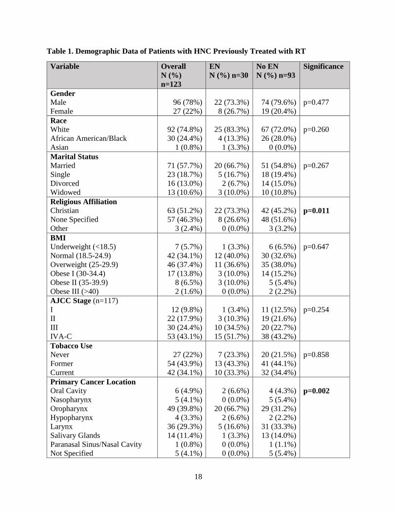
Additional demographic information for patients included in the study (n=123) are presented in
Table 1.
The results of Mann-Whitney U tests showed no significant difference in percent weight
change between diagnosis and initiation of RT between the EN and NEN groups. It did show a
significant difference in percent weight change between diagnosis and completion of RT
between EN and NEN groups (p=0.01) as well as between the beginning and end of RT
(p=0.001). Additionally, results of related-samples Wilcoxon Signed Rank testing showed a
significant difference between percent weight change before and after EN initiation (-9% before,
-1% after; p=0.004). Results for percent weight change at five different analysis intervals are
presented in Table 2.
Spearman’s correlational analysis was performed with all demographic and weight
change variables to investigate relationships with EN initiation (Hypothesis 1). After this
univariate analysis, three variables were significantly correlated to EN initiation: percent weight
17
change between diagnosis and completion of RT (p=0.007), percent weight change between start
and completion of RT (p=0.001), and religious affiliation (p=0.008). However, religious
affiliation had 9 categories, which were then condensed down to three for analysis purposes:
Christian (n=63), none specified (n=57), and other (n=3). Due to the small sample size, “other”
was not included in analysis. After condensing the religious affiliation variable, Spearman’s
correlation was performed again, with religious affiliation remaining significant, indicating that
patients that self-identified as Christians were more likely to initiate EN than those who did not
specify a religion (R= -0.202; p=0.011). Therefore patients that identified as Christian were more
likely to initiate EN than patients who did not specify a religion. Other demographic variables
(age, gender, marital status, race) were not found to be significantly related to EN initiation.
The three significant variables from the univariate analysis were entered into a linear
regression model to identify predictors of EN initiation. As the variables ‘percent weight change
between diagnosis and completion of RT’ and ‘percent weight change between start and
completion of RT’ exhibit multicollinearity, percent weight change between start and completion
of RT was used in linear regression analysis along with religious affiliation. In the regression
model, religious affiliation and percent weight change between RT start and completion
remained significant (p=0.012, p=0.004, respectively). To investigate the relationship of
demographic variables with timing of EN, measured as days from start of RT date to date of EN
initiation (Hypothesis 2), Spearman’s correlation analyses were performed. No variables
collected were found to be associated with the number of days from RT initiation to EN
initiation.
18
Table 1. Demographic Data of Patients with HNC Previously Treated with RT
Variable Overall
N (%)
n=123
EN
N (%) n=30
No EN
N (%) n=93
Significance
Gender
Male
Female
96 (78%)
27 (22%)
22 (73.3%)
8 (26.7%)
74 (79.6%)
19 (20.4%)
p=0.477
Race
White
African American/Black
Asian
92 (74.8%)
30 (24.4%)
1 (0.8%)
25 (83.3%)
4 (13.3%)
1 (3.3%)
67 (72.0%)
26 (28.0%)
0 (0.0%)
p=0.260
Marital Status
Married
Single
Divorced
Widowed
71 (57.7%)
23 (18.7%)
16 (13.0%)
13 (10.6%)
20 (66.7%)
5 (16.7%)
2 (6.7%)
3 (10.0%)
51 (54.8%)
18 (19.4%)
14 (15.0%)
10 (10.8%)
p=0.267
Religious Affiliation
Christian
None Specified
Other
63 (51.2%)
57 (46.3%)
3 (2.4%)
22 (73.3%)
8 (26.6%)
0 (0.0%)
42 (45.2%)
48 (51.6%)
3 (3.2%)
p=0.011
BMI
Underweight (<18.5)
Normal (18.5-24.9)
Overweight (25-29.9)
Obese I (30-34.4)
Obese II (35-39.9)
Obese III (>40)
7 (5.7%)
42 (34.1%)
46 (37.4%)
17 (13.8%)
8 (6.5%)
2 (1.6%)
1 (3.3%)
12 (40.0%)
11 (36.6%)
3 (10.0%)
3 (10.0%)
0 (0.0%)
6 (6.5%)
30 (32.6%)
35 (38.0%)
14 (15.2%)
5 (5.4%)
2 (2.2%)
p=0.647
AJCC Stage (n=117)
I
II
III
IVA-C
12 (9.8%)
22 (17.9%)
30 (24.4%)
53 (43.1%)
1 (3.4%)
3 (10.3%)
10 (34.5%)
15 (51.7%)
11 (12.5%)
19 (21.6%)
20 (22.7%)
38 (43.2%)
p=0.254
Tobacco Use
Never
Former
Current
27 (22%)
54 (43.9%)
42 (34.1%)
7 (23.3%)
13 (43.3%)
10 (33.3%)
20 (21.5%)
41 (44.1%)
32 (34.4%)
p=0.858
Primary Cancer Location
Oral Cavity
Nasopharynx
Oropharynx
Hypopharynx
Larynx
Salivary Glands
Paranasal Sinus/Nasal Cavity
Not Specified
6 (4.9%)
5 (4.1%)
49 (39.8%)
4 (3.3%)
36 (29.3%)
14 (11.4%)
1 (0.8%)
5 (4.1%)
2 (6.6%)
0 (0.0%)
20 (66.7%)
2 (6.6%)
5 (16.6%)
1 (3.3%)
0 (0.0%)
0 (0.0%)
4 (4.3%)
5 (5.4%)
29 (31.2%)
2 (2.2%)
31 (33.3%)
13 (14.0%)
1 (1.1%)
5 (5.4%)
p=0.002

19
Unknown 3 (2.4%) 0 (0.0%) 3 (3.2%)
Chemotherapy
Yes
No
82 (66.7%)
41 (33.3%)
27 (90.0%)
3 (10.0%)
55 (40.8%)
38 (59.1%)
p=0.002
Chemotherapy Types (n=82)
CISplatin
Erubix
Carboplatin
Combination (2 or more)
57 (69.5%)
15 (18.3%)
1 (1.2%)
9 (10.9%)
22 (81.5%)
4 (14.8%)
0 (0.0%)
1 (3.7%)
35 (63.6%)
11 (20.0%)
1 (1.8%)
8 (14.5%)
p=0.069
Table 2. Percent Weight Change
% Weight Change for All Patients
(n=123)
Mean (+/- SD) Range Significance
(1) Diagnosis to Radiation Start
EN
No EN
0.02% (3.6)
0.39% (4.7)
-0.09% (3.2)
-15% - +21%
p=0.759
(2) Diagnosis to Radiation End
EN
No EN
-8.13 (6.0)
-13.2% (17.7)
-9.9% (18.3)
-24% - +11.46%
p=0.01*
(3) Radiation Start to Radiation End*
EN
No EN
-8.08% (5.3)
-15.0%(30.9)
-10.2% (14.5)
-22.6% - +6.43%
p=0.001*
% Weight Change for EN Patients
(n=30)
Before and After EN initiation (4) Diagnosis to EN initiation
(5) EN initiation to RT end
-9.02% (6.78)
-1.03% (8.1)
-26% – +6.84%
-11% – +23.3%
p=0.004**
*Mann-Whitney test for statistically significant differences between patients with EN versus
patients without EN
** Wilcoxon test for statistically significant differences before and after EN initiation
20
CHAPTER 5
DISCUSSION
The primary aim of this study was to investigate the relationship between demographic
variables (age, ethnicity, and marital status) and the initiation of EN. Additionally, the
relationship between demographic variables (age, ethnicity, and marital status) and the timing of
EN was examined. The results of this investigation did not support the study hypotheses which
were:
Hyp 1: Demographic variables (age, ethnicity, and marital status) are significantly related
to EN initiation in HNC patients receiving radiotherapy.
Hyp 2: Demographic variables (age, ethnicity, and marital status) are significantly
associated to the timing of EN initiation in HNC patients receiving radiotherapy.
Contrary to our original hypotheses, these demographic variables were not found to be
significant predictors of EN initiation or timing of EN initiation.
The secondary aim was to investigate the relationship between religious affiliation and gender
and initiation of enteral initiation. Although gender was not associated with feeding tube
placement, religious affiliation was a significant independent predictor. In particular, patients
who identified with the Christian faith were more likely to initiate EN.
The primary hypotheses were established based on results from previous studies. Sachdev
et al44 found that age was the single independent predictor of EN initiation after multivariate
21
analysis (p=0.003) and patients >60 years of age were 4.18 times more likely to initiate EN
compared with patients <60 years of age (p=0.0019)44. Cheng et al42 also found increased age to
be related to EN initiation (Odds Ratio 1.3, p=0.02)42. Although this study had similar median,
mean, and range of ages, the current study did not find the same relationship between increased
age and increased EN initiation. Cheng et al42 did not include protocols for recommending
NG/PEG tube placement, which could explain the difference in the results of the study, as PEG
tubes in the current study were recommended specifically based on percent body weight loss.
Results from a SEER-Medicare survey (n=8,306) showed that unmarried patients with
HNC (including separated, divorced, widowed) were more likely to initiate prophylactic EN.49
This current study did not show this result, possibly due to a much smaller sample size (n=123).
Magnuson et al48 found a relationship between ethnicity and feeding tube dependence in
univariate analysis, but this association was not present in multivariate analysis considering other
factors. This lack of result in the multivariate analysis is consistent with the results of our study.
Magnuson’s study was conducted in Birmingham, AL; therefore both of these studies were
conducted in a similar geographical area. This lends strength to the result of this study that
ethnicity is not related to EN initiation. However, in studies looking at nursing home patients
with severe cognitive impairment, African American patients were more likely to initiate EN
than white patients.51,52 This ethnic preference to initiate EN has not been found in studies in
patients with HNC. A potential reason for this difference could be the lower number of African
Americans in the present study.
However, this study used a convenience sample of the patients available at the MCC. The
ethnicities of the study population (74.8% white and 24.4% African American) do not accurately
reflect that of Tuscaloosa and the surrounding counties, which average 57.1% white and 41.0%
22
Black/African American according to the 2015 US Census Bureau.53 This discrepancy can
potentially explain the null result related to ethnicity. More significantly, this discrepancy points
to a larger issue of access to care. This disparity in results points to a possible barrier to
accessing proper medical care for African American patients. Further research should fully
identify and address this issue.
Another reason could be the difference in purpose for utilizing EN in HNC compared
with end-of-life scenarios. In HNC, patients generally can resume oral intake, and EN is a
temporary measure. However, at the end-of-life, patients and their caregivers ponder ethical
questions such as whether EN is prolonging life or prolonging death.54 This ethnic disparity
based on end-of-life situations is also seen in the Coping with Cancer study which included 606
patients with advanced cancer from across 5 states.55 This study showed that black and Latino
patients were more likely than non-Latino white patients to prefer life-prolonging care, such as a
feeding tube, if it would extend life for 1 day (46% black patients, 41% Latino patients, 26%
non-Latino white patients; p<0.001).55 These results show that future studies should stratify the
decision to initiate EN by prognosis.
With regards to the secondary aim, religious affiliation was found to be significantly
related to EN initiation after univariate analysis (p=0.008). After condensing the nine categories
into Christian, none-given, and other, religious affiliation was still found to be significantly
associated with EN initiation (R= -0.202, p=0.027). There was a negative correlation, showing
that patients who identified as Christians were more likely to initiate EN compared with patients
who did not specify a religion.
Religion, faith, and spirituality are well documented to affect medical decisions.56-58 In
fact, in a study with 100 patients with advanced lung cancer and 257 medical oncologists,
23
Silvestri et al59 found that, after the oncologist, the patient’s faith in God was the most important
factor affecting their treatment decisions. However, no previous studies have examined this in
relation to EN. There are a few possible explanations for the correlation between self-identified
Christians and EN initiation. One reason is that patients who specified Christian religion may
have more social support through their church. One of the barriers to receiving EN is fear and
anxiety surrounding the procedure and management of the feeding tube.60 Religion can provide a
social support for a patient that may ease the anxiety of initiating EN.56,57
A final possibility is that a pillar of Christianity is the sanctity of life.50 Therefore, a
patient who identifies as Christian may be more willing to begin EN to improve their life because
their life is sacred according to their beliefs. A study examining spiritual coping mechanisms at
the end of life found that patients who used spiritual coping were more likely to choose life-
sustaining measures.61
After bivariate analysis, weight change between the start and completion of radiation
treatment was also found to be significantly related to EN initiation (p=0.004). An informal
interview with the radiation oncologists at MCC revealed that the oncologists recommended
reactive EN on a case-by-case basis. Specifically, if a patient lost 5% of his/her body weight,
special attention would be given, and by a 10% loss in body weight, EN would be placed. This is
not a written hospital policy, but rather a general guideline that the radiation oncologists follow.
The results of this study suggest that physicians were following this guideline. Additionally,
based on the lack of significant differences between age, gender, ethnicity, and marital status and
EN initiation, this informal guideline seems to have reduced potential bias in EN placement.
The results of this study have also shown that EN initiation reduced weight loss from 9%
before EN initiation to 1% after EN initiation (p=0.004). This confirms similar results from

24
Burney et al9 that showed a reduction in weight loss after EN placement (23 lbs before, 2.3 lbs
after). Previous studies have shown that weight loss in patients with HNC is associated with a
reduced QoL.13,14 Specifically, Languis et al14 showed a 10% weight loss was associated with a
reduced QoL.
Because EN is shown to reduce weight loss, which can improve outcomes and QoL, there
is an argument to implement a standard policy in which EN is recommended before a patient has
a 10% weight loss. The informal guidelines that the oncologists at MCC follow (monitoring a
patient at 5% weight loss, if there is no improvement, then recommend EN initiation)
demonstrate the ability to reduce a patient’s weight loss and reduce bias in EN placement.
This study was limited by the small amount of patients who received EN. This limits the
ability to draw conclusions about the population that did receive EN. However, no other
variables tested were close to being significant, and the authors are confident that the religious
affiliation and weight change were the only significant predictors of EN initiation in this
population.
Another limitation was the retrospective design. This limited the variables available to be
collected. Religious affiliation was a question on an initial admittance questionnaire, and if a
patient did not fill out a religious affiliation, it is unclear as to the reason. Thus, the meaning of
the association between religious affiliation and EN initiation cannot be fully explained. As there
was such a small sample size of patients with EN (n=30), future studies should include a larger
amount to further investigate demographic correlates of the timing of enteral initiation (days
from RT start to EN initiation).
25
As demonstrated above, a policy based on weight loss appears beneficial in reducing bias
related to EN initiation. In particular, a policy in which patients with a 5% weight loss are
closely monitored, and if weight does not stabilize EN is initiated, was helpful. Therefore, a
larger, prospective study is warranted using this policy. Patient attitudes towards the
recommendations should be collected, as this will add to the literature.
In conclusion, while age, ethnicity, and marital status were not associated with EN
initiation or timing of EN initiation, patients who identify as Christian tend to start EN more than
patients who do not specify a religion, and percent weight loss is a predictor of EN initiation.
Because of an informal guideline monitoring and recommending EN to patients with >5%
weight loss, bias was reduced. Hospitals would benefit from implementing a formal policy to
initiate EN in patients with this weight loss.

26
REFERENCES
1. Head and Neck Cancers. National Cancer Institute website.
https://www.cancer.gov/types/head-and-neck/head-neck-fact-sheet#r29. Reviewed
February 1, 2013. Accessed February 18, 2017.
2. Siegel RL, Miller KD, Jemal A. Cancer statistics, 2017. CA Cancer J Clin. 2017;67:7-30.
3. American Cancer Society. Cancer Facts and Figures 2016. Atlanta, GA: American
Cancer Society; 2016. https://www.cancer.org/research/cancer-facts-statistics/all-cancer-
facts-figures/cancer-facts-figures-2016.html. Accessed February 18, 2017.
4. Hashibe M, Brennan P, Benhamou S, et al. Alcohol drinking in never users of tobacco,
cigarette smoking, in never drinkers, and the risk of head and neck cancer: pooled
analysis in the International Head and Neck Cancer Epidemiology Consortium. J Natl
Cancer Inst. 2007;99(10):777-89.
5. Chaturvedi AK, Engels EA, Pfeiffer RM, et al. Human papillomavirus and rising
oropharyngeal cancer incidence in the United States. J Clin Oncol. 2011;29(32):4294-
301.
6. Radiation Therapy. National Cancer Institute website. https://www.cancer.gov/about-
cancer/treatment/types/radiation-therapy#WRRT. Updated February 2, 2017. Accessed
February 20, 2017.
7. Unsal D, Mentes B, Akmansu M, et al. Evaluation of nutrition status in cancer patients
receiving radiotherpy: A prospective study. Am J Clin Oncol. 2006;29:183-188.
8. Mahan LK, Escott-Stump S, Raymond JL. Food and Nutrient Delivery: Nutrition Support
Methods. Krause’s Food and the Nutrition Care Process. 13th ed. St. Louis, MS:
Elsevier Saunders; 2012:309-14.
9. Mercuri A, Lim Joon D, Wada M, et al. The effect of an intensive nutritional program on
daily set-up variations and radiotherapy planning margins of head and neck cancer
patients. J Med Imaging Radiat Oncol. 2009;53:500-505.
10. Burney RE, Bryner BS. Safety and long-term outcomes of percutaneous endoscopic
gastrostomy in patients with head and neck cancer. Surg Endosc. 2015;29:3689-3689.
11. Assenat E, Thezenas S, Flori N, et al. Prophylactic percutaneous endoscopic gastrosotmy
in patients with advanced head and neck tumors treated by combined chemoradiotherapy.
J Pain Symptom Manage. 2011;42(4):548-556.
27
12. Bahl M, Siu LL, Pond GR, et al. Tolerability of the Intergroup 0099 (INT 0099) Regimen
in locally advanced nasopharyngeal cancer with a focus on patients’ nutritional status. Int
J Radiation Oncology Biol Phys. 2004;60(4):1127-1136.
13. Rutter CE, Yovino S, Taylor R, et al. Impact of early percutaneous endoscopic
gastrostomy tube placement on nutritional status and hospitalization in patients with head
and neck cancer receiving definitive chemoradiation therapy. Head and Neck. 2011;10.
14. Morton RP, Crowder VL, Mawdsley R, et al. Elective gastrostomy, nutritional status and
quality of life in advanced head and neck cancer patients receiving chemoradiotherapy.
ANZ J Surg. 2009;79:713-718.
15. Languis J, van Dijk A, Doornaert P, et al. More than 10% weight loss in head and neck
cancer patients during radiotherapy is independently associated with deterioration in
quality of life. Nutr Cancer. 2013;65:76-83.
16. Lis CG, Gupta D, Lammersfeld CA. Role of nutritional status in predicting quality of life
outcomes in cancer – a systematic review of the epidemiological literature. Nutr J.
2012;11:27.
17. Osborne JB, Collin LA, Posluns EC, et al. The Experience of head and neck cancer
patients with a percutaneous endoscopic gastrostomy tube at a Canadian cancer center.
Nutr Clin Pract. 2012;27(5):661-668.
18. Amerman EC. The Digestive System. Human Anatomy & Physiology. New York, NY:
Pearson Education, Inc; 2016: 852-859.
19. Amerman EC. The Digestive System. Human Anatomy & Physiology. New York, NY:
Pearson Education, Inc; 2016:124-132.
20. Sanderson R, Ironside AD. Sqamous cell carcinomas of the head and neck. BMJ.
2002;325:822-7.
21. Lopez F, Williams MD, Slavova A. How phenotype guides management of the most
common malignant salivary neoplasms of the larynx? Adv Ther. 2017;34:813-825.
22. Fakhry C, Westra WH, Li S, et al. Improved survival of patients with human
papillomavirus-positive head and neck squamous cell carcinoma in a prospective clinical
trial. J Natl Cancer Inst. 2008;100:261-9.
23. Cancer Staging System. American Joint Committee on Cancer website.
https://cancerstaging.org/references-tools/Pages/What-is-Cancer-Staging.aspx. Updated
2017. Accessed February 19, 2017.
24. Surgery. National Cancer Institute website. https://www.cancer.gov/about-
cancer/treatment/types/surgery. Updated April 29, 2015. Accessed February 20, 2017.
25. Locher JL, Bonner JA, Carroll WR. Gastrostomy tube placement use in patients with
head and neck cancer. Head & Neck. 2011.
26. Murphy BA, Gilbert J. Dysphagia in head and neck cancer patients treated with radiation:
Assessment, sequelae, and rehabilitation. Semin Radiat Oncol. 2009;19:35-42.

28
27. Schindler A, Denaro N, Russi EG. Dysphagia in head and neck cancer patients treated
with radiotherapy and systemic therapies: Literature review and consensus. Crit Rev Onc.
2015;96:372-384.
28. Chemotherapy. National Cancer Institute website. https://www.cancer.gov/about-
cancer/treatment/types/chemotherapy. Updated April 29, 2015. Accessed February 20,
2017.
29. Targeted Cancer Therapies. National Cancer Institute website.
https://www.cancer.gov/about-cancer/treatment/types/targeted-therapies/targeted-
therapies-fact-sheet#q7. Reviewed April 25, 2014. Accessed February 20, 2017.
30. Corry J, Poon W, McPhee N. Prospective study of percutaneous endoscopic gastrostomy
tubes versus nasogastric tubes for enteral feeding in patients with head and neck cancer
undergoing (Chemo)Radiation. Head & Neck;2008.
31. Nasogastric Feeding Tube versus Percuataneous Endoscopic gastrostomy for patients
with head or neck cancer: A review of clinical effectiveness and guidelines. Rapid
Response Report: Summary with Critical Appraisal. Ottawa(ON): Canadian Agency for
Drugs and Technologies in Health;2014.
32. Lewis SL, Brody R, Touger-Decker R, et al. Feeding tube use in patients with head and
neck cancer. Head and Neck. 2014;29.
33. Cady J. Nutrition Support During Radiotherapy for head and neck cancer: the role of
prophylactic feeding tube placement. Clin J Oncol Nurs. 2006;11(6):875-880.
34. Wiggenraad, RGJ, Flierman, L, Goossens, A. Prophylactic gastrostomy placement and
early tube feeding may limit loss of weight during chemoradiotherapy for advanced head
and neck cancer, a preliminary study. Clin Otolaryngol. 2007;32:384-390.
35. Beer KT, Krause KB, Zuercher T. Early percutaneous endoscopic gastrostomy insertion
maintains nutritional state with aerodigestive tract cancer. Nutr Cancer. 2005;52(1):29-
34.
36. Mayre-Chilton KM, Talwar BP, Goff LM. Different experiences and perspectives
between head and neck cancer patients and their care-givers on their daily impact of a
gastrostomy tube. J Hum Nutr Diet. 2011;24:449-459.
37. Ehrsson YT, Sundberg K, Laurell G, Langius-Eklof A. Head and neck cancer patients’
perceptions of quality of life and how it is affected by the disease and enteral tube feeding
during treatment. Ups J Med Sci. 2015;120:280-289.
38. Roger SN, Thomson R, O’Toole P, Lowe D. Patients experience with long-term
percutaneous endosopic gastrostomy feeding following primary surgery for oral and
oropharyngeal cancer. Oral Oncol. 2007;43:499-507.
39. Mangar S, Slevin N, Mais K, Sykes A. Evaluating predictive factors for determining
enteral nutrition in patients receiving radical radiotherapy for head and neck cancer: A
retrospective review. Rad Onc. 2006;78:152-158.

29
40. Strom T, Trotti AM, Kish J, et al. Risk factors for percutaneous endoscopic gastrostomy
tube placement during chemoradiotherapy for oropharyneal cancer. Otolaryngol Head
Neck Surg. 2013;139(11):1242-1246.
41. Gokhale AS, McLaughlin BT, Flickinger JC, et al. Clinical and dosimetric factors
associated with a prolonged feeding tube requirement in patients treated with
chemoradiotherapy (CRT) for head and neck cancers. Annals of OncologyI. 2010;21:145-
151.
42. Cheng SS, Terrell JE, Bradford CR, et al. Variables associated with feeding tube
placement in head and neck cancer. Arch Otolarngol Head Neck Surg. 2006;132(6):655-
661.
43. Bhayani MK, Hutchenson KA, Barringer DA, et al. Gastrostomy tube placement in
patients with oropharyngeal carcinoma treated with radiotherapy or chemotherapy:
Factors affecting placement and dependence. Head Neck. 2013;35:1634-1640.
44. Sachdev S, Refaat T, Bacchus ID, Sathiaseelan V, Mittal BB. Age most significant
predictor of requiring enteral feeding in head-and-neck cancer patients. Rad Onco.
2015;10:93.
45. Massa ST, Osazuwa-Peters N, Christopher KM, et al. Competing causes of death in the
head and neck cancer population. Oral Oncol. 2017;65:8-15.
46. Choi SH, Terrell JE, Fowler KE, et al. Socioeconomic and other demographic disparities
predicting survival among head and neck cancer patients. PLoS ONE.
2016;11(3):e0149886.
47. Konski AA, Pajak TF, Movsas B. Disadvantage of men living alone participating in
radiation therapy oncology group head and neck trials. J Clin Oncol. 2006;24:4177-4183.
48. Magnuson JS, Durst J, Rosenthal EL. Long-term gastrostomy tube dependence more
likely in head and neck cancer survivors without partners. Head Neck. 2013;35(3):420-
425.
49. Locher JL, Bonner JA, Carroll WR, et al. Patterns of prophylactic gastrostomy tube
placement in head and neck cancer patients: A consideration of the significance of social
support and practice variation. Laryngoscope. 2013;123(8):1918-25.
50. Clarfield AM, Gordon M, Markwell H, Alibhai SMH. Ethical issues in end-of-life
geriatric care: The approach of three monotheistic religions – Judaism, Catholicism, and
Islam. J Am Geriatr Soc. 2003;51:1149-1154.
51. Gessert CE, Curry NM, Robinson A. Ethnicity and end-of-life care: the use of feeding
tubes. Ethn Dis. 2001;11(1):97-106.
52. Mitchell S, Teno J, Roy J, et al. Clinical and organizational factors associated with
feeding tube use among nursing home residents with advanced cognitive impairment.
JAMA. 2003;290(1):73-80.

30
53. QuickFacts. The United States Census Bureau. https://www.census.gov/quickfacts/
table/PST045215 /01125. Accessed May 4, 2017.
54. Farrow AM, McCallum TJ, Messinger-Rapport BJ. Preferences of older African-
Americans for long-term tube feeding at the end of life. Aging Ment Health.
2004;8(6):530-534.
55. Garrido MM, Harrington ST, Prigerson HG. End-of-life treatment preferences: A key to
reducing ethnic/racial disparities in advance care planning? Cancer. 2014;120(24):3981-
3986.
56. Koenig HG. Religion, spirituality, and medicine: Research findings and implications for
clinical practice. South Med J. 2004;97(12):1194-1200.
57. Puchalski CM. The role of spirituality in health care. BUMC Proceedings. 2001;14:352-
7.
58. Johnson J, Hayden T, True J, et al. The impact of faith beliefs on perceptions of end-of-
life care and decision making among African American church members. J Palliat Med.
2016;19(2)143-8.
59. Silvestri GA, Knitting S, Zoller JS, Nietert PJ. Importance of faith on medical decisions
regarding cancer care. J Clin Oncol. 2003;21:1379-1382.
60. Jaafar MH, Mahadeva S, Morgan K, Tan MP. Systematic review of qualitative and
quantitative studies on the attitudes and barriers to percutaneous endoscopic gastrostomy
feeding. Clin Nutr. 2016;35(6):1226-1235
61. True Gala, Phipps EJ, Braitman LE, et al. Treatment preferences and advance care
planning at end of life: The role of ethnicity and spiritual coping in cancer patients. Ann
Behav Med. 2005;30(2):174-9.

31
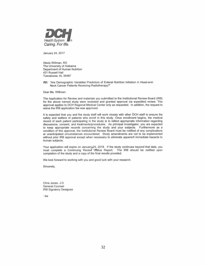
APPENDIX A:
Research Approval Letter from DCH Institutional Review Board
32

33
APPENDIX B:
Research Approval Letter from UA Institutional Review Board

34

35
APPENDIX C:
Review Outcome for Revisions to Approved Study from DCH Institutional Review Board
36

37
APPENDIX D:
Approval of Protocol Revision from UA Institutional Review Board

38