dengue hemorrhagic fever in infants: research ... · in infants: research opportunities ignored ......
TRANSCRIPT
PERSPECTIVE
1474 Emerging Infectious Diseases • Vol. 8, No. 12, December 2002
Dengue Hemorrhagic Fever in Infants: Research
Opportunities Ignored Scott B. Halstead,* Nguyen Trong Lan,† Thein Thein Myint,‡ Than Nu Shwe,‡
Ananda Nisalak,§ Siripen Kalyanarooj¶, Suchitra Nimmannitya,¶ Soegeng Soegijanto,# David W. Vaughn,§ and Timothy P. Endy§
The age distribution of cases of dengue hemorrhagic fever and dengue shock syndrome (DHF/DSS) ininfants under the age of 1 year are reported from Bangkok, Thailand, and for the first time for Ho Chi MinhCity, Vietnam; Yangon, Myanmar; and Surabaya, Indonesia. The four dengue viruses were isolated fromThai infants, all of whom were having a primary dengue infection. Progress studying the immunologicallydistinct infant DHF/DSS has been limited; most contemporary research has centered on DHF/DSS accom-panying secondary dengue infections. In designing research results obtained in studies on a congruentanimal model, feline infectious peritonitis virus (FIPV) infections of kittens born to FIPV-immune queensshould be considered. Research challenges presented by infant DHF/DSS are discussed.
ince World War II, the four dengue viruses (formal name:Dengue virus [DENV]) have progressively spread geo-
graphically throughout the tropics, resulting in a global pan-demic with tens of millions of infections annually, includingseveral hundred thousand hospitalizations for dengue hemor-rhagic fever (DHF) and dengue shock syndrome (DSS) (1).The size and spread of the dengue pandemic, the unpredict-ability of epidemic occurrences, and the circulation of virulentand nonvirulent strains make DHF/DSS a model for an emerg-ing infectious disease.
Ample evidence suggests that DHF/DSS accompanies sec-ondary dengue infections in children older than 1 year (1–3).Less well-known are the epidemiologic and clinical studiesthat document an identical severe syndrome in infants duringtheir first dengue infection (4,5). Ignoring these data, contem-porary models of dengue immunopathogenesis focus on thesequential dengue viral infection phenomenon; such modelssuggest that severe disease results from amplified cytokinerelease caused by dengue infections occurring in the presenceof T-cell memory (6). However, that model cannot explainDHF/DSS during a first dengue infection.
That dengue in infants is not often studied is understand-able. Small subjects pose technical difficulties in obtainingsamples required by research protocols, and human use proto-cols may be constraining. Yet infants represent 5% or more ofall DHF/DSS patients (7). Uniquely, infants with DHF/DSSpresent an opportunity to obtain both the causative virus andthe preinfection antibodies as research reagents in a hospital
setting without recourse to a time-consuming and expensiveprospective cohort study. The all-important preinfection anti-bodies can be collected from the mother, as her serum is a sur-rogate for cord blood (8).
Enhancement of infant infectious diseases by cord bloodantibodies is not described for human infections other thandengue. However, such a phenomenon occurs naturally ininfected kittens born to queens immune to feline infectiousperitonitis virus (FIPV) (9–11). To refocus attention on theresearch opportunities afforded by this immunopathologicentity, we provide evidence that infants with DHF/DSS areregularly admitted to hospitals in four of the largest dengue-endemic countries. The age distribution of all these infantDHF/DSS patients is similar. Most of those studied serologi-cally had had primary dengue infections. Because of FIPV’scongruence to infant dengue, a short literature review is pro-vided on that animal model.
Materials and Methods
PatientsData on infants, ages <12 months, hospitalized with a clin-
ical diagnosis of DHF were obtained from four hospitals: Chil-dren’s Hospital No. 1, Ho Chi Minh City, Vietnam; the QueenSirikit National Institute of Child Health, also referred to asBangkok Children’s Hospital, Bangkok, Thailand; Children’sHospital, Yangon, Myanmar; and the Department of Pediat-rics, Dr. Soetomo Hospital, Surabaya, Indonesia. In this study,data for 4 consecutive years, either 1995–1998 or 1996–1999were combined. Patients were under the routine care of one ormore of the authors, each an experienced senior academicinfectious diseases pediatrician. All diagnoses of DHF/DSS ininfants conformed to World Health Organization case defini-tions. In Bangkok, serum samples from all infants and childrenhospitalized for DHF were sent for routine diagnostic study to
*Uniformed Services University of the Health Sciences, Bethesda,Maryland, USA; †Children’s Hospital No. 1, Ho Chi Minh City, Vietnam;‡Yangon Children’s Hospital, Yangon, Myanmar; §Armed ForcesResearch Institute of the Medical Sciences, Bangkok, Thailand;¶Queen Sirikit National Institute of Child Health, Bangkok, Thailand;and #Dr. Soetomo Hospital, Airlangga University Medical School, Sura-baya, Indonesia
S
Emerging Infectious Diseases • Vol. 8, No. 12, December 2002 1475
PERSPECTIVE
the Virology Department, Armed Forces Research Institute ofMedical Sciences (AFRIMS). For nearly 30 years, AFRIMShas provided such dengue diagnostic services to BangkokChildren’s Hospital. Similar routine diagnostic tests were pro-vided for infants and children admitted to Children’s Hospital,Yangon, by the Virology Department, Department of MedicalResearch. Fiscal constraints limited the number of serologictests performed. Individual data were disassociated from anyidentifiers and are presented here only in aggregate.
Virus IsolationAs described, DENV isolations were attempted from
acute-phase plasma or serum samples from Thai children byinoculation into C6/36 cells or intrathoracically in mosquitoes(Toxorrhynchites splendens) (12).
Viral IdentificationDENV was identified in C6/36 cells by an antigen-specific
enzyme-linked immunosorbent assay (ELISA) with a panel ofmonoclonal antibodies against DENV (13).
SerologyPlasma or serum samples were tested for serologic evi-
dence of acute DENV infection by immunoglobulin (Ig) Mand IgG ELISA, hemagglutination inhibition (HAI) assays, orboth (14). For single specimens, >5 days after onset of fever40 U of IgM to DENV was considered evidence of a DENVinfection. A DENV IgM-to-IgG ratio >1.8 defined a primaryinfection. A ratio <1.8 defined a secondary DENV infection.With serial specimens, twofold increase in IgG to DENV withan absolute value of >100 U indicated a secondary infection inthe absence of IgM to DENV of >40 U.
In Bangkok, HAI antibody against DENV types 1–4 andJapanese encephalitis virus were measured in all sera (15). Afourfold increase was considered positive for acute flavivirusinfection. The infection was diagnosed as primary if titers >1week after onset of illness were <1:1,280 or as secondary ifantibody titers were >1:1,280 (16).
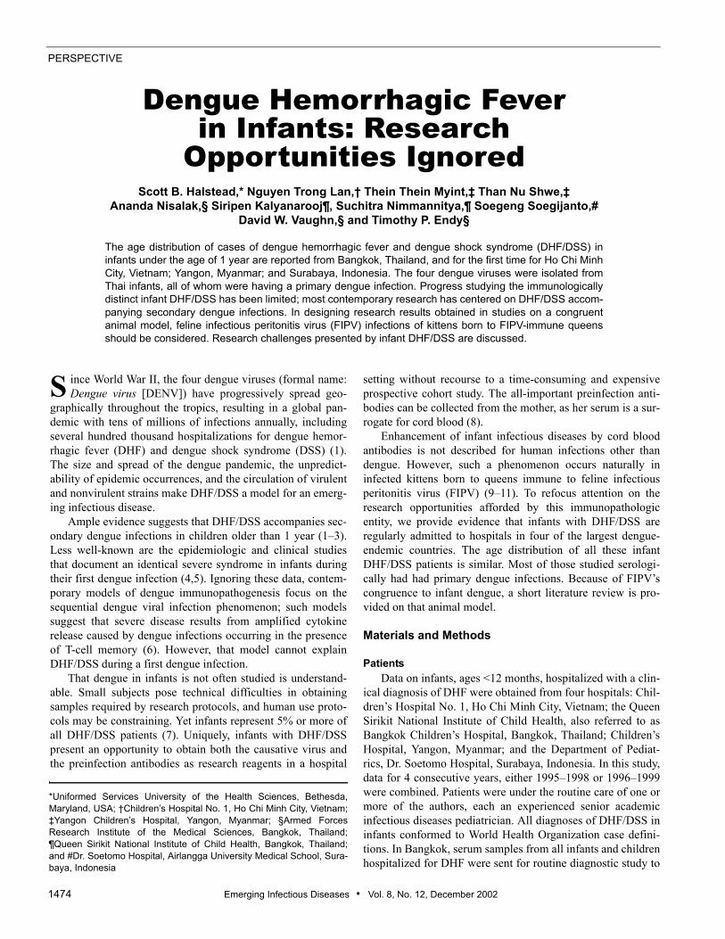
ResultsInfants are at high risk for DHF/DSS. Figure 1 provides
data from the only published study to estimate age-specificdengue hospitalization rates for the Bangkok metropolitanarea. In 1964, 17/1,000 seven-month-old infants, more than1% of the population that age, were hospitalized for DHF/DSS(17). This modal rate was two times higher than the 1964modal hospitalization rate for children (age 4 years, data notshown) in Bangkok during the same year (17). In our presentstudy, infant DHF/DSS constituted 4.9%, 4.6%, 5.0%, and4.9% of 4,872; 14,053; 8,938; and 2,057 Thai, Vietnamese,Myanmar, and Indonesian infants and children hospitalizedwith DHF in 1995–1998, respectively.
During a 4-year period, 237, 652, 449, and 101 infantswith presumptive DHF/DSS were admitted to hospitals inBangkok, Ho Chi Minh City, Yangon, and Surabaya. No sig-
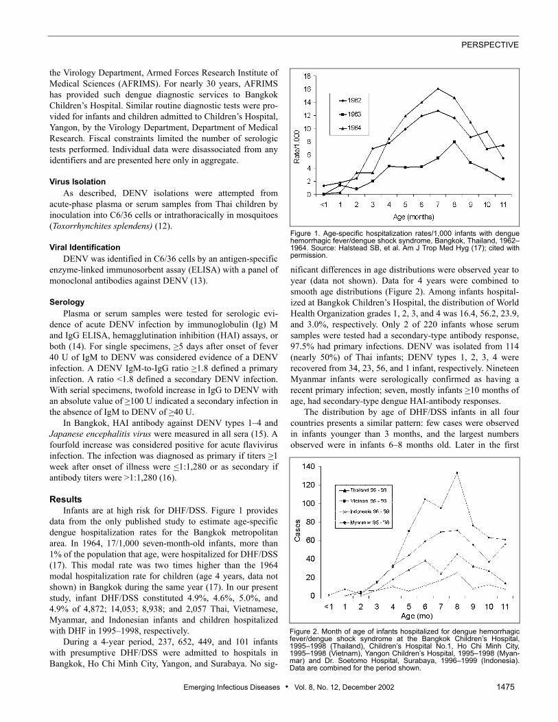
nificant differences in age distributions were observed year toyear (data not shown). Data for 4 years were combined tosmooth age distributions (Figure 2). Among infants hospital-ized at Bangkok Children’s Hospital, the distribution of WorldHealth Organization grades 1, 2, 3, and 4 was 16.4, 56.2, 23.9,and 3.0%, respectively. Only 2 of 220 infants whose serumsamples were tested had a secondary-type antibody response,97.5% had primary infections. DENV was isolated from 114(nearly 50%) of Thai infants; DENV types 1, 2, 3, 4 wererecovered from 34, 23, 56, and 1 infant, respectively. NineteenMyanmar infants were serologically confirmed as having arecent primary infection; seven, mostly infants >10 months ofage, had secondary-type dengue HAI-antibody responses.
The distribution by age of DHF/DSS infants in all fourcountries presents a similar pattern: few cases were observedin infants younger than 3 months, and the largest numbersobserved were in infants 6–8 months old. Later in the first
Figure 1. Age-specific hospitalization rates/1,000 infants with denguehemorrhagic fever/dengue shock syndrome, Bangkok, Thailand, 1962–1964. Source: Halstead SB, et al. Am J Trop Med Hyg (17); cited withpermission.
Figure 2. Month of age of infants hospitalized for dengue hemorrhagicfever/dengue shock syndrome at the Bangkok Children’s Hospital,1995–1998 (Thailand), Children’s Hospital No.1, Ho Chi Minh City,1995–1998 (Vietnam), Yangon Children’s Hospital, 1995–1998 (Myan-mar) and Dr. Soetomo Hospital, Surabaya, 1996–1999 (Indonesia).Data are combined for the period shown.
PERSPECTIVE
1476 Emerging Infectious Diseases • Vol. 8, No. 12, December 2002
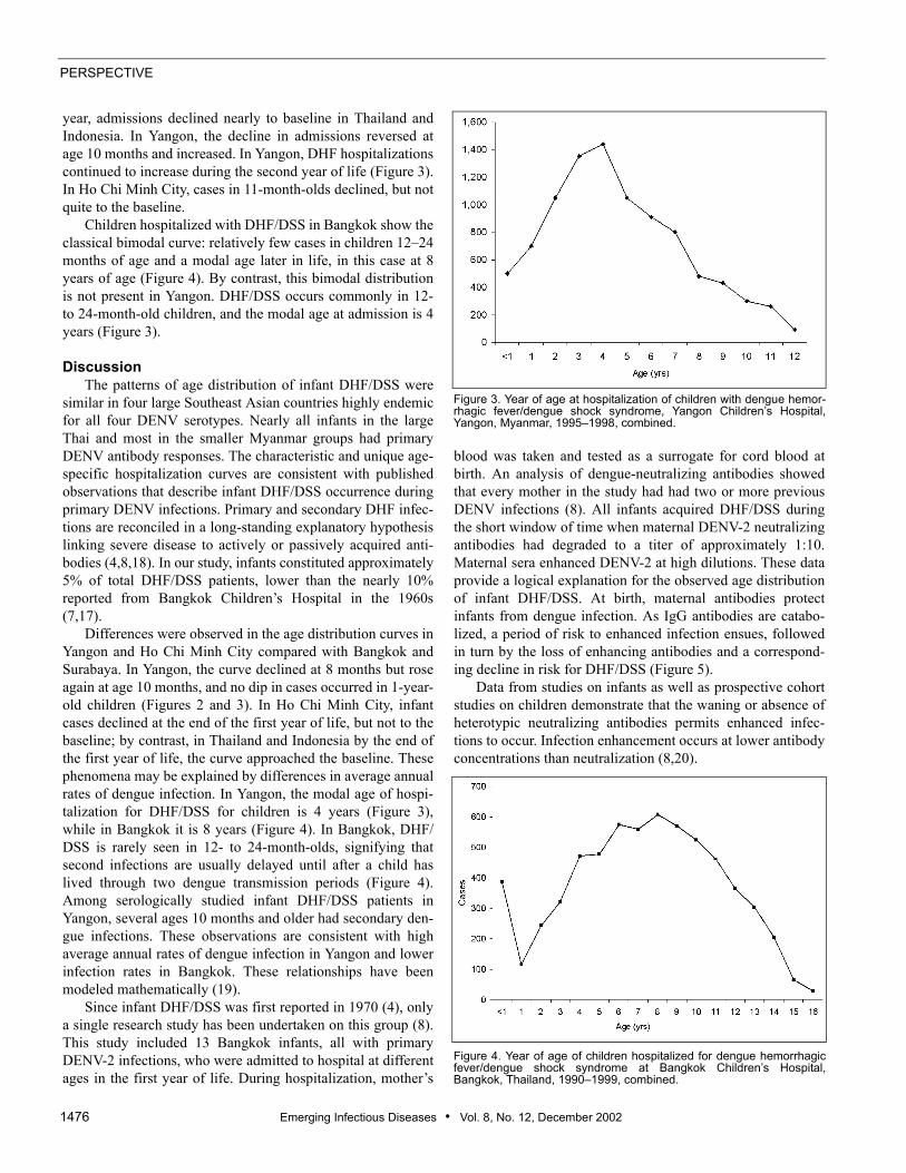
year, admissions declined nearly to baseline in Thailand andIndonesia. In Yangon, the decline in admissions reversed atage 10 months and increased. In Yangon, DHF hospitalizationscontinued to increase during the second year of life (Figure 3).In Ho Chi Minh City, cases in 11-month-olds declined, but notquite to the baseline.
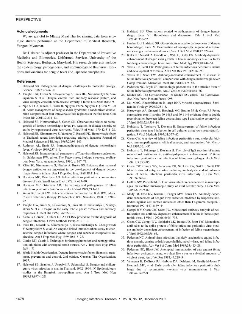
Children hospitalized with DHF/DSS in Bangkok show theclassical bimodal curve: relatively few cases in children 12–24months of age and a modal age later in life, in this case at 8years of age (Figure 4). By contrast, this bimodal distributionis not present in Yangon. DHF/DSS occurs commonly in 12-to 24-month-old children, and the modal age at admission is 4years (Figure 3).
DiscussionThe patterns of age distribution of infant DHF/DSS were
similar in four large Southeast Asian countries highly endemicfor all four DENV serotypes. Nearly all infants in the largeThai and most in the smaller Myanmar groups had primaryDENV antibody responses. The characteristic and unique age-specific hospitalization curves are consistent with publishedobservations that describe infant DHF/DSS occurrence duringprimary DENV infections. Primary and secondary DHF infec-tions are reconciled in a long-standing explanatory hypothesislinking severe disease to actively or passively acquired anti-bodies (4,8,18). In our study, infants constituted approximately5% of total DHF/DSS patients, lower than the nearly 10%reported from Bangkok Children’s Hospital in the 1960s(7,17).
Differences were observed in the age distribution curves inYangon and Ho Chi Minh City compared with Bangkok andSurabaya. In Yangon, the curve declined at 8 months but roseagain at age 10 months, and no dip in cases occurred in 1-year-old children (Figures 2 and 3). In Ho Chi Minh City, infantcases declined at the end of the first year of life, but not to thebaseline; by contrast, in Thailand and Indonesia by the end ofthe first year of life, the curve approached the baseline. Thesephenomena may be explained by differences in average annualrates of dengue infection. In Yangon, the modal age of hospi-talization for DHF/DSS for children is 4 years (Figure 3),while in Bangkok it is 8 years (Figure 4). In Bangkok, DHF/DSS is rarely seen in 12- to 24-month-olds, signifying thatsecond infections are usually delayed until after a child haslived through two dengue transmission periods (Figure 4).Among serologically studied infant DHF/DSS patients inYangon, several ages 10 months and older had secondary den-gue infections. These observations are consistent with highaverage annual rates of dengue infection in Yangon and lowerinfection rates in Bangkok. These relationships have beenmodeled mathematically (19).
Since infant DHF/DSS was first reported in 1970 (4), onlya single research study has been undertaken on this group (8).This study included 13 Bangkok infants, all with primaryDENV-2 infections, who were admitted to hospital at differentages in the first year of life. During hospitalization, mother’s
blood was taken and tested as a surrogate for cord blood atbirth. An analysis of dengue-neutralizing antibodies showedthat every mother in the study had had two or more previousDENV infections (8). All infants acquired DHF/DSS duringthe short window of time when maternal DENV-2 neutralizingantibodies had degraded to a titer of approximately 1:10.Maternal sera enhanced DENV-2 at high dilutions. These dataprovide a logical explanation for the observed age distributionof infant DHF/DSS. At birth, maternal antibodies protectinfants from dengue infection. As IgG antibodies are catabo-lized, a period of risk to enhanced infection ensues, followedin turn by the loss of enhancing antibodies and a correspond-ing decline in risk for DHF/DSS (Figure 5).
Data from studies on infants as well as prospective cohortstudies on children demonstrate that the waning or absence ofheterotypic neutralizing antibodies permits enhanced infec-tions to occur. Infection enhancement occurs at lower antibodyconcentrations than neutralization (8,20).
Figure 3. Year of age at hospitalization of children with dengue hemor-rhagic fever/dengue shock syndrome, Yangon Children’s Hospital,Yangon, Myanmar, 1995–1998, combined.
Figure 4. Year of age of children hospitalized for dengue hemorrhagicfever/dengue shock syndrome at Bangkok Children’s Hospital,Bangkok, Thailand, 1990–1999, combined.

Emerging Infectious Diseases • Vol. 8, No. 12, December 2002 1477
PERSPECTIVE
Immunopathogenesis mechanisms have been more exten-sively studied in a remarkably similar viral infection of cats,FIPV, a highly fatal coronavirus disease of domestic and exoticcats (9–11). Most cats naturally exposed as adults to FIPVdevelop antibody titers without showing clinical signs.Lesions in sick cats are believed to result from immunologi-cally mediated responses (9,10,21). Kittens receiving appar-ently competent neutralizing antibodies to FIPV, transferred incolostrum from immune queens, develop a fatal disease a fewdays after infection with wild-type virus (22). Passive transferof antibody by other routes produces the same result (22,23).This phenomenon is called the early death syndrome. FIPV inantibody-negative kittens occurs less reliably and is delayedfor several weeks until animals develop their own antibodyresponse to the virus. FIPV in kittens is characterized bythrombocytopenia and elevated ALT, AST, and serum biliru-bin (22).
The coronaviruses, pathogens of mammals and birds, are alarge family of enveloped RNA viruses with a nonsegmented,positive-stranded genome that is 27–32 kb in length (24). Oneof the most intriguing aspects of coronavirus replication is theoccurrence of high-frequency homologous RNA recombina-tion (25). Together with porcine Transmissible gastroenteritisvirus (TGEV), canine coronavirus, and human coronavirus229E (HCV), the feline coronaviruses form a separate clusterwithin the genus Coronavirus, including Feline enteric coro-navirus, and FIPV (26). Coronavirus virions possess threestructural proteins, a large spike glycoprotein (S), a small inte-gral membrane glycoprotein (M), and a nucleocapsid protein(N) (24). These proteins are analogous to the envelope (E), M,and nucleocapsid (C) proteins of the flaviviruses. The feline
coronaviruses can be divided into two serotypes, I and II, onthe basis of cross-reactivity to canine coronavirus in virus neu-tralization assays (26). Type I viruses grow poorly in tissuecultures and show virtually no neutralization with anti–caninecoronavirus sera (27). Type II viruses grow readily in vitro(28). Analysis of gene structure suggests that type II virusesare derived from recombination of type I feline coronavirusand canine coronavirus (26). The two serotypes circulate astwo pathotypes, the avirulent enteric viruses and the virulentFIPV (26). High-frequency mutations may help coronavirusesescape neutralization and promote infection enhancement inFc receptor–bearing cells.
Antibody-dependent enhancement of FIPV has been dem-onstrated in vitro in feline macrophages as well as in stablehuman and mouse macrophage cell lines (29). More cells areinfected in the presence compared with the absence of anti-body; the rates of viral entry and viral replication are similarunder both conditions (30). Coronaviruses appear to entermononuclear phagocytes by means of the plasma membranewithout marked involvement of phagocytic or endosomalpathways (28). Some researchers have surmised that, as withDENV, when antibody-virus complexes attach to Fc-receptors,viruses are brought close to cell surfaces, where they enter thecells by normal mechanisms (31,32). Enhancement is medi-ated by clusters of epitopes on the S protein (33,34). Resultswith FIPV suggest that feline IgG2a antibodies mediate bothneutralization and enhancement (33). Antibody-dependentenhancement in FIPV demonstrates a bell-shaped curve withincreasing dilutions; maximal enhancement occurs at subneu-tralizing titers (34).
Antibody-dependent enhancement is believed to be thecause of vaccine failure after immunization with live (35,36)or recombinant (37) vaccines. Inoculation of cats with arecombinant vaccinia virus expressing the S protein FIPV 79-1146 sensitized cats and led to accelerated disease after FIPVchallenge (37), while inoculation with recombinant vacciniaviruses expressing the M or N proteins did not (38). Immuni-zation with vaccines made from other members of the felinecoronavirus group, TGEV or canine coronavirus, also sensi-tizes cats to early death syndrome (39).
Flaviviridae do not appear to be subject to as high rates ofhomologous recombination as are the Coronaviridae; nonethe-less, during evolutionary history four dengue serotypes haveemerged. A phenomenon reminiscent of the feline coronavi-ruses is the evidence that DENV also circulate as two bio-types: DENV-2 American genotype does not cause DHF/DSS,while DENV-2 SE Asian genotype does (40). As with felinecoronaviruses, the severity of disease with the two DENV bio-types may be regulated by cross-reactive antibodies (41).Focused research on viral-antibody interactions at the struc-tural level might clarify early pathogenesis events in DHF/DSS. Infants may provide an accessible and inexpensivemodel to study mechanisms controlling the severity of dengueinfections. Workers actively involved in developing denguevaccines may benefit from lessons learned in the FIPV model.
Figure 5. Relationship between the age distributions of infants hospital-ized for dengue hemorrhagic fever/dengue shock syndrome (DHF/DSS) and the protective and infection-enhancing effects of maternaldengue antibodies. Shown are mean age specific hospitalization rate/1,000 for Bangkok and Thonburi, 1962–1964 (see Figure 1). At birth,antibodies are at protective concentrations. With the passage of time,maternal immunoglobulin G antibodies are catabolized to concentra-tions that result in antibody-dependent enhancement (ADE) of infec-tions. By the end of the first year of life, ADE antibodies are catabolizedto concentrations below the ADE threshold, and DHF/DSS cases disap-pear.
PERSPECTIVE
1478 Emerging Infectious Diseases • Vol. 8, No. 12, December 2002
AcknowledgmentsWe are grateful to Mlaing Myat Thu for sharing data from sero-
logic studies performed at the Department of Medical Research,Yangon, Myanmar.
Dr. Halstead is adjunct professor in the Department of PreventiveMedicine and Biometrics, Uniformed Services University of theHealth Sciences, Bethesda, Maryland. His research interests includethe epidemiology, pathogenesis, and immunology of flavivirus infec-tions and vaccines for dengue fever and Japanese encephalitis.
References 1. Halstead SB. Pathogenesis of dengue: challenges to molecular biology.
Science 1988;239:476–81. 2. Vaughn DW, Green S, Kalayanarooj S, Innis BL, Nimmannitya S, Sun-
tayakorn S, et al. Dengue viremia titer, antibody response pattern, andvirus serotype correlate with disease severity. J Infect Dis 2000;181:2–9.
3. Ngo NT CX, Kneen R, Wills B, Nguyen VMN, Nguyen TQ, Chu VT, etal. Acute management of dengue shock syndrome: a randomized double-blind comparison of four intravenous fluid regimens in the first hour. ClinInfect Dis 2001;32:204–13.
4. Halstead SB, Nimmannitya S, Cohen SN. Observations related to patho-genesis of dengue hemorrhagic fever. IV. Relation of disease severity toantibody response and virus recovered. Yale J Biol Med 1970;42:311–28.
5. Halstead SB, Nimmannitya S, Yamarat C, Russell PK. Hemorrhagic feverin Thailand; recent knowledge regarding etiology. Japanese Journal ofMedical Science and Biology 1967;20:96–103.
6. Rothman AL, Ennis FA. Immunopathogenesis of dengue hemorrhagicfever. Virology 1999;257:1–6.
7. Halstead SB. Immunological parameters of Togavirus disease syndromes.In: Schlesinger RW, editor. The Togaviruses, biology, structure, replica-tion. New York: Academic Press; 1980. p. 107–73.
8. Kliks SC, Nimmannitya S, Nisalak A, Burke DS. Evidence that maternaldengue antibodies are important in the development of dengue hemor-rhagic fever in infants. Am J Trop Med Hyg 1988;38:411–9.
9. Horzinek MC, Osterhaus AD. Feline infectious peritonitis: a coronavirusdisease of cats. Small Anim Pract 1978;19:623–30.
10. Horzinek MC, Osterhaus AD. The virology and pathogenesis of felineinfectious peritonitis: brief review. Arch Virol 1979;59:1–15.
11. Weiss RC, Scott FW. Feline infectious peritonitis. In: Kirk RW, editor.Current veterinary therapy. Philadelphia: W.B. Saunders; 1980. p. 1288–92.
12. Vaughn DW, Green S, Kalayanrooj S, Innis BL, Nimmannitya S, Suntay-akorn S, et al. Dengue in the early febrile phase: viremia and antibodyresponses. J Infect Dis 1997;176:322–30.
13. Kuno G, Gomez I, Gubler DJ. An ELISA procedure for the diagnosis ofdengue infections. J Virol Methods 1991;33:101–13.
14. Innis BL, Nisalak A, Nimmannitya S, Kusalerdchariya S, ChongswasdiV, Suntayakorn S, et al. An enzyme-linked immunosorbent assay to char-acterize dengue infections where dengue and Japanese encephalitis co-circulate. Am J Trop Med Hyg 1989;40:418–27.
15. Clarke DH, Casals J. Techniques for hemagglutination and hemagglutina-tion inhibition with arthropod-borne viruses. Am J Trop Med Hyg 1958;7:561–73.
16. World Health Organization. Dengue haemorrhagic fever: diagnosis, treat-ment, prevention and control. 2nd edition. Geneva: The Organization;1997.
17. Halstead SB, Scanlon J, Umpaivit P, Udomsakdi S. Dengue and chikun-gunya virus infection in man in Thailand, 1962–1964: IV. Epidemiologicstudies in the Bangkok metropolitan area. Am J Trop Med Hyg1969;18:997–1021.
18. Halstead SB. Observations related to pathogenesis of dengue hemor-rhagic fever. VI. Hypotheses and discussion. Yale J Biol Med1970;42:350–62.
19. Fischer DB, Halstead SB. Observations related to pathogenesis of denguehemorrhagic fever. V. Examination of age-specific sequential infectionrates using a mathematical model. Yale J Biol Med 1970;42:329–49.
20. Kliks SC, Nisalak A, Brandt WE, Wahl L, Burke DS. Antibody-dependentenhancement of dengue virus growth in human monocytes as a risk factorfor dengue hemorrhagic fever. Am J Trop Med Hyg 1989;40:444–51.
21. Weiss RC, Scott FW. Pathogenesis of feline infectious peritonitis: natureand development of viremia. Am J Vet Res 1981;42:382–90.
22. Weiss RC, Scott FW. Antibody-mediated enhancement of disease infeline infectious peritonitis: comparisons with dengue hemorrhagic fever.Comp Immunol Microbiol Infect Dis 1981;4:175–88.
23. Pederson NC, Boyle JF. Immunologic phenomena in the effusive form offeline infectious peritonitis. Am J Vet Res 1980;41:868–76.
24. Siddell SG. The Coronaviridae. In: Siddell SG, editor. The Coronaviri-dae. New York: Plenum Press;1995.
25. Lai MMC. Recombination in large RNA viruses: coronaviruses. Semi-nars in Virology 1996;7:381–8.
26. Herrewegh AA, Smeenk I, Horzinek MC, Rottier PJ, de Groot RJ. Felinecoronavirus type II strains 79-1683 and 79-1146 originate from a doublerecombination between feline coronavirus type I and canine coronavirus.J Virol 1998;72:4508–14.
27. Hohdatsu T, Tatekawa T, Koyama H. Enhancement of feline infectiousperitonitis virus type I infection in cell cultures using low-speed centrifu-gation. J Virol Methods 1995;51:357–62.
28. Olsen CW. A review of feline infectious peritonitis virus: molecular biol-ogy, immunopathogenesis, clinical aspects, and vaccination. Vet Micro-biol 1993;36:1–37.
29. Hohdatsu T, Tokunaga J, Koyama H. The role of IgG subclass of mousemonoclonal antibodies in antibody-dependent enhancement of felineinfectious peritonitis virus infection of feline macrophages. Arch Virol1994;139:273–85.
30. Olsen CW, Corapi WV, Jacobson RH, Simkins RA, Saif LJ, Scott FW.Identification of antigenic sites mediating antibody-dependent enhance-ment of feline infectious peritonitis virus infectivity. J Gen Virol1993;74:745–9.
31. Gollins SW, Porterfield JS. Flavivirus infection enhancement in macroph-ages: an electron microscopic study of viral cellular entry. J Gen Virol1985;66:1969–82.
32. Mady BJ, Erbe DV, Kurane I, Fanger MW, Ennis FA. Antibody-depen-dent enhancement of dengue virus infection mediated by bispecific anti-bodies against cell surface molecules other than Fc-gamma receptor. JImmunol 1991;147:3139–44.
33. Corapi WV, Olsen CW, Scott FW. Monoclonal antibody analysis of neu-tralization and antibody-dependent enhancement of feline infectious peri-tonitis virus. J Virol 1992;66:6695–705.
34. Olsen CW, Corapi WV, Ngichabe CK, Baines JD, Scott FW. Monoclonalantibodies to the spike protein of feline infectious peritonitis virus medi-ate antibody-dependent enhancement of infection of feline macrophages.J Virol 1992;66:956–65.
35. Pedersen NC. Animal virus infections that defy vaccination: equine infec-tious anemia, caprine arthritis-encephalitis, maedi-visna, and feline infec-tious peritonitis. Adv Vet Sci Comp Med 1989;33:413–28.
36. Pedersen NC, Black JW. Attempted immunization of cats against felineinfectious peritonitis, using avirulent live virus or sublethal amounts ofvirulent virus. Am J Vet Res 1983;44:229–34.
37. Vennema H, DeGroot RJ, Harbour DA, Dalderup M, Gruffydd-Jones T,Horzinek MC, et al. Early death after feline infectious peritonitis chal-lenge due to recombinant vaccinia virus immunization. J Virol1990;64:1407–9.
Emerging Infectious Diseases • Vol. 8, No. 12, December 2002 1479
PERSPECTIVE
38. Vennema H, DeGroot RJ, Harbour DA, Horzinek M, Spaan WJM. Pri-mary structure of the membrane and nucleocapsid protein genes of felineinfectious peritonitis virus and immunogenicity of recombinant vaccineviruses in kittens. Virology 1991;181:327–35.
39. Chalmers WSK, Horsburgh BC, Baxendale W, Brown TDK. Enhance-ment of FIP in cats immunize with vaccina virus recombinants expressingCCV and TGEV spike glycoproteins. In: Laude H, Vautherot JF, editors.Coronoviruses. New York: Plenum Press; 1994. p. 359–64.
40. Watts DM, Porter KR, Putvatana P, Vasquez B, Calampa C, Hayes CG, etal. Failure of secondary infection with American genotype dengue 2 tocause dengue haemorrhagic fever [see comments]. Lancet1999;354:1431–4.
41. Kochel TJ, Watts DM, Halstead SB, Hayes CG, Espinosa A, Felices V, etal. Neutralization of American genotype dengue 2 viral infection by den-gue l antibodies may have prevented dengue hemorrhagic fever in Iqui-tos, Peru. Lancet 2002;360:310–2.
Address for correspondence: Scott B. Halstead, 5824 Edson Lane, Rockville,MD 20852, USA; fax: 301-984-8042; e-mail: [email protected]
Search past issues of EID at www.cdc.gov/eid