dengue virus infection perturbs lipid homeostasis in
TRANSCRIPT
Dengue Virus Infection Perturbs Lipid Homeostasis inInfected Mosquito CellsRushika Perera1, Catherine Riley1., Giorgis Isaac2., Amber S. Hopf-Jannasch3., Ronald J. Moore2,
Karl W. Weitz2, Ljiljana Pasa-Tolic4, Thomas O. Metz2, Jiri Adamec3¤, Richard J. Kuhn1,3*
1 Markey Center for Structural Biology, Department of Biological Sciences, Purdue University, West Lafayette, Indiana, United States of America, 2 Biological Sciences
Division, Pacific Northwest National Laboratory, Richland, Washington, United States of America, 3 Bindley Bioscience Center, Purdue University, West Lafayette, Indiana,
United States of America, 4 Environmental Molecular Sciences Laboratory, Pacific Northwest National Laboratory, Richland, Washington, United States of America
Abstract
Dengue virus causes ,50–100 million infections per year and thus is considered one of the most aggressive arthropod-borne human pathogen worldwide. During its replication, dengue virus induces dramatic alterations in the intracellularmembranes of infected cells. This phenomenon is observed both in human and vector-derived cells. Using high-resolutionmass spectrometry of mosquito cells, we show that this membrane remodeling is directly linked to a unique lipid repertoireinduced by dengue virus infection. Specifically, 15% of the metabolites detected were significantly different between DENVinfected and uninfected cells while 85% of the metabolites detected were significantly different in isolated replicationcomplex membranes. Furthermore, we demonstrate that intracellular lipid redistribution induced by the inhibition of fattyacid synthase, the rate-limiting enzyme in lipid biosynthesis, is sufficient for cell survival but is inhibitory to dengue virusreplication. Lipids that have the capacity to destabilize and change the curvature of membranes as well as lipids that changethe permeability of membranes are enriched in dengue virus infected cells. Several sphingolipids and other bioactivesignaling molecules that are involved in controlling membrane fusion, fission, and trafficking as well as molecules thatinfluence cytoskeletal reorganization are also up regulated during dengue infection. These observations shed light on theemerging role of lipids in shaping the membrane and protein environments during viral infections and suggest membrane-organizing principles that may influence virus-induced intracellular membrane architecture.
Citation: Perera R, Riley C, Isaac G, Hopf-Jannasch AS, Moore RJ, et al. (2012) Dengue Virus Infection Perturbs Lipid Homeostasis in Infected Mosquito Cells. PLoSPathog 8(3): e1002584. doi:10.1371/journal.ppat.1002584
Editor: Mark T. Heise, University of North Carolina at Chapel Hill, United States of America
Received September 19, 2011; Accepted January 27, 2012; Published March 22, 2012
Copyright: � 2012 Perera et al. This is an open-access article distributed under the terms of the Creative Commons Attribution License, which permitsunrestricted use, distribution, and reproduction in any medium, provided the original author and source are credited.
Funding: This work was supported by the NIH P01 AIO55672 to R. J.K. and NIH R21 AI083984 to R.J.K. from the National Institute of Allergy and InfectiousDiseases. Portions of this research were performed at the Environmental Molecular Sciences Laboratory, a national scientific user facility located at PacificNorthwest National Laboratory (PNNL) and sponsored by the U. S. Department of Energy (DOE) Office of Biological and Environmental Research. PNNL is operatedby Battelle for the DOE under Contract No. DE-AC06-76RLO-1830. The funders had no role in study design, data collection and analysis, decision to publish, orpreparation of the manuscript.
Competing Interests: The authors have declared that no competing interests exist.
* E-mail: [email protected]
¤ Current address: Department of Biochemistry, University of Nebraska-Lincoln, Nebraska, United States of America.
. These authors contributed equally to this work.
Introduction
In the past 20 years, it has become increasingly evident that
lipids are important bioactive molecules that mediate signalling
cascades and regulatory events in the cell. The ability to synthesize
lipids predisposes an organism to function as a host to parasites
that have lost or lack this trait [1]. Viruses as obligate parasites rely
exclusively on the host to fulfill their membrane and lipid
requirements. This is especially true for enveloped viruses since
they utilize host-derived lipid membranes to facilitate release from
infected cells by budding as well as to enter cells through
membrane fusion. Lipids also form an integral structural
component of the virus particle.
For most viruses that replicate in the cytoplasm of infected cells,
lipids are essential for the replication of viral genomes. Both
enveloped and non-enveloped viruses induce extensive ultrastruc-
tural changes in infected cells. Host-derived membranes are
rearranged to provide extensive platforms that help to assemble
arrays of replication factories [2–6]. Some of these factories are
housed in specialized membrane compartments that assist in
evading host antiviral defense mechanisms [2–4,7]. These
compartments also function to increase the local concentration
of molecules necessary for efficient viral RNA replication and
particle assembly. Recent advances in electron tomography
techniques have been instrumental in providing a three-dimen-
sional perspective of these virus-induced membranes [2–4,7].
However, the metabolic cost to the host or vector and the
contribution of lipid biosynthesis and trafficking to the formation
of these replication factories is yet in its early stages of investigation
[8–12].
In this study, we have focused on the importance of lipid
biosynthesis on dengue virus (DENV) replication. DENV is one of
the most aggressive re-emerging pathogens worldwide [13]. Over
two and a half billion people in more than 100 endemic countries
are at risk for contracting dengue fever. Currently 50–100 million
cases of dengue fever are estimated annually [14]. Since DENV
replicates within the mosquito vector as well as the human host,
the spread of the virus can be greatly reduced by controlling the
PLoS Pathogens | www.plospathogens.org 1 March 2012 | Volume 8 | Issue 3 | e1002584
vector. Much effort has been placed in understanding the
dynamics of virus transmission and replication in the mosquito
vector, including identification of host proteins in the midgut and
salivary glands that are regulated by DENV infection [15–17].
Less is known about the global impact of DENV on host metabolic
pathways.
Previous electron microscopy studies on DENV infected
mosquito cells have shown that virus-induced membrane struc-
tures similar to those observed in human cells are prevalent [18–
19]. This extensive requirement in both host and vector, for
intracellular membranes that support viral RNA replication and
assembly suggest that quantifiable changes may exist in the lipid
repertoire of the infected cell to assist in the formation of these
membranes. Identifying these lipid changes that occur during
infection is a necessary first step to discovering how DENV and its
constituent proteins modify the lipid metabolism of cells. The
reason(s) for such modifications has yet to be described, but can be
pursued with a knowledge of which lipid changes occur.
Furthermore, novel therapeutics that modify or inhibit these lipid
changes and lipid-protein interactions could conceivably result in
inhibition of virus replication.
To investigate these possibilities, we used high-resolution mass
spectrometry methods to profile the lipidome of DENV infected
mosquito cells. We have identified several lipid classes that are
regulated by DENV infection. Many of these lipids have
characteristic roles in influencing membrane architecture as well
as functioning in cellular signal transduction pathways. Specifi-
cally, we have identified differences induced in the lipid profile
upon virus binding and entry alone compared to those induced by
viral RNA replication, assembly and egress. We have also profiled
the lipidome of cells treated with an inhibitor of de novo
phospholipid biosynthesis. Through this we have identified a lipid
environment that supports cell survival but is yet inhibitory to
DENV replication.
Results
We had previously shown that fatty acid biosynthesis was a key
target of DENV in human cells and that the rate limiting enzyme,
fatty acid synthase (FAS), was both required and re-localized to
sites of viral RNA replication during DENV infection [20]. In this
study, using an inhibitor of FAS, C75, we determined that this
requirement for fatty acid biosynthesis was conserved between the
host and its vector during DENV infection. In the presence of C75
DENV replication was significantly reduced in C6/36 mosquito
cells indicating that fatty acid biosynthesis is important for virus
viability (Figure 1A). Furthermore, a time course of addition of
C75 (Figure 1B) indicated that while pre-treatment or treatment of
cells with C75 during viral adsorption reduced virus replication by
,10–100 fold, the most significant effect occurred upon addition
of the drug at 4 and 8 hr post-infection (,1000 fold). This
suggested that a post-entry step was affected by the inhibition of
FAS. A comparison of virus released into the supernatant to
intracellular virus indicated that an accumulation of intracellular
virus was not occurring in the C75 treated cells (data not shown).
Thus, the block in replication was not at the level of virus assembly
or release.
Analysis of the whole cell lipidomeBased on the observation that DENV induces significant
rearrangements in the membrane architecture of infected cells,
together with its distinct susceptibility to inhibitors of FAS, it was
our hypothesis that lipid biosynthesis was important to DENV
replication. Therefore, to investigate whether the intracellular lipid
composition was altered during DENV infection, we carried out
LC-MS-based analyses of the global lipidome of mosquito cells
infected with DENV. To differentiate between changes to the lipid
profile that occurred upon exposure to infectious virus versus that
induced by virus entry alone, we included UV-inactivated DENV
(UV-DENV) in the studies. This inactivated virus is only capable
of binding, entry and initial rounds of viral RNA translation but
does not have the ability to replicate its genome.
Figure 2 shows a principle component analysis (PCA) plot of the
overall lipid abundance in C6/36 mosquito cells infected with
either DENV or UV-DENV at 36 and 60 hr post-infection. The
36 hr time point was chosen to represent a peak in viral replication
while the 60 hr time point represents late stages of replication as
well as increased cellular stress. Our previous analysis of earlier
time points (ie. 24 hr post-infection) indicated that concurrent with
increasing RNA synthesis activity and virus release, there were
substantial changes in the lipidome of infected cells. Some of these
changes were greater (higher fold changes) than those observed at
the 36 hr time point. However, the overall intensities of the
expressed lipids was lower contributing to a low signal to noise
ratio. Furthermore, the number of species expressed at significant
levels (p,0.05) were limited. Therefore, we chose to pursue the
later time points. Under conditions that ensured that all cells were
infected (multiplicity of infection of 20), optimal viral RNA
synthesis occurred between 24–36 hr post infection (data not
shown). A total of 7217 features observed in the LC-MS analyses
were included in the PCA analyses. The plot shows specific
segregation of lipid profiles between DENV and UV-DENV
exposed cells compared to uninfected cells (mock). A temporal
regulation of the lipid profile was also observed. However, this is
more discernible upon analysis of individual lipid species (Figure 3)
rather than in the PCA analysis. Overall, in this whole cell analysis
Author Summary
Dengue virus is one of the most aggressive humanpathogens worldwide. It causes 50–100 million infectionsper year but there is no vaccine or antiviral that is currentlyeffective against the disease. The virus is spread by Aedesaegyptii and Aedes albopictus mosquitoes and viralreplication within the mosquito vector is required fortransmission to a new human host. During this replicationcycle, the virus causes significant changes to the mem-brane organization of infected cells. These virus-inducedmembrane alterations help to assemble arrays of viralreplication factories and aid the virus to evade hostantiviral defense mechanisms. Previously, much effort hasbeen placed in trying to identify viral and cellular proteineffectors that aid virus replication. In this study we haveexplored the role of lipids in the formation of theseextensive membrane platforms in mosquito cells. Usinghigh-resolution mass spectrometry we have profiled thelipid composition of dengue virus infected mosquito cellsand compared it to uninfected cells. Through this we haveidentified several lipid classes that are differentiallyregulated during dengue virus replication. Using inhibitorsof lipid biosynthesis we have also identified a lipidrepertoire that is inhibitory to viral replication. Knowledgeof how dengue virus utilizes cellular lipids and down-stream signaling pathways to facilitate its replication willprovide novel targets that could be utilized for developingeffective antivirals. This study is also a forerunner for futurecomparative analyses of the human host and vectormembrane environments required for viral replication.
Dengue Virus and Lipid Metabolism in Insect Cells
PLoS Pathogens | www.plospathogens.org 2 March 2012 | Volume 8 | Issue 3 | e1002584
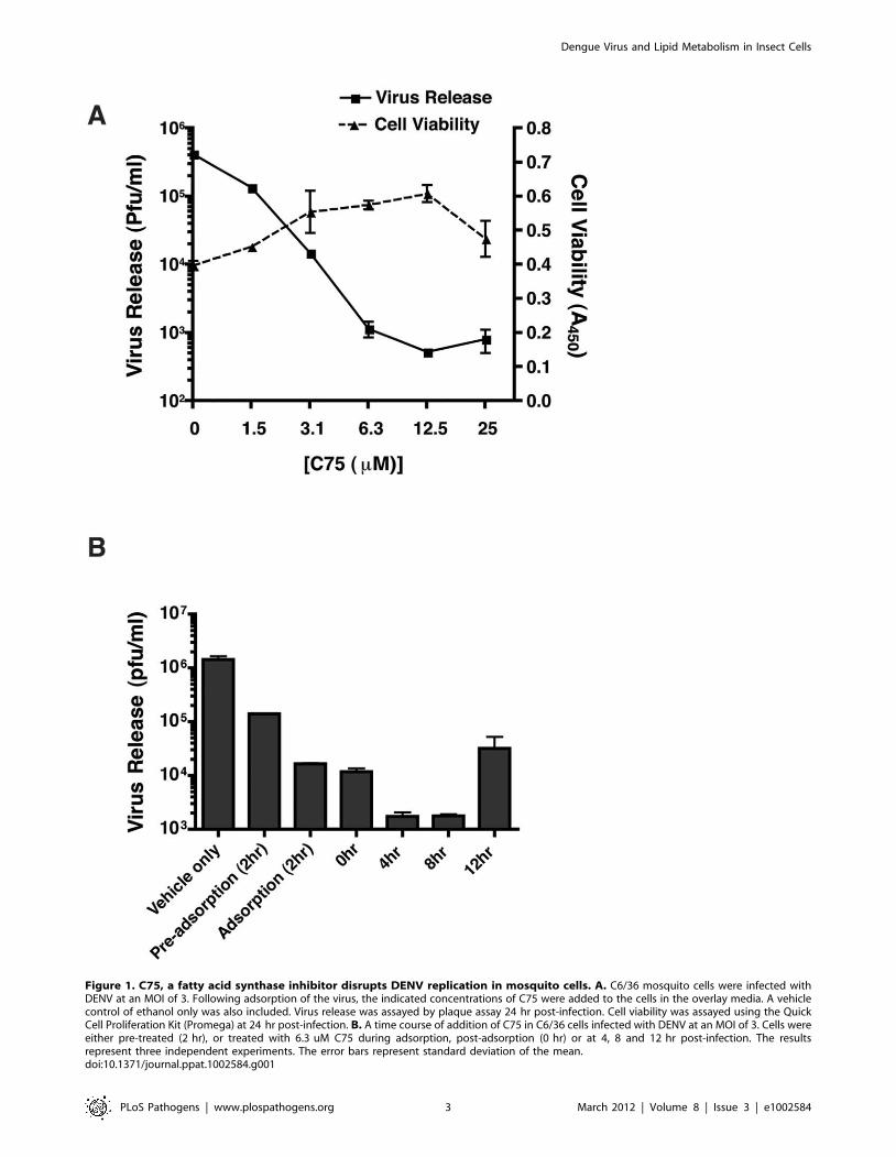
Figure 1. C75, a fatty acid synthase inhibitor disrupts DENV replication in mosquito cells. A. C6/36 mosquito cells were infected withDENV at an MOI of 3. Following adsorption of the virus, the indicated concentrations of C75 were added to the cells in the overlay media. A vehiclecontrol of ethanol only was also included. Virus release was assayed by plaque assay 24 hr post-infection. Cell viability was assayed using the QuickCell Proliferation Kit (Promega) at 24 hr post-infection. B. A time course of addition of C75 in C6/36 cells infected with DENV at an MOI of 3. Cells wereeither pre-treated (2 hr), or treated with 6.3 uM C75 during adsorption, post-adsorption (0 hr) or at 4, 8 and 12 hr post-infection. The resultsrepresent three independent experiments. The error bars represent standard deviation of the mean.doi:10.1371/journal.ppat.1002584.g001
Dengue Virus and Lipid Metabolism in Insect Cells
PLoS Pathogens | www.plospathogens.org 3 March 2012 | Volume 8 | Issue 3 | e1002584

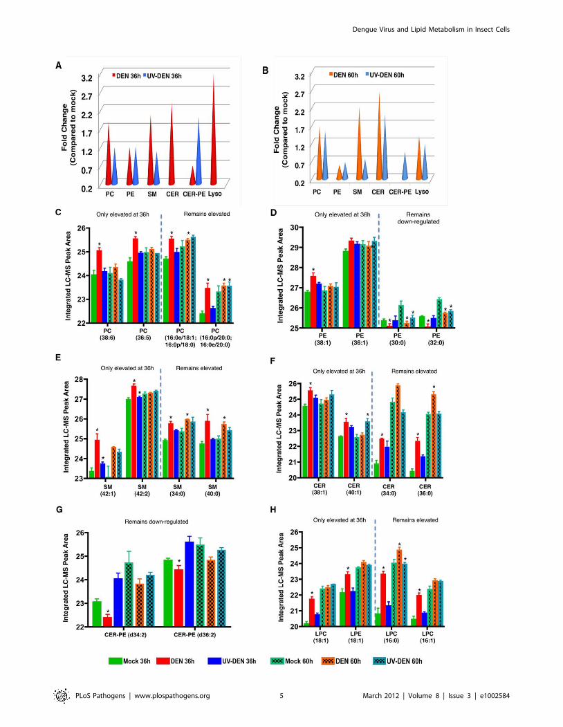
15% of the metabolites identified were significantly (Anova
p,0.05) different between virus infected cells and the mock
control.
Based on the total number of lipids that were significantly
regulated between groups (uninfected and infected, Anova
p,0.05) and subsequently structurally identified (Table S1 and
Methods) the overall abundance of lipid classes is as follows:
phosphotidylcholine (PC), 39.2%; phosphatidylethanolamine (PE),
31.2%; phosphatidylserine (PS), 0.8%; sphingomyelins (SM),
18.5%; ceramide (CER), 5.3%; lysophospholipids, 0.8% and
ceramide phosphoethanolamine (CER-PE), 4%. This distribution
is similar to the membrane lipid composition of eukaryotic cells
where the most abundant phospholipid is PC [21]. It is also
consistent with the membrane lipid composition of the Diptera
species where PE is a predominant PL [22]. CER-PE, which is
preferentially expressed in insect cells, is also observed here [23–
24]. Several of these lipids are differentially regulated upon
exposure of cells to DENV or UV-DENV (Figure 3). It was
noticeable however, that several negatively charged lipid classes
such as phosphatidylglycerol (PG), phosphatidylinositol (PI), and
Cardiolipin, were absent from this list. These lipids have
previously been reported to account for 6–13% of the lipidome
of mosquito cells [25,26]. However, in this whole cell analysis,
these lipids were not significantly regulated during virus infection.
A comparative analysis of the overall abundance of lipid species
(relative to the mock control) in DENV and UV-DENV exposed
cells is shown in Figure 3A and B. In all of the lipid classes with
significantly regulated lipids (Anova p,0.05), DENV-infected cells
have a unique expression pattern (expressed lipid molecular
species) compared to UV-DENV infected cells. For a complete list
of the differentially expressed lipids see Table S1.
Phospholipids (PL). In this whole cell analysis, the primary
PLs that were significantly regulated (Anova p,0.05) (and
subsequently structurally identified) were mostly neutral or
zwitterionic. PS was the only acidic lipid significantly regulated.
At 36 hr post-infection, there was an ,2 fold up-regulation of PC
species in DENV-infected cells compared to UV-DENV and mock
controls (Figure 3A). At the later time point (60 hr post-infection),
the relative levels of PC remained elevated in DENV-infected cells,
however the differential expression between DENV and UV-
DENV-exposed cells was not as evident (Figure 3B). Selected PC
species were up regulated only at the 36 hr time point while others
were up regulated at both time points (Figure 3C). Interestingly, a
majority (,80%) of the PC species that were up regulated had
unsaturated fatty acyl chains.
Analysis of the overall fold change in PE at the 36 hr time point
(Figure 3A) does not show a difference between the virus-exposed
(DENV and UV-DENV) cells and the mock. This is due to an
Figure 2. A plot of the principle components analysis scores shows segregation of the global lipid profile between uninfected andDENV-infected mosquito cells. The abundance of lipids in C6/36 cells infected with either DENV (MOI 20) or UV-DENV was measured at 36 and60 hr post-infection and compared to uninfected controls (mock). Each experiment included 4 independent replicates. A total of 7217 features werecompared by principle component analysis (PCA). The plot shows differences that are specific to infectious virus (DENV), a non-replicating virus(DENV-UV) and the mock control.doi:10.1371/journal.ppat.1002584.g002
Dengue Virus and Lipid Metabolism in Insect Cells
PLoS Pathogens | www.plospathogens.org 4 March 2012 | Volume 8 | Issue 3 | e1002584
Dengue Virus and Lipid Metabolism in Insect Cells
PLoS Pathogens | www.plospathogens.org 5 March 2012 | Volume 8 | Issue 3 | e1002584
equal number of individual lipid molecular species being up
regulated as were down regulated (Figure 3D). At the 60 hr time
point the relative levels of PE in virus exposed cells were lower
than mock levels (Figure 3B and D). There was also limited
overlap between the specific PE molecular species regulated
between DENV- and UV-DENV-exposed cells (Table S1). Given
that insect cells have a high abundance of PE in their membranes
(40–50%), it is interesting that there is a selective requirement for
PC (over PE) in DENV-infected mosquito cells [22].
Another group of PLs that were preferentially expressed in
DENV-infected cells were the lysophospholipids (LPLs) (Figure 3A
and B). These lipids result primarily from the hydrolysis of PC by
phospholipase A2-type enzymes (PLA2) and represent a PL that is
missing an acyl chain [27–30]. In DENV-infected cells, there was
a preferential up regulation (,3 fold) of these lipids compared to
both UV-DENV and mock controls at the 36 hr time point. This
expression was slightly down regulated at the 60 hr time point to
about ,1.5 fold above the mock. In UV-DENV exposed cells,
LPLs were undetectable at the 36 hr time point, while at the later
time point, they were similar or slightly above the mock levels. As
shown in figure 3H, LPLs with C16 acyl chains were up regulated
at both time points while there was a selective up regulation of
LPLs containing C18 acyl chains only at the early time point
(Figure 3H). The C16 LPLs have been implicated in pro-apoptotic
signaling pathways. This observation of selected LPL expression
presents an attractive hypothesis that chain length differences may
dictate specific roles for LPLs during virus infection.
Since LPLs result from the activity of PLA2-type enzymes, we
investigated whether DENV infected cells displayed a higher
activity of PLA2 compared to uninfected cells. Utilizing a
fluorescent PC substrate (BODIPY-PC) we used mass spectrom-
etry to monitor the hydrolysis of PC to LPC by intracellular PLA2.
Essentially, we monitored the production of BODIPY-LPC
(resulting from PLA2 mediated metabolism of PC) during a time
course of DENV infection (Figure S1) (method from [31]). The
assay indicated that following 24 hr of infection and longer, PLA2
activity was elevated in DENV-infected cells compared to the
controls, with the highest activity occurring at 48 and 72 hr post-
infection. Therefore, the elevated levels of PLA2 could be the
source of the LPL detected in DENV-infected cells.
Sphingolipids. Although originally known as vital
components of barrier membranes, sphingolipids are also potent
bioactive molecules that regulate cell death, growth, differentiation
and intracellular trafficking [32–33]. The primary sphingolipids
regulated during DENV infection were sphingomyelin (SM) and
ceramide (CER). In DENV infected cells, SM was up regulated by
,2-fold compared to the controls (UV-DENV and mock) and
remained elevated at both 36 hr and 60 hr time points (Figure 3A
and B). Furthermore, this temporal expression varied depending
on the specific molecular species being expressed (Figure 3E and
Table S1). UV-DENV exposed cells showed similar levels of SM
compared to the mock at the 36 hr time point, but these levels
decreased at the later time point. Interestingly, an analog of SM
known as ceramide phosphoethanolamine (CER-PE), was
preferentially expressed (up regulated by ,2 fold) in UV-DENV
exposed cells at the 36 hr time point (compared to DENV and
mock) (Figure 3G). However, this expression level dropped below
the mock at the later time point. In comparison, DENV infected
cells showed very low expression of this lipid at the early time point
(,0.7 fold compared to mock) and its expression was undetectable
at the later time point (Figure 3A and B). This lipid is expressed
more abundantly in insect cells rather than in mammalian cells
[23–24].
Another bioactive sphingolipid, CER, results from either the
degradation of SM by sphingomyelinases or de novo synthesis
through the condensation of palmitate and serine [32,34]. These
lipids were preferentially up regulated in DENV-infected cells at
both the 36 and 60 hr time points (,2-fold compared to the
mock). Since SM was also up regulated similarly in DENV
infected cells, the rise in CER is possibly the result of de novo
synthesis rather than SM degradation. UV-DENV-exposed cells
showed undetectable CER levels at 36 hr, while at 60 hr the
expression was (,2 fold) above the mock control. Since there was
a concurrent decrease in SM in UV-DENV exposed cells at 60 hr,
the increased levels of CER in this case could be due to the
degradation of SM resulting from increased cellular stress
(resulting from incubation of the cells for 60 hr). Similar to other
lipid species, CER also showed temporal regulation depending on
the molecular species expressed (Figure 3F).
An overall comparison of the PLs regulated at the 36 and 60 hr
time points indicated that in all lipid classes that were significantly
regulated (Anova p,0.05) during DENV infection, selected
molecular species were regulated only at the 36 hr time point
while others were regulated at both time points. Representative
examples for each lipid class are shown (Figure 3C–H). These
observations may represent lipidome differences in cells sustaining
early but active DENV replication (36 hr) compared to those
experiencing advanced cellular stress (60 hr). It is also interesting
to note that any up regulation observed in UV-DENV exposed
cells (at either time point) was only in the range of 1–1.5 fold
compared to the mock control, while DENV infected cells showed
much higher levels of expression.
Lipidomic analysis of replication complex membranesIn addition to evaluating the lipid composition of whole cells, we
also explored the possibility of profiling the lipid repertoire of
specific membranes induced by DENV. Given the extensive
interconnectivity of the membranes induced in DENV infected
cells, isolation of morphologically distinct membranes proved
challenging. Therefore, we carried out subcellular fractionation of
C6/36 cells and isolated post-nuclear supernatants that were
further fractionated to provide a total membrane fraction (16K
pellet) enriched in viral replication components and a remaining
cytoplasmic extract (CE) [35]. To confirm that the 16K pellet was
indeed the membrane fraction enriched in the replication complex
components, we used quantitative RT-PCR to measure the ratio
Figure 3. Whole cell lipidomics reveal an altered lipid composition in DENV infected cells. Panel A and B represent an average expression(fold change) of the total number of individual lipids significantly expressed (p,0.05) per lipid class at 36 and 60 hr post-infection, respectively. Thefold changes represent DENV-infected cells or UV-DENV exposed cells compared to the mock control. A lack of cones indicates that the expressionlevel of those specific lipids were not significant (p,0.05). Panels C–H are representative lipid molecular species from specific lipid classes significantlyregulated at the two different time points. The data are plotted as the integrated LC-MS peak abundance, in log 2 scale with standard deviation. PC,phosphatidylcholine; PE, phosphatidylethanolamine; SM, sphingomyelin; CER, ceramide; CER-PE, ceramide phosphoethanolamine; Lyso, lysopho-spholipids. See supplementary table S1 for a complete list of lipid features detected in this study. Four replicates were included in the lipidomicanalyses. The error bars represent standard deviation of the mean. The blue dashed line separates species that remain elevated at both time points(36 and 60 hr) from species that are only elevated at the 36 hr time point. Infections were carried out using an MOI of 20 in C6/36 cells. Significantlyexpressed lipids species are shown denoted with an asterisk (*).doi:10.1371/journal.ppat.1002584.g003
Dengue Virus and Lipid Metabolism in Insect Cells
PLoS Pathogens | www.plospathogens.org 6 March 2012 | Volume 8 | Issue 3 | e1002584
of viral RNA to lipid in both 16K and CE fractions. Lipids were
quantified by pulse-chase analysis using 14C-acetate to label newly
synthesized lipids and the results are shown in Figure 4. In post-
nuclear supernatants, there was increase in the incorporation of14C-acetate into lipids in the DENV infected cells compared to the
mock control (Figure 4A). This increase was observed at both
36 hr and 60 hr post-infection with the latter time point showing a
greater difference between DENV and mock. A more detailed
analysis of the lipid distribution in the subcellular fractions (16K
and CE) is shown in Figure 4B. At the 36 hr time point, the CE
fraction was enriched in newly synthesized lipids (compared to the
16K fraction) in both the DENV and mock samples, with DENV
showing an almost 2-fold increase compared to mock. This
distribution changed at the 60 hr time point where the 16K pellet
was more enriched in newly synthesized lipids, however, the fold
change (DENV compared to mock) was not as significant.
To identify the primary fraction containing viral induced
membranes that included the viral replication complex, we carried
out a comparison of the viral RNA genome copies to labeled lipid
in the 16K and CE subcellular fractions (Figure 4C). The results
indicated that the 16K membrane fraction was enriched (a ,3-
fold increase) in viral RNA (compared to CE) at the 36 hr time
point while at the 60 hr time point the CE fraction had slightly
more viral RNA per labeled lipid (compared to the 16K fraction).
Based on the 14C-acetate labeling studies above, the CE fraction
showed an increased amount of newly synthesized lipids
(compared to the 16K fraction at the 36 hr time point). In lipid
biosynthesis pathways, acetate is a precursor to both phospholipid
as well as sterol biosynthesis. Therefore, the labeled lipids (in CE)
represent not only phospholipids in membranes, but also
cholesterol and triglycerides that form lipid droplets [36]. In this
study, lipidomic analyses were carried out on both the 16K and
CE subcellular fractions isolated from the 36 hr time point from
DENV infected cells, UV-DENV exposed cells and the mock
control. The primary lipids observed in the CE fraction were
sphingolipids. Cholesterol and triglycerides were not successfully
separated using our mass spectrometry analyses and very few
phospholipids were observed to be statistically significant (p,0.05)
in virus infected cells over the mock control (data not shown).
Therefore, we focused our primary lipidomic analysis on lipids
extracted from the 16K pellet at the 36 hr time point. Similar to
the whole cell analysis, DENV infected cells displayed a unique
expression profile compared to UV-DENV and mock infected
cells (Figure S2). Over 85% of the metabolites detected were
significantly regulated in virus-infected cells compared to the
controls. Analysis of the membrane fraction also highlighted
several lipid species not detected in our whole cell analysis. An
overall fold change (compared to mock) of the significantly
(p,0.05) expressed lipid classes in the membrane fraction is shown
in Figure 5. For a complete list of differentially expressed lipids see
Table S2.
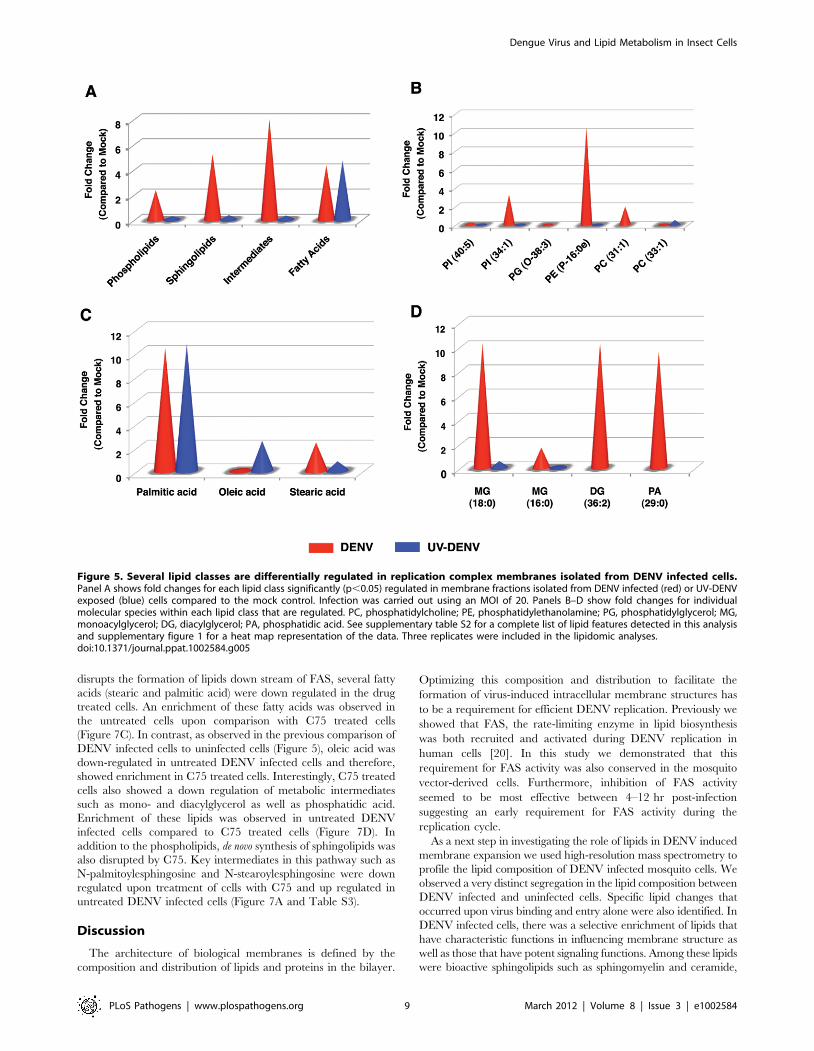
Phospholipids. There was an up regulation of
phospholipids, sphingolipids and several bioactive intermediates
in DENV infected cells compared to UV-DENV and mock
controls (Figure 5A). Amongst the specific phospholipids
expressed, PE was selectively up regulated in membrane
fractions isolated from DENV infected cells (Figure 5B).
However, the primary PE molecular species up regulated was a
lysophospholipid (PE P-16:0e). Both DENV and UV-DENV
exposed cells showed similar expression of fatty acids (Figure 5A).
However, the individual molecular species expressed differed
between the two viruses (Figure 5C). For instance, stearic acid was
up regulated in DENV infected cells while oleic acid was up
regulated in UV-DENV exposed cells. Palmitic acid had a similar
level of expression in both DENV and UV-DENV exposed cells.
Several bioactive intermediates such as monoacylglycerol (MG),
diacylglycerol (DG) and phosphatidic acid (PA) were also
prominently up regulated in DENV infected cells compared to
the controls (Figure 5D).
Sphingolipids. Although the Blygh and Dyer method [37]
utilized for lipidomic analyses of whole cells or membrane
fractions detected the expression of sphingolipids, Merrill et al
optimized a protocol dedicated to analyzing sphingolipids without
the interference from co-purifying PLs [38]. Utilizing this protocol,
we carried out mass spectrometry analysis of these bioactive lipids
from mosquito cells fractionated to provide three subcellular
fractions: nuclear (N), cytoplasmic (CE) and membrane (16K).
Similar to the above analyses, UV-DENV and mock infected cells
were included as controls. The specific sphingolipids monitored
included CER, SM, monohexosylceramide (GlcCER) and
dihexosylceramide (GalCER).
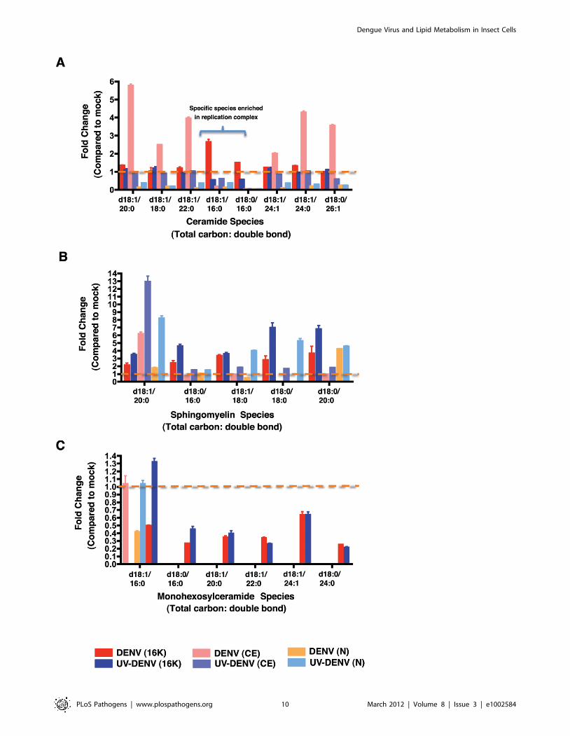
As shown in Figure 6A, CER levels in C6/36 cells showed clear
elevation in DENV-infected cells compared to mock-infected or
UV-DENV exposed cells. Comparison of the different subcellular
fractions indicated that the elevated CER was primarily concen-
trated in the CE fraction with the exception of two species (d18:1/
16:0 and d18:0/16:0) which were enriched in the 16K pellet. These
two species consist of the most abundant fatty acids observed in cells.
The analysis of SM levels in mosquito cells (Figure 6B) indicated
that virus exposed cells have a higher level of SM compared to
mock-infected cells. This is most evident in the 16K pellet with the
exception of the most abundant species (d18:1/20:0) that showed
the highest levels in the CE. Comparison of infectious DENV to
UV-DENV exposed cells indicates that the latter which is incapable
of replication maintained higher levels of SM overall.
GlcCER were primarily detected in the 16K pellet of both
DENV infected and UV-DENV exposed cells (Figure 6C).
However, the overall levels were down-regulated compared to
the mock control with the exception of d18:1/16:0, which showed
slight elevation in the UV-DENV exposed cells. Dihexosylcer-
amides were not detected to significant levels in any of the samples.
Analysis of lipid re-distribution upon C75 treatment ofmosquito cells
The FAS inhibitor, C75 is a potent inhibitor of DENV
replication in both mosquito (Figure 1) and mammalian cells
[20]. Cells treated with non-cytotoxic concentrations of C75
(,50 mM) induce an environment that has redistributed its lipid
repertoire to support cell survival in the absence of FAS activity,
and yet, this environment does not support viral replication. To
determine the basis for this exclusion of virus replication, we
profiled the lipidome of cells treated with 25 mM C75. Membrane
fractions isolated from DENV infected and mock control cells in
the presence or absence of C75 were profiled. A hierarchical
clustering analysis of the lipidomic data (Figure S3) indicated that
the lipid environment of DENV infected cells treated with C75
clustered very closely to the mock C75 treated control and was
furthest from DENV infected cells suggesting that the two
represent significantly different environments. A comparative
analysis of the lipid species expressed in the two environments
indicated that a majority of the lipids that were up regulated in
DENV infected cells were down regulated upon treatment of those
cells with C75.
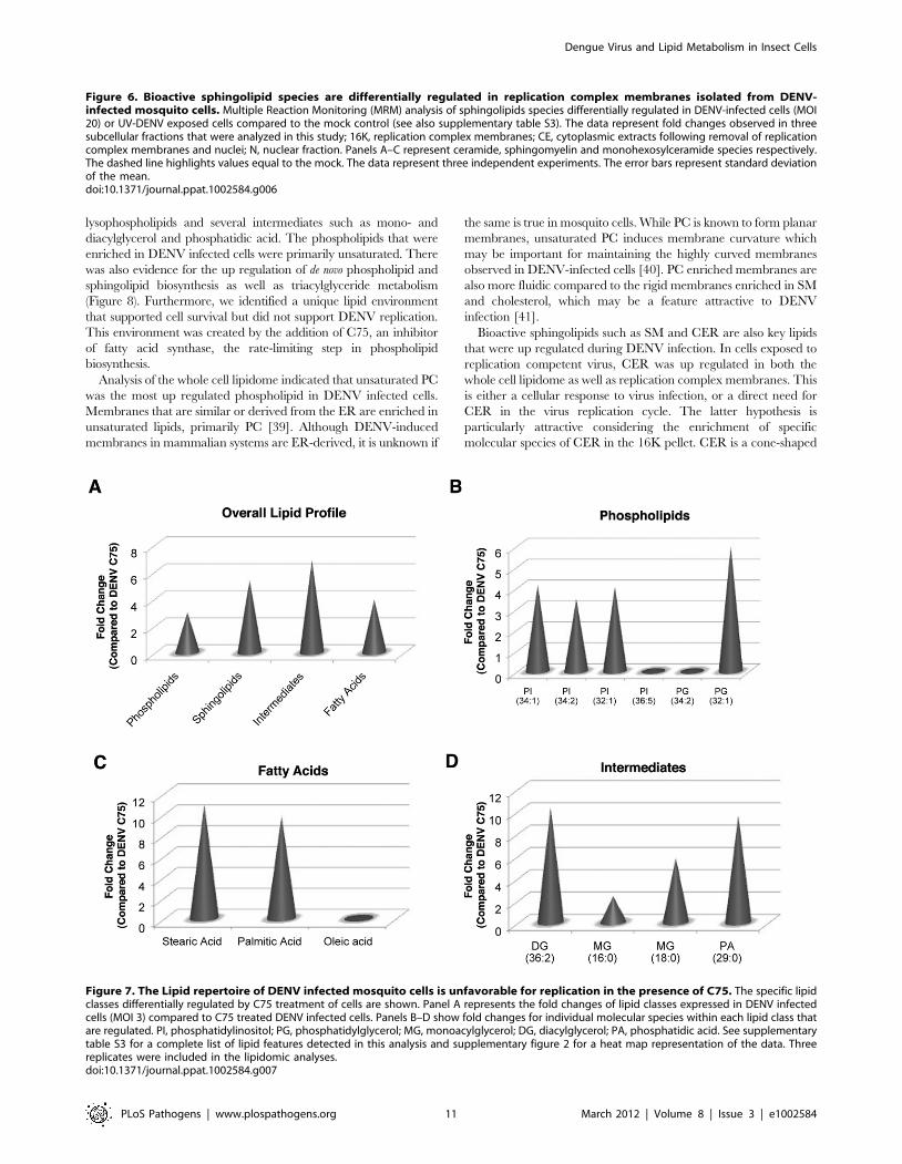
Amongst the phospholipids, several phosphatidylinositol species
were up regulated 3–4 fold in the untreated DENV infected cells
compared to the C75 treated cells (Figure 7B). However, none of
these PI species were phosphorylated. A single species of PG was
also up regulated by over 5 fold in the untreated cells. Since C75
Dengue Virus and Lipid Metabolism in Insect Cells
PLoS Pathogens | www.plospathogens.org 7 March 2012 | Volume 8 | Issue 3 | e1002584
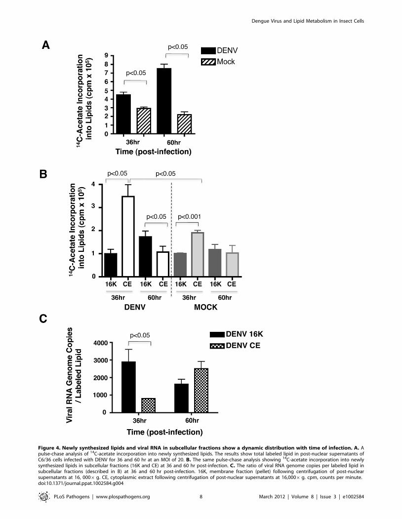
Figure 4. Newly synthesized lipids and viral RNA in subcellular fractions show a dynamic distribution with time of infection. A. Apulse-chase analysis of 14C-acetate incorporation into newly synthesized lipids. The results show total labeled lipid in post-nuclear supernatants ofC6/36 cells infected with DENV for 36 and 60 hr at an MOI of 20. B. The same pulse-chase analysis showing 14C-acetate incorporation into newlysynthesized lipids in subcellular fractions (16K and CE) at 36 and 60 hr post-infection. C. The ratio of viral RNA genome copies per labeled lipid insubcellular fractions (described in B) at 36 and 60 hr post-infection. 16K, membrane fraction (pellet) following centrifugation of post-nuclearsupernatants at 16, 0006g. CE, cytoplasmic extract following centrifugation of post-nuclear supernatants at 16,0006g. cpm, counts per minute.doi:10.1371/journal.ppat.1002584.g004
Dengue Virus and Lipid Metabolism in Insect Cells
PLoS Pathogens | www.plospathogens.org 8 March 2012 | Volume 8 | Issue 3 | e1002584
disrupts the formation of lipids down stream of FAS, several fatty
acids (stearic and palmitic acid) were down regulated in the drug
treated cells. An enrichment of these fatty acids was observed in
the untreated cells upon comparison with C75 treated cells
(Figure 7C). In contrast, as observed in the previous comparison of
DENV infected cells to uninfected cells (Figure 5), oleic acid was
down-regulated in untreated DENV infected cells and therefore,
showed enrichment in C75 treated cells. Interestingly, C75 treated
cells also showed a down regulation of metabolic intermediates
such as mono- and diacylglycerol as well as phosphatidic acid.
Enrichment of these lipids was observed in untreated DENV
infected cells compared to C75 treated cells (Figure 7D). In
addition to the phospholipids, de novo synthesis of sphingolipids was
also disrupted by C75. Key intermediates in this pathway such as
N-palmitoylesphingosine and N-stearoylesphingosine were down
regulated upon treatment of cells with C75 and up regulated in
untreated DENV infected cells (Figure 7A and Table S3).
Discussion
The architecture of biological membranes is defined by the
composition and distribution of lipids and proteins in the bilayer.
Optimizing this composition and distribution to facilitate the
formation of virus-induced intracellular membrane structures has
to be a requirement for efficient DENV replication. Previously we
showed that FAS, the rate-limiting enzyme in lipid biosynthesis
was both recruited and activated during DENV replication in
human cells [20]. In this study we demonstrated that this
requirement for FAS activity was also conserved in the mosquito
vector-derived cells. Furthermore, inhibition of FAS activity
seemed to be most effective between 4–12 hr post-infection
suggesting an early requirement for FAS activity during the
replication cycle.
As a next step in investigating the role of lipids in DENV induced
membrane expansion we used high-resolution mass spectrometry to
profile the lipid composition of DENV infected mosquito cells. We
observed a very distinct segregation in the lipid composition between
DENV infected and uninfected cells. Specific lipid changes that
occurred upon virus binding and entry alone were also identified. In
DENV infected cells, there was a selective enrichment of lipids that
have characteristic functions in influencing membrane structure as
well as those that have potent signaling functions. Among these lipids
were bioactive sphingolipids such as sphingomyelin and ceramide,
Figure 5. Several lipid classes are differentially regulated in replication complex membranes isolated from DENV infected cells.Panel A shows fold changes for each lipid class significantly (p,0.05) regulated in membrane fractions isolated from DENV infected (red) or UV-DENVexposed (blue) cells compared to the mock control. Infection was carried out using an MOI of 20. Panels B–D show fold changes for individualmolecular species within each lipid class that are regulated. PC, phosphatidylcholine; PE, phosphatidylethanolamine; PG, phosphatidylglycerol; MG,monoacylglycerol; DG, diacylglycerol; PA, phosphatidic acid. See supplementary table S2 for a complete list of lipid features detected in this analysisand supplementary figure 1 for a heat map representation of the data. Three replicates were included in the lipidomic analyses.doi:10.1371/journal.ppat.1002584.g005
Dengue Virus and Lipid Metabolism in Insect Cells
PLoS Pathogens | www.plospathogens.org 9 March 2012 | Volume 8 | Issue 3 | e1002584
Dengue Virus and Lipid Metabolism in Insect Cells
PLoS Pathogens | www.plospathogens.org 10 March 2012 | Volume 8 | Issue 3 | e1002584
lysophospholipids and several intermediates such as mono- and
diacylglycerol and phosphatidic acid. The phospholipids that were
enriched in DENV infected cells were primarily unsaturated. There
was also evidence for the up regulation of de novo phospholipid and
sphingolipid biosynthesis as well as triacylglyceride metabolism
(Figure 8). Furthermore, we identified a unique lipid environment
that supported cell survival but did not support DENV replication.
This environment was created by the addition of C75, an inhibitor
of fatty acid synthase, the rate-limiting step in phospholipid
biosynthesis.
Analysis of the whole cell lipidome indicated that unsaturated PC
was the most up regulated phospholipid in DENV infected cells.
Membranes that are similar or derived from the ER are enriched in
unsaturated lipids, primarily PC [39]. Although DENV-induced
membranes in mammalian systems are ER-derived, it is unknown if
the same is true in mosquito cells. While PC is known to form planar
membranes, unsaturated PC induces membrane curvature which
may be important for maintaining the highly curved membranes
observed in DENV-infected cells [40]. PC enriched membranes are
also more fluidic compared to the rigid membranes enriched in SM
and cholesterol, which may be a feature attractive to DENV
infection [41].
Bioactive sphingolipids such as SM and CER are also key lipids
that were up regulated during DENV infection. In cells exposed to
replication competent virus, CER was up regulated in both the
whole cell lipidome as well as replication complex membranes. This
is either a cellular response to virus infection, or a direct need for
CER in the virus replication cycle. The latter hypothesis is
particularly attractive considering the enrichment of specific
molecular species of CER in the 16K pellet. CER is a cone-shaped
Figure 6. Bioactive sphingolipid species are differentially regulated in replication complex membranes isolated from DENV-infected mosquito cells. Multiple Reaction Monitoring (MRM) analysis of sphingolipids species differentially regulated in DENV-infected cells (MOI20) or UV-DENV exposed cells compared to the mock control (see also supplementary table S3). The data represent fold changes observed in threesubcellular fractions that were analyzed in this study; 16K, replication complex membranes; CE, cytoplasmic extracts following removal of replicationcomplex membranes and nuclei; N, nuclear fraction. Panels A–C represent ceramide, sphingomyelin and monohexosylceramide species respectively.The dashed line highlights values equal to the mock. The data represent three independent experiments. The error bars represent standard deviationof the mean.doi:10.1371/journal.ppat.1002584.g006
Figure 7. The Lipid repertoire of DENV infected mosquito cells is unfavorable for replication in the presence of C75. The specific lipidclasses differentially regulated by C75 treatment of cells are shown. Panel A represents the fold changes of lipid classes expressed in DENV infectedcells (MOI 3) compared to C75 treated DENV infected cells. Panels B–D show fold changes for individual molecular species within each lipid class thatare regulated. PI, phosphatidylinositol; PG, phosphatidylglycerol; MG, monoacylglycerol; DG, diacylglycerol; PA, phosphatidic acid. See supplementarytable S3 for a complete list of lipid features detected in this analysis and supplementary figure 2 for a heat map representation of the data. Threereplicates were included in the lipidomic analyses.doi:10.1371/journal.ppat.1002584.g007
Dengue Virus and Lipid Metabolism in Insect Cells
PLoS Pathogens | www.plospathogens.org 11 March 2012 | Volume 8 | Issue 3 | e1002584
molecule that promotes inward budding of membranes (negative
curvature) [42]. Some DENV-induced membranes show a double
membrane morphology indicating that negative curvature modify-
ing lipids such as CER may be active in their formation.
As a second messenger, CER can also induce apoptosis and
autophagy [34]. However in mosquito cells, DENV maintains a
persistent infection and antagonizes apoptotic pathways [19,43].
Therefore, CER up regulation may not be due to the prevalence
of an apoptotic response but may be utilized to up regulate
autophagy during DENV infection. It has been shown in
mammalian cells that autophagy is up regulated and necessary
for DENV replication. The prevalence of several intermediates in
CER biogenesis, including N-palmitoylsphingosine and diacylgly-
cerol, suggest that the observed CER could result from either de
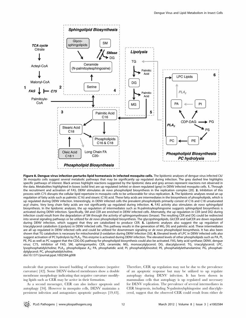
Figure 8. Dengue virus infection perturbs lipid homestasis in infected mosquito cells. The lipidomic analyses of dengue virus infected C6/36 mosquito cells suggest several metabolic pathways that may be significantly up regulated during infection. The grey dashed line highlightsspecific pathways of interest. Black arrows highlight reactions suggested by the lipidomic data and grey arrows represent reactions not observed inthe data. Metabolites highlighted in boxes (solid line) are up regulated (white) or down regulated (grey) in DENV infected mosquito cells. 1. Throughthe recruitment and activation of FAS, DENV stimulates de novo phospholipid biosynthesis in the replication complex [20]. 2. Inhibition of thisprocess with C75 disrupts the cellular lipid repertoire in mosquito cells to be unfavorable for virus replication. 3. The lipidomic analyses reveal an upregulation of fatty acids such as palmitic (C16) and stearic (C18) acid. These fatty acids are intermediates in the biosynthesis of phospholipids, which isup regulated during DENV infection. Interestingly, in DENV infected cells the prevalent phospholipids primarily consist of C16 and C18 unsaturatedacyl chains. Very long chain fatty acids are not significantly up regulated during infection. 4. FAS activity also stimulates de novo sphingolipidbiosynthesis. In the lipidomic analyses, the up regulation of intermediates such as N-palmitoylesphingosine suggests sphingolipid biosynthesis isactivated during DENV infection. Specifically, SM and CER are enriched in DENV infected cells. Alternately, the up regulation in CER (and DG) duringinfection could result from the degradation of SM through the activity of sphingomyelinases (Smase). The resulting CER and DG could be redirectedinto several signaling pathways or be utilized for de novo phospholipid biosynthesis. The glycopshingolipids, GlcCER and GalCER are down regulatedduring DENV infection, which suggest that they are catabolized to produce CER. 5. Lipidomic analyses also suggest the up regulation oftriacylglycerol catabolism (Lipolysis) in DENV infected cells. This pathway results in the generation of MG, DG and palmitic acid. These intermediatesare all up regulated in DENV infected cells and could be utilized for downstream signaling or de novo phospholipid biosynthesis. It has also beenshown that TG catabolism is necessary for mitochondrial b-oxidation during DENV infection [50]. 6. Elevated levels of LPC in DENV infected cells alsosuggest activation of PC hydrolysis by PLA2. This enzyme is activated during DENV infection. The elevated levels of other phospholipids such as PA, PI,PE, PG as well as PC suggest that the CDG-DG pathway for phospholipid biosynthesis could also be activated. FAS, fatty acid synthase; DENV, denguevirus; C75, inhibitor of FAS; SM, sphingomyelin; CER, ceramide; MG, monoacylglycerol; DG, diacylglycerol; TG, triacylglycerol; LPC,lysophosphatidylcholine; PLA2, phospholipase A2; PA, phosphatidic acid; PI, phosphatidylinositol; PE, phosphatidylethanolamine; PG, phosphati-dylglycerol; PC, phosphatidylcholine.doi:10.1371/journal.ppat.1002584.g008
Dengue Virus and Lipid Metabolism in Insect Cells
PLoS Pathogens | www.plospathogens.org 12 March 2012 | Volume 8 | Issue 3 | e1002584
novo synthesis by serine palmitoyltransferase (SPTLC) in the ER
or catabolism of SM by sphingomyelinases (SMases) at the plasma
membrane [44]. The lipidomic analyses also indicated a down
regulation of GlcCER, which may suggest that a salvage pathway
that metabolizes GlcCER to CER may also be active.
Inverted cone-shaped lipids such as LPLs were also up regulated
during DENV infection. Primarily, LPC was up regulated in the
whole cell analysis. It results from the hydrolysis of PC by PLA2
[45]. We have shown that this enzyme is activated during DENV
infection. Many other viruses have also shown a dependency for
PLA2 in their life cycle [31,46]. Due to its inverted cone-shaped
structure, asymmetric incorporation of LPC into membranes
causes positive curvature and induces vesicle fission and budding
[47]. Structural analyses of DENV-induced membrane structures
in mosquito cells indicate the presence of highly curved
membranes and smaller vesicles [18–19]. Therefore, vesiculation
through fission and budding as well as enrichment of molecules
that increase membrane curvature may be an underlying
mechanism for forming these membrane structures.
LPC also increases the permeability of membranes [48]. This is
an attractive concept given that DENV replication complexes are
encased within membrane barriers that need to facilitate the
exchange of components to and from the cytosol for genome
replication and virus assembly. Therefore, leaky membranes may
be favored in DENV-infected cells through the selective
incorporation of molecules such as LPC. Another observation
that gives credence to this hypothesis is that the transmembrane
permeabilizing effect of LPC is synergistically enhanced in the
presence of palmitic acid, which is also up regulated in DENV-
infected cells. As a signaling molecule, LPC is both a pro-survival
as well as an inflammatory molecule [46]. Therefore, its expression
in DENV infected cells could be a cellular response to viral
infection.
Evidence for lipogenesisIt has been previously reported that de novo lipid biosynthesis was
up regulated during DENV infection [20]. Consistent with these
observations, several primary fatty acids (palmitic, stearic and oleic
acid) involved in the biosynthesis of higher order PLs were
detected in our lipidomic analyses. Palmitic acid is the first fatty
acid of the lipogenesis pathway and stearic acid (C18:0), is
immediately downstream of palmitic acid. They are both up
regulated in DENV infected cells. The latter is the precursor to the
biosynthesis of oleic acid (C18:1) or long chain fatty acids (with
acyl chains of C20 or greater). Interestingly, since oleic acid is
down regulated in DENV-infected cells stearic acid may be
stimulating long chain FA biosynthesis. However, a majority of the
PLs expressed in DENV infected cells have C16- or C18-acyl
chains with varying degrees of unsaturation. Therefore, it seems
that desaturation of palmitic and stearic acid rather than
elongation may be the chosen pathway for lipogenesis during
DENV infection.
Evidence for lipolysisSeveral intermediates in lipid catabolism were also detected in
the replication complex membranes (palmitic acid, MG and DG).
Analysis of these intermediates suggested the up regulation of
pathways for PC hydrolysis and triacylglycerol (TG) metabolism.
The latter pathway is responsible for regulating lipid homeostasis
and energy production in the cell through the metabolism of lipid
droplets [49]. Through the action of lipases the degradation of
lipid droplets results in the release of palmitic acid, DG and MG
[36]. These metabolic products are substrates for mitochondrial b-
oxidation as well as intermediates (fatty acids) in the synthesis of
new lipids. DENV infected cells show a high prevalence of these
species compared to mock infected cells.
While several acidic phospholipid species were detected in the
16K membrane fraction very few were significantly regulated
during infection. Previous studies in mammalian cells have
indicated the importance of acidic lipids in DENV infection
[46,50]. These negatively charged lipids are also implicated in
influencing membrane structure.
The most striking results were obtained by the comparison of
DENV infected cells in the presence and absence of the FAS
inhibitor, C75. It is well established that treatment of cells with this
inhibitor disrupts de novo phospholipid biosynthesis [51]. To
circumvent this inhibition, the cell redistributes its lipid repertoire
to ensure cell survival. However, this redistribution does not
support DENV replication. A comparison of the different lipid
environments indicated that many of the lipids that were up
regulated in DENV infected cells compared to cells treated with
C75, were similar to those previously observed when infected cells
were compared to mock (uninfected) controls. Therefore,
comparison of DENV infected cells to two different controls
(uninfected cells and C75 treated infected cells) highlighted the
same lipids as being up regulated during an active DENV
infection.
In these studies we utilized UV-DENV as a control to identify
lipid metabolic changes that occurred upon virus binding and
entry alone. This virus is incapable of replication but immuno-
fluorescence microscopy with anti-NS3 antibodies have indicated
that there is a very low level of translation that occurs within the
first 24 hr (data not shown). Analysis of the lipidome of UV-
DENV exposed cells showed a significant difference in the
phospholipid and sphingolipid content in comparison to DENV
infected cells and the mock control. It is possible that binding,
membrane fusion and entry alone, or the expression of viral
proteins from the incoming viral RNA triggers the activation of
lipases or lipid transport mechanisms that result in the differences
observed in the lipidome of the cells. This has been shown for
other viruses. Jan et al. have shown that binding and entry alone of
Sindbis virus triggered the activation of sphingomyelinases that
degraded membrane bound SM to Cer [52]. It has also been
shown that viruses induced apoptotic-signaling cascades upon
binding and entry alone [53–55]. These cascades may either result
from or induce lipolysis or lipid transfer between compartments in
the cell.
The most pronounced changes are those observed in the 16K
pellet analyses where a significant decrease in phospholipids,
sphingolipids and lipid intermediates are observed (compared to
mock cells, Figure 5A). A comparison of SM and CER in the
whole cell analysis at the 36 hr and 60 hr time points (Figure 1A
and 1B) indicated a reduction in SM with a corresponding
increase in CER with time. This could be due to a similar scenario
to SINV where a non-replicating UV-DENV is capable of
inducing lipid conversion in membranes [52]. Therefore, SM
and other sphingolipids may be converted to CER that is
transported away from the 16K membrane fraction to other
locations in the cell. This is also evident from the increased content
of fatty acids compared to mock cells that may result from the
degradation of lipids. UV-DENV exposed cells do not show
significant regulation of MG or DG, but show an up regulation of
palmitic acid (C16:0). Virus binding and entry alone could
stimulate lipid droplet hydrolysis (catabolism of TG) resulting in
the release of palmitic acid. These studies have shown that UV-
DENV could be a versatile tool to pursue mechanistic studies on
the metabolic pertabations that occur upon virus binding,
membrane fusion, and entry.
Dengue Virus and Lipid Metabolism in Insect Cells
PLoS Pathogens | www.plospathogens.org 13 March 2012 | Volume 8 | Issue 3 | e1002584

In summary, using high-resolution mass spectrometry, we have
determined that DENV drastically alters the lipid profile of
infected cells. Specifically, DENV infection elevates the expression
of lipids that have the capacity to change the physical properties of
the bilayer such as bilayer curvature, permeability, and the
recruitment and assembly of protein complexes in the membrane.
Several of the identified molecules also function as bioactive
messengers that control signaling and membrane trafficking
pathways in the cells. They represent molecules that result from
the activation of cellular stress pathways that respond to viral
infections. Based on these findings, the next steps will be to
investigate the mechanisms of how these lipid species play a role in
DENV replication, as well as identify the control points in these
pathways that may be influenced by viral gene products.
Methods and Materials
Cell culture and virus infectionsC6/36 (Aedes albopictus) cells (ATCC) were maintained in
Minimal Essential Medium (MEM) supplemented with glutamine
(2 mM), non-essential amino acids, 25 mM Hepes and 10% heat
inactivated fetal calf serum. Dengue virus 2, strain 16681 (obtained
from Richard Kinney, CDC. Ft. Collins) stocks were amplified in
C6/36 cells in MEM (supplemented as above) and 2% heat
inactivated fetal calf serum. UV-inactivated DENV was obtained
by exposing the same virus stock (used for infection) to UV-light
for 3 hr and then conformation of inactivation by two blind
passages of the virus on cells for 60 hr per passage. Lack of
infectivity was confirmed by plaque assay and immunofluores-
cence assay.
Lipidomic sample preparation and mass spectrometryanalyses of whole cells
Sample preparation. For infection, ,56108 C6/36 cells
were infected with DENV at a MOI of 20 at 30uC. Infection of all
cells (required for lipidomic analyses) was confirmed by
immunofluorescence analyses. Each experiment included three
biological replicates. Following infection, cells were harvested at 36
and 60 hr post-infection, pelleted and resuspended in 100 ml of
100 mM ammonium bicarbonate. Lipids were extracted from an
equal number of cells using a modified Bligh and Dyer protocol.
Briefly, a mixture of 2:1 Chloroform:methanol, 0.1% acetic acid
and 0.01% butylated hydroxy toluene (BHT) were added to the
cell suspension in ammonium bicarbonate such that there was a
4:1 ratio of organic solvent to cells. Following lipid extraction, the
organic phase was separated from the aqueous phase by
centrifugation and dried down under a N2 stream in low
retention microfuge tubes (Fisher). The dried lipids were
resuspended in 75 ml of methanol and vortexed for 10 s. The
samples were then centrifuged at 13,4006 g for 5 min to remove
any particulates.
Liquid chromatography/mass spectrometry. Lipid
molecular species were profiled using a dual-column
nanocapillary LC system equipped with 75 mm665 cm columns,
each packed with 3 mm Jupiter particles (Phenomenex, Torrance,
CA). The mobile phases were (A) 10 mM ammonium acetate in
50:50 water/methanol (v/v) and (B) 10 mM ammonium acetate in
50:50 methanol/acetonitrile (v/v). The LC system was
equilibrated at 10,000 psi with mobile phase A prior to injecting
0.4 mL of sample. Exponential gradient elution was initiated
50 min after sample loading with an initial column flow of
,300 nL/min. After 90 min of gradient separation, the mobile
phase mixer was purged with 3 mL of mobile phase B, followed by
a 5 min column wash. Finally, the mobile phase mixer was purged
with 10 mL of mobile phase A, which represented the end of one
separation cycle. While gradient elution was performed on one
column, the other column was equilibrated with mobile phase A.
The LC system was coupled to a hybrid linear ion-trap-Orbitrap
mass spectrometer (ThermoFisher, San Jose, CA), and the
capillary temperature and electrospray voltage were 200uC and
+2.2 kV, respectively. The Orbitrap was used as the mass analyzer
during MS survey scans over the m/z range 200–2000 with a duty
cycle of ,1.2 s. Data-dependant MS/MS was performed in the
LTQ for the top 5 ions, with a normalized collision energy of
,35%. Dynamic exclusion in the LTQ during data-dependant
MS/MS experiments was enabled as follows: repeat count of 2,
repeat duration of 30 s, exclusion list size of 250, and exclusion
duration of 60 s.
Data processing. LC-MS datasets, defined as the data
obtained from a single LC-MS analysis, were processed using
the PRISM Data Analysis system [56], a series of software tools
freely available at http://ncrr.pnl.gov/software/ and developed
in-house. The first step involved deisotoping of the raw MS data to
give the monoisotopic mass, charge state, and intensity of the
major peaks in each mass spectrum using Decon2LS [16]. The
data were next examined in a 2-D fashion using MultiAlign to
identify groups of mass spectral peaks that were observed in
sequential spectra using an algorithm [57] that computes a
Euclidean distance in n-dimensional space for combinations of
peaks. Each group, generally ascribed to one detected species and
referred to as a ‘‘feature’’, has a median monoisotopic mass,
central normalized elution time (NET), and abundance estimate
computed by summing the intensities of the MS peaks that
comprise the entire LC-MS feature. LC-MS features were then
chromatographically aligned across all datasets using the
LCMSWARP algorithm [58] in MultiAlign, and the lipid
identities of detected features were initially determined by
comparing their measured monoisotopic masses and NETs to
calculated monoisotopic masses and observed NETs for lipids in
an AMT tag database [59] within search tolerances of 63 ppm
and 60.03 NET for monoisotopic mass and elution time,
respectively. Subsequent identifications of lipid features that did
not match to entries in the AMT tag database were made by
searching their molecular weights against entries in the Lipid
MAPS database followed by manual confirmation based on MS/
MS spectra.
Statistical analysis of processed data. Following
chromatographic alignment and database matching, the
abundances of all detected features (both AMT tag database
matched and unmatched) were loaded into DAnTE [60] for
statistical analysis. Feature abundances were transformed to log2
scale then subjected to central tendency normalization [61].
Comparative data analysis was then performed and statistically
significant differences between the lipid profiles of the samples
were determined using ANOVA. Partial least-squares (PLS) [62]
analysis was also performed using the data matrices containing
either AMT tag database matched features alone or all features
(both database matched and unmatched).
Pulse-chase analyses and quantitative RT-PCRVirus infection of C6/36 cells was carried out as described for
the whole cell analyses. To label newly synthesized lipids, Acetic
Acid, Sodium Salt, [1-14C] (Perkin Elmer) was added to cell
supernatants at 8 hr post-infection. At 12 hr post-infection, the
label containing supernatant was removed and new medium was
added to the cells. At 36 and 60 hr post-infection, cells were
harvested by trypsinization and washed with buffer A (35 mM
Hepes, pH 7.4, 146 mM NaCl, 11 mM glucose). Cells were then
Dengue Virus and Lipid Metabolism in Insect Cells
PLoS Pathogens | www.plospathogens.org 14 March 2012 | Volume 8 | Issue 3 | e1002584
osmotically shocked in buffer B (20 mM Hepes, pH 7.4, 10 mM
KCl, 1.5 mM MgOAc and 1 mM DTT) for 30 min on ice,
homogenized and centrifuged at 15006 g, at 4uC for 5 min to
remove nuclei. Post-nuclear supernatants were centrifuged at
160006g for 30 min at 4uC to obtain a membrane pellet (referred
to as the 16K pellet). The supernatant post nuclear and membrane
fractionation is referred to as the cytoplasmic extract (CE). Total
RNA was extracted from each fraction (16K and CE) using Trizol
reagent (Sigma) according to the manufacturer’s instructions.
Following RNA extraction, total lipids were extracted using the
Bligh and Dyer method mentioned above and counted using a
Beckman LS 6000 scintillation counter. The viral RNA in each
fraction was measured using the SuperScript III Platinum SYBR
Green One-Step qPCR Kit with ROX and DENV specific
primers; (forward) 59 ACAAGTCGAACAACCTGGTCCAT 39
and (reverse) 59 GCCGCACCATTGGTCTTCTC 39 on a
Applied Biosystems 7300 Real Time PCR machine. The labeled
lipid in subcellular fractions was standardized to the total RNA
isolated from each fraction prior to determining the viral RNA
genome copies per labeled lipid in subcellular fractions.
Lipidomic sample preparation and mass spectrometryanalyses of replication complex membranes
Sample preparation. For profiling the lipidome of the
replication complex membranes, lipids were extracted from the
16K pellet using the same modified Blygh and Dyer protocol
described above.
Liquid chromatography/mass spectrometry profiling
analysis of lipid content. A LTQ Orbitrap XL instrument
(Thermo Fisher Scientific San Jose, CA) was used for the analysis.
It was coupled to an Agilent 1100 series LC (Agilent Technologies,
Santa Clara, CA) equipped with a micro well plate auto sampler
and binary pumping device. Reverse phase liquid chromatography
was used to analyze the samples. An Agilent Eclipse XDB-C8
column with 2.16150 mm, 3.5 mm dimensions was used for the
separation. Solvent A consisted of water +0.1% piperidine. Solvent
B contained acetonitrile : methanol (50:50 v/v) +0.1% piperidine.
The flow rate was 300 mL/minute. A volume of 10 mL was loaded
onto the column. The gradient was as follows: time 0 minutes,
50% B; time 25 minutes, 95% B; time 45 minutes, 95% B; time
50 minutes, 50% B; time 60 minutes, 50% B. The MS analysis
used negative polarity electrospray ionization. The source voltage
was 4.0 kV, source current 100 mA, capillary voltage 230.0 V,
tube lens voltage 2100.0 V. The capillary temperature was
200uC, sheath gas flow was 35, auxiliary and sweep gas were both
set to 0. Data were acquired using data dependent scanning mode.
FTMS resolution of 60,000 with a mass range of 70–1200 was
used for full scan analysis and the ITMS was used for MS/MS
data acquisition. The top three most intense ions were acquired
with a minimum signal of 500, isolation width of 2, normalized
collision energy of 35, default charge state of 1, activation Q of
0.250, and an activation time of 30.0. The samples were evaluated
with Thermo XCalibur software (version 2.1.0) and downstream
alignment done with an in-house data processing package called
Omics Discovery Pipeline [63,64].
Liquid chromatography/mass spectrometry for targeted
sphingolipid analysis. The method was slightly modified from
[65]. Briefly, each sample was extracted according to published
protocol then dried and reconstituted in 100 mL of methanol :
water : formic acid (74:25:1) containing 5 mM ammonium
formate. An Agilent 6400 QQQ (Agilent Technologies, Santa
Clara, CA) was used for analysis coupled to an 1100 Series LC
equipped with HPLC Chip interface (Agilent Technologies Santa
Clara, CA). Solvent A consisted of methanol : water : formic acid
(74:25:1) containing 5 mM ammonium formate and solvent B
methanol : formic acid (99:1) containing 5 mM ammonium
formate. The gradient was as follows: time 0 minutes, 20% B; time
1 minute, 20% B; time 3 minutes, 100% B; time 23 minutes,
100% B; time 24 minutes, 20% B; time 30 minutes, 20% B. The
flow rate was 0.4 mL/min. Data were acquired in MRM mode
with transitions and collision energy according to Merrill et al [65]
with the following source conditions: gas temperature 300uC, gas
flow 4 L/minute, capillary voltage 1900 V. The fragmentor
voltage was set to 180 V in all cases. Data were processed with
Agilent Mass Hunter software version B03.01.
C75 inhibitor studiesC6/36 cells were infected with DENV at an MOI of 3.
Following adsorption, virus was removed and the cells were
washed and overlayed with media containing varying concentra-
tions of C75 [51]. The vehicle for C75 was ethanol. Virus
supernatants were harvested at 24 hr post-infection and virus titer
assayed by plaque assay. Cytotoxicity of C75 was simultaneously
assayed using the Quick Cell Proliferation Kit (Abcam). TIME OF
ADDITION OF C75: C6/36 cells were infected with DENV as
described above. At the indicated time points, 6.3 mM C75 was
added to the cells. Cells were harvested at 24 hr post-infection and
the amount of released virus was determined by plaque assay. For
the lipidomic studies, cells were infected with DENV (as described
above) or left uninfected. Following adsorption of the virus at room
temperature for 2 hr, C75 was added to the media in the overlay,
and cells were incubated at 30uC for 36 hr. Sample preparation,
lipid extraction and mass spectrometry analyses were carried out
as described above for the fractionated (16K pellet) samples.
Phospholipase A (PLA) analysesPhospholipase A2 activity was monitored using a Red/Green
BODIPY PC-A2 substrate (Invitrogen) using a similar method as
described in [31]. C6/36 cells were infected with DENV at an
MOI = 3 in 6-well plates. Following virus adsorption, the cells
were overlayed with MEM (supplemented as above) and 10% heat
inactivated fetal calf serum (2 ml/well). At the indicated time
points, the media were removed, and new media (1 ml/well)
containing 6 mM of the fluorogenic phospholipase A substrate
(BODIPY-PC) were added to the cells. The cells were incubated at
30uC for 30 min. Following incubation, media was removed and
the cells were washed with 16PBS. The lipids were then extracted
using butanol-1 (1 ml/well). The aqueous fraction was discarded
and the lipids in the butanol phase were analyzed by mass
spectrometry to monitor the conversion of BODIPY-PC to
BODIPY LCP by PLA2.
Liquid chromatography/mass spectrometry for targeted
Phospholipase A2 activity analysis. The lipid samples were
reconstituted in 100 mL of methanol : water : formic acid (74:25:1)
containing 5 mM ammonium formate and analyzed with an
Agilent 6400 QQQ (Agilent Technologies, Santa Clara, CA)
coupled to an 1100 Series LC equipped with HPLC Chip interface
(Agilent Technologies Santa Clara, CA). Solvent A consisted of
methanol : water : formic acid (74 : 25 : 1) containing 5 mM
ammonium formate and solvent B methanol : formic acid (99 : 1)
containing 5 mM ammonium formate. The gradient was as
follows: time 0 minutes, 40% B; time 5 minute, 70% B; time
7 minutes, 100% B; time 9 minutes, 100% B; time 9 minutes,
40% B; time 13 minutes, 40% B. The flow rate was 0.4 mL/min.
Data were acquired for parent ions corresponding to the
BODIPY-PC (986.9 m/z) and BODIPY LCP (320.2 m/z) in
single ion monitoring (SIM) mode with the following source
conditions: gas temperature 300uC, gas flow 4 L/minute, capillary
Dengue Virus and Lipid Metabolism in Insect Cells
PLoS Pathogens | www.plospathogens.org 15 March 2012 | Volume 8 | Issue 3 | e1002584
voltage 1900 V. The fragmentation voltage was set to 200 V in all
cases. Data were processed with Agilent Mass Hunter software
version B03.01.
Supplemental dataSupplemental data include three figure and three tables.
Supporting Information
Figure S1 PLA2 is activated in DENV-infected mosquitocells. C6/36 cells were either mock-infected or infected with
DENV at an MOI = 3. At the indicated time points, media was
removed, and new media containing the fluorogenic phospholi-
pase A substrate (BODIPY-PC) was added to the cells. The cells
were incubated at 30uC for 30 min. Following the incubation, cells
were washed and lipids were extracted and analyzed by mass
spectrometry to monitor the conversion of BODIPY-PC to
BODIPY LCP by PLA2. The graph represents the ratio of
BODIPY-LPC/PC with time.
(TIF)
Figure S2 Lipid homeostasis is altered in DENVinfected mosquito cells. A hierarchical clustering analysis of
the results from the mass spectrometry analysis of lipid extracts
from the 16K membrane fraction isolated at the 36 hr time point
post treatment of C6/36 cells with the different conditions. The
conditions included: uninfected cells (Mock), DENV-infected cells
(DENV), UV-inactivated DENV treated cells (UV-DENV). Each
condition included 3 replicates (denoted 1-3). Each horizontal row
represents a differentially regulated metabolite. Each vertical row
represents an individual sample. The samples from left to right
include: DEN_1-3; cells infected with DENV, Mock_1-3;
uninfected cells, UV-DEN_1-3; cells exposed to UV-inactivated
DENV (UV-DENV). The Row Z-Score was calculated by
subtracting the mean of the row from every value and then
dividing the resulting values by the standard deviation of the row.
The heatmap was created using the heatmap.2 function of the
‘gplots’ package in R [64].
(TIF)
Figure S3 The FAS inhibitor, C75 alters Lipid homeo-stasis in DENV infected mosquito cells. A hierarchical
clustering analysis of the results from the mass spectrometry
analysis of lipid extracts from the 16K membrane fraction isolated
at the 36 hr time point post treatment of C6/36 cells with the
different viruses and drug conditions. The conditions included:
uninfected cells (Mock), uninfected cells treated with 25 mM C75
(C75 Mock), DENV-infected cells (DENV), DENV-infected cells
treated with 25 mM C75 (C75 DENV), UV-inactivated DENV
treated cells (UV-DENV). Each treatment included 3 replicates
(denoted 1-3). Each horizontal row represents a differentially
regulated metabolite. Each vertical row represents an individual
sample. The Row Z-Score was calculated by subtracting the mean
of the row from every value and then dividing the resulting values
by the standard deviation of the row. The heatmap was created
using the heatmap.2 function of the ‘gplots’ package in R [64].
(TIF)
Table S1 Select list of lipid species from the whole cellanalysis significantly regulated across conditions andtime points (Anova p,0.05). Conditions: Mock (uninfected
cells), DENV (infectious dengue virus type 2, strain 16681), UV-
DENV (UV-inactivated dengue virus type 2, strain 16681). Time
points: 36 and 60 hr post-infection. A total of 7216 features were
detected in the mass spectrometry analysis. Following Anova
analysis, 677 features had a p,0.05. Structural identification was
carried out on 100 lipids. For each condition and time point, the
following information is provided: Treatment P; p-values from the
Anova analysis on minimum observations data (p,0.1), Exact
mass; mass of each lipid from http://www.lipidmaps.org,
[M+H]+; Protonated molecular ion, NET; normalized elution
time, total carbon/double bond; corresponds to each lipid species
detected, LM_ID; identification for each lipid as displayed in
LIPID MAPS, Formula, elemental composition of each lipid, PPM
Error; difference in experimental mass compared to exact mass,
Fold DENV/Mock; fold change of average abundance from 4
replicates of each treatment. Identity abbreviations were made for
phoshatidylcholine (PC; O- fatty acid chain number means that an
alkyl acyl linkage to the glycerol chain is present for the respective
PC), phosphatidylethnolamine (PE), phosphatidylserine (PS),
sphingomyelin (SM), ceramide (Cer), ceramide phosphoethanola-
mine (Cer-PE), lysophosphatidylcholine (LPC). The notation
further indicates total number of carbons and double bonds
however it does not discern redundancy associated with varying
fatty acid composition for the same molecular weight. Accession
numbers (LM_ID) were obtained from the Lipid Maps Gateway
(lipidmaps.org).
(PDF)
Table S2 Select list of lipid species from the analysis ofthe 16K membrane fraction (isolated from mosquitocells treated with conditions described below) regulatedacross conditions (p,0.05). Conditions: Mock (uninfected
cells), DENV (infectious dengue virus type 2, strain 16681), UV-
DENV (UV-inactivated dengue virus type 2, strain 16681). A total
of 484 features were detected in the mass spectrometry analysis
following normalization of Mock, DENV and UV-DENV treated
samples and 415 of these features were significantly regulated
(p,0.05). Identification was successful for 68 lipids (ppm error
,10). For each condition the following information is provided:
pttest and pwilcox; p-values on minimum observation data (a
measurement must be present in two of three replicates to be
considered present), Exact mass; mass of each lipid from http://
www.lipidmaps.org, M/Z; mass to charge ratio, RT; retention
time, Formula, elemental composition of each lipid, PPM Error;
difference in experimental mass compared to exact mass, Fold
DENV/Mock; Fold UV-DENV/Mock; fold change of average
abundance from 3 replicates of each treatment. Identity
abbreviations were made for phoshatidylcholine (PC), phosphati-
dylethnolamine (PE), phosphatidylglycerol (PG, O- fatty acid
chain number means that an alkyl acyl linkage to the glycerol
chain is present for the respective PG), sphingomyelin (SM),
monoacylglycerol (MG), diacylglycerol (DG), phosphatidic acid
(PA). Accession numbers were obtained from the Lipid Maps
Gateway (lipidmaps.org) and the Human Metabolome Database
(hmdb.ca).
(PDF)
Table S3 Select list of lipid species from the analysis ofthe 16K membrane fraction (isolated from mosquitocells treated with conditions described below) regulatedacross conditions (p,0.05). Conditions: DENV (cells infected
with DENV in the presence of vehicle only), DENV C75 (cells
infected with DENV and treated with the FAS inhibitor C75). A
total of 366 features were detected in the mass spectrometry
analysis following normalization of DENV and DENV C75
treated samples and 314 of these features were significantly
regulated (p,0.05). Identification was successful for 27 lipids (ppm
error ,10). For each condition the following information is
provided: pttest and pwilcox; p-values on minimum observation
data (a measurement must be present in two of three replicates to
Dengue Virus and Lipid Metabolism in Insect Cells
PLoS Pathogens | www.plospathogens.org 16 March 2012 | Volume 8 | Issue 3 | e1002584

be considered present), Exact mass; mass of each lipid from
http://www.lipidmaps.org, M/Z; mass to charge ratio, RT;
retention time, Formula, elemental composition of each lipid,
PPM Error; difference in experimental mass compared to exact
mass, Fold DENV/DENV C75; fold change of average abun-
dance from 3 replicates of each treatment. Identity abbreviations
were made for phoshatidylinositol (PI), phosphatidyllglycerol (PG),
monoacylglycerol (MG), diacylglycerol (DG), phosphatidic acid
(PA). Accession numbers were obtained from the Lipid Maps
Gateway (lipidmaps.org) and the Human Metabolome Database
(hmdb.ca).
(PDF)
Acknowledgments
We thank Erik Gough and Ali M. Roumani for assistance with the Omics
Discovery Pipeline and Anita Robinson for clerical and administrative
support. We thank Richard Kinney for supplying dengue virus type 2
(strain 16681). We thank Sonja Best for critical reading of the manuscript.
The authors would like to acknowledge the Metabolite Profiling Facility
(MPF) and the Computational Life Sciences and Informatics Core at the
Bindley Bioscience Center at Purdue University for their contributions to
this research.
Author Contributions
Conceived and designed the experiments: RP CR ASHJ GI TOM JA
RJK. Performed the experiments: RP CR ASHJ GI KWW RJM. Analyzed
the data: RP CR ASHJ GI TOM. Contributed reagents/materials/
analysis tools: RP TOM LPT RJK. Wrote the paper: RP CR ASHJ GI
TOM JA RJK. Contributed capabilities: LPT.
References
1. Visser B, Le Lann C, den Blanken FJ, Harvey JA, van Alphen JJ, et al. (2010)
Loss of lipid synthesis as an evolutionary consequence of a parasitic lifestyle. Proc
Natl Acad Sci U S A 107: 8677–82.
2. Diaz A, Wang X, Ahlquist P (2010) Membrane-shaping host reticulon proteins
play crucial roles in viral RNA replication compartment formation and function.
Proc Natl Acad Sci U S A 107: 16291–6.
3. Gillespie LK, Hoenen A, Morgan G, Mackenzie JM (2010) The endoplasmic
reticulum provides the membrane platform for biogenesis of the flavivirus
replication complex. J Virol 84: 10438–47.
4. Welsch S, Miller S, Romero-Brey I, Merz A, Bleck CK, et al. (2009)
Composition and three-dimensional architecture of the dengue virus replication
and assembly sites. Cell Host Microbe 5: 365–75.
5. Jackson WT, Giddings TH, Jr., Taylor MP, Mulinyawe S, Rabinovitch M, et al.
(2005) Subversion of cellular autophagosomal machinery by RNA viruses. PLoS
Biol 3: 156.
6. Lyle JM, Bullitt E, Bienz K, Kirkegaard K (2002) Visualization and functional
analysis of RNA-dependent RNA polymerase lattices. Science 296: 2218–22.
7. Knoops K, Kikkert M, Worm SH, Zevenhoven-Dobbe JC, van der Meer Y, et
al. (2008) SARS-coronavirus replication is supported by a reticulovesicular
network of modified endoplasmic reticulum. PLoS Biol 6: 1957–1974.
8. Munger J, Bennett BD, Parikh A, Feng XJ, McArdle J, et al. (2008) Systems-
level metabolic flux profiling identifies fatty acid synthesis as a target for antiviral
therapy. Nat Biotechnol 26: 1179–86.
9. Diamond DL, Syder AJ, Jacobs JM, Sorensen CM, Walters KA, et al. (2010)
Temporal proteome and lipidome profiles reveal hepatitis C virus-associated
reprogramming of hepatocellular metabolism and bioenergetics. PLoS Pathog 6:
1–18.
10. Alvisi G, Madan V, Bartenschlager R (2011) Hepatitis C virus and host cell
lipids: an intimate connection. RNA Biol 8: 258–6.
11. Spencer CM, Schafer XL, Moorman NJ, Munger J (2011) Human
cytomegalovirus induces the activity and expression of acetyl-coenzyme A
carboxylase, a fatty acid biosynthetic enzyme whose inhibition attenuates viral
replication. J Virol 85: 5814–24.
12. Wang X, Diaz A, Hao L, Gancarz B, den Boon JA, et al. (2011) Intersection of
the multivesicular body pathway and lipid homeostasis in RNA replication by a
positive-strand RNA virus. J Virol 85: 5494–50.
13. Morens DM, Fauci AS (2008) Dengue and hemorrhagic fever: a potential threat
to public health in the United States. JAMA 299: 214–6.
14. WHO report on global surveillance of epidemic –prone infectious disease –
dengue and dengue haemorrhagic fever. Available: http://www.who.int/csr/
resources/publications/dengue/CSR_ISR_2000_1/en/index.html.
15. Zhang M, Zheng X, Wu Y, Gan M, He A (2010) Quantitative analysis of
replication and tropisms of dengue virus type 2 in Aedes albopictus. Am J Trop
Med Hyg 83: 700–7.
16. Cao-Lormeau VM (2009) Dengue viruses binding proteins from Aedes aegypti and
Aedes polynesiensis salivary glands. Virol J 6: 35.
17. Tchankouo-Nguetcheu S, Khun H, Pincet L, Roux P, Bahut M, et al. (2010)
Differential protein modulation in midguts of Aedes aegypti infected with
chikungunya and dengue 2 viruses. PLoS One 5: e13149.
18. Hase T, Summers PL, Eckels KH, Baze WB (1987) An electron and
immunoelectron microscopic study of dengue-2 virus infection of cultured
mosquito cells: maturation events. Arch Virol 92: 273–91.
19. Ko KK, Igarashi A, Fukai K (1979) Electron microscopic observations on Aedes
albopictus cells infected with dengue viruses. Arch Virol 62: 41–52.
20. Heaton NS, Perera R, Berger KL, Khadka S, Lacount DJ, et al. (2010) Dengue
virus nonstructural protein 3 redistributes fatty acid synthase to sites of viral
replication and increases cellular fatty acid synthesis. Proc Natl Acad Sci U S A
107: 17345–50.
21. Van Meer G, Voelker DR, Feigenson GW (2008) Membrane lipids: where they
are and how they behave. Nat Rev Mol Cell Biol 9: 112–24.
22. Jenkin HM, McMeans E, Anderson LE, Yang TK (1975) Comparison of
phospholipid composition of Aedes aegypti and Aedes albopictus cells obtained fromlogarithmic and stationary phases of growth. Lipids 10: 686–94.
23. Dennis RD, Wiegandt H (1993) Glycosphingolipids of the invertebrata asexemplified by a cestode platyhelminth, Taenia crassiceps, and a dipteran insect,
Calliphora vicina. Adv Lipid Res 26: 321–35.
24. Abeytunga DT, Glick JJ, Gibson NJ, Oland LA, Somogyi A, et al. (2004)
Presence of unsaturated sphingomyelins and changes in their compositionduring the life cycle of the moth Manduca sexta. J Lipid Res 45: 1221–31.
25. Townsend D, Jenkin HM, Yang TK (1972) Lipid analysis of Aedes aegypti cells
cultivated in vitro. Biochim Biophys Acta 260: 20–5.
26. Butters TD, Hughes RC (1981) Phospholipids and Glycolipids in Subcellular
Fractions of Mosquito Aedes aegypti Cells. In Vitro 17: 831–838.
27. Cullis PR, de Kruijff B (1979) Lipid polymorphism and the functional roles of
lipids in biological membranes. Biochim Biophys Acta 559: 399–420.
28. Janmey PA, Kinnunen PK (2006) Biophysical properties of lipids and dynamicmembranes. Trends Cell Biol 16: 538–546.
29. Rivera R, Chun J (2008) Biological effects of lysophospholipids. Rev Physiol
Biochem Pharmacol. 160: 25–46.
30. Hishikawa D, Shindou H, Kobayashi S, Nakanishi H, Taguchi R, et al. (2008)
Discovery of a lysophospholipid acyltransferase family essential for membraneasymmetry and diversity. Proc Natl Acad Sci U S A 105: 2830–5.
31. Allal C, Buisson-Brenac C, Marion V, Claudel-Renard C, Faraut T, et al. (2004)Human cytomegalovirus carries a cell-derived phospholipase A2 required for
infectivity. J Virol 78: 7717–26.
32. Hannun YA, Obeid LM (2008) Principles of bioactive lipid signaling: lessons
from sphingolipids. Nat Rev Mol Cell Biol 9: 139–5.
33. Gault CR, Obeid LM, Hannun YA (2010) An overview of sphingolipidmetabolism: from synthesis to breakdown. Adv Exp Med Biol 688: 1–2.
34. Hannun YA, Obeid LM (2011) Many Ceramides. J Biol Chem 286: 27855–62.
35. Uchil PD, Satchidanandam V (2003) Architecture of the flaviviral replicationcomplex. Protease, nuclease, and detergents reveal encasement within double-
layered membrane compartments. J Biol Chem 278: 24388–98.
36. Ducharme NA, Bickel PE (2008) Minireview: Lipid Droplets in Lipogenesis and
Lipolysis. Endocrinol 149: 942–949.
37. Bligh EG, Dyer WJ (1959) A rapid method of total lipid extraction andpurification. Can J Biochem Physiol 37: 911–7.
38. Merrill AH, Jr., Sullards MC, Allegood JC, Kelly S, Wang E (2005)Sphingolipidomics: high-throughput, structure-specific, and quantitative analysis
of sphingolipids by liquid chromatography tandem mass spectrometry. Methods36: 207–24.
39. Van Meer G (1989) Lipid traffic in animal cells. Annu Rev Cell Biol 5: 247–75.
40. Martinez-Seara H, Rog T, Pasenkiewicz-Gierula M, Vattulainen L,Karttunen M, et al. (2008) Interplay of Unsaturated Phospholipids and
Cholesterol in Membranes: Effect of the Double-Bond Position. Biophys J 95:
73295–330.
41. Roux A, Cuvelier D, Nassoy P, Prost J, Bassereau P, et al. (2005) Role ofcurvature and phase transition in lipid sorting and fission of membrane tubules.
EMBO J 24: 1537–45.
42. Utermohlen O, Herz J, Schramm M, Kronke M (2008) Fusogenicity of
membranes: the impact of acid sphingomyelinase on innate immune responses.Immunobiol 213: 307–14.
43. Chen WJ, Chen SL, Fang AH (1994) Phenotypic characteristics of dengue 2virus persistently infected in a C6/36 clone of Aedes albopictus cells. Intervirol 37:
25–30.
44. Smith WL, Merrill AH, Jr. (2002) Sphingolipid metabolism and signalingminireview series. J Biol Chem 277: 25841–2.
Dengue Virus and Lipid Metabolism in Insect Cells
PLoS Pathogens | www.plospathogens.org 17 March 2012 | Volume 8 | Issue 3 | e1002584
45. Linkous A, Yazlovitskaya E (2010) Cytosolic phospholipase A2 as a mediator of
disease pathogenesis. Cell Microbiol 10: 1369–77.46. Zadori Z, Szelei J, Lacoste MC, Li Y, Gariepy S (2001) A viral phospholipase A2
is required for parvovirus infectivity. Dev Cell 1: 291–302.
47. Chernomordik L (1996) Non-bilayer lipids and biological fusion intermediates.Chem Phys Lipids 81: 203–13.
48. Davidsen J, Mouritsen OG, Jørgensen K (2002) Synergistic permeabilityenhancing effect of lysophospholipids and fatty acids on lipid membranes.
Biochim Biophys Acta 1564: 256–62.
49. Lass A, Zimmermann R, Oberer M, Zechner R (2011) Lipolysis - a highlyregulated multi-enzyme complex mediates the catabolism of cellular fat stores.
Prog Lipid Res 50: 14–27.50. Zaitseva E, Yang ST, Melikov K, Pourmal S, Chernomordik LV (2010) Dengue
virus ensures its fusion in late endosomes using compartment-specific lipids.PLoS Pathog 6: e1001131.
51. Kuhajda FP, Pizer ES, Li JN, Mani NS, Frehywot GL, et al. (2000) Synthesis
and antitumor activity of an inhibitor of fatty acid synthase. Proc. Natl AcadSci U S A 97: 3450–3454.
52. Jan JT, Chatterjee S, Griffin DE (2000) Sindbis virus entry into cells triggersapoptosis by activating sphingomyelinase, leading to the release of ceramide.
J Virol 74: 6425–3.
53. Brojatsch J, Naughton J, Rolls MM, Zingler K, Young JAT (1996) CAR1, aTNFR-related protein, is a cellular receptor for cytopathic avian leukosis-
sarcoma viruses and mediates apoptosis. Cell 87: 845–855.54. Hanon E, Meyer G, Vanderplasschen A, Dessy-Doize C, Thiry E, et al. (1998)
Attachment but not penetration of bovine herpesvirus 1 is necessary to induceapoptosis in target cells. J Virol 72: 7638–7641.
55. Ramsey-Ewing A, Moss B (1998) Apoptosis induced by a postbinding step of
vaccinia virus entry into Chinese hamster ovary cells. Virol 242: 138–149.
56. Kiebel GR, Auberry KJ, Jaitly N, et al. (2006) PRISM: a data management
system for high-throughput proteomics. Proteom 6: 1783–1790.57. Monroe ME, Tolic N, Jaitly N, Shaw JL, Adkins JN, et al. (2007) VIPER: an
advanced software package to support high-throughput LC-MS peptide
identification. Bioinform 23: 2021–2023.58. Jaitly N, Monroe ME, Petyuk VA, Clauss TR, Adkins JN, et al. (2006) Robust
algorithm for alignment of liquid chromatography-mass spectrometry analysesin an accurate mass and time tag data analysis pipeline. Anal Chem 78:
7397–7409.
59. Ding J, Sorensen CM, Jaitly N, et al. (2008) Application of the accurate massand time tag approach in studies of the human blood lipidome.
J Chromatogr B Analyt Technol Biomed Life Sci 871: 243–252.60. Polpitiya AD, Qian WJ, Jaitly N, et al. (2008) DAnTE: a statistical tool for
quantitative analysis of -omics data. Bioinform 24: 1556–1558.61. Callister SJ, Barry RC, Adkins JN, et al. (2006) Normalization approaches for
removing systematic biases associated with mass spectrometry and label-free
proteomics. J Proteome Res 5: 277–286.62. Trygg J, Holmes E, Lundstedt T (2007) Chemometrics in metabonomics.
J Proteome Res 6: 469–479.63. Riley CP, Gough ES, He J, Jandhyala SS, Kennedy B, et al. (2007) The
Proteome Discovery Pipeline - A Data Analysis Pipeline for Mass Spectrometry-
Based Differential Proteomics Discovery. Open Proteomics J 3: 8–19.64. R Development Core Team (2007) R: A language and environment for
statistical computing. R Foundation for Statistical Computing, Vienna, Austria.ISBN 3-900051-07-0 (Available at http://www.R-project.org).
65. Merrill AH, Jr., Sullards MC, Allegood JC, Kelly S, et al. (2005)Sphingolipidomics: high-throughput, structure-specific, and quantitative analysis
of sphingolipids by liquid chromatography tandem mass spectrometry. Methods
36: 207–24.
Dengue Virus and Lipid Metabolism in Insect Cells
PLoS Pathogens | www.plospathogens.org 18 March 2012 | Volume 8 | Issue 3 | e1002584