dennis - amazon web servicescdocs-magazine.s3-website-us-east-1.amazonaws.com/2012/cerec... · we...
TRANSCRIPT

Dennis FasbinDerD.D.s., M.a.G.D., a.b.G.D.
The cerecDocTors.coM inTerview
By Dr. Sameer Puri

2 | cerecDoctorS.com | quarter 1 • 2012
WINTER LAB
T 877 422 4123THEWINTERLAB.COM
EVERY DOCTOR NEEDS A LAB PARTNER THEY CAN TRUST
STAINED AND GLAZED
MONOLITHIC UNITSSTARTING AT
$165
FOR CEREC OWNERS: ADD “THE WINTER LABORATORY” TO YOUR FAVORITES ON WWW.CEREC-CONNECT.COM
FOCUSING ON FIT, FORM AND FUNCTION, OUR TECHNICIANS TRANSFORM EACH UNIT UNDER MAGNIFICATION, CREATING BEAUTIFUL, LUSTROUS, LIFELIKE RESTORATIONS.
“I founded The Winter Laboratory to provide a unique collaborative experience for clinicians of all levels. We are with you through every step on every case, helping you deliver superlative restorations.”
- Dr. Robert Winter
0112_09SWL-fin.indd 1 2/6/12 11:04 AM

quarter 1 • 2012 | cerecDoctorS.com | 3
WINTER LAB
T 877 422 4123THEWINTERLAB.COM
EVERY DOCTOR NEEDS A LAB PARTNER THEY CAN TRUST
STAINED AND GLAZED
MONOLITHIC UNITSSTARTING AT
$165
FOR CEREC OWNERS: ADD “THE WINTER LABORATORY” TO YOUR FAVORITES ON WWW.CEREC-CONNECT.COM
FOCUSING ON FIT, FORM AND FUNCTION, OUR TECHNICIANS TRANSFORM EACH UNIT UNDER MAGNIFICATION, CREATING BEAUTIFUL, LUSTROUS, LIFELIKE RESTORATIONS.
“I founded The Winter Laboratory to provide a unique collaborative experience for clinicians of all levels. We are with you through every step on every case, helping you deliver superlative restorations.”
- Dr. Robert Winter
0112_09SWL-fin.indd 1 2/6/12 11:04 AM
conTenTs | QUarTer 1, 2012
4 From The editorby Mark Fleming, D.D.s. | changing and evolving
6 Making it big in the age of the consumer-driven Practice
by imtiaz Manji
49 caD/Toons!by brian Thornton, D.D.s.
ProFiLes
8 evolution of a complete Dentistby imtiaz Manji | Dr. Michael skramstad on how CEREC fits into the bigger picture
16 Dr. Dennis Fasbinder: The cerecdoctors.com interview
by sameer Puri, D.D.s.
40 “cerec saved My Practice!”by Mark Fleming, D.D.s. | Dr. brian Toorani is a biG fan of cerec
DiscUssion ForUM
42 Full arch cerec connect case
haPPeninGs in The worLD oF caD/caM
50 sirona Plans another Great Party in Las vegas
by sameer Puri, D.D.s.
CSCS DFDF
CCCCPP
CSCS DFDF
CCCCPP
CSCS DFDF
CCCCPPCSCS DFDF
CCCCPP
case sTUDies
10 The effects of Desensitizing agents in vitro
by edward a. McLaren, D.D.s., M.D.c. and Yair whiteman, D.D.s.
24 CEREC-Planned, Lab-Executed, Esthetic & Durable Restorations
by Mike skramstad, D.D.s.
28 clinical aspects of all-ceramicsby Gerwin arnetzl, Dr. med. Dent.
30 Veneering of Oxide Ceramic Bridge Frameworks Using vita rapid Layer Technology
by Gerhard werling, D.D.s.
35 restoring opposing restorations with cerec
by sameer Puri, D.D.s.
16
40
24 30
“Dentistry is poised to enter an era of unprecedented expectations. This is not the time to limit the scope of your abilities.”iMTiaz Manji
6
Dr. Dennis
FasbinDer
quarter 1 • 2012 | cerecDoctorS.com | 3
Dr. brian Toorani
4 | cerecDoctorS.com | quarter 1 • 2012
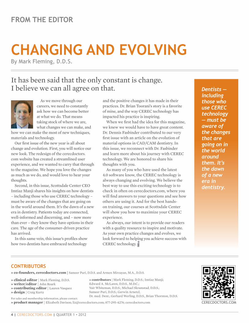
chanGinG anD evoLvinGBy mark Fleming, D.D.S.
As we move through our careers, we need to constantly ask how we can become better at what we do. That means taking stock of where we are, what changes we can make, and
how we can make the most of new techniques, materials and technology.
Our first issue of the new year is all about change and evolution. First, you will notice our new look. The redesign of the cerecdoctors.com website has created a streamlined user experience, and we wanted to carry that through to the magazine. We hope you love the changes as much as we do, and would love to hear your thoughts.
Second, in this issue, Scottsdale Center CEO Imtiaz Manji shares his insights on how dentists – including those who use CEREC technology – must be aware of the changes that are going on in the world around them. It’s the dawn of a new era in dentistry. Patients today are connected, well-informed and discerning, and – now more than ever – they know they have options in their care. The age of the consumer-driven practice has arrived.
In this same vein, this issue’s profiles show how two dentists have embraced technology
FroM The eDiTor
It has been said that the only constant is change. I believe we can all agree on that.
and the positive changes it has made in their practices. Dr. Brian Toorani’s story is a favorite of mine, and the way CEREC technology has impacted his practice is inspiring.
When we first had the idea for this magazine, we knew we would have to have great content. Dr. Dennis Fasbinder contributed to our very first issue with an article on the evolution of material options in CAD/CAM dentistry. In this issue, we reconnect with Dr. Fasbinder and learn more about his journey with CEREC technology. We are honored to share his thoughts with you.
As many of you who have used the latest 4.0 software know, the CEREC technology is always changing and evolving. We believe the best way to use this exciting technology is to check in often on cerecdoctors.com, where you will find answers to your questions and see how others are using it. And for the best hands-on training, our courses at Scottsdale Center will show you how to maximize your CEREC experience.
As always, our intent is to provide our readers with a quality resource to inspire and motivate. As your own practice changes and evolves, we look forward to helping you achieve success with CEREC technology.
Dentists — including those who use CEREC technology — must be aware of the changes that are going on in the world around them. It’s the dawn of a new era in dentistry.
» contributors | Mark Fleming, D.D.S.; Imtiaz Manji; Edward A. McLaren, D.D.S., M.D.C.;Yair Whiteman, D.D.S.; Michael Skramstad, D.D.S.; Sameer Puri, D.D.S.; Gerwin Arnetzl, Dr. med. Dent.; Gerhard Werling, D.D.S., Brian Thornton, D.D.S.
» co-founders, cerecdoctors.com | Sameer Puri, D.D.S. and Armen Mirzayan, M.A., D.D.S.
conTribUTors
» clinical editor | Mark Fleming, D.D.S.» writer/editor | John Roark» contributing editor | Lauren Vasquez» design | Craig Kurtz
For sales and membership information, please contact: » product manager | Elizabeth Davison; [email protected]; 877-295-4276; cerecdoctors.com cerecDocTors.coM
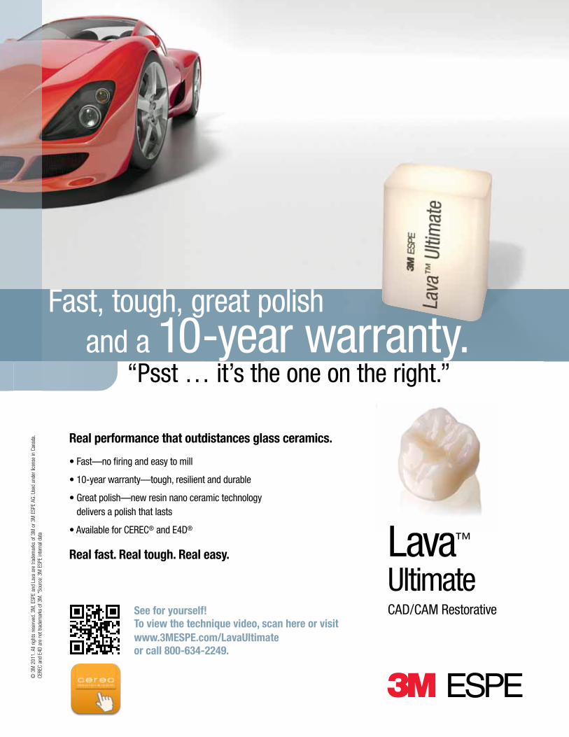
Fast, tough, great polish and a 10-year warranty.
© 3
M 2
011.
All
right
s re
serv
ed. 3
M, E
SPE
and
Lava
are
trad
emar
ks o
f 3M
or 3
M E
SPE
AG. U
sed
unde
r lic
ense
in C
anad
a.
CERE
C an
d E4
D ar
e no
t tra
dem
arks
of 3
M. *
Sour
ce: 3
M E
SPE
inte
rnal
dat
a
Real performance that outdistances glass ceramics.
• Fast—no fi ring and easy to mill
• 10-year warranty—tough, resilient and durable
• Great polish—new resin nano ceramic technology delivers a polish that lasts
• Available for CEREC® and E4D®
Real fast. Real tough. Real easy.
“Psst … it’s the one on the right.”
See for yourself! To view the technique video, scan here or visit www.3MESPE.com/LavaUltimate or call 800-634-2249.
Lava™ UltimateCAD/CAM Restorative
7032-911_LavaUltimate_CERECDrs.indd 1 9/8/11 4:55 PM

6 | cerecDoctorS.com | quarter 1 • 2012
MakinG iT biG in The aGe oF The consUMer-Driven PracTiceBy imtiaz manji
championing this incredible technology in my workshops and presentations for years. That’s why Scottsdale Center for Dentistry has partnered with the leading educators at cerecdoctors.com to present a comprehensive, progressive curriculum of CEREC education. That’s why CEREC is a prominent feature throughout our facilities.
And that’s why, I suppose, many dentists were surprised when we launched The Winter Laboratory, a partnership between Dr. Bob Winter, prosthodontist and master ceramist, and Spear. People wanted to know: If you’re so excited about the possibilities of CEREC, why are you investing in a dental lab? It may sound contradictory, but we put our heart and soul into the lab with the same passion that we do with CEREC — and for the same reason. Let me explain.
I’ve been lobbying dentists long and hard about mastering CEREC because not only have I seen with my own eyes just how life-changing it can be for clinicians to explore its full potential, but because I see how only a minority of CEREC owners are getting anywhere near the full value from this knowledge. CEREC can be a catalyst to phenomenal clinical growth — you can honestly re-invent yourself as a dentist with this technology — but too many CEREC users have no clear vision of what they are capable of achieving with that machine in the corner of the operatory.
At the same time, I’m willing to bet
The coMPLeTe DenTisT
I am as enthusiastic a supporter of CEREC as you are going to find, because I truly believe in its ability to transform dentistry. That’s why I have been
that those same dentists who are not getting full value from CEREC now were probably not getting full value from lab-based dentistry then. How many of them ever really worked in close partnership with a lab, in a way that elevated their game and drove them to new heights of patient care? How many of them think that buying a CEREC means that one day soon they’ll never work with a lab again?
The point is, it shouldn’t be about CEREC versus a lab. That’s why CEREC Connect was developed — so dentists can case-select what should be done with CEREC, and what should be done using a lab. The mission I support is about delivering great dentistry, period.
It should be about transforming your practice and the lives of your patients, and that has many components: being able to diagnose at a high level; getting patients excited; creating the right clinical environment (and that includes CEREC); and working with the right clinical partners (and that includes your specialists and your lab). That’s why we developed a progressive curriculum for mastery of both clinical and value excellence, featuring both CEREC and lab-based fabrications.
I stand behind both CEREC and The Winter Lab because I believe in dentists using every resource in their arsenals to the fullest extent, as part of the quest to become the best dentists they can be.
That’s always been a good approach to professional fulfillment, but it’s about to become crucially important because dentistry is poised to enter an era of unprecedented expectations. This is not the time to limit the scope of your abilities.
wanT To see YoUr FUTUre? Look aT The worLD aroUnD YoUEvery education provider (and Spear is no exception) will tell you that it’s important to stay on top of new techniques, new materials and new technology. That’s what it means to be clinically current in the practice.
But just as important, from my perspective, are the developments happening outside
“Dentistry is poised to enter an era of unprecedented expectations. This is not the time to limit the scope of your abilities.”iMTiaz Manji
quarter 1 • 2012 | cerecDoctorS.com | 7
the practice — the ones that are changing the way people think and behave. Think of it this way: when their mouths are open, the people who come into your practice are patients; when their mouths are closed, they’re consumers. And we’re witnessing the beginning of a revolution that is going to take consumer awareness about the possibilities of today’s dentistry to exciting new levels.
When you think about it, those of us inside dentistry have been talking in terms of full-mouth restoration and esthetic ideals for about 20 years. And in the last five years or so we have seen the technological leaps toward making that vision of dentistry a common reality. But that sense of what today’s dentistry is all about has only begun to trickle down into the consciousness of the average consumer. In the minds of a great many people, a visit to the dentist is still about discomfort and nuisance. It’s about drill-and-fill and metal crowns and unpleasant procedures that somebody else (i.e., insurance) pays for.
That’s all changing, though. And to understand how it’s changing (and how fast) all you have to do is take a look at the world around you.
worD-oF-MoUTh MULTiPLieDFor decades in this country, among the most powerful people in any given city were those who owned the newspapers, because they controlled a good deal of the flow of information in town. For decades, millions of television viewers watched what the executives of three networks decided they would watch.
Compare that with what’s happening today. Newspapers everywhere are either dying off or struggling for relevance and survival. The average TV viewer has dozens of specialty channels to choose from. And, perhaps most notably, “citizen bloggers” and the friends we follow through social media are having a tremendous influence on the decisions we
make in all areas of our lives. We’re getting more information from more sources than ever, including from among our peers. And that is an exciting development for dentists.
Dental practices have always been about person-to-person interaction — that’s the nature of the profession — and they have always grown and thrived on word-of-mouth endorsements: the relative with the story of her incredible dentist and the co-worker with the transformed smile and the heartfelt recommendation. That’s what our practices’ success has always been built on. But now, these patients aren’t just telling the person in the next cubicle. They’re writing on their blogs, they’re tweeting pictures of their new smiles, they’re posting Facebook updates before they’re even out of your office. This is word-of-mouth multiplied and accelerated, and it’s what is going to make the next era of dentistry so exciting.
When opportunities for exposure multiply, it creates new levels of awareness, and that means new levels of expectations. As I said, patients are also consumers, and today’s plugged-in consumers are well-informed and demanding. They’re starting to realize that dentistry today is about high-tech convenience, and that’s where CEREC comes in. They’re also realizing that a skilled dentist today can not only restore their smile, they can make it better than what nature originally bestowed on them—and that’s where a mastery of a full range of procedures, including lab-based restorations, comes in.
Whatever the economy, there are people willing to pay for high-quality, lifestyle-enhancing services and products. Just ask Apple, or Starbucks. Dentistry has already made significant inroads in this way in recent years, but we’ve really only seen the beginning. We’re about to see a new era. The era of the consumer-driven practice, where it’s about always being ready to fulfill patients’ needs in a way that compares with what they’ve come to expect in the greater marketplace. The
dentists who rise to the top in the years to come will be the ones who are ready to compete in this new reality.
The TriUMPh oF The coMPLeTe DenTisTAnd that brings us back to my original point. CEREC (along with CEREC Connect) is going to play a big part in this new era. In fact, I believe no dentist will be able to seriously compete without it. So it makes sense to do everything you can to optimize its full value. But CEREC is still just one technology, and if it’s going to serve you and your patients in an ideal way, it needs to be integrated into a comprehensive picture of what it means to be a dedicated clinician today.
A while ago at the Center I spoke with a dentist who was a passionate CEREC enthusiast. He was an early adopter, an alpha tester and devoted student of the technology. He has committed to being a top CEREC dentist, and as a result he is seeing significant success. When I suggested he consider using The Winter Lab for some of his more complex cases, he was resistant. In fact, he was almost shocked at the idea, “Why would I use a lab when I can do everything with CEREC?” But I persisted, and eventually, when the right case came along, he decided to try working with the lab. He was delighted and astonished with the results.
Since then, he has worked with the lab on other challenging cases, and he has taken additional education in other advanced clinical areas. He’s developing his skills in diagnosis and value creation, his practice-management techniques and his team development to match his devotion to CEREC mastery. Now his goal is not just to be the best CEREC dentist he can be. His commitment is to be the best dentist he can be.
He is becoming a complete dentist, and he’s ready for the next generation of consumer/patients who are looking for the best.

8 | cerecDoctorS.com | quarter 1 • 2012
The evoLUTion oF a coMPLeTe DenTisTDr. Michael Skramstad on how CEREC fits into the bigger pictureBy imtiaz manji
Dr. Michael Skramstad is the ultimate CEREC enthusiast, and he was an early and eager adopter of the technology. He has lectured widely on CEREC, and is a cerecdoctors.com faculty member who teaches at Scottsdale Center for Dentistry. He is a basic and advanced trainer for Patterson Dental, as well as one of very few alpha/beta testers for Sirona. He’s also devoted to being the best clinician he can be, which is why he recently decided to see how he could integrate CEREC into a greater world of clinical possibilities.
Q: You’re one of the leading CEREC dentists in the country. How is it you came to re-discover working with a lab?A: Imtiaz Manji [Spear CEO and cerecdoctors.com magazine contributor] made me do it. He’d been telling me about what they were doing at The Winter Lab and it all sounded good, but I kept saying, “I can do just about anything with CEREC; I don’t really need to worry that much about working with a lab.” But he was persistent, and I finally agreed to send them a case.
Q: How much did you know about the lab before you did that?A: I went to visit them myself. I met with Dr. Bob Winter and a number of people there and I was really impressed. So I thought, “OK, I’ll give this a try.”
Q: What was your impression of the lab?A: It was just phenomenal. The attention to detail, and the way they work with the
ProFiLe
dentist — it was great to feel that sense of partnership with people who had their own unique expertise, and it was a real eye-opener to see the quality of work they produce.
Q: How did that experience change things for you?A: Well, CEREC will always be a big part of my clinical repertoire. I really believe in it passionately. But what I learned was that it is easy to get so into what CEREC can do that you can forget the other tools you have at your disposal as a clinician. The whole point is you want to be able to do a complete range of cases, and do them in the best possible way, and often that means partnering with a lab you trust.
I take pride in being a CEREC leader, but I have a new appreciation for how being a complete dentist today is about more than just CEREC.
Q: What advice do you have for a CEREC dentist as far as training and education?A: Don’t restrict yourself. If you want to get the most from CEREC, get involved with a progressive curriculum, like the one we offer at Scottsdale Center for Dentistry. It’s too great a technology to just use at a surface level. But make sure you do that within the context of a comprehensive advanced program like the Spear continuum. Become a master of CEREC, but learn how to really work with a lab partner, too, and how to integrate CEREC into a complete clinical philosophy.
“I take pride in being a CEREC leader, but I have a new appreciation for how being a complete dentist today is about more than just CEREC.”Dr. MichaeL skraMsTaD
CSCS DFDF
CCCCPP

GLIDEWELLLABORATORIES
v Conventional or adhesive cementation
v Virtually perfect contacts and occlusion
v IPS Empress®-like esthetics
800-854-7256 www.glidewelldental.com
Big Savings for CEREC® Connect Users
$79/unitvia CEREC Connect*
$79/unitvia CEREC Connect*
IPS e.max ®
IPS e.max and IPS Empress are registered trademarks of Ivoclar Vivadent. CEREC is a registered trademark
of Sirona Dental Systems Inc.
*Price valid for model-free monolithic BruxZir or IPS e.max restorations fabricated from a digital file via CEREC connect.
Price does not include $7 overnight return shipping.
®
SOLID ZIrcOnIa crOwnS & BrIDgeS
Premium Products - Outstanding Value
v Save $20 per unit
v Save $10 by not using impression material & tray
v Save $7 on inbound case shipping
Total savings: $37
Milled with the Sirona inLab MC XL and Enhanced CEREC
3D Software.
brux_e.max_CEREC_March.indd 1 1/25/12 2:16 PM

10 | cerecDoctorS.com | quarter 1 • 2012
The eFFecTs oF DesensiTizinG aGenTs in viTroBy edward a. mcLaren, D.D.S., m.D.c. and yair Whiteman, D.D.S.
CSCS DFDF
CCCCPP
to provide more stable, durable and long-lasting bonds. Among the first available are still in use today, including total-etch techniques and materials. These are known to effectively remove the smear layer and re-open dentin tubules with a 30 percent to 40 percent phosphoric acid etchant.1 Total-etch techniques have also proven useful in facilitating bonds to un-cut enamel in minimal-to-no-preparation, all-ceramic restorative cases.2 Additionally, these materials and techniques provide dentists the ability to etch sclerotic dentin.1
Because of this, well-proven total-etch techniques and materials have remained among the most effective in indirect restorative cases.3 However, a recent shift away from total-etch techniques and materials is occurring.1
Because of the technique sensitivity of total-etch systems, many dentists have sought products and techniques requiring fewer steps and simpler placement to reduce clinical challenges and the likelihood of operator error. Typically requiring separate application of the etchant and primer, total-etch techniques demand extra steps and the time required for the bonding process is increased.1 Further, because the phosphoric acid in the etchant is relatively strong, careful observation of acid exposure times to different substrates is required to prevent over-etching.1 Finally, one of the most significant consequences has remained postoperative sensitivity.1,4
case sTUDY
Over the last 25 years, the techniques and materials used to place indirect restorations have evolved
Postoperative sensitivity, or dentin hypersensitivity, does not result from defects within the tooth or other pathological causes, but is related to the loss of the protective enamel layer through dysfunction, parafunctional habits, disease, or mechanical and chemical preparation.4,5 Triggering pain, the exposed dentin becomes sensitive to a variety of chemical, thermal, tactile and osmotic stimuli.4,5 The phosphoric acid used in total-etch techniques has been shown to cause hypersensitivity when the dentin is not sealed prior to etching or bonding, often requiring removal and replacement of indirect restorations.4,5
To address these concerns, newer generations of materials and techniques have attempted to reduce incidences of postoperative and technique sensitivity.4 Promising greater ease-of-use, self-etch or all-in-one materials combine the acid, primer and adhesive in one bottle.1,4
Although they have gained popularity, there is reason for skepticism regarding the efficacy, viability and longevity of the bonds they create.4,6 Coinciding with their use, increased rates of fracture, de-bonding, marginal leakage and postoperative sensitivity led to questions about the ability of self-etch and all-in-one materials to properly etch tooth substrates (Fig. 1).6 Therefore, it has been suggested that total-etch techniques and materials be used for indirect restorations, rather than self-etch and all-in-one adhesive materials.6
Through a greater understanding of the chemical and mechanical aspects of bonding, techniques have improved and material sciences evolved. By sealing, disinfecting and desensitizing the dentin, these newer-generation materials reduce or completely eliminate the risk of postoperative sensitivity.7 Although desensitizing agents have demonstrated a long history of success, key opinion leaders and researchers have struggled to qualify when these materials should be placed. Consequently, confusion has arisen over which techniques offer the greatest benefit and whether sealing should be delayed or completed immediately with these agents.
Gluma (Heraeus) has been specially formulated to penetrate exposed dentin tubules up to 200µ, while reducing the permeability of the dentin by sealing the peripherals of the tubules.8 By preventing the flow of fluid during osmotic changes, postoperative pain is significantly reduced, and the material also acts as a microbial barrier by forming a hermetic
Fig. 1: Clinical example of leakage at an enamel margin where a self-etch system was used with no enamel etching
1

quarter 1 • 2012 | cerecDoctorS.com | 11
than 5 percent postoperative sensitivity using self-etch. Marginal micro-leakage and staining was evident at enamel margins over time with the self-etch but not the total-etch. In another patient group, a 5 percent Glutaraldehyde and 35 percent HEMA desensitizer was added to the total-etch technique. Patient-reported postoperative sensitivity decreased drastically to less than 5 percent, consistent with the self-etch technique. Due to etching of the enamel, there has been no observable marginal leakage in this patient population.
in viTro bonD sTrenGTh TesTinG MeThoDsTo test the effects of a desensitizing agent (5 percent Glutaraldehyde and 35 percent HEMA; Gluma and Gluma PowerGel desensitizer; Heraeus) on the adhesive effect of bond strength to dentin, 40 extracted teeth were mounted and the axial dentin was exposed just below the dento-enamel junction (DEJ) (Fig. 2). The 40 teeth were randomly assigned to four groups. The process used in Groups 1, 2 and 3 has been referred to as the delayed dentin sealing technique (DDS), which performs dentin sealing at the time of cementation
of the final prosthesis. The specimens in groups 1, 2 and 3 were sprayed with CEREC Opti-Spray (Fig. 3) on the exposed dentin and then stored in water at 37 degrees Celsius for one hour. After one hour, the specimens from all three groups were treated by three different methods, and IPS Empress (Ivoclar Vivadent) ceramic rods were adhesively bonded to the dentin.
In Group 1, the dentin surface was rinsed thoroughly with water and then dried for two seconds, a 32 percent phosphoric acid was applied for 30 seconds (Fig. 4), and a fourth-generation bonding agent (All-Bond 3, Bisco) was applied by first placing two coats of the All-Bond 3 primer for 15 seconds (Fig. 5), drying for 10 seconds in air, and then applying the All-Bond 3 filled adhesive (Fig. 6) that was then thinned in air. A dual-cure cement (Duo-Link) was then applied to a ceramic rod, placed on the dentin and photopolymerized for one minute (Fig. 7, next page). In Group 2, the exact same steps were followed, except sandblasting was added after the initial rinsing but prior to the acid etch. The dentin surface was sandblasted at 20 psi with 50-micron aluminous oxide for 10 seconds. In Group 3, all steps were
Fig. 2: Specimen of freshly extracted tooth with dentin exposed, ready for bonding
Fig. 3: Tooth specimens sprayed with cerec opti-spray
Fig. 4: Specimen with 32% H2Po4 Uni-tech (Bisco) applied to exposed dentin
Fig. 5: Applying dentin primer
Fig. 6: Applying filled adhesive
seal that inhibits bacterial growth.9 Additionally, Gluma does not affect bond strength and can be used safely in conjunction with adhesive bonding agents and resin cements.8
Here, we discuss using Gluma as a desensitizer in the adhesive process, and test its effect on dentin bond strengths in conjunction with the CEREC chair-side technique in vitro.
in vivo cLinicaL observaTions in The UcLa cenTer For esTheTic DenTisTrYTo observe the effects of a desensitizing agent (5 percent Glutaraldehyde and 35 percent HEMA; Gluma and Gluma Power Gel desensitizer, Heraeus) on the adhesive process, following clinical observations were undertaken in 2010.
Total-etch and self-etch techniques were compared to determine which offered the greatest benefit and the least clinical challenges. Standard total-etch and self-etch techniques were accomplished by graduate students. Patients reported at least some postoperative sensitivity in approximately 20 percent of the cases using total-etch without a desensitizer and less
2
4
3
5 6
12 | cerecDoctorS.com | quarter 1 • 2012
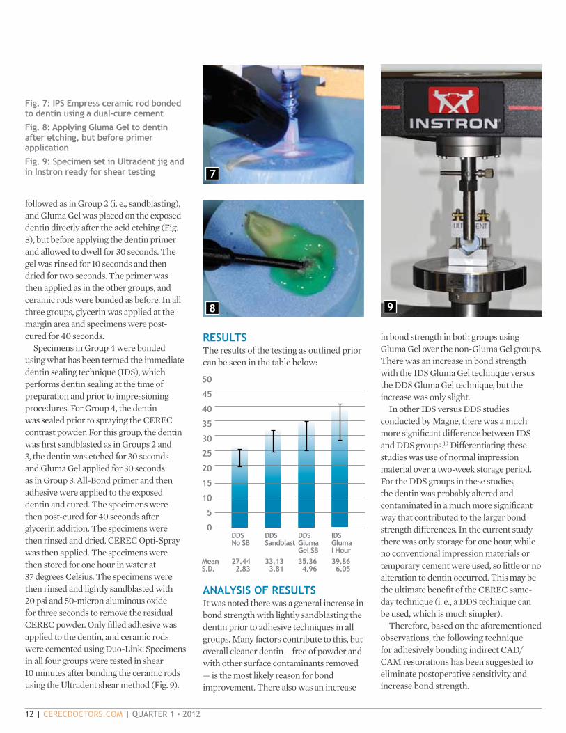
resULTsThe results of the testing as outlined prior can be seen in the table:
50
45
40
35
30
25
20
15
10
5
0 DDs DDs DDs iDs No SB Sandblast Gluma Gluma Gel sb i hourMean 27.44 33.13 35.36 39.86 s.D. 2.83 3.81 4.96 6.05
followed as in Group 2 (i. e., sandblasting), and Gluma Gel was placed on the exposed dentin directly after the acid etching (Fig. 8), but before applying the dentin primer and allowed to dwell for 30 seconds. The gel was rinsed for 10 seconds and then dried for two seconds. The primer was then applied as in the other groups, and ceramic rods were bonded as before. In all three groups, glycerin was applied at the margin area and specimens were post-cured for 40 seconds.
Specimens in Group 4 were bonded using what has been termed the immediate dentin sealing technique (IDS), which performs dentin sealing at the time of preparation and prior to impressioning procedures. For Group 4, the dentin was sealed prior to spraying the CEREC contrast powder. For this group, the dentin was first sandblasted as in Groups 2 and 3, the dentin was etched for 30 seconds and Gluma Gel applied for 30 seconds as in Group 3. All-Bond primer and then adhesive were applied to the exposed dentin and cured. The specimens were then post-cured for 40 seconds after glycerin addition. The specimens were then rinsed and dried. CEREC Opti-Spray was then applied. The specimens were then stored for one hour in water at 37 degrees Celsius. The specimens were then rinsed and lightly sandblasted with 20 psi and 50-micron aluminous oxide for three seconds to remove the residual CEREC powder. Only filled adhesive was applied to the dentin, and ceramic rods were cemented using Duo-Link. Specimens in all four groups were tested in shear 10 minutes after bonding the ceramic rods using the Ultradent shear method (Fig. 9).
resULTsThe results of the testing as outlined prior can be seen in the table below:
anaLYsis oF resULTsIt was noted there was a general increase in bond strength with lightly sandblasting the dentin prior to adhesive techniques in all groups. Many factors contribute to this, but overall cleaner dentin —free of powder and with other surface contaminants removed — is the most likely reason for bond improvement. There also was an increase
in bond strength in both groups using Gluma Gel over the non-Gluma Gel groups. There was an increase in bond strength with the IDS Gluma Gel technique versus the DDS Gluma Gel technique, but the increase was only slight.
In other IDS versus DDS studies conducted by Magne, there was a much more significant difference between IDS and DDS groups.10 Differentiating these studies was use of normal impression material over a two-week storage period. For the DDS groups in these studies, the dentin was probably altered and contaminated in a much more significant way that contributed to the larger bond strength differences. In the current study there was only storage for one hour, while no conventional impression materials or temporary cement were used, so little or no alteration to dentin occurred. This may be the ultimate benefit of the CEREC same-day technique (i. e., a DDS technique can be used, which is much simpler).
Therefore, based on the aforementioned observations, the following technique for adhesively bonding indirect CAD/CAM restorations has been suggested to eliminate postoperative sensitivity and increase bond strength.
Fig. 7: IPS Empress ceramic rod bonded to dentin using a dual-cure cement
Fig. 8: Applying Gluma Gel to dentin after etching, but before primer application
Fig. 9: Specimen set in Ultradent jig and in instron ready for shear testing 7
98

.Order your supply today:
T: 1.877.APEX123 | www.apexdentalmaterials.com
interfaceThe Fastest Way to Etch and Silanate a CEREC® Restoration
•PreparesCEREC®restorationsfordeliveryorrepairsinonesimpleapplication
•Eliminateshydrofluoricacid
•Easy...Savestime
•Alwaysfresh...3yearshelflife
•Theonlymaterialforreliableceramicrepairs
•Compatiblewithallcementsandadhesives

14 | cerecDoctorS.com | quarter 1 • 2012
UcLa cerec aDhesive TechniQUeAfter fabrication of the CEREC restoration using the standard chair-side CEREC technique, contaminants were removed from the preparation through light sandblasting with 50-micron aluminum oxide at 20 psi. Although pumice solutions may be used, sandblasting cleans more effectively and enhances the bond strength. However, careful consideration is necessary when sandblasting, since pressure above 20 psi may damage prior restorations and cause gingival bleeding.
Immediately following sandblasting, the enamel and dentin were both etched with 32 percent phosphoric acid for 30 seconds to standardize. The large bolus of etch is removed by suction, and the preparation is rinsed for 10 seconds and then dried for two seconds. A 2 percent chlorhexidene solution is applied and left to dwell for 10 seconds, after which the excess is removed with suction. Recent research has shown that rinsing with chlorhexidine may further increase final bond strengths and aid Gluma when re-wetting the dentin.
Using a delayed sealing technique and a nylon brush, Gluma was then burnished on the preparation for 20 to 30 seconds. If using Gluma PowerGel, it is necessary to leave the gel on the dentin for 45 seconds, since it takes a bit longer to soak into the dentin. Excess was removed using suction, rather than air-drying, since Gluma may burn soft tissues, specifically the mucosa. When using the gel, it is necessary to rinse slightly for five seconds to remove the gel residue. Although the burns are often minor and will heal quickly and fully, they can be painful. Immediately following removal of excess, compressed nitrogen was used for two seconds to remove excess water that remained on the dentin from the Gluma.
A fourth-generation dentin bonding agent (All-Bond 3) is then applied to the Gluma-treated preparation. First, the
dentin primer All-Bond 3 is applied and burnished into the dentin for 10 seconds. Then, compressed nitrogen is used to evaporate the ethanol solvent for 10 seconds. The surface should still be shiny; if it isn’t, the All-Bond 3 should be re-applied. This is not cured. Then, a filled adhesive is applied and nitrogen thinned, but again not cured. To seat the restoration, a highly filled, dual-cured resin cement is used, since it offers a more stable bond on dentin.
The restoration is then seated on the preparation and firm pressure applied. Prior to initial light-curing, excess was removed from the margins and interproximal areas. After initial curing, remaining excess cement was removed and the restoration underwent final curing. Occlusion was then adjusted as necessary, and the restoration was finished and polished.
In cases where the CEREC technique is used but there will be extended time between preparation and cementation (e. g., temporaries will be fabricated and several days or longer will pass before cementation), the IDA technique will be performed as described in Group 4 from the in vitro study.
concLUsionWhen placing indirect restorations, the authors believe that total-etch techniques and materials should be chosen over self-etching for a variety of reasons.7 Although the literature has demonstrated that self-etch materials offer sound dentin bonding in the short term, research on their efficacy in the long term remains inconclusive.6 Further, it is known that self-etching materials lack the ability to bond to enamel long term.6 While initial bond strength appears promising, marginal leakage frequently leads to restorative failure.6
Currently, the trend in adhesive dentistry has shifted toward the use of self-etch and all-in-one materials and techniques for simpler placement of indirect restorations.
However, the inherent risks must be considered. Providing greater strength, durability, stability and longevity on both enamel and dentin, total-etch techniques remain among the most proven.7 Although these materials are more technique-sensitive, the time and additional steps are nominal when compared to restorative failure and replacement.6 Combined with the proper techniques and materials, desensitizing materials such as Gluma offer the greatest benefit to both dentist and patient.7
Conflict of Interest Disclosure Statement: Bisco, Ivoclar, 3M/ESPE, Nobelbiocare, Vident, and Heraeus have all provided UCLA with grant support for research projects conducted by the UCLA Center for Esthetic Dentistry
reFerences
1 Tay Fr, Pashley Dh. have dentin adhesives become too hydrophilic. J Can Dent Assoc. 2003;69(11):726-31.
2 Triolo PT jr, swift ej, jr. , Mudgil a, Levine A. Effects of etching time on enamel bond strengths. Am J Dent. 1993;6(6):302-4.
3 Pashley Dh, ciucchi b, sano h, horner ja. Permeability of dentin to adhesive agents. Quintessence Int. 1993;24(9):618-31.
4 Perdigão j, Geraldeli s, hodges js. Total-etch versus self-etch adhesive: effect on postoperative sensitivity. j am Dent assoc. 2003;134(12):1621-9.
5 Rosing CK, Fiorini T, Liberman DN, Cavagni J. Dentine hypersensitivity: analysis of self-care products. Braz Oral Res. 2009;23 Suppl 1:56-63.
6 Swift EJ Jr. Dentin bonding: what is the state of the art. compend contin educ Dent. 2001;22(12 Suppl):4-7; quiz 18.
7 bertolotti rL. Total-etch the rational dentin bonding protocol. J Esthet Dent. 1991;3(1):1-6.
8 aranha ac, Pimenta La, Marchi GM. clinical evaluation of desensitizing treatments for cervical dentin hypersensitivity. braz oral Res. 2009;23(3):333-9.
9 christensen rP. effectiveness of Glutaraldehyde as a chemosterilizer used in a wrapping technique on simulated metal instruments. J Dent Res. 1977;56(7):822-6.
10 Magne P, kim Th, cascione D, Donovan Te. immediate Dentin sealing improves bond strength of indirect restorations. j Prosth Dent 2005(94)511-519.
11 Ricci HA, Sanabe ME, de Souza Costa CA, Pashley DH, Hebling J. Chlorhexidine increases the longevity of in vivo resin-dentin bonds. Eur J Oral Sci. 2010;118(4):411-6.

withPFM Bridge BruxZir® Bridge
3175 Range Ave. • Santa Rosa, CA 95403
(800) 368-8482www.dentalmasters.com
Build Bridges
Model Milled from CEREC Impression Model Milled from CEREC Impression
Milled Wax-Up BruxZir® Bridge After Milling
Final PFM Bridge on Model Final BruxZir® Bridge on Model
e.max® is a registered trademark of Ivoclar VivadentBruxZir® is a registered trademark of Glidewell Laboratories, Inc.*Certificates valid for cases received via CEREC Connect. One offer per customer.
You know the benefits CEREC already has in your practice. Digital impressions eliminate discomfort and offer superior accuracy. With CEREC Connect, you can bring these benefits to more patients on more cases.
Send in your digital impression, and our team of highly experienced technicians can create any restoration you prescribe.
From CAD options like e.max® and BruxZir® to conventional PFMS, cast gold and veneers.
Experience digital solutions from Dental Masters
CEREC Connect
$300Receive in CEREC eCertificates!Call or visit www.GoCerec.com
$129
• e.max CAD• BruxZir• PressTek (NP) PFM
Consistent Success from People You Trust™
per unit

16 | cerecDoctorS.com | quarter 1 • 2012
ProFiLe
CSCS DFDF
CCCCPP
The cerecDocTors.coM inTerview
DR. DENNISFASBINDER
16 | cerecDoctorS.com | quarter 1 • 2012

quarter 1 • 2012 | cerecDoctorS.com | 17
bY saMeer PUri, D.D.s.For this issue, it was my distinct privilege to interview one of the most prolific minds in CEREC-related re-search. Dr. Dennis Fasbinder has been involved in numerous studies on CEREC materials and serves as a consultant to major manufacturers on materials used by CEREC clinicians. We are honored to share his insights on the uses of the CEREC technology.
Q: How long have you been a CEREC user? Describe your
experience with it.
A: I was introduced to the CEREC system in 1992, shortly after
taking a faculty position at the University of Michigan School of
Dentistry. My department chair at the time, Dr. Joseph Dennison,
had been looking for areas in which to broaden our research focus,
and thought the new CAD/CAM process offered a number of
potential areas for investigation. Someone needed to learn to use
the system, and I was interested in its clinical potential, so I took
on the task.
CAD/CAM dentistry has been the focus of my professional career
for the past 20 years. I have been fortunate to meet and collaborate
with some tremendously talented, skilled and passionate people.
These collaborations, more than anything else, has kept me engaged
in the continued development of the technology.
q: in the years that you have been involved with the cerec
technology, what is the one feature that you feel has been the
most innovative?
A: The ability to do ceramic restorations in a single appointment
is the greatest advantage of CEREC technology. Ceramic materials
have significant advantages over other direct restorative materials.
DR. DENNIS
quarter 1 • 2012 | cerecDoctorS.com | 17
18 | cerecDoctorS.com | quarter 1 • 2012
And to me, the ability to accurately and
efficiently record the
cavity preparation is the most
innovative feature. Without an
accurate digital file, the rest of the
process is of little value. The digital file
has so far been applied to fabricating
restorations; however, I see the
potential for other diagnostic and
treatment-planning uses of the digital
file as being even greater. From my
very first course, the CEREC system
was presented as an impression-free
system, and the more the technology
allows me to treat my patients without
physical impressions — including
diagnostic impressions — the better.
Q: You are one of the most prolific
researchers for the CEREC technology
in regard to materials. How did you
get started with this?
A: I have always had an interest in
understanding the physical properties
of the materials I work with. I think
it helps me to make better clinical
decisions on their application with my
patients. And I have had the opportunity
to be mentored by several excellent
dental-material researchers during my
career, including Dr. Joseph Dennison
and Dr. John Burgess. Dental-material
research is really about exploring the
capabilities and limits of our materials
to help us solve patient problems.
CEREC technology was being
introduced to the North American
marketplace about the same time I was
learning the system, which was also
when I started my academic career
at the University of Michigan. This
innovative system was being touted to
the dental profession, but with little
evidence of how well it worked beyond
some early laboratory studies. I was
expected to engage in scholarly activity
as a part of my academic position, so
many of my early research projects on
CEREC dealt with obvious questions,
such as how predictable the process
was, how well restorations fit, and
how strong the materials were. As I
attended courses and meetings, many
clinicians advocated techniques,
processes and materials to be
successful with CEREC. I often found
myself asking, “How do you know that
works?” And this helped me develop
the foundation for ongoing research
projects with CEREC.
Q: Currently, what are the advantages
that milled restorations have over
traditionally fabricated restorations?
What are the disadvantages?
A: Milled restorations have obvious
advantages by virtue of the block
fabrication process. For example,
polymerization shrinkage is not an
issue when milling a restoration from
a composite block. The industrial
fabrication process of the block
maximizes the physical properties
of the materials. There are no voids,
inclusions or similar imperfections
in ceramic blocks compared to those
made conventionally. CAD/CAM has
evolved into a core process for our
ceramic materials, especially when
one considers the two most popular,
zirconia and lithium disilicate, are both
millable materials.
Disadvantages can also be traced
to the block fabrication process.
The ability to customize shades and
translucencies may require a cut-back
or layered technique. And all blocks
require significant capital investment
in the equipment to mill them.

quarter 1 • 2012 | cerecDoctorS.com | 19
Q: What innovations would you like to
see in regard to materials for CEREC?
A: I think dentists would like to be more
conservative in tooth reduction, so this
is driving the search for more durable
esthetic materials that can be used in
thinner cross-sections. The efficiency of
the process of handling the material after
milling could also be improved, relative
to the ease of finishing and polishing.
I also think that there has been
considerable expansion in what doctors
are doing with CEREC technology. We
have moved beyond the usual posterior
restorations to complete smile designs,
and on to implant restorations, and
using it instead of traditional posterior
composite restorations. This has also
created a greater demand for a wider
variety of materials.
Q: In your view, the perfect materials
would possess what properties?
A: I doubt my “optimal material” is
much different from any other dentist.
I would like a material that has the
esthetics of ceramics, the strength and
longevity of gold, and the chair-side
handling of composite resin. And,
since I am asking for the moon, how
about it self-adheres to the tooth with a
material surface activation process that
eliminates the need for cement.
Q: What would you, personally, like to
test in your next research project?
A: There have been a number of
very well-done, controlled clinical
studies published over the last 10
years that document the success of
CEREC technology. These studies have
generally been done in well-controlled
clinical environments. As many of my
colleagues kid me, I can take four hours
to do a research restoration, but we and
our patients cannot afford that length
of time in a private-practice setting.
Controlled clinical studies are often
designed to maximize the potential
outcome of the clinical technique. If
everything is handled correctly, this
can be the expected outcome. But we
all realize that this type of control is not
always possible with our patients.
I would be very interested to move
my research to a practice-based
network involving a number of private
practices with a similar outcome
assessment that is used in controlled
clinical studies. Clinicians use clinical
research findings to help them make
decisions in their offices. It would be
very interesting to see if the outcomes
in those offices are similar to those we
20 | cerecDoctorS.com | quarter 1 • 2012
see in controlled clinical studies, or
how and why they may differ.
Q: What studies do you currently
have ongoing, and can you share any
significant findings you have had?
A: We recently published the 10-year
results of a randomized clinical trial
on Paradigm MZ100 and Vita Mark
II inlays. At the end of 10 years, six
of the ceramic inlays had fractured,
while only one of the composite inlays
fractured. There was no significant
difference in the materials for all other
categories evaluated. There may be
some interesting expanded treatment
options for polymer-based restorations.
We also have a clinical study on e.max
CAD crowns using several different
types of adhesive resin cements; we
will be reporting the five-year recall
data in March 2012. The interesting
finding in this project has been the lack
of chipping, surface fracture or occlusal
wear of the e.max CAD crowns over
time. This may be why some clinicians
are considering more conservative
preparation designs for e.max CAD
restorations. And this would make for
an interesting follow-up study as well.
Q: How does the CEREC hardware
compare to other systems — including
the camera and the milling unit?
A: CAD/CAM units basically need
three pieces of equipment: a camera
device to record the dentition, a
software program to design the
restoration and a milling unit to
fabricate the restoration. The Bluecam
is an efficient and accurate camera, but
it is limited to line-of-sight perspective.
A video camera may be the next step
to expand the capability of the system.
And the milling chamber is a limiting
step in that there is a limit to how
fast it can mill without damaging the
material; so how much faster can it be
done or how much smoother? Can it be
moved to a printer style?
Q: What suggestions can you give
to an office considering adding
CEREC to their restorative arsenal?
A: CEREC is a process, not an
outcome. The outcome you need to
understand and embrace is adhesive
ceramic and composite restorations.
The process has obvious advantages
of efficiency for both doctors and
patients. However, if the doctor
has concerns about the longevity of
adhesive ceramic restoration or its
durability, they will naturally limit their
use of the system. Once the outcome is
understood and embraced, the process
is fairly straightforward to fabricate the
desired restoration.
Q: In which situations do you feel
that it’s inappropriate for an office to
use CEREC?
A: I am not sure I am the person
to suggest what is appropriate or
inappropriate for a colleague to choose
to do with their practice. I would suggest
that there are types of offices that would
not enjoy a similar level of success with
CEREC technology as others. Those
offices that would limit their use of
adhesive ceramic restorations in favor
of alternative treatments would then
minimally use CEREC technology
for their patients. And those offices
that view the implementation of a
new technology as an insurmountable
hurdle would struggle with learning the
CEREC process as well.
Q: What does the future hold for
Dr. Dennis Fasbinder?
A: I have never been very good at
predicting my future; for example,
I started college as a history major.
Opportunities have generally shaped
the arc of my professional career. I
enlisted in the U.S. Air Force out of
dental school to do some traveling in
Japan and the far east. Instead, I ended
up taking an assignment in Germany,
doing my graduate training and getting
an opportunity to be involved in
graduate dental education. I truly enjoy
the complementary roles of research:
discovering new information/patient
care; applying the new discoveries/
education; and sharing the outcomes
with students and colleagues. It is
a great ride to be on and I am in no
hurry for it to stop. I look forward to
finding challenges presented by new
opportunities, but I have no idea what
they may turn out to be!
quarter 1 • 2012 | cerecDoctorS.com | 21
CEREC MASTERY – RAPID INTEGRATION INTO YOUR PRACTICE WITH 4.0 SOFTWAREAfter completing Patterson Basic Training, it’s time to acquire the skills necessary to use the software to its fullest capabilities. Also, gain an understanding of what it takes to make the CEREC process
2012: May 17-18; May 19-20 (Limited Seats); June 7-8
POSTERIOR QUADRANT PROFICIENCYAND RESTORING IMPLANTSLearn to produce highly esthetic posterior restorations with perfect occlusion and contacts, whether you are treating a single tooth or complete quadrant.
2012: Apr. 14-15; May 17-18; June 9-10
MASTERING CEREC ANTERIORSThoroughly understand how to complete any anterior case with ease, achieving esthetic results that rival high-quality labs.
2012: March 22-23; March 24-25 (Sold Out); May 19-20
CEREC INLAB PROFICIENCY AND MASTERY ESTHETICSIncrease your knowledge of the inLab software and improve your ability to better manipulate porcelain. Designed for doctors who already have the CEREC system, and who have completed the core cerecdoctors.com training curriculum.
2012: April 12-13 (Limited Seats); June 29-30; Sept. 15-16
FUNDAMENTALS OF CEREC AND
GALILEOS INTEGRATIONMaster all aspects of guided-implant planning using the GALILEOS
guided systems that are available to work with the CEREC and GALILEOS Integration protocol.
2012: April 14-15; Nov. 8-9
CREATING THE “YES” PRACTICECEREC doctors and their teams will learn how to work with, talk about and create value for CEREC on a daily basis.
2012: Oct. 26-27
FOR MORE INFORMATION AND TO REGISTER, CONTACT SHAYNA PHIPPS AT:480.588.9101 OR [email protected]
2012 COURSE DATESWE HAVE A COURSE PERFECTLY SUITED TO MEET YOUR NEEDS.
VISIT WWW.CERECDOCTORS.COMFOR MORE INFORMATION
NO MATTER WHAT YOUR SKILL LEVEL, WE’LL SHOW YOU HOWYOU CAN DO MORE!
JAY B. REZNICK D.M.D., M.D.
MIKE SKRAMSTAD D.D.S.
MARK FLEMING D.D.S.
FACULTY
B
ARMEN MIRZAYAN M.A., D.D.S.
SAMEER PURI D.D.S.
LEVEL 1PATTERSON BEGINNING ORIENTATION OFFERED
DIRECTLY THROUGH PATTE)RSON DENTAL1LEVEL
LEVEL 1PATTERSON BEGINNING ORIENTATION OFFERED
DIRECTLY THROUGH PATTERSON DENTAL
CEREC MASTERY -RAPID INTEGRATION INTO YOUR PRACTICE WITH 4.0 SOFTWARE2LEVEL
LEVEL 1PATTERSON BEGINNING ORIENTATION OFFERED
DIRECTLY THROUGH PATTERSON DENTAL
POSTERIOR QUADRANT PROFICIENCY AND RESTORING IMPLANTS3LEVEL
SON BEGINNING ORIENTATION OFFEREDDIRECTLY THROUGH PATTERSON DENTAL
MASTERING MULTIPLECEREC ANTERIORS4LEVEL
CEREC INLAB PROFICIENCYAND MASTERY ESTHETICS5LEVEL
PATTERSON BEGINNING ORIENTATION (OFFERED DIRECTLY THROUGH PATTERSON DENTAL WHEN YOUPURCHASE YOUR CEREC)
1LEVEL
LEVEL 1PATTERSON BEGINNING ORIENTATION OFFERED
DIRECTLY THROUGH PATTERSON DENTAL
2LEVEL
3LEVEL 4LEVEL
5LEVEL 6LEVEL
LEVEL 1PATTERSON BEGINNING ORIENTATION OFFERED
DIRECTLY THROUGH PATTE)RSON DENTAL1LEVEL
LEVEL 1PATTERSON BEGINNING ORIENTATION OFFERED
DIRECTLY THROUGH PATTERSON DENTAL
CEREC MASTERY -RAPID INTEGRATION INTO YOUR PRACTICE WITH 4.0 SOFTWARE2LEVEL
LEVEL 1PATTERSON BEGINNING ORIENTATION OFFERED
DIRECTLY THROUGH PATTERSON DENTAL
POSTERIOR QUADRANT PROFICIENCY AND RESTORING IMPLANTS3LEVEL
SON BEGINNING ORIENTATION OFFEREDDIRECTLY THROUGH PATTERSON DENTAL
MASTERING MULTIPLECEREC ANTERIORS4LEVEL
CEREC INLAB PROFICIENCYAND MASTERY ESTHETICS5LEVEL
PATTERSON BEGINNING ORIENTATION (OFFERED DIRECTLY THROUGH PATTERSON DENTAL WHEN YOUPURCHASE YOUR CEREC)
1LEVEL
LEVEL 1PATTERSON BEGINNING ORIENTATION OFFERED
DIRECTLY THROUGH PATTERSON DENTAL
2LEVEL
3LEVEL 4LEVEL5LEVEL 6LEVEL
LEVEL 1PATTERSON BEGINNING ORIENTATION OFFERED
DIRECTLY THROUGH PATTE)RSON DENTAL1LEVEL
LEVEL 1PATTERSON BEGINNING ORIENTATION OFFERED
DIRECTLY THROUGH PATTERSON DENTAL
CEREC MASTERY -RAPID INTEGRATION INTO YOUR PRACTICE WITH 4.0 SOFTWARE2LEVEL
LEVEL 1PATTERSON BEGINNING ORIENTATION OFFERED
DIRECTLY THROUGH PATTERSON DENTAL
POSTERIOR QUADRANT PROFICIENCY AND RESTORING IMPLANTS3LEVEL
SON BEGINNING ORIENTATION OFFEREDDIRECTLY THROUGH PATTERSON DENTAL
MASTERING MULTIPLECEREC ANTERIORS4LEVEL
CEREC INLAB PROFICIENCYAND MASTERY ESTHETICS5LEVEL
PATTERSON BEGINNING ORIENTATION (OFFERED DIRECTLY THROUGH PATTERSON DENTAL WHEN YOUPURCHASE YOUR CEREC)
1LEVEL
LEVEL 1PATTERSON BEGINNING ORIENTATION OFFERED
DIRECTLY THROUGH PATTERSON DENTAL
2LEVEL
3LEVEL 4LEVEL5LEVEL 6LEVEL
2LEVEL 1
PATTERSON BEGINNING ORIENTATION OFFEREDDIRECTLY THROUGH PATTERSON DENTAL
POSTERIOR QUADRANT PROFICIENCY AND RESTORING IMPLANTS3LEVEL
SON BEGINNING ORIENTATION OFFEREDDIRECTLY THROUGH PATTERSON DENTAL
MASTERING MULTIPLECEREC ANTERIORS4LEVEL
CEREC INLAB PROFICIENCYAND MASTERY ESTHETICS5LEVEL
1LEVEL
LEVEL 1PATTERSON BEGINNING ORIENTATION OFFERED
DIRECTLY THROUGH PATTERSON DENTAL
2LEVEL
3LEVEL 4LEVEL
5LEVEL 6LEVEL
DIRECTLY THROUGH PATTERSON DENTALMASTERING MULTIPLECEREC ANTERIORS4LEVEL
CEREC INLAB PROFICIENCYAND MASTERY ESTHETICS5LEVEL
1LEVEL 2LEVEL
3LEVEL 4LEVEL
5LEVEL 6LEVEL
0911-09fin.indd 1 2/7/12 10:35 AM
predictable and efficient.predictable and efficient.
and the CEREC, and gain a complete understanding of all the different

22 | cerecDoctorS.com | quarter 1 • 2012
facebook.com/CERECbySirona @CERECbySirona #C27aah
CEREC® has never been about waiting. After the success of CEREC 25, we knew we couldn’t wait five more years for our 30th Anniversary, and so CEREC 27 and a half was born! Join us in Vegas for A-list entertainment, world-class speakers and three days of action-packed education and fun. An event like this only comes around every few and a half years.
Join us at The Venetian!AUGUST 16-18, 2012
CEREC27andahalf.com 855.237.3248
Scan the QR code to register and get up-to-date
news on C27aah!
T h e D e n t a l C o m p a n y
CER001l CEREC Doctors C27aah Ad V1 2-3 1/18/12 5:04 PM

quarter 1 • 2012 | cerecDoctorS.com | 23
facebook.com/CERECbySirona @CERECbySirona #C27aah
CEREC® has never been about waiting. After the success of CEREC 25, we knew we couldn’t wait five more years for our 30th Anniversary, and so CEREC 27 and a half was born! Join us in Vegas for A-list entertainment, world-class speakers and three days of action-packed education and fun. An event like this only comes around every few and a half years.
Join us at The Venetian!AUGUST 16-18, 2012
CEREC27andahalf.com 855.237.3248
Scan the QR code to register and get up-to-date
news on C27aah!
T h e D e n t a l C o m p a n y
CER001l CEREC Doctors C27aah Ad V1 2-3 1/18/12 5:04 PM

24 | cerecDoctorS.com | quarter 1 • 2012
cerec-PLanneD, Lab-execUTeD, esTheTic & DUrabLe resToraTionsBy mike Skramstad, D.D.S.
CSCS DFDF
CCCCPP
with innovative treatment options for procedures including veneers, crowns and three-unit bridges.1-3 CEREC high-performance milling machines reduce time and cost for both dentists and patients while providing predictable and esthetic outcomes.4 Intuitive and user-friendly software makes it easy for dentists to fabricate 3-D models, it is designed to automatically adjust occlusion and provides color coding to ensure correct proximal contacts before milling.4 Available for both chair-side and laboratory applications,1,2
dentists can use CEREC to plan and carry out the case, or, if unable to fabricate the restoration chair-side, the procedure can be digitally planned using CEREC and relayed for the laboratory to execute the case.
To meet the individual needs of patients, these systems accommodate various restorative materials, such as metal-ceramic, composite and all-ceramic materials, based on the indication being performed.1,2
Designated for use with CAD/CAM technology, Telio CAD acrylate polymer blocks are designed for medium- to long-term temporary crowns and bridges.5
By eliminating the challenges associated with traditional temporization processes, including mixing errors, impression errors, polymerization shrinkage and clean-up, Telio CAD makes it easy to fabricate provisional restorations of various modalities. These include temporary bridges with up to two pontics, anterior and posterior crowns, and temporary restorations on implants.5,6 An
case sTUDY
Computer-aided design and manufacture (CAD/CAM) technologies contribute significantly to restorative dentistry today by providing clinicians
ideal material for long-term temporary placement due to its durability, Telio CAD is designed with a flexural modulus of 3,200± 300 MPa, and a high flexural strength of 130± 10 MPa.7 Demonstrating lifelike fluorescence and durable shade stability, Telio CAD allows dentists and laboratory technicians to fabricate long-lasting and esthetic temporary restorations.5
Based on Telio CAD provisionals, laboratory ceramists can then fabricate highly esthetic and durable restorations using pressable ingots. IPS e.max Press (Ivoclar Vivadent) offers optimal strength and esthetics compared to traditional all-ceramic materials.8-11 Due to its structural integrity — developed from 70-percent-by-volume needle-like crystals in a glassy matrix, with controlled size, shape and density — lithium disilicate demonstrates considerable strength and durability.8,9 Lithium disilicate can be either pressed or milled. Pressable lithium disilicate, designed with a flexural strength of 400 MPa, is pressed using the wax hot-press technique (IPS e.max Press).9-11 Available in a variety of translucencies and opacities, pressable lithium disilicate is designed with a lower refractive index and exceptional optical properties to achieve lifelike esthetics.8-12
case sTUDYA 30-year-old man presented with two three-unit bridges (#6-#8 and #9-#11). The bridges had been placed 13 years prior to replace congenitally missing lateral
incisors. Unhappy with the color, shape and overall esthetics of the bridges, single-unit crowns and pontics with implants were discussed. Due to the congenitally missing
teeth, however, additional procedures such as bone grafting would be required. Not interested in undergoing the additional time and cost of the procedure, the patient chose to have the bridges replaced with two three-unit lithium disilicate (IPS e.max) pressed bridges to achieve maximum esthetics.
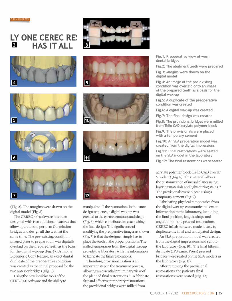
cLinicaL ProTocoLPrior to preparing the teeth for bridge restorations, the entire preoperative condition was digitally duplicated using the new CEREC 4.0 software (Fig. 1). The abutment teeth were then prepared and isolated, and more images were taken of the preparations and the maxillary arch
2
1
quarter 1 • 2012 | cerecDoctorS.com | 25
ONLY ONE CEREC RESOURCE HAS IT ALL
CONTACT LIZ DAVISON AT 877.295.4276 OR [email protected]
HANDS-ONLEARNING
DIGITAL LEARNING
PEER-TO-PEER LEARNING
THE WORLD’S MOST POWERFUL ONLINE CEREC COMMUNITY
acrylate polymer block (Telio CAD, Ivoclar Vivadent) (Fig. 8). This material allows the customization of incisal planes using layering materials and light-curing stains.13 The provisionals were placed using a temporary cement (Fig. 9).
Fabricating physical temporaries from the digital wax-up communicated exact information to the laboratory, including the final position, length, shape and angulation of the pressed restorations. CEREC inLab software made it easy to duplicate the final and anticipated design.
An SLA preparation model was created from the digital impressions and sent to the laboratory (Fig. 10). The final lithium disilicate (IPS e.max Press) pressed bridges were seated on the SLA models in the laboratory (Fig. 11).
After removing the provisional restorations, the patient’s final restorations were seated (Fig. 12).
Fig.1: Preoperative view of worn dental bridges
Fig.2: The abutment teeth were prepared
Fig.3: Margins were drawn on the digital model
Fig.4: An image of the pre-existing condition was overlaid onto an image of the prepared teeth as a basis for the digital wax-up
Fig.5: A duplicate of the preoperative condition was created
Fig.6: A digital wax-up was created
Fig.7: The final design was created
Fig.8: The provisional bridges were milled from Telio CAD acrylate polymer block
Fig.9: The provisionals were placed with a temporary cement
Fig.10: An SLA preparation model was created from the digital impressions
Fig.11: Final restorations were seated on the SLA model in the laboratory
Fig.12: The final restorations were seated
4
3
5
7
6
9
8
10
12
11
(Fig. 2). The margins were drawn on the digital model (Fig. 3).
The CEREC 4.0 software has been designed with two additional features that allow operators to perform Correlation bridges and design all the teeth at the same time. The pre-existing condition, imaged prior to preparation, was digitally overlaid on the prepared teeth as the basis for the digital wax-up (Fig. 4). Using the Biogeneric Copy feature, an exact digital duplicate of the preoperative condition was created as the initial proposal for the two anterior bridges (Fig. 5).
Using the new intuitive tools of the CEREC 4.0 software and the ability to
manipulate all the restorations in the same design sequence, a digital wax-up was created to the correct contours and shape (Fig. 6), which contributed to establishing the final design. The significance of modifying the preoperative images as shown (Fig. 7) is that the designer simply has to place the teeth in the proper positions. The milled temporaries from the digital wax-up provide the laboratory with the information to fabricate the final restorations.
Therefore, provisionalization is an important step in the treatment process,
allowing an essential preliminary view of the planned final restorations.13 To fabricate fast and effective temporary restorations, the provisional bridges were milled from

26 | cerecDoctorS.com | quarter 1 • 2012
concLUsionContinuous technological and material advancements in dentistry allow today’s dentists to address cases with greater predictability and more effective techniques.14 Products designed for use with CEREC CAD/CAM systems such as Telio CAD acrylate polymer blocks enable dentists to achieve indications such as crowns, veneers and bridges quickly and easily, saving the patient chair time and cost.14 With CEREC CAD/CAM technology, whether performed chair-side, in-lab or both, patients and practitioners can expect highly esthetic and unequivocal results. Eliminating the fear of failure, new materials, techniques and technologies help dental professionals hone their artistic and clinical skills while achieving success with even the most difficult cases. 14
Dr. Skramstad would like to thank the specialists at The Winter Lab in Laguna Beach, Calif., for their collaboration on this case.
For questions or additional information, Dr. Skramstad can be reached at [email protected]
reFerences
1 Miyazaki T, hotta Y, kunii j, kuriyama s, Tamaki Y. A review of dental cad/cam: current status and future perspectives from 20 years of experience. Dent Mater J. 2009;28(1):44-56.
2 Christensen GJ. In-office cad/cam milling of restorations. j am Dent assoc. 2008;139(1):83-85.
3 Culp L, McLaren EA. Lithium disilicate: the restorative material of multiple options. Compend Contin Educ Dent. 2010;31(9):716-20, 722, 724-5.
4 Poticny DJ, Klim J. Cad/cam in-office technology: innovations after 25 years for predictable, esthetic outcomes. J Am Dent Assoc. 2010;141(Suppl 2):5S-9S.
5 ivoclar vivadent introduces Telio. Dentistry iQ. 2010; april.
6 Telio cs, Telio caD instructions for use. ivoclar vivadent aG, schaan / Liechtenstein; 2010: 1-36.
7 Telio temporary restorations out of one hand: scientific documentation. Telio CAD. Amherst, NY: Ivoclar Vivadent; 2010: 2-11.
8 McLaren EA, Phong TC. Ceramics in dentistry: classes of materials. inside Dentistry. 2009;5(9):94-103.
9 Tysowsky GW. The science behind lithium disilicate: a metal-free alternative. Dent Today. 2009;28(3):112-13.
10 Reynolds JA, Roberts M. Lithium-disilicate pressed veneers for diastema closure. inside Dentistry. 2010;6(5):46-52.
11 helvey Ga. chairside cad-cam. Lithium disilicate restoration for anterior teeth made simple. Inside Dentistry. 2009;5(10):58-66.
12 ritter rG, culp L. ingot selection for aesthetic restorations using contemporary pressed ceramics. Pract Proced Aesthet Dent. 2002;14(6):472-78; quiz 470.
13 Telio. ivoclar vivadent. schaan Liechtenstein 2010; 1-11.
14 Fasbinder DJ, Dennison JB, Heys D, Neiva G. A clinical evaluation of chairside lithium disilicate cad/cam crowns: a two-year report. J Am Dent Assoc. 2010;141(Suppl 2):10S-4S.
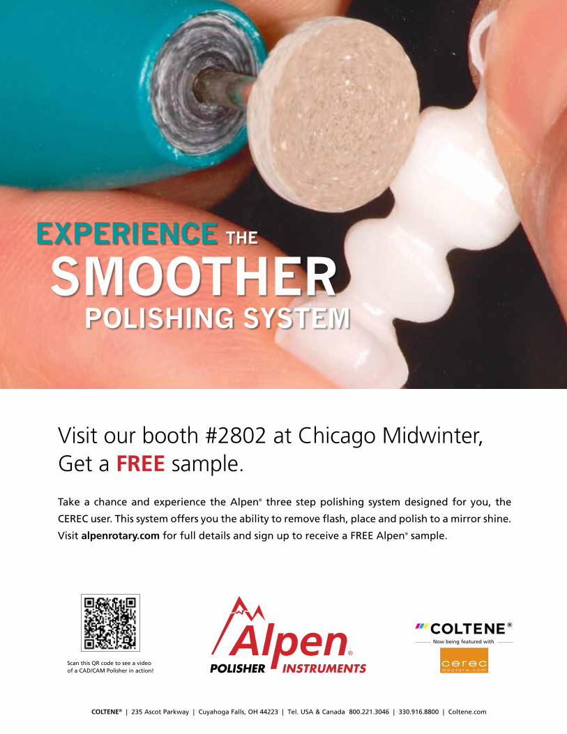
Take a chance and experience the Alpen® three step polishing system designed for you, the
CEREC user. This system offers you the ability to remove flash, place and polish to a mirror shine.
Visit alpenrotary.com for full details and sign up to receive a FREE Alpen® sample.
COLTENE® | 235 Ascot Parkway | Cuyahoga Falls, OH 44223 | Tel. USA & Canada 800.221.3046 | 330.916.8800 | Coltene.com
Visit our booth #2802 at Chicago Midwinter, Get a FREE sample.
Now being featured with
SMOOTHEREXPERIENCE THE
POLISHING SYSTEM
Scan this QR code to see a video of a CAD/CAM Polisher in action!

28 | cerecDoctorS.com | quarter 1 • 2012
cLinicaL asPecTs oF aLL-ceraMics By Gerwin arnetzl, Dr. med. Dent.
CSCS DFDF
CCCCPP
success in treating the various indications, the material properties and the corresponding requirements in terms of preparation and restoration design must be taken comprehensively into
consideration.This article aims
to help promote appreciation for “thinking in ceramic dimensions.”
basic reQUireMenTsThe key to the long-
term success of all-ceramic restorations is shape design that is appropriate to the
case sTUDY
All-ceramic restorations have become a standard part of everyday dental procedure. However, in order to reliably achieve a high level of long-term
material in question. A convex cavity base design, for example, helps prevent notch stress and allows tensile stress to be converted to compressive stress (Fig. 1). Box-shaped preparation should generally be avoided, as it causes tensile stress on the side opposite to where the force is generated, and also results in notch stress in the box area in the case of rounded box-shaped preparation (Fig. 2, 3, 4 and 5). At the same time, attention should be paid to ensuring a simple design for the restoration without deep fissures; sharp edges must always be rounded and changes in the cross-section may only be gradual, not sudden (Fig. 6 and 7). In this way, several
prerequisites crucial to clinical success are already fulfilled.
GeneraL noTes on PreParaTionPreparation must take the specifics of the dentition, material and technology in question into consideration. With regard to all-ceramics, this means that sufficient space should be provided for esthetic rehabilitation and for implementing a stable framework, while focusing on the motto “As much as necessary, as little as possible.” The amount of space sufficient for ensuring structural retention depends on the type of ceramic and indication. Flexible positioning and freedom of rotation must
arnetzl
1
2 4
3 5
76

quarter 1 • 2012 | cerecDoctorS.com | 29
also be ensured. Furthermore, preparation must be performed with a view to ensuring alignment with the tooth axes as well as maintaining a residual dentine thickness of 0.7 to 1 mm in all areas. If fabrication is performed using computer technology, additional parameters must be taken into consideration in the CAD/CAM system used, such as software specifications, geometry of axes of the milling system, diameter of the smallest processing tool, etc.
The requirements generally applicable to the clinical procedure, such as sufficient cooling during preparation, as well as preventing exposure to heat caused by high pressure, remain valid. Also important is that the preparation margin is not placed subgingivally: in light of periodontal-physiological considerations, the aim should be to define a supragingival preparation border. Esthetics may also require that the preparation border be located in the paramarginal area.
recoMMenDeD TYPes oF PreParaTionIn the case of all-ceramic crowns, a chamfer or shoulder with a rounded inner angle can be prepared. Uniform and smooth surfaces
are recommended; all transitions from the axial to the occlusal or incisal surfaces should be rounded. A wax-up and the fabrication of silicone keys to control the preparation are helpful for the diagnosis and the clinical application in order to ensure defect-oriented preparation.
Particularly in the case of all-ceramic anterior crowns, a minimum incisal wall thickness of 1.5 mm (circumferential 1.0 mm) must be observed. The tapering crown margin must be at least 1.0 mm (Fig. 8). For posterior crowns, the tooth should be prepared with a cone of 4 - 6° with undercuts blocked out (Fig. 9). The width of the chamfer or shoulder with a rounded inner angle should be 0.8 mm in the approximal area of premolars and the lingual area of the lower molars, and 1.0 mm in all other areas. Reduce circumferentially by 1.5 mm for optimum esthetic results. In the cusp and fissure area, a reduction of 1.5 to 2 mm is important for static reasons.
For inlay and onlay restorations, an opening angle of > 10° as well as a minimum layer thickness of 1.5 mm in the fissure area and 2.0 mm in the cusp area must be ensured (Fig. 10) during preparation. The minimum width in the area of the isthmus is also 2.0 mm, while the posterior residual wall of the residual tooth substance should be at least 2.0 to 2.5 mm.
sUMMarYThe brochure “Clinical Aspects of All-Ceramics” provides a wide-ranging and comprehensive overview of the preparation required for all-ceramic restorations. A PDF of the brochure can also be downloaded from www.vita-zahnfabrik.de under “News & Press releases” > VITA Product News > VITA All-ceramics.
From “ZWR Das deutsche Zahnärzteblatt,” May 2011, Germany
98 10

30 | cerecDoctorS.com | quarter 1 • 2012
veneerinG oF oxiDe ceraMic briDGe FraMeworks UsinG viTa raPiD LaYer TechnoLoGY By Gerhard Werling, D.D.S.
CSCS DFDF
CCCCPP
systems (both Sirona Dental Systems, D-Bensheim) to create a veneering structure for oxide ceramic bridge frame- works with the fine-structure feldspar ceramic VITABLOCS TriLuxe forte
TF-40/19 (VITA Zahnfabrik, D-Bad Säckingen) in a computer-aided procedure. Since framework and veneering structure are optimally adjusted to each other and classic layering
technique is not used, the risk of chipping is minimized. This technology was developed for bridges with up to four units, mainly placed in the posterior region.
For computer-aided veneering, the multilayer software module of the inLab Software (>V 3.80), which is part of the CEREC or inLab MC XL system, automatically generates the separate data sets for the production of the framework and veneering structure. This is done within several seconds and as soon as full-contour design is completed. After the milling process, both elements can be bonded to one another quickly and easily using an adhesive composite.
Here, the different process steps will be explained using a case study.
case sTUDY
VITA Rapid Layer Technology (RLT) enables CAD/CAM users of the CEREC or inLab MC XL
case sTUDYThe female patient presented with a loose bridge in region 47 (#31) (Fig. 1 and 2). On the basis of the previous medical records and the laboratory liquidation she brought along, it was possible to reconstruct that the bridge had been produced with a zirconia framework using the CAD/CAM system of the company Etkon. The framework had received a ceramic veneering, and the all-ceramic restoration had been incorporated in 2004. The patient reported that immediately after its placement, chipping was detected in the basal area of the bridge units 45 (#29) and 46 (#30).
PLanninG PhaseWe started with a precise analysis of the potential errors which could have led to the failure of the restoration. We used this approach to achieve the goal of avoiding the same mistakes and the resulting failures. Thus, the loosening of the bridge was explained by the fact that the bridge abutments were too short and that tooth 47 (#31) had a large composite core build-up. Certainly, one of the reasons for loosening was the lacking bond of the resin cement with the restorative that was used for the build-up. In addition, the occurring torsion forces may have been one of the reasons for de-bonding, in our opinion.
Due to the wide span of the bridge, and by taking into consideration the short abutment teeth as well as the large size of the build-ups, the preferred solution was a bridge with a torsion attachment to compensate for the distortion of the mandible. This would have been an adequate alternative solution to a rigid zirconia bridge construction veneered with porcelain. However, the patient was allergic to several substances including metal, latex and gluten, and therefore desired a restoration made of a biocompatible all-ceramic material. Even the failure of the all-ceramic bridge that had just been removed did not deter her from requesting an all-ceramic bridge. Conjointly, it was decided to replace the old restoration using
Fig. 1: occlusal view of the initial situation
Fig. 2: buccal view of the initial situation
werling
2
1

quarter 1 • 2012 | cerecDoctorS.com | 31
a bridge with a zirconia framework which is veneered with feldspar ceramic material in a computer-aided procedure. The production technique of choice was VITA Rapid Layer Technology.
DesiGnInitially, the bridge surface (Fig. 3) and the antagonist teeth (Fig. 4) were matted intraorally (VITA Powder Scan Spray, VITA Zahnfabrik) in order to take a buccal bite registration with the CEREC Bluecam (Sirona Dental Systems). Together with the images of the preparation, this data was transmitted to the dental laboratory via the CEREC Connect Portal.
After removal of the zirconia bridge with the ATD Bridge Remover (Anthogyr,
F-Sallanches), it became clearly visible that tooth 47 (#31) had a large core build-up with composite (Fig. 5). At the bridge, basal chipping became apparent at teeth 46 (#30) and 45 (#29). Also of interest was the large amount of resin cement in the approximal area between the bridge units 47 (#31) and 46 (#30) (Fig. 6).
In the next step, the bridge was virtually designed using the inLab 3-D software. For the three-dimensional design, the restoration and the design mode Multilayer were selected in the software. The model was generated and, afterwards, VITA In-Ceram YZ was activated for the framework and VITABLOCS TriLuxe forte TF-40/19 for the veneering porcelain in the material selection dialog. After
preparation-margin detection and definition of the insertion direction, the software generated a design proposal. It was
possible to check this Zirkoproposal also by fading in the antagonist teeth in full size (Fig. 7). Figure 8 shows the bridge design in transparent view and Figure 9 the occlusal view of the design near completion. The framework was automatically reduced without producing any undercuts. Immediately after having switched to the milling preview, the bridge was graphically separated into a framework and a veneering structure – each of the parts fulfilling the defined minimal wall thickness and strength of the connectors (Fig. 10 and 11).
aFTer The MiLLinG ProcessAfter milling, the zirconia bridge framework was shaded using VITA
Fig. 3: Intraoral powdering of the bridge surface
Fig. 4: Intraoral powdering of the antagonist teeth
Fig. 5: Tooth 47 (#31) was restored with a large composite build-up
Fig. 6: Chipping at the removed bridge
Fig. 7: Full view of the antagonist teeth
Fig. 8: Transparent representation of the bridge design for control of the minimum thicknesses
Fig. 9: Occlusal view of the design prior to completion
Fig. 10: Milling preview of the bridge framework which was milled from zirconia with schematic representation of the porcelain structure
Fig. 11: View of the veneering structure which was milled from viTabLocs TriLuxe forte TF-40/19
4
3
5
7
6
9
8
10 11

32 | cerecDoctorS.com | quarter 1 • 2012
A strong and safe bond between the framework and the veneering structure is achieved by using the adhesive bonding technique. In preparation for the bonding step, the outer surfaces of the framework were sandblasted with Al2O3, and the inner surfaces of the veneering structure etched with fluoric acid gel (5 percent) and silanized after cleaning. Subsequently, the two elements were bonded with Panavia 21 (Kuraray, D-Frankfurt a. M.) (Figs. 16, 17 and 18).
Figures 19 and 20 show the completed bridge 44-47 (#28-#31) after polymerization and characterization.
PerManenT ceMenTaTion oF The briDGeIn the subsequent step, the bridge was placed on the abutment teeth. For this, the inner surface was previously sandblasted with alumina. Silicatization of the composite build-ups of the abutment teeth followed with Micro-Etcher (Dynaflex, USSt. Ann) and Rocatec Korund 30 µm (3M ESPE, DSeefeld), while they were silanized with Monobond Plus (Ivoclar Vivadent, FL-Schaan). With these procedures, a maximally strong bond between the resin cement and the composite build-ups is ensured during adhesive cementation of the bridge on the stumps (Fig. 21).
12
1413
15
17
16
19
18
20
Fig. 12: View of the milled, sintered framework made of viTa in-ceram Yz and the veneering structure made of VITABLOCS TriLuxe forte TF-40/19Fig. 13: Try in of the framework and ...Fig. 14: ... the veneering structure placed on it in the mouthFig. 15: Finalization of the veneering porcelain with stain pastesFig. 16: Application of Panavia 21 on the basal areas of the veneering structureFig. 17: Placement of the framework into the veneering structureFig. 18: Removal of the excess cementFigs. 19,20: Final bridge that was produced with rapid Layer Technology
In-Ceram YZ Coloring Liquid (VITA Zahnfabrik), and then sintered. Figure 12 (following page) shows the two milled structures: the zirconia framework and the feldspar ceramic veneering structure.
Afterwards, the framework was tried in at first (Fig. 13) and subsequently, the porcelain work was placed on it and tried in as well in the mouth (Fig. 14). Occlusion and fit of the restoration were checked cautiously. Thanks to the bio-generic reconstruction method, no/only minor adjustments of the veneering porcelain are required in the occlusal area.
Characterization of the surfaces was done with the staining technique using VITA AKZENT / VITA SHADING PASTE (both VITA Zahnfabrik) (Fig. 15). Since veneering and framework are bonded to one another with composite, characterization with the staining technique or individualization with layering technique – if required – generally has to be done prior to connection with the zirconia framework. In addition, the basal areas of the framework should be treated with glaze prior to bonding as well.

quarter 1 • 2012 | cerecDoctorS.com | 33
on the basal parts of the bridge units by an inadequate design of the former zirconia bridge framework, as well as by load on the framework caused by torsion. This is due to distortion of the mandible during occlusion. In this case, we opted for a restoration produced with VITA Rapid Layer Technology. Since the parts are connected to one another in the cold state using adhesive composite, the risk of stresses between framework and veneering structure is excluded. The idea behind this method is to create a buffer with the adhesive composite – between veneering structure and framework on the one hand, and in order to compensate for stresses and torsion caused by chewing forces on the other. We assume that, for this reason, chipping of the veneering structure will not occur. Moreover, the bending strength of the industrially produced monolithic feldspar ceramic is 150 MPa and thus higher than that of veneering porcelain conventionally produced in the dental laboratory.
A further benefit of restorations produced with Rapid Layer Technology is the option of producing them in the dental practice. In order to implement this, however, a sintering furnace is required for zirconia. Alternatively, the sintering process can be done in collaboration with a laboratory owning a sintering furnace.
Further finalization steps such as staining of the veneering structure and glaze firing are common procedures already performed by many CEREC users in the context of producing single-tooth restorations. For these steps, a mere conventional ceramic furnace is necessary.
The connection in the cold state using composite and curing of the dual-cure composite with a UV light are part of routine dental procedures and easily performed chair-side without further investment in additional equipment. The bridges which have been produced using Rapid Layer Technology and placed in our
Fig. 21: Preparations for the placement of the bridge
Fig. 22: after adhesive cementation of the bridge
Fig. 23: Final situation
practice until now have not shown any failures caused by chipping or fracture of the framework. The esthetic results are excellent – of which we are accustomed from years of experience with multilayer, single-tooth restorations. The problem of a white margin — which does exist with multilayer restorations due to the design process — are solved in our practice by:
• Compensation in software design, which leads to a cervical margin of minimum width;
• Shading of the zirconia material or selection of a tooth-colored zirconia material;
• Staining of veneering structure and framework prior to stain or glaze firing.
concLUsionIn connection with a sintering furnace and the utilization of VITA Rapid Layer Technology, it is possible today to produce bridges in a semi-chair-side procedure in the dental practice. This workflow is realized by using the software inLab 3-D and the CEREC system.
This means that – if the preparation is done in the morning – the permanent cementation of the bridge can be done the same day. In this calculation, all required work steps and production times are taken into consideration.
The connection of the veneering structure and the framework is not regarded as a critical step by us. We assume that, opposed to veneering ceramics, this connection offers a compensation mechanism acting against internal material stresses as well as stresses and torsion forces which occur in static and dynamic occlusion.
For questions and more information, Dr. Gerhard Werling can be reached at [email protected].
Editor’s Note: This piece originally appeared in the German periodical “Digital Dental News” (May 2011).
21
22
23
Cementation was done with RelyX Unicem (3M ESPE). Figures 22 and 23 show the final situation after having checked the articulation and occlusion.
consiDeraTionsWe explain the occurrence of chipping

SUBSCRIBE NOW
Contact us at 1.866.781.0072
INSTANT ACCESSONLY $2900
TOMORROW’S EDUCATION, AVAILABLE TODAY.
THE NEW SPEAR DIGITAL PLATFORM IS A
REVOLUTIONARY METHOD TO ACCELERATE
YOUR CLINICAL LEARNING.
PEERLESS CONTENT. Imagine a searchable index of the best minds in dentistry. At your fingertips 24/7/365.
SELF-DIRECTED, LAYERED LEARNING. Learn at your own pace. Track your progress online, take quizzes, and earn CE credits.
INSTANT ACCESS. Forget booking, schedules, travel, waiting. Available now, on-demand from anywhere at any time on any device.
speareducation.com/digital
SPR_DL_Ad_d1.indd 1 2/8/12 9:55 AM

quarter 1 • 2012 | cerecDoctorS.com | 35
21
resTorinG oPPosinG resToraTions wiTh cerecBy Sameer Puri, D.D.S.
CSCS DFDF
CCCCPP
when it comes to in-office CAD/CAM systems. While earlier versions of the CEREC technology limited clinicians to the restoration of one or maybe two teeth, advances are increasing the capabilities of the software, therefore allowing users to fabricate multiple restorations simultaneously (Fig. 1). In fact, not only can users fabricate an unlimited number of restorations chair-side in any arch, the 4.0 software allows the operator to fabricate restorations on opposing arches as well (Fig. 2).
While software advances continue to increase the application of the technology, materials evolution has allowed for an increased range of indications for in-office CAD/CAM by increasing the clinical
case sTUDY
Evolution in materials and software have created an increased range of indications for clinicians. The CEREC system continues to lead the pack
indications where the technology can be used. Definitive restorations have been successfully fabricated for years, and improvements in all-ceramic technology in newer materials such as lithium disilicate (e.max) from Ivoclar have allowed clinicians to have great success with CEREC and basically replicate the restorations they receive from the laboratory.
In addition to fabricating definitive restorations, one often overlooked and underutilized aspect of the CEREC process is to use the CEREC to create long-term provisional restorations. A new material from 3M called LAVA Ultimate, based on nano-ceramic technology, allows for the material to be used for numerous clinical scenarios.
While clinically indicated for definitive crowns and onlays, another potential indication of this material may be for its use in the long-term provisionalization of teeth. Teeth that need to undergo periodontal therapy prior to definitive restorations could be one indication where this nano-ceramic technology could be applied. Large rehab cases could initially be milled with the LAVA Ultimate blocks, and then converted to a different material once the patient has stabilized. Another indication could be to fabricate restorations on implants where the softer, more resilient nano-ceramic material could aid in the progressive loading of the implants with a material softer than an all-ceramic restoration.

36 | cerecDoctorS.com | quarter 1 • 2012
4
3
5
6
7
8

quarter 1 • 2012 | cerecDoctorS.com | 37
This article provides a clinical case including the step-by-step procedures outlining how to restore multiple teeth provisionally when the two teeth are opposing each other in the patient’s mouth. The CEREC 4.0 software was used to fabricate two provisional restorations on teeth that were indicated for periodontal therapy and were not ready for the definitive restorations.
case sTUDYThe patient was a healthy male in his 40s who presented with a defective restoration on #14 as well as a debonded all-ceramic crown that was placed approximately 12 years earlier. Because the patient was to undergo periodontal therapy prior to the placement of the definitive restorations, the plan was to address teeth #14 and #19 in this first appointment so that he would be able to function properly on the left side of his mouth. The teeth were to be prepared and temporized with LAVA Ultimate nano-ceramic restorations as provisional restorations (Figs. 3-5).
The software was opened and the appropriate teeth were indicated on the Administration Tab. Teeth #14 and #19 were selected for full-coverage crowns. The appropriate material to mill the restorations from was also selected at this stage (Fig. 6).
Once the administrative information had been entered, scans of the arches were taken with the CEREC Bluecam. The automatic image capture allowed the software to capture an entire quadrant in approximately 15 to 20 seconds (Figs. 7, 8). These intraoral scans should contain all the information needed to fabricate the restoration, including visible and clear margins, as well as appropriate data of the adjacent teeth needed to design
9
10
11

38 | cerecDoctorS.com | quarter 1 • 2012
14 15
16
the contours of the restorations.
Once the scans had been processed by the software, virtual models of both arches were created by the software (Figs. 9, 10). These virtual models allowed the design of the desired restorations to
commence. As with all CEREC restorations, the
following steps were taken to design the restorations: the virtual model was created, the margin was placed on the preparation, the insertion axis was corrected so that the virtual model was
concLUsionCEREC has been used for decades to fabricate definitive restorations for patients. Evolutions in materials and software technology now allow clinicians to work not only on multiple teeth in the same arch, but also on multiple teeth on opposing arches. This results in an increased capability of the software and an increased range of indications for clinicians.
Additionally, improvements in material technology allow for the use of the technology in an increased range of indications in addition to the definitive restorations of the teeth.
13
12
oriented in the correct direction, and the virtual model was rendered.
In this case, because there were two restorations being designed, the appropriate tooth to be designed was selected on the Dock Bar at the bottom and the steps were repeated. Once proposed, the restorations could be articulated on the virtual models and adjusted as needed (Fig. 11).
Each restoration could be viewed in occlusion with its opposing pair, or worked on individually by selecting the appropriate tooth in the Dock Bar (Figs. 12, 13). Once designed, the restorations were milled in the LAVA Ultimate and cemented with a provisional cement (Figs. 14-16).
© Vident 2012. Easyshadeand 3D-Master are registered trademarks of VITA Zahnfabrik.
Order now from Patterson Dental at 800-873-7683
Instant Shades in the Palm of Your Hand
800-828-3839 | www.vident.com
Cordless, sleek and incredibly fast, the VITA Easyshade® Compact delivers the fastest, easiest way to measure tooth shades under any lighting condition. Easyshade Compact gives you the ability to conquer all shade challenges all day, every day, instantly and accurately.
Now you can measure natural teeth, check restoration shades and track bleaching progress with the push of a button. Order today and instantly prescribe VITA 3D-Master® and Classical shades.
UPDATED SOFTWARE NOW AVAILABLE!VITA Easyshade® Compact
Featuring the all-new VITABLOCS mode that accurately determines
the shade for all of your CEREC restorations

40 | cerecDoctorS.com | quarter 1 • 2012
“cerec saveD MY PracTice!”Profile by Mark Fleming, D.D.S.
He examined his hefty lab bill only to realize it was costing as much as $11,000 a month. He took the initiative to learn everything he could about CEREC technology – including having a procedure done on himself. Now this Orange County, Calif.-based D.D.S. boasts an active patient list of 2,500 in a thriving 4,000-sq.-ft. office. And he’s the first to say, “CEREC saved my practice!”
Q: How long have you been in practice? A: I graduated from the University of the Pacific School of Dentistry in 1999. I was an associate for one year in my current practice before buying in as a 50 percent partner in 2001. I bought out my partner in 2006 and became full owner.
Q: What is the size of your practice?A: We moved from a 1,850-sq.-ft. office to a 4,000-sq.-ft. location in January of 2008. We currently have about 2,500 fully active patients with one GP doctor (myself ), one part-time endodontist associate, one full-time hygienist, one part-time hygienist, two full-time assistants, and two full-time business coordinators.
Q: How many operatories do you have? A: We have a total of eight operatories. I work out of two to three, we use another two to three for hygiene, and two operatories are used for surgeries and endo.
Q: What type of dentistry do you do? A: General dentistry, implants (surgically
ProFiLe
Dr. Brian Toorani was struggling to cover the costs of his dream office when he had a reality check that led him to right-size his business.
placing and restoring), Invisalign, cosmetic, family, conservative/non-invasive restorations, and CEREC single, multiple and full-mouth restorations. Our whole purpose is to be unique and provide a level of service unlike any other office in our area. We love our family of patients and our purpose for existing is to change how dentistry is seen by the general public.
Q: Why was CEREC your CAD/CAM choice?A: I had heard about this CEREC “thing” and decided to check it out. I went to a prospective buyer event and asked lots of questions. I then saw a live demo at the CEREC 25 Discovery Event. I even had a CEREC done on myself! I was amazed with how great it was.
Based on what I had read and seen, CEREC was the most efficient and fastest system with the best (i.e., the most predictable) results. I also liked the fact that there was so much support and education available.
I decided to invest in the future of our practice. The best part was that when I told my wife the cost and how much it could save us, her response was, “What are you waiting for, and why didn’t you get it sooner?”
Q: How does this technology fit into your office philosophy?A: Our practice philosophy is strictly based on providing the highest level of care and service. We treat our patients with
compassion and kindness, and we are dedicated to doing the best, providing the best, and using only the best on our patients. CEREC has helped us meet and exceed that commitment.
Q: How does CEREC impact your practice?A: I believe that traditional dentistry can become repetitive and boring. Think about it: you poke, you drill, you take a goopy impression, you temporize and you ask the patient to come back a few weeks later. Then you poke them again, work in an irritated environment, keep your fingers crossed that the lab did a great job, adjust contact, adjust occlusion, polish and cement.
Now, with CEREC it all changes. Dentistry is no longer boring! It truly feels like I get paid to play a video game all day. The best part is, you have less sensitivity, no temporary to come off, no irritation of the tissue, and perfect occlusion, color and contact.
Further, CEREC has given me the full control of my restoration. I don’t have to depend on anyone but myself and my direct team.
Additionally, I have significantly reduced my lab bill. With what I have saved (after taking into account the additional payment for my CEREC machine), I will be able to pay off my CEREC in less than 18 months.
I believe, more than ever before and in
CSCS DFDF
CCCCPP
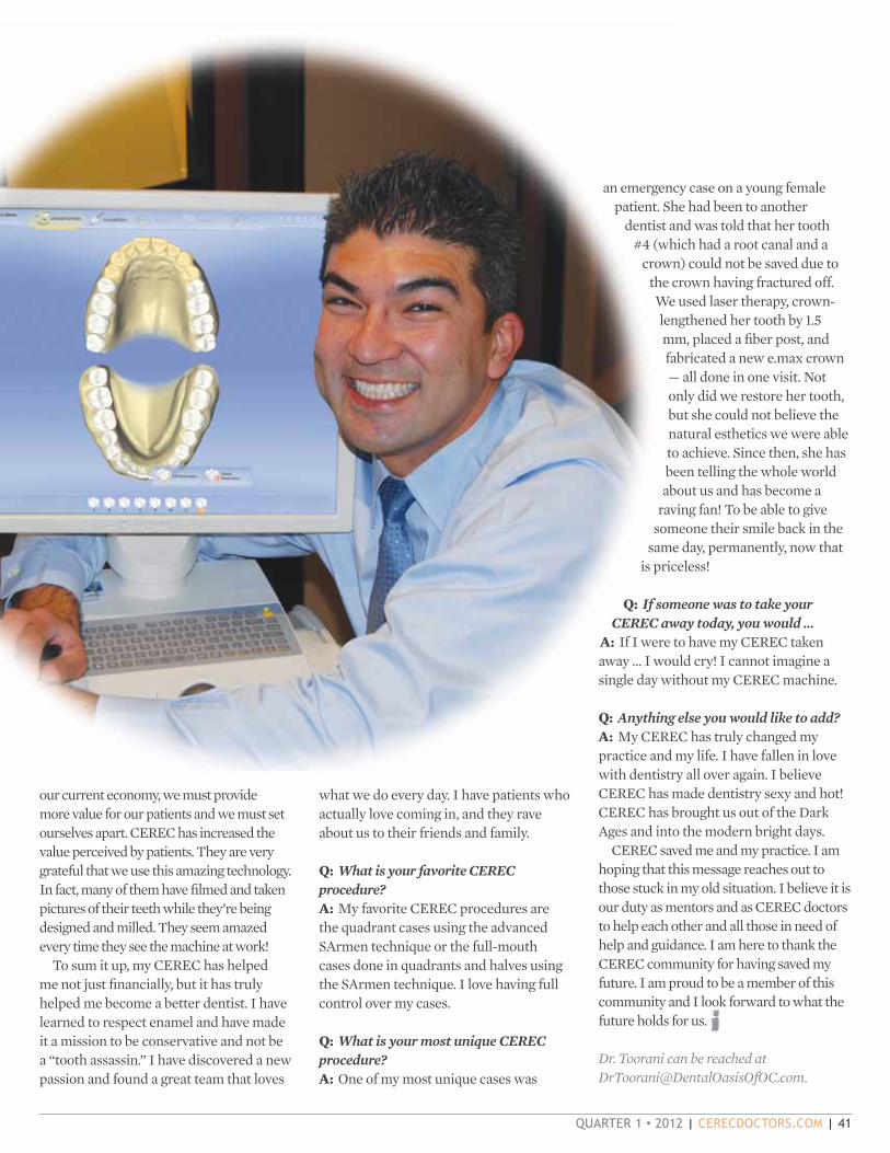
quarter 1 • 2012 | cerecDoctorS.com | 41
an emergency case on a young female patient. She had been to another
dentist and was told that her tooth #4 (which had a root canal and a
crown) could not be saved due to the crown having fractured off. We used laser therapy, crown-lengthened her tooth by 1.5 mm, placed a fiber post, and fabricated a new e.max crown — all done in one visit. Not only did we restore her tooth, but she could not believe the natural esthetics we were able to achieve. Since then, she has been telling the whole world about us and has become a
raving fan! To be able to give someone their smile back in the
same day, permanently, now that is priceless!
Q: If someone was to take your CEREC away today, you would …
A: If I were to have my CEREC taken away … I would cry! I cannot imagine a single day without my CEREC machine.
Q: Anything else you would like to add?A: My CEREC has truly changed my practice and my life. I have fallen in love with dentistry all over again. I believe CEREC has made dentistry sexy and hot! CEREC has brought us out of the Dark Ages and into the modern bright days.
CEREC saved me and my practice. I am hoping that this message reaches out to those stuck in my old situation. I believe it is our duty as mentors and as CEREC doctors to help each other and all those in need of help and guidance. I am here to thank the CEREC community for having saved my future. I am proud to be a member of this community and I look forward to what the future holds for us.
Dr. Toorani can be reached at [email protected].
our current economy, we must provide more value for our patients and we must set ourselves apart. CEREC has increased the value perceived by patients. They are very grateful that we use this amazing technology. In fact, many of them have filmed and taken pictures of their teeth while they’re being designed and milled. They seem amazed every time they see the machine at work!
To sum it up, my CEREC has helped me not just financially, but it has truly helped me become a better dentist. I have learned to respect enamel and have made it a mission to be conservative and not be a “tooth assassin.” I have discovered a new passion and found a great team that loves
what we do every day. I have patients who actually love coming in, and they rave about us to their friends and family.
Q: What is your favorite CEREC procedure?A: My favorite CEREC procedures are the quadrant cases using the advanced SArmen technique or the full-mouth cases done in quadrants and halves using the SArmen technique. I love having full control over my cases.
Q: What is your most unique CEREC procedure?A: One of my most unique cases was

42 | cerecDoctorS.com | quarter 1 • 2012
FULL arch cerec connecT caseCompiled by cerecdoctors.com In this recurring section of cerecdoctors.com magazine, we like to share a sample of the different conversations that are occurring online.
DiscUssion ForUM
CEREC enthusiast Nathan Tilman of Newport, R.I., shares an impressive and complicated case he successfully completed using CEREC Connect, and asks the community for ideas on alternative methods for bonding.
So, I’m all set to deliver a 13-unit maxillary arch case that’s a mix of a three-unit bridge, crowns and veneers. I wanted to give it a try: imaging the case with CEREC Connect to see if I could. I was in 3.8 Connect, and it was challenging imaging across the midline on both arches. But so far everything looks great. I can’t wait to see it in the mouth.
I’ll be sure to update with post-op photos next week.
I wasn’t sure how I was going to capture and verify the bite, but since she has a stable occlusion and I don’t want to change her vertical, I first prepped #s 12 and 13 and took a bite registration of these preps. I then prepped #14 and took my buccal bite image. Then, I made temps for 12-14 and adjusted to her occlusion. Then, I prepared 4 and 5 and took a bite registration for the right side. I finished the right side posterior preps and made temps for those teeth. I finished preparing the anterior teeth and imaged the upper and lower arch.
I sent the two bite registrations to my lab to verify the mounting on the SLA models. My lab tells me that the buccal bite and the physical bite registrations were spot on.
CSCS DFDF
CCCCPP
nathan Tilman | Newport, r.i.
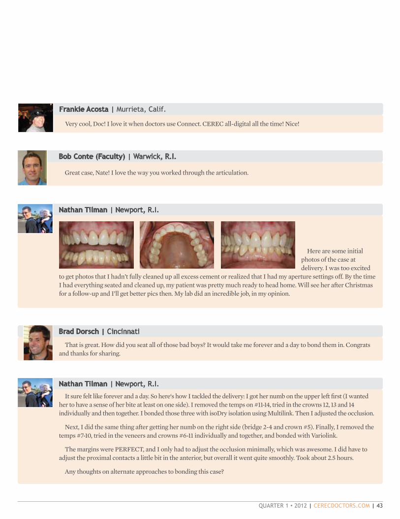
quarter 1 • 2012 | cerecDoctorS.com | 43
Here are some initial photos of the case at delivery. I was too excited
to get photos that I hadn’t fully cleaned up all excess cement or realized that I had my aperture settings off. By the time I had everything seated and cleaned up, my patient was pretty much ready to head home. Will see her after Christmas for a follow-up and I’ll get better pics then. My lab did an incredible job, in my opinion.
Bob Conte (Faculty) | Warwick, r.i.
Great case, Nate! I love the way you worked through the articulation.
nathan Tilman | Newport, r.i.
brad Dorsch | cincinnati
That is great. How did you seat all of those bad boys? It would take me forever and a day to bond them in. Congrats and thanks for sharing.
nathan Tilman | Newport, r.i.
It sure felt like forever and a day. So here‘s how I tackled the delivery: I got her numb on the upper left first (I wanted her to have a sense of her bite at least on one side). I removed the temps on #11-14, tried in the crowns 12, 13 and 14 individually and then together. I bonded those three with isoDry isolation using Multilink. Then I adjusted the occlusion.
Next, I did the same thing after getting her numb on the right side (bridge 2-4 and crown #5). Finally, I removed the temps #7-10, tried in the veneers and crowns #6-11 individually and together, and bonded with Variolink.
The margins were PERFECT, and I only had to adjust the occlusion minimally, which was awesome. I did have to adjust the proximal contacts a little bit in the anterior, but overall it went quite smoothly. Took about 2.5 hours.
Any thoughts on alternate approaches to bonding this case?
Frankie acosta | murrieta, calif.
Very cool, Doc! I love it when doctors use Connect. CEREC all-digital all the time! Nice!

44 | cerecDoctorS.com | quarter 1 • 2012
Daniel Vasquez | oceanside, calif.
Nathan: Beautiful case, well executed. If possible, can you post the screen shot from your buccal bite? Will be good to know how you control the vertical dimension in this case.
Michael saso | ottawa, ontario, canada
Awesome case.
It is still unclear how you took the buccal bite. I don’t see any “similar data” for the program to stitch the buccal bite image to the prepared image.
Also, did you do the buccal bite on both sides? Seems like a ridiculous question.
Your steps for the temporizing and bite registration seem fine. Since you had some stops in the anterior remaining, I would likely have prepared one quadrant, taken a bite, prepared the other quadrant, taken another bite with the first bite in place. I would have temporized the whole thing at the same time. No real benefit over what you did, except maybe a bit more time efficient. For a big case though, it is absolutely most important to do it the way you are most comfortable. No amount of time efficiency or money can make up for a big case that you find yourself “lost in,” or so I have heard.
Armen Mirzayan (cerecdoctors.com Co-founder): This is not an easy case; superb execution! You should be proud. Good job with the buccal bite and keeping track of the bite the whole time. Well done.
Thanks, Armen. Not easy at all, but so much fun. Couldn’t have done it without a great lab either! My lab is relatively recently on board with CEREC Connect so it’s great to let them see what is possible.
nathan Tilman | Newport, r.i.
armen Mirzayan (cerecdoctors.com co-founder) | Los angeles
Gene Messenger | North adams, mass.
Enjoyed seeing this.
This is not an easy case; superb execution! You should be proud. Good job with the buccal bite and keeping track of the bite the whole time. Well done.
Michael Saso: Awesome case. It is still unclear how you took the buccal bite. I don’t see any
“similar data” for the program to stitch the buccal bite image to the prepared image.
Also, did you do the buccal bite on both sides? Seems like a ridiculous question.
Your steps for the temporizing and bite registration seem fine. Since you had some stops in the anterior remaining, I would likely have prepared one quadrant, taken a bite, prepared the other quadrant, taken another bite with the first bite in place. I would have temporized the whole thing at the same time. No real benefit over what you did, except maybe a bit more time efficient.
nathan Tilman | Newport, r.i.
Continued...

ONLY ONE CEREC RESOURCE HAS IT ALL
CONTACT LIZ DAVISON AT 877.295.4276 OR [email protected]
HANDS-ONLEARNING
DIGITAL LEARNING
PEER-TO-PEER LEARNING
THE WORLD’S MOST POWERFUL ONLINE CEREC COMMUNITY
Continued...

46 | cerecDoctorS.com | quarter 1 • 2012
Michael saso | ottawa, ontario, canada
I can’t believe the model articulated both arches with a bite on one side only! Is this how it is actually taught? Great that it worked for you.
How would one manage this case if all the teeth were prepared with full coverage crowns, re: taking a buccal bite.
nathan Tilman | Newport, r.i.
Distinctive Dental: Awesome case! Just wondering what material you decided to use: e.max, or something else?
Everything was e.max except for the three-unit bridge which was e.max pressed to zirconia.
Peter Gardell (Faculty) | Stamford, conn.
Thanks. Just trying to figure out how to unleash the full potential of this beast!Michael – The way Nathan managed it is perfect since he was keeping the VD Prep a side and take the BB images.
Just a few, maybe three. This will be enough to articulate both arches.
jeffrey caso (Faculty) | merrick, N.y.
Michael,
Taking the bite on one side generally works very well, with a few exceptions that hopefully will get worked out in the software soon (see Javier’s thread on how to avoid this). Full preps will work just fine. In fact, next time you prep a full crown, image the buccal bite on the prep, while you will see some spikes, the software cleans it up well and the stitch will be good. Generally, avoiding the full prep is best for bb.
It blows my mind the way articulating a full arch was done and can be done, super accurately. It’s a world apart in terms of technique.
Distinctive Dental | Vancouver, Wash.
Awesome case! Just wondering what material you decided to use: e.max, or something else?
For a big case though, it is absolutely most important to do it the way you are most comfortable. No amount of time efficiency or money can make up for a big case that you find yourself “lost in,” or so I have heard.
So here’s my buccal bite image. I wasn’t quite sure if my approach was going to work, but it worked great. She had a stable bite that I didn’t want to change at all, and I wanted to capture the buccal bite for stitching in CEREC Connect. I also wanted to capture a physical bite registration so my lab could verify the mounting of the models once they arrived from InfiniDent. I think I described it earlier in the thread, but if not, here’s how I sequenced it:
I removed the existing crowns on #s 12 and 13, finalized my preps and took a physical bite registration (leaving #14 intact to be sure I had the most stable bite possible). Then I removed the crown on #14, finalized that prep and took my CEREC buccal bite images. I temporized 12-14 and adjusted the occlusion on the temps before preparing the right side.
With the left-side temps in place, I removed the bridge from 2-4, finalized those preps and took a physical bite registration of the right side for lab verification. I did not take a CEREC buccal bite of the right side.
I then finished off my preps, imaged the upper and lower arches, and temporized the rest of the upper arch.
It took some time to do it that way, but made sure I had as accurate physical bite registrations as possible for the lab, and made sure that I didn’t “lose her bite” in the process.
nathan Tilman | Newport, r.i.
Continued from page 44

quarter 1 • 2012 | cerecDoctorS.com | 47
nathan Tilman | Newport, r.i.
I will have some more images just after the first of the year. I’m on vacation this week and will see this patient early next week for a post-op evaluation, any needed occlusal adjustments, delivery of an occlusal guard and photos.
Prior to this case, I hadn’t had much experience with full arch optical impressions and I did have a little bit of difficulty crossing the midline. I imaged this case a couple of days before having 4.0 installed. I found it more difficult crossing the midline on the lower arch than the upper arch. Despite having to make multiple attempts at getting the full arch scans, it took about the same amount of time as a traditional impression would have taken ... maybe a couple of minutes longer. I know for sure that my patient LOVED not having the PVS impression taken.
Since installing 4.0 and discovering the ability to image two quadrants and stitch them together, I have practiced that on some of my team members and it works much easier, although I did still have some difficulty with the lower arch with 4.0 as well. As with anything we do, practice makes perfect and the more times I take these scans the easier I expect it will be.
As for suggestions for someone doing this for the first time, I would say for the optical image taking, practice on one of your team members or take larger models when working on some routine CEREC cases to extend across the midline. This way you can avoid getting frustrated trying to get this large of a model.
As for suggestions for the rest of the case planning, I think it’s really important to keep track of your occlusion the way I did and be sure to capture a physical bite registration that can be used by the lab to confirm the “virtual" mounting. I think there are a few different approaches that could be used to keep track of and record the bite and vertical dimension, so you’ll have to plan out what will work best in your hands.
Hopefully those suggestions will help anyone about to tackle a case like this. Nicely managed by both you and the lab. What did you do for a temp on this? Did you get pre-made temps or a putty matrix or just take a pre-op impression and use that?
Long zhao | Singapore
Great case!
What I do think may be a minor improvement is investing in a soft tissue model. This could have been easily done by ordering extra SLA models and transfering the gingival information to the sectioned die model; and with it, you might nail the inter-proximal contour just a tad better. The anteriors are quite triangular, and that can be quite challenging if you under-contoured the cervical third and end up with minor black triangle. I am interested to know how you manage to prescribe any occlusal scheme to this single arch rehab as the SLA models are simple hinge articulators? Expecting to see the final pic at your review though.
jeffrey caso (Faculty) | merrick, N.y.
The more I look at this case, the more I like it. It was a challenging case in so many ways. You said that you took a physical bite registration, and sent that to the lab to confirm the bite. Did they comment on the accuracy of the bite as it was received by them right after the models were milled and mounted? I assume it was perfect because of the minimal adjustments you had.
Thanks, Jeff
Richard Rosenblatt (Faculty) | Highland Park, ill.
I used a putty matrix that I made from a diagnostic wax-up. Worked pretty well. I think a pre-op impression would have worked just as well for the posteriors.

48 | cerecDoctorS.com | quarter 1 • 2012
nathan Tilman | Newport, r.i.
sameer Puri (cerecdoctors.com co-founder)
Nathan Tilman: I will have some more images just after the first of the year. I’m on vacation this week
and will see this patient early next week for a post-op evaluation, any needed occlusal adjustments, delivery
of an occlusal guard and photos.
Prior to this case, I hadn’t had much experience with full arch optical impressions and I did have a little bit of difficulty crossing the mid-line. I imaged this case a couple of days before having 4.0 installed. I found it more difficult crossing the midline on the lower arch than the upper arch. Despite having to make multiple attempts at getting the full arch scans, it took about the same amount of time as a traditional impression would have taken ... maybe a couple of minutes longer. I know for sure that my patient LOVED not having the PVS impression taken.
Lower arch impressions are much more difficult to do. The ridge on the lower arch is more narrow, and you have to deal with the moving floor of the mouth as well as the lips, where as on the uppers you only have to deal with the palate, which does not move. You can always use the quadrant stitching feature in 4.0 and that will help you take large images.
Nathan Tilman: Since installing 4.0 and discovering the ability to image two quadrants and stitch them together, I have practiced that on some of my team members and it works much easier although I did still have some difficulty with the lower arch with 4.0 as well. As with anything we do, practice makes perfect and the more times I take these scans the easier I expect it will be.
Technically, you can do this in 3.8 as well by taking your images in CEREC Connect and importing the case into CEREC Chair-side.
Nathan Tilman: As for suggestions for someone doing this for the first time, I would say for the optical image taking, practice on one of your team members or take larger models when working on some routine CEREC cases to extend across the midline. This way you can avoid getting frustrated trying to get this large of a model.
Good idea.
Nathan Tilman: As for suggestions for the rest of the case planning, I think it’s really important to keep track of your occlusion the way I did and be sure to capture a physical bite registration that can be used by the lab to confirm the “virtual” mounting. I think there are a few different approaches that could be used to keep track of and record the bite and vertical dimension, so you’ll have to plan out what will work best in your hands.
Another good idea. Initially at least, a physical bite is not a bad idea at all. Once you get more comfortable, I think this step can be skipped.
Nathan Tilman: Hopefully those suggestions will help anyone about to tackle a case like this.
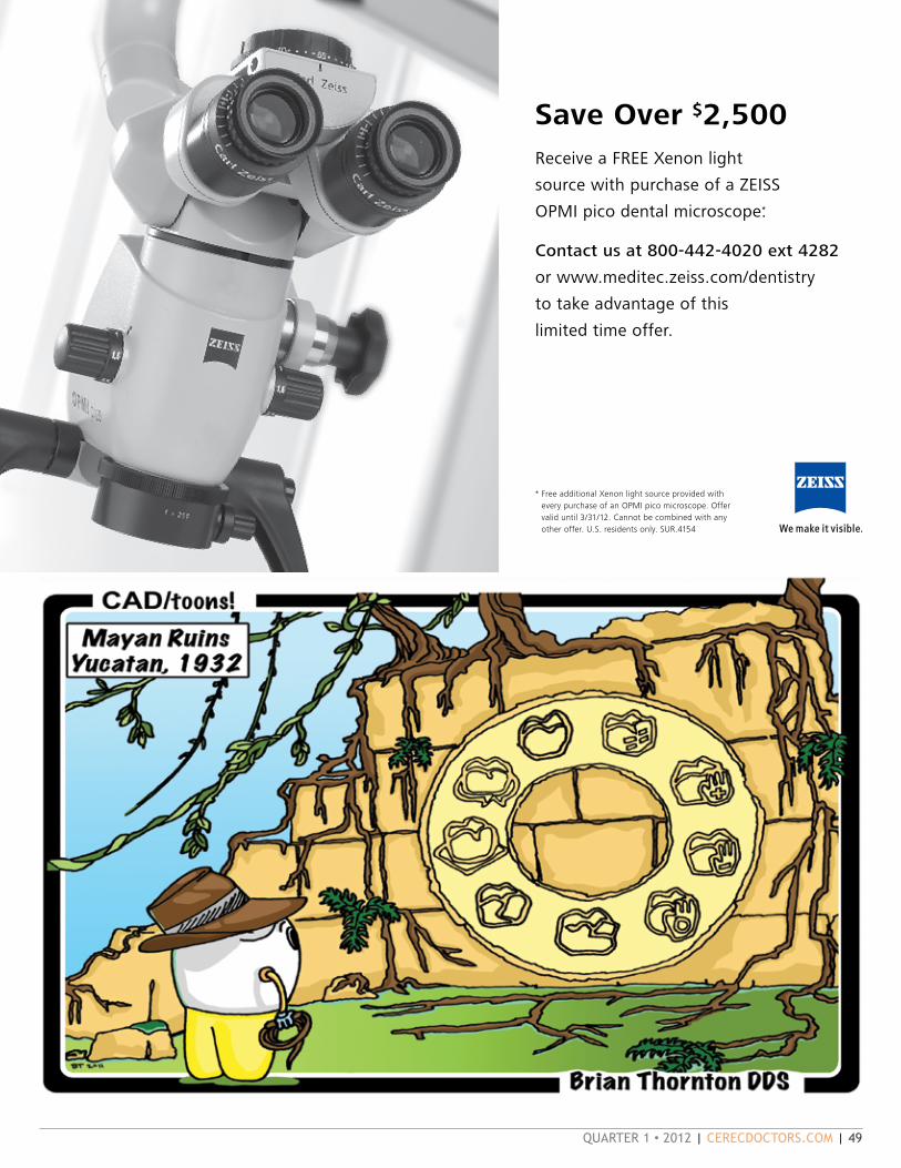
quarter 1 • 2012 | cerecDoctorS.com | 49
Save Over $2,500Receive a FREE Xenon light
source with purchase of a ZEISS
OPMI pico dental microscope.*
Contact us at 800-442-4020 ext 4282
or www.meditec.zeiss.com/dentistry
to take advantage of this
limited time offer.
* Free additional Xenon light source provided with every purchase of an OPMI pico microscope. Offer valid until 3/31/12. Cannot be combined with any other offer. U.S. residents only. SUR.4154
CerecDoctors_MicroscopeOffer_Ad_012512_2.indd 1 1/25/12 8:47 PM
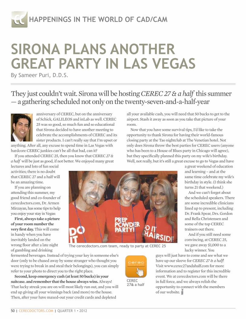
50 | cerecDoctorS.com | quarter 1 • 2012
sirona PLans anoTher GreaT ParTY in Las veGasBy Sameer Puri, D.D.S.
anniversary of CEREC, but on the anniversary of Schick, GALILEOS and inLab as well. CEREC 25 was so good, so much fun and so educational that Sirona decided to have another meeting to celebrate the accomplishments of CEREC and its sister products. I can’t really say that I’m upset or
anything. After all, any excuse to spend time in Las Vegas with hardcore CEREC junkies can’t be all that bad, can it?
If you attended CEREC 25, then you know that CEREC 27 & a half will be just as good, if not better. We enjoyed many great lectures and lots of fun social activities; there is no doubt that CEREC 27 and a half will be an amazing time.
If you are planning on attending this summer, my good friend and co-founder of cerecdoctors.com, Dr. Armen Mirzayan, has some tips to help you enjoy your stay in Vegas:
First, always take a picture of your room number the very first day. This will come in handy when you have inevitably landed on the wrong floor after a late night of gambling and drinking fermented beverages. Instead of trying your key in someone else’s door (only to be chased away by some stranger who thought you were trying to break in and steal their belongings), you can simply refer to your photo to direct you to the right place.
Second, keep emergency cash (at least 50 bucks) in your suitcase. and remember that the house always wins. Always! That lucky streak you are on will most likely run out, and you will end up giving all your winnings back (and more) to the house. Then, after your have maxed-out your credit cards and depleted
haPPeninGs in The worLD oF caD/caM
They just couldn’t wait. Sirona will be hosting CEREC 27 & a half this summer — a gathering scheduled not only on the twenty-seven-and-a-half-year
all your available cash, you will need that 50 bucks to get to the airport. Stash it away as soon as you take that picture of your room.
Now that you have some survival tips, I’d like to take the opportunity to thank Sirona for having their world-famous closing party at the Tao nightclub at The Venetian hotel. Not only does Sirona throw the best parties for CEREC users (anyone who has been to a House of Blues party in Chicago will agree), but they specifically planned this party on my wife’s birthday. Well, not really, but it’s still a great excuse to go to Vegas and have
a great weekend of education and learning – and at the same time celebrate my wife’s birthday in style. (I think she turns 21 that weekend.)
And we can’t forget about the scheduled speakers. There are some incredible clinicians lined up to present, including Dr. Frank Spear, Drs. Gordon and Rella Christensen and some of the top CEREC trainers out there.
And if you still need some convincing, at CEREC 25, we gave away $1,000 to a lucky winner. You
guys will just have to come and see what we have up our sleeve for CEREC 27 & a half! Visit www.cerec27andahalf.com for more information and to register for this incredible event. We at cerecdoctors.com will be there in full force, and we always relish the opportunity to connect with the members of our website.
The cerecdoctors.com team, ready to party at cerec 25
CSCS DFDF
CCCCPP
cerec 27& a half

quarter 1 • 2012 | cerecDoctorS.com | 51
Multi-tooth design
Simultaneous design in multiple quadrants
Direct-on-tooth application tools
Streamlined workflow
Biogeneric anatomical slider for customized proposal
CEREC Software 4.0 represents a major enhancement and redesign of the CEREC software platform, bringing unprecedented power and performance to the CEREC process from start to finish. Dental CAD/CAM is now more predictable and profitable than ever before!
facebook.com/CERECbySirona @CERECbySirona youtube.com/cerectube
Call 800.873.7683 or visit us at www.cereconline.com
Contact your Patterson representative for more information.
Join us at The Venetian!AUGUST 16-18, 2012
CEREC27andahalf.com 855.237.3248
CER001l CEREC Doctors 4.0 Single Page Ad V1.indd 1 1/19/12 10:21 AM

52 | cerecDoctorS.com | quarter 1 • 2012
all ceramic
all you need
From
left:
G. U
bass
y, D
enta
l Tec
hnic
ian,
Fra
nce
| M. R
ober
ts, D
enta
l Tec
hnic
ian,
USA
| M
. Tem
pera
ni, D
enta
l Tec
hnic
ian,
Ital
y | D
. Hor
nbro
ok, D
entis
t, U
SA |
O. B
rix, D
enta
l Tec
hnic
ian,
Ger
man
y | U
. Bro
dbec
k, D
entis
t, Sw
itzer
land
| G
. Gür
el, D
entis
t, Tu
rkey
| C.
Coa
chm
an, D
entis
t, Ce
ram
ist,
Braz
il |
A. S
hepp
erso
n, D
entis
t, N
ew Z
eala
nd |
A. B
rugu
era,
Den
tal T
echn
icia
n, S
pain
| S.
Kat
aoka
, Den
tal T
echn
icia
n, Ja
pan
| S. K
ina,
Den
tist,
Braz
il
THE WORLD SPEAKS e.max.
AND SO DOES THE SCIENCE...• 10 YEARS OF CLINICAL EVIDENCE.
• 96.8% SUCCESS RATE.
• 40 MILLION RESTORATIONS.
• 1 PROVEN SYSTEM: e.max.IPS
100% CUSTOMER SATISFACTIONG U A R A N T E E D !
Call us toll free at 1-800-533-6825 in the U.S., 1-800-263-8182 in Canada.© 2012 Ivoclar Vivadent, Inc. IPS e.max is a registered trademark of Ivoclar Vivadent.
ivoclarvivadent.com
* The IPS e.max Scientific Report Vol. 01 (2001 – 2011) is now available at: www.ivoclarvivadent.us/emax/science
*
IPS e.max SCIENCE - CEREC DRS FEB 2012.indd 1 1/25/12 8:35 AM