dental conference - mid periodontal disease november 11, 2004
Post on 18-Dec-2015
214 views
TRANSCRIPT
Dental Conference - MID
Periodontal Disease
November 11, 2004
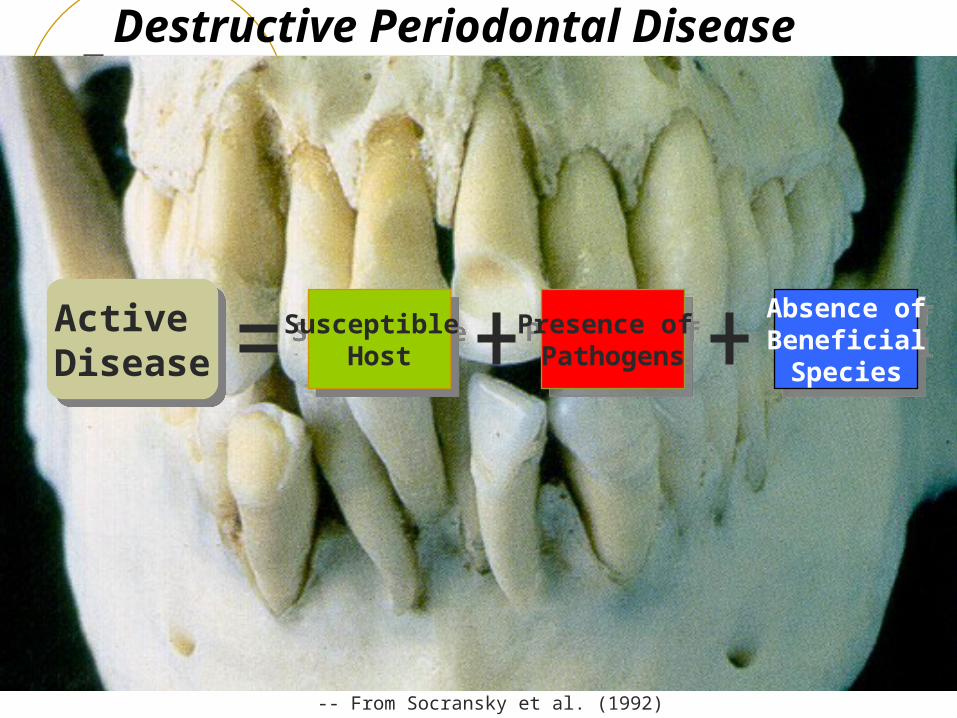
Destructive Periodontal Disease
Active Disease
Active Disease
Susceptible Host
Susceptible Host
Presence of Pathogens
Presence of Pathogens
Absence ofBeneficial
Species
Absence ofBeneficial
Species
-- From Socransky et al. (1992)
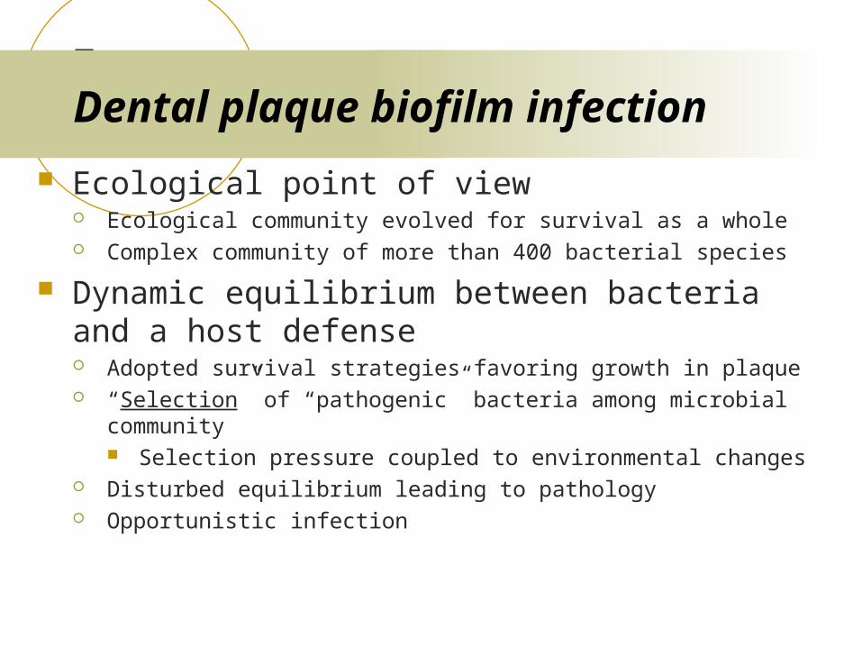
Dental plaque biofilm infection
Ecological point of view Ecological community evolved for survival as a whole Complex community of more than 400 bacterial species
Dynamic equilibrium between bacteria and a host defense Adopted survival strategies favoring growth in plaque “Selection” of “pathogenic” bacteria among microbial community
Selection pressure coupled to environmental changes Disturbed equilibrium leading to pathology Opportunistic infection
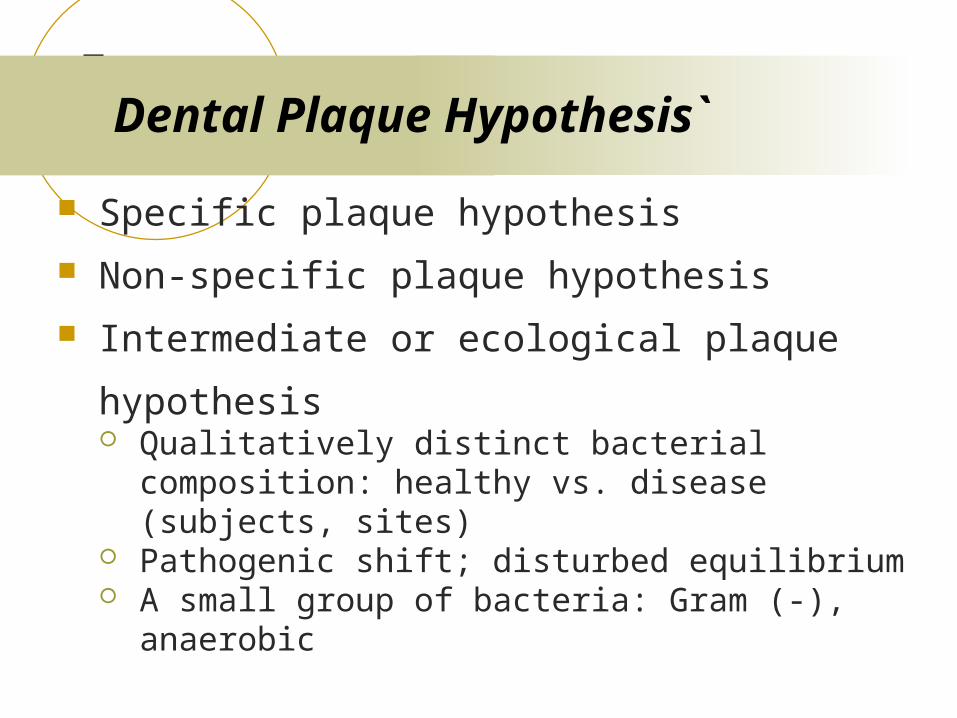
Dental Plaque Hypothesis`
Specific plaque hypothesis
Non-specific plaque hypothesis
Intermediate or ecological plaque hypothesis Qualitatively distinct bacterial composition:
healthy vs. disease (subjects, sites) Pathogenic shift; disturbed equilibrium A small group of bacteria: Gram (-), anaerobic
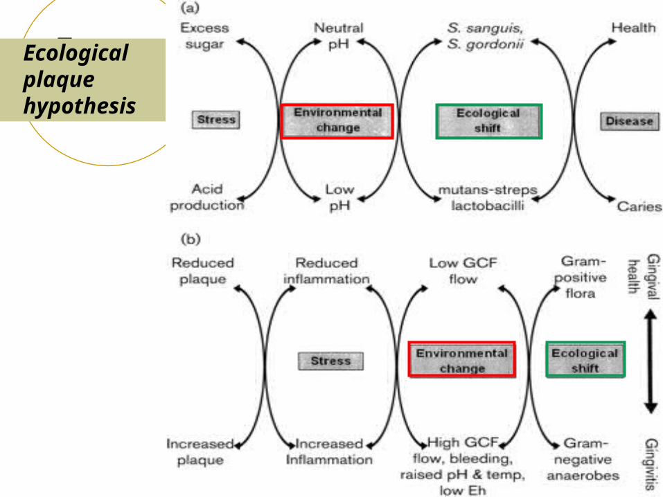
Ecological plaque hypothesis
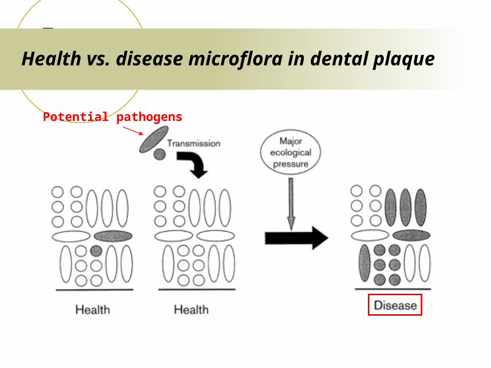
Health vs. disease microflora in dental plaque
Potential pathogens
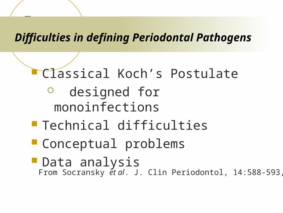
Difficulties in defining Periodontal Pathogens
Classical Koch’s Postulate designed for monoinfections
Technical difficulties Conceptual problems Data analysis
From Socransky et al. J. Clin Periodontol, 14:588-593, 1987
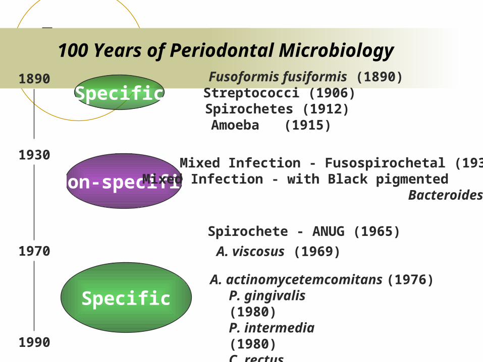
100 Years of Periodontal Microbiology
Specific
Non-specific
Specific
1890
1930
1970
Fusoformis fusiformis (1890)Streptococci (1906)Spirochetes (1912)Amoeba (1915)
Mixed Infection - Fusospirochetal (1930)Mixed Infection - with Black pigmented Bacteroides (1955)
Spirochete - ANUG (1965)
A. viscosus (1969)
A. actinomycetemcomitans (1976)P. gingivalis (1980)P. intermedia (1980)C. rectusB. forsythus
1990
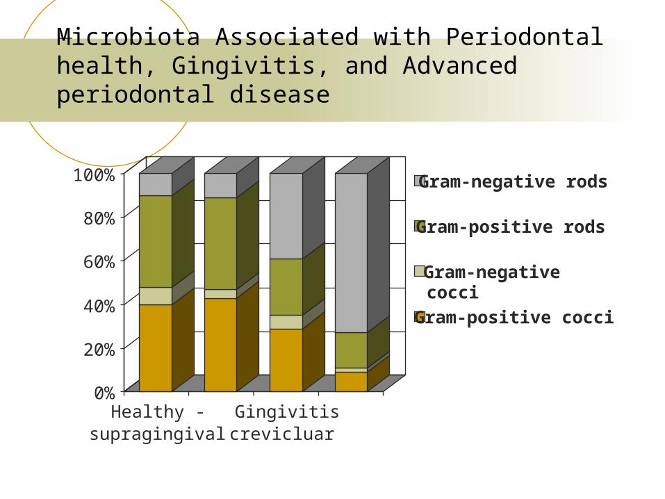
Microbiota Associated with Periodontal health, Gingivitis, and Advanced periodontal disease
0%
20%
40%
60%
80%
100%
Healthy -supragingival
Gingivitiscrevicluar
Gram-negative rods
Gram-positive rods
Gram-negativecocci
Gram-positive cocci
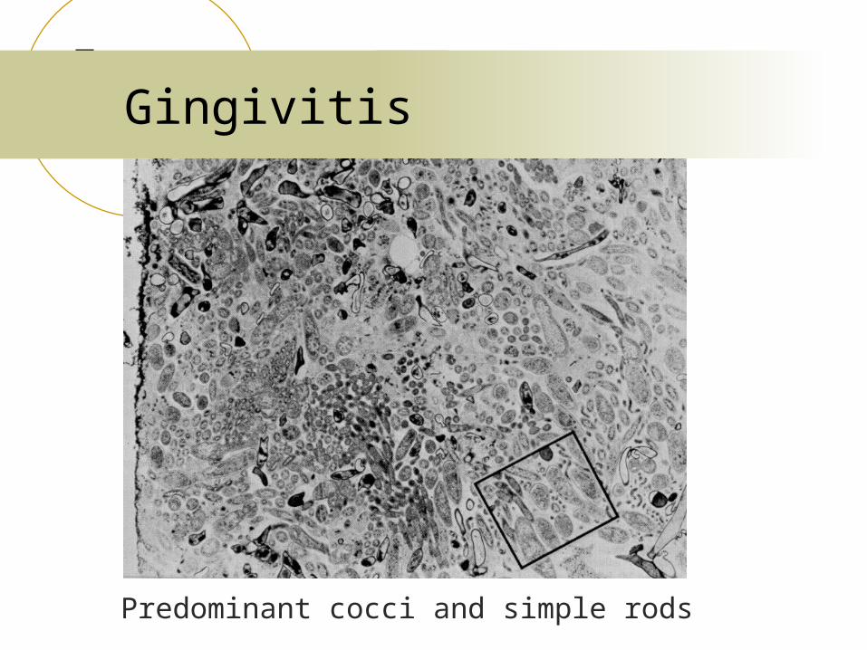
Gingivitis
Predominant cocci and simple rods
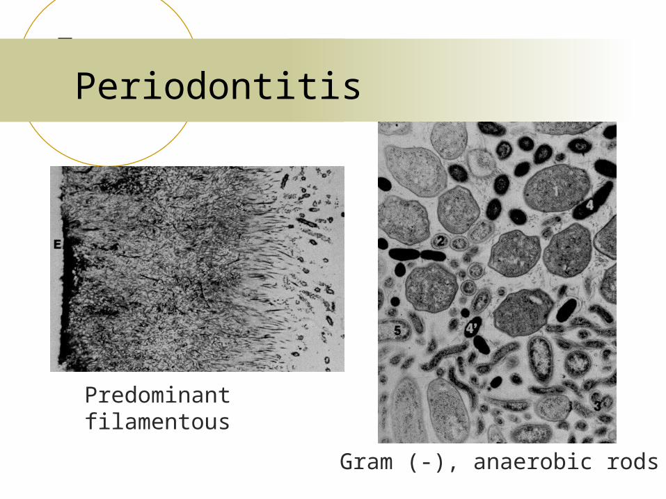
Periodontitis
Predominant filamentous
Gram (-), anaerobic rods
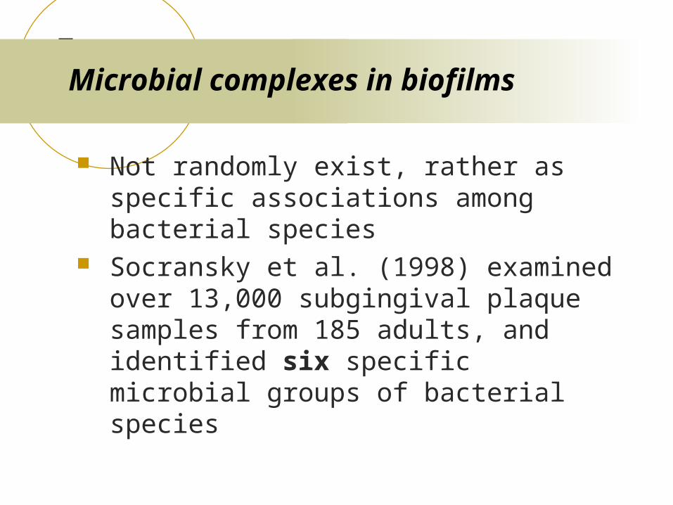
Microbial complexes in biofilms
Not randomly exist, rather as specific associations among bacterial species
Socransky et al. (1998) examined over 13,000 subgingival plaque samples from 185 adults, and identified six specific microbial groups of bacterial species
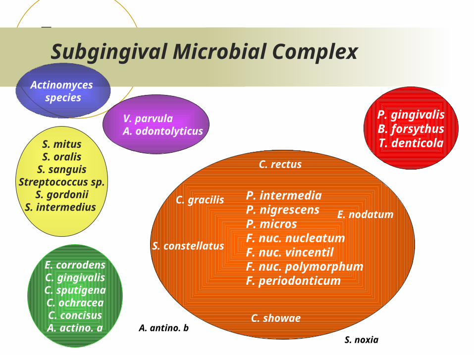
S. mitusS. oralis
S. sanguisStreptococcus sp.
S. gordoniiS. intermedius
S. noxia A. antino. b
Subgingival Microbial Complex
P. intermediaP. nigrescensP. microsF. nuc. nucleatumF. nuc. vincentil F. nuc. polymorphum F. periodonticum
P. gingivalisB. forsythusT. denticola
V. parvulaA. odontolyticus
E. corrodensC. gingivalisC. sputigenaC. ochraceaC. concisusA. actino. a
Actinomyces species
S. constellatus
C. gracilis
C. rectus
E. nodatum
C. showae
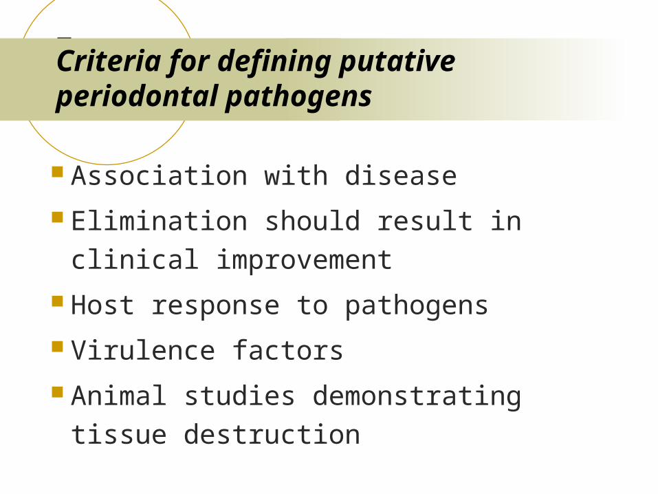
Criteria for defining putative periodontal pathogens
Association with disease
Elimination should result in clinical
improvement
Host response to pathogens
Virulence factors
Animal studies demonstrating tissue destruction
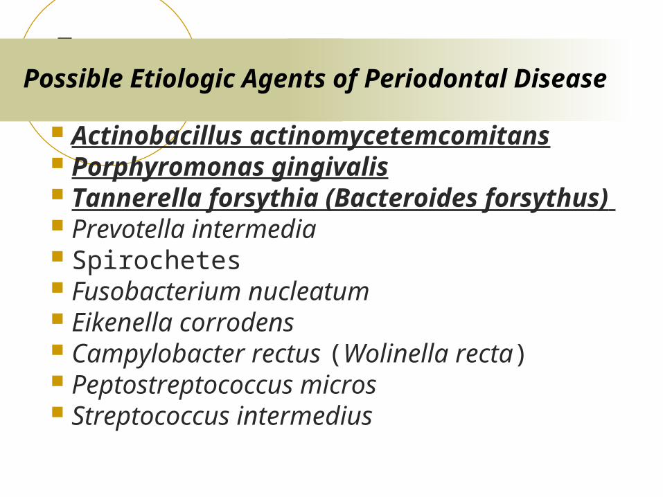
Possible Etiologic Agents of Periodontal Disease
Actinobacillus actinomycetemcomitans Porphyromonas gingivalis Tannerella forsythia (Bacteroides forsythus) Prevotella intermedia Spirochetes Fusobacterium nucleatum Eikenella corrodens Campylobacter rectus (Wolinella recta) Peptostreptococcus micros Streptococcus intermedius
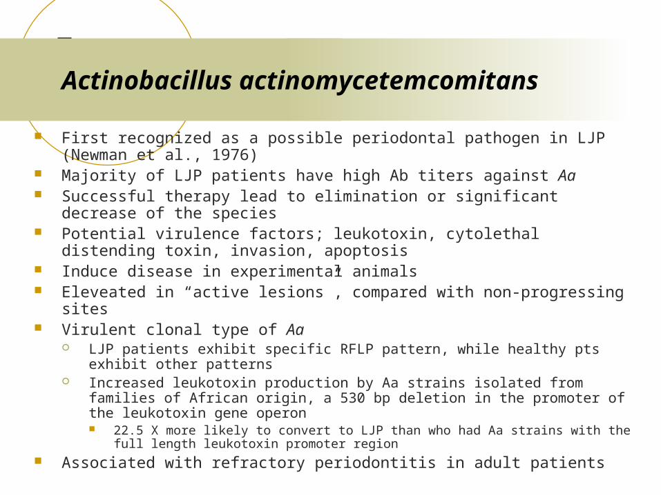
Actinobacillus actinomycetemcomitans
First recognized as a possible periodontal pathogen in LJP (Newman et al., 1976)
Majority of LJP patients have high Ab titers against Aa Successful therapy lead to elimination or significant decrease of the species Potential virulence factors; leukotoxin, cytolethal distending toxin, invasion,
apoptosis Induce disease in experimental animals Eleveated in “active lesions”, compared with non-progressing sites Virulent clonal type of Aa
LJP patients exhibit specific RFLP pattern, while healthy pts exhibit other patterns
Increased leukotoxin production by Aa strains isolated from families of African origin, a 530 bp deletion in the promoter of the leukotoxin gene operon 22.5 X more likely to convert to LJP than who had Aa strains with the full length
leukotoxin promoter region Associated with refractory periodontitis in adult patients
Porphyromonas gingivalis
Gram (-), anaerobic, asaccharolytic, black-pigmented bacterium
Suspected periodontopathic microorganismAssociation
Elevated in periodontal lesions, rare in health Elimination or suppression resulted in successful therapy
Immunological correlation Elevated systemic and local antibody in periodontitis
Animal pathogenicity Monkey, dog, and rodent models
Putative virulent factors
Spirochetes
G (-), anaerobic, spiral, highly motile ANUG Increased numbers in deep periodontal pockets Difficulty in distinguishing individual species
15 subgingival spirochetes described Obscure classification - Small, medium, or large
T. denticola More common in diseased, subgingival site
Uncultivated “pathogen-related oral spirochetes Detected by Ab cross-reactivity to T. pallidum antibody
Prevotella intermedia/Prevotella nigrescens
Strains of “P. intermedia” separated into two species, P. intermedia and P. nigrescins
Hemagglutination activity Adherence activity Induce alveolar bone loss In certain forms of periodontitis Successful therapy leads to decrease in P.
intermedia
G(-), anaerobic, spindle-shaped rod Has been recognized as part of the subgingival
microbiota for over 100 years The most common isolate found in cultural studies of
subgingival plaque samples:7-10% of total isolates Prevalent in subjects with periodontitis and periodontal
abscess Invasion of epithelial cell Apoptosis activity
Fusobacterium nucleatum
Other species
Campylobacter rectus Produce leukotoxin Contains the S-layer Stimulate gingival fibroblast to produce IL-6 and IL-8
Eikenella corrodens Peptostreptococcus micros
G(+), anaerobic, small asaccharolytic Long been associated with mixed anaerobic infections
Selemonas species Curved shape, tumbling motility S. noxia found in deep pockets, conversion from healthy to disease site
Eubacterium specues The “milleri” streptococci
S. anginosus, S. constellatus, S. intermedius
Periodontal disease as an infectious disease
Events in all infectious disease: Encounter Entry Spread Multiplication Damage Outcome
Virulence factors
Gene products that enhance a microorganism’s potential to cause disease
Involved in all steps of pathogenicityAttach to or enter host tissueEvade host responsesProliferateDamage the hostTransmit itself to new hosts
Define “the pathogenic personality” Virulence genes
Expression of virulence factors
Constitutive Under specific environmental signals
Can be identified by mimicking environmental signals in the laboratory
Many virulence-associated genes are coordinately regulated by environmental signals
Only in vivo Cannot be identified in the laboratory Anthrax toxin, cholera toxin
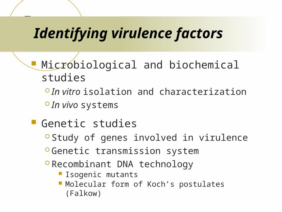
Identifying virulence factors
Microbiological and biochemical studies In vitro isolation and characterization In vivo systems
Genetic studies Study of genes involved in virulence Genetic transmission system Recombinant DNA technology
Isogenic mutants Molecular form of Koch’s postulates (Falkow)
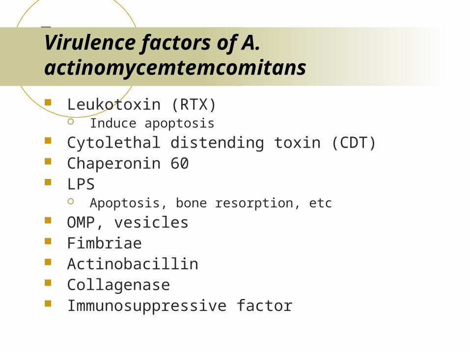
Virulence factors of A. actinomycemtemcomitans
Leukotoxin (RTX) Induce apoptosis
Cytolethal distending toxin (CDT) Chaperonin 60 LPS
Apoptosis, bone resorption, etc OMP, vesicles Fimbriae Actinobacillin Collagenase Immunosuppressive factor
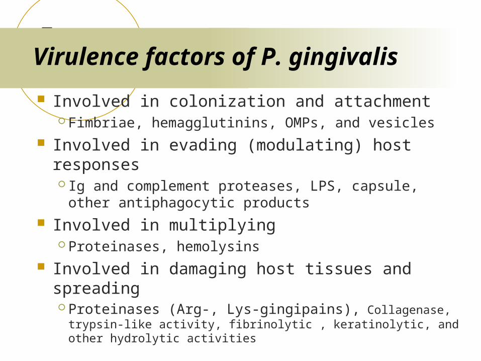
Virulence factors of P. gingivalis
Involved in colonization and attachment Fimbriae, hemagglutinins, OMPs, and vesicles
Involved in evading (modulating) host responses Ig and complement proteases, LPS, capsule, other
antiphagocytic products Involved in multiplying
Proteinases, hemolysins Involved in damaging host tissues and spreading
Proteinases (Arg-, Lys-gingipains), Collagenase, trypsin-like activity, fibrinolytic , keratinolytic, and other hydrolytic activities
An Example of Studying Microbial Pathogenesis
Hypothesis
S-layer of T. forsythia is a virulence factor
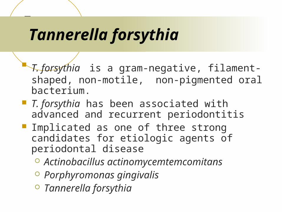
Tannerella forsythia
T. forsythia is a gram-negative, filament-shaped, non-motile, non-pigmented oral bacterium.
T. forsythia has been associated with advanced and recurrent periodontitis
Implicated as one of three strong candidates for etiologic agents of periodontal disease Actinobacillus actinomycemtemcomitans Porphyromonas gingivalis Tannerella forsythia
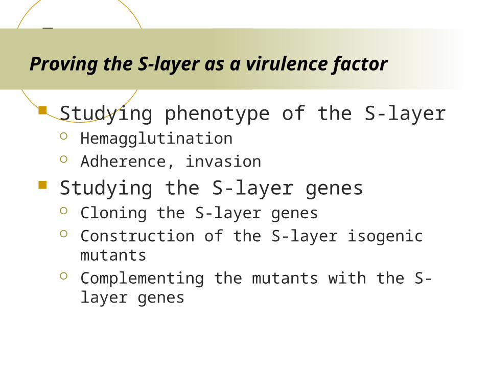
Proving the S-layer as a virulence factor
Studying phenotype of the S-layer Hemagglutination Adherence, invasion
Studying the S-layer genes Cloning the S-layer genes Construction of the S-layer isogenic mutants Complementing the mutants with the S-layer genes
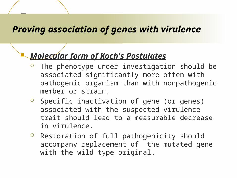
Proving association of genes with virulence
Molecular form of Koch's Postulates The phenotype under investigation should be associated
significantly more often with pathogenic organism than with nonpathogenic member or strain.
Specific inactivation of gene (or genes) associated with the suspected virulence trait should lead to a measurable decrease in virulence.
Restoration of full pathogenicity should accompany replacement of the mutated gene with the wild type original.