dental status, dental rehabilitation procedures, demographic and
TRANSCRIPT
Niewald et al. Radiation Oncology 2013, 8:227http://www.ro-journal.com/content/8/1/227
RESEARCH Open Access
Dental status, dental rehabilitation procedures,demographic and oncological data as potentialrisk factors for infected osteoradionecrosis of thelower jaw after radiotherapy for oral neoplasms: aretrospective evaluationMarcus Niewald1*, Jochen Fleckenstein1, Kristina Mang2, Henrik Holtmann3, Wolfgang J Spitzer3
and Christian Rübe1
Abstract
Purpose: Retrospective evaluation of the dental status of patients with oral cancer before radiotherapy, the extentof dental rehabilitation procedures, demographic and radiotherapy data as potential risk factors for development ofinfected osteoradionecrosis of the lower jaw.
Methods: A total of 90 patients who had undergone radiotherapy for oral cancer were included into thisretrospective evaluation. None of them had distant metastases. After tumour surgery the patients were referred toan oral and maxillofacial surgeon for dental examination and the necessary dental rehabilitation proceduresinclusive potential tooth extraction combined with primary soft tissue closure. Adjuvant radiotherapy was startedafter complete healing of the gingiva (> 7 days after potential extraction). The majority of patients (n = 74) wastreated with conventionally fractionated radiotherapy with total doses ranging from 50-70Gy whereas further 16patients received hyperfractionated radiotherapy up to 72Gy. The records of the clinical data were reviewed.Furthermore, questionnaires were mailed to the patients’ general practitioners and dentists in order to get moredata concerning tumour status and osteoradionecrosis during follow-up.
Results: The patients’ dental status before radiotherapy was generally poor. On average 10 teeth were present, sixof them were regarded to remain conservable. Extensive dental rehabilitation procedures included a mean of 3.7tooth extractions. Chronic periodontitis with severe attachment loss was found in 40%, dental biofilm in 56%. Aninfected osteoradionecrosis (IORN) grade II according to (Schwartz et al., Am J Clin Oncol 25:168-171, 2002) wasdiagnosed in 11 of the 90 patients (12%), mostly within the first 4 years after radiotherapy. We could not findsignificant prognostic factors for the occurrence of IORN, but a trendwise correlation with impaired dental status,rehabilitation procedures, fraction size and tumour outcome.
Conclusion: The occurrence of IORN is an important long-term side effect of radiotherapy for oral cancers. Fromthis data we only can conclude that a poor dental status, conventional fractionation and local tumour progressionmay enhance the risk of IORN which is in concordance with the literature.
Keywords: Dental status, Dental rehabilitation procedures, Radiotherapy, Fractionation, Infected osteoradionecrosis
* Correspondence: [email protected] of Radiotherapy and Radiooncology, Saarland UniversityMedical Center, Kirrberger Str. 1, D-66421 Homburg, GermanyFull list of author information is available at the end of the article
© 2013 Niewald et al.; licensee BioMed Central Ltd. This is an Open Access article distributed under the terms of the CreativeCommons Attribution License (http://creativecommons.org/licenses/by/2.0), which permits unrestricted use, distribution, andreproduction in any medium, provided the original work is properly cited.

Table 1 Patients’ demographical and oncological data(n = 90)
Item Mean value Minimumvalue
Maximumvalue
Remarks
Age 56.97 21.1 84.0 Years
Karnofskyperformancestatus
78% 50% 100%
Follow-up 3.5 0 12.25 Years
Item Value (%)
T-stage
T1 14 (16%)
T2 33 (37%)
T3 8 (9%)
T4 35 (38%)
N-stage
N0 20 (22%)
N1 24 (27%)
N2 46 (51%)
N3 0
UICC stage
I 7 (8%)
II 6 (7%)
III 11 (12%)
IV 66 (73%)
Pre-treatment
None 27 (30%)
Surgery 61 (70%)
Surgery to the lower jaw
Partial resection 16 (23%)
Continuity resection 10 (14%)
Chemotherapy 17 (19%)
Radiotherapy – fractionation
Conventional 74 (82%)
Hyperfractionation 16 (18%)
Fraction size Gy/day
1 × 2.0 72 (80%)
1 × 3.0 2 (2%)
2 × 1.2 15 (17%)
2 × 1.4 1 (1%)
total dose – conventionally fractionated (n = 75) Gy
30 1 (1%)
36 1 (1%)
50 7 (9%)
58 2 (2%)
60 31 (44%)
64 9 (13%)
70 23 (31%)
Niewald et al. Radiation Oncology 2013, 8:227 Page 2 of 12http://www.ro-journal.com/content/8/1/227
BackgroundThe dental status of patients with neoplasms of the headand neck region is known to be more unfavourable com-pared to healthy persons. Some reasons may be the fre-quent abuse of nicotine and alcohol and limited dentalhygiene. These factors may increase the risk for developinginfected osteoradionecrosis (IORN) after radiotherapy as atypical long-term side effect [1]. To our knowledge, IORNcan only be sufficiently avoided performing by extensivedental rehabilitation procedures including extraction ofteeth. Nevertheless, IORN occurs in a frequency rangingfrom 0-22% [2]. Because of the necrosis of the gingiva, ero-sion and sequestration of the jaw bone and dentoalveolarabscess formation sufficient chewing and thus nutrition ofthe patient can be problematic and thus impair the qualityof life of those patients by a large extent.16 years ago we have published a retrospective evalu-
ation of the frequency and risk factors for IORN [2].The frequency of IORN in patients after conventionallyfractionated radiotherapy (total dose 60-70Gy) was 8.6%while the same rate amounted to 22.9% in patients withhyperfractionated radiotherapy (total dose 82.8Gy). Weconcluded that this excessive occurrence of IORN in pa-tients having been treated with a hyperfractionated ir-radiation regimen was probably caused by the high totaldose and a too short interfraction interval.In the presented analysis, we performed a second
evaluation of frequency and risk factors for IORN in atotally different collective of patients having undergoneradiotherapy in the years 1993–2001.
MethodsNinety patients with histologically proven squamous-cellcancers of the oral cavity treated in the years 1993–2001were included into this retrospective evaluation. Themajority had primary tumors, one a local recurrence buthad not been irradiated before, none had distant metas-tases. The mean age at the beginning of radiotherapywas 57 years, the mean Karnofsky performance index7.8. Sixty-four patients had undergone prior surgery, theremaining 24 had not.Dental examination and treatment procedures were
performed as early as possible with a minimal time intervalof 7–10 days from the last procedure to the beginning ofradiotherapy. All dental extractions were performedaccording to a written protocol under “special care” (pri-mary tissue closure, perioperative antibiotics for 7–10 daysbeginning one day before surgery). In the nineties all pa-tients were advised not to wear their dental prostheses upto 6–12 months after radiotherapy (today after completehealing of mucositis) [3,4].After completion of all dental examination and rehabilita-
tion procedures (Tab. 3), all patients underwent radio-therapy. 74 patients were treated in a conventionally
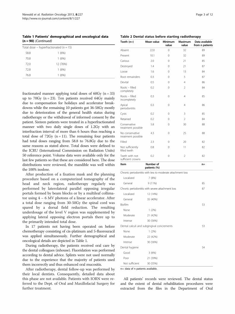
Table 2 Dental status before starting radiotherapyTeeth (n=) Mean value Minimum
valueMaximumvalue
Data availablefrom n patients
Absent 22.0 0 32 89
Present 10.1 0 32 89
Carious 2.0 0 21 85
Destroyed 1.4 0 21 87
Loose 1.6 0 13 84
Root remainders 0.3 0 5 87
Devital 0.5 0 4 86
Roots – filledcompletely
0.2 0 2 84
Roots – filledincompletely
0.3 0 4 85
Apicalperiodontitis
0.3 0 4 86
Cysts 0.2 0 3 85
Retained 0.2 0 2 84
Conservativetreatment possible
5.8 0 30 88
No conservativetreatment possible
4.3 0 25 88
Filled 2.3 0 20 82
Not sufficientlyfilled teeth
0.8 0 11 82
Teeth with notsufficient crowns
0.2 0 2 82
Item Number ofpatients (%)
n=
Chronic periodontitis with less to moderate attachment loss
Localized 7 (8%)
General 9 (11%) 85
Chronic periodontitis with severe attachment loss 87
Localized 12 (14%)
General 35 (40%)
Biofilm 53
None 1 (2%)
Moderate 21 (42%)
Intense 30 (56%)
Dental calculi and subgingival concrements 53
None 1 (2%)
Moderate 22 (42%)
Intense 30 (56%)
Dental hygiene 54
Good 3 (6%)
Poor 21 (39%)
Not sufficient 30 (55%)
n=: data of n patients available.
Table 1 Patients’ demographical and oncological data(n = 90) (Continued)
Total dose – hyperfractionated (n = 15)
58.8 1 (6%)
70.8 1 (6%)
72.0 12 (76%)
72.8 1 (6%)
76.8 1 (6%)
Niewald et al. Radiation Oncology 2013, 8:227 Page 3 of 12http://www.ro-journal.com/content/8/1/227
fractionated manner applying total doses of 60Gy (n = 33)up to 70Gy (n = 23). Ten patients received 64Gy mainlydue to compensation for holidays and accelerator break-downs while the remaining 10 patients got 36-58Gy mostlydue to deterioration of the general health status duringradiotherapy or the withdrawal of informed consent by thepatient. Sixteen patients were treated in a hyperfractionatedmanner with two daily single doses of 1.2Gy with aninterfraction interval of more than 6 hours thus reaching atotal dose of 72Gy (n = 11). The remaining four patientshad total doses ranging from 58.8 to 76.8Gy due to thesame reasons as stated above. Total doses were defined tothe ICRU (International Commission on Radiation Units)50 reference point. Volume data were available only for thelast few patients so that these are considered here. The dosedistributions were reviewed, the mandible was well withinthe 100% isodose.After production of a fixation mask and the planning
procedure based on a computerized tomography of thehead and neck region, radiotherapy regularly wasperformed by laterolateral parallel opposing irregularportals formed by beam blocks or by a multileaf collima-tor using 4 – 6 MV photons of a linear accelerator. Aftera total dose ranging from 30-50Gy the spinal cord wasspared by a dorsal field reduction. The resultingunderdosage of the level V region was supplemented byapplying lateral opposing electron portals there up tothe primarily intended total dose.In 17 patients not having been operated on before
chemotherapy consisting of cis-platinum and 5-fluorouracilwas applied simultaneously. Further demographical andoncological details are depicted in Table 1.During radiotherapy, the patients received oral care by
the dental colleagues (inhouse). Fluoridation was performedaccording to dental advice. Splints were not used normallydue to the experience that the majority of patients usedthem incorrectly and thus enhanced oral mucositis.After radiotherapy, dental follow-up was performed by
their local dentists. Consequently, detailed data aboutthis phase are not available. Patients with IORN were re-ferred to the Dept. of Oral and Maxillofacial Surgery forfurther treatment.
All patients’ records were reviewed. The dental statusand the extent of dental rehabilitation procedures wereextracted from the files in the Department of Oral

Table 3 Dental treatment procedures
Teeth (n=) Mean value Minimum value Maximum value Data available from n patients
Endodontic treatment 0.05 0 2 88
Removal of root remainders with primary tissue closure 0.2 0 5 88
Tooth extraction with primary tissue closure 3.7 0 22 89
Conserving treatment 0.6 0 6 88
Cystectomy 0.1 0 2 87
Healthy teeth remaining after dental rehabilitation 6.2 0 30 89
Edentulous after dental treatment 47 patients (52%)
Niewald et al. Radiation Oncology 2013, 8:227 Page 4 of 12http://www.ro-journal.com/content/8/1/227
and Maxillofacial Surgery. Furthermore, all x-rays (ortho-pantomograms) were re-examined. Infected osteo-radionecrosis (IORN) was (at minimum) defined here asinfected mucosal ulcers with eroded mandibular boneunderneath (grade II according to Schwartz et al. [5]). Theradiooncological data were extracted from the files in theDepartment of Radiotherapy and Radiooncology. Addition-ally, standardized questionnaires were mailed to the pa-tients’ medical doctors, dentists and local authorities threetimes within the observation period in order to achieveadditional data concerning tumor outcome, occurrence ofIORN and general health status.All data were entered into a medical database
(MEDLOG, Parox Comp., Münster, Germany). Distribu-tions and means were computed. For survival curvesand failure curves (occurrence of IORN) the KaplanMeier estimate was used. These curves were comparedusing the Mantel Haensel test.Prognostic parameters for IORN were analyzed uni-
variately by comparison of means and distributions in agroup containing the patients with IORN compared to an-other group with the patients who never experiencedIORN using the t-test, u-test and chi-square test in the ap-propriate variables. Multivariate search for independentprognostic factors was performed by logistic regression.All patients had given their written informed consent
to surgery, dental rehabilitation procedures and radio-therapy before treatment. The approval by the local eth-ics committee was dispensable due to the retrospectiveevaluation performed here. This research is in concord-ance with the Declaration of Helsinki.
Table 4 Dental treatment after radiotherapy
Treatment Number ofpatients (%)
None 73 (81%)
Conservative treatment 2 (2%)
Tooth extraction with primary tissue closure 14 (16%)
Tooth extraction with primary tissue closure andconservative treatment
1 (1%)
ResultsGeneral remarksUp to July 2013, 58 patients were dead with a meanfollow-up of 2.4 [0–8.8] years. The patients known to bealive were seen irregularly, the most recent informationresulted from questionnaires, nearly all patients werelost to follow-up after on average 7.4 [0–15] years.
Dental findings before radiotherapyThe patients’ dental status was generally poor. On average,only 10 teeth were present (and thus 22 teeth missing) atthe beginning of therapy in the oral cavity. Of those, onaverage 2.0 were carious, 1.5 loose (clinical grade II-III) and1.4 deeply destroyed. As a result of the meticulous dentalexamination only a mean of six teeth was regarded to re-main conservable on average. 11% of the patients showedchronic periodontitis with less to moderate attatchmentloss while in 40% chronic periodontitis with severeattatchment loss was diagnosed.Additionally, plaque was found frequently; 56% of the pa-
tients had dental biofilm and dental calculi and subgingivalconcrements, respectively. The general dental hygiene wasclassified separately by a dentist as poor in 55% of the pa-tients. These findings are based on the data of 53 patients,further information was not available in the files. The de-tailed data are depicted in Table 2.Data on the use of dental prostheses are available for
65/90 patients. Partial prostheses were used by six pa-tients in the upper jaw and by 22 patients in the lowerjaw. Complete prostheses were used by 34 patients inthe upper jaw and by 15 patients in the lower jaw. Thepatients had been counselled not to wear their pros-theses during radiotherapy and 12 months afterwards.
Dental rehabilitation procedures before and afterradiotherapyAll patients were referred to the dentist and oro-maxillofacial surgeon for dental and surgical treatment,89/90 have been seen there. The dental rehabilitation pro-cedures necessary before radiotherapy were very extensive:on average 3.7 teeth had to be extracted followed by pri-mary soft tissue closure of the extraction alveoles. The

0 2 4 6 8 10 12 14 16 18 200
0.1
0.2
0.3
0.4
0.5
0.6
0.7
0.8
0.9
1.0
Time in years
Pro
bab
ility
of
IOR
N
Occurrence of IORN
All patients
Figure 1 Occurrence of IORN over time (Kaplan-Meier estimate and 95% confidence interval).
Niewald et al. Radiation Oncology 2013, 8:227 Page 5 of 12http://www.ro-journal.com/content/8/1/227
extraction was followed up by an interval of at least 7–10 days using soft diet, valid antibiosis and prosthodonticabstention. The detailed data to this point are summarizedin Table 3.During follow-up after radiotherapy additional dental
treatment procedures were necessary in 17 patients –the data are depicted in Table 4.
Frequency, risk factors and therapy of infectedosteoradionecrosis11 patients (12%) were found to have developed infectedosteoradionecrosis during follow-up. The one-year preva-lence was 5%, the two- and three-year prevalence 15%.All of them had been treated by conventionally frac-
tionated radiotherapy applying doses of 50Gy (1 pat.),60Gy (4 pats.), 64Gy (3 pats.), and 70Gy (3 pats.), re-spectively. Nine of these patients had undergone tumourresection, two had not. Additionally, two patients hadundergone a partial resection of the lower jaw, furtherthree patients a continuity resection of the lower jaw. Intotal, 9/64 patients (14%) having been operated on hadIORN compared to only 8% in the non-surgical patients.In the group treated by hyperfractionation no patient
with IORN could be identified. The Kaplan Meier esti-mate shows that IORN normally occurs within the firstfour years after radiotherapy, after that time new IORNcases were very rare (Figure 1).All parameters mentioned in Tables 1, 2, 3 and 4 were
tested univariately for potential prognostic significancefor the occurrence of IORN. Results of this analysis aredepicted in Table 5. The number of carious teeth andof odontogenic cysts in the lower jaw were found
significant prognostic factors, for the numbers of presentteeth before dental treatment and of remaining teethafter dental treatment a trend can be assumed. Multi-variate analysis showed “odontogenic cysts” as the onlysignificant factor.The treatment of infected osteoradionecrosis consisted
of non-continuously bone resection in two patients, sur-gery without bone resection in further three. Two pa-tients were treated conservatively while one underwenthyperbaric oxygen therapy. The remaining three patientswere not treated at all.
Oncological resultsDue to the patients’ very limited compliance the out-come data were incomplete. The following numbersshow the frequency of recurrence or progression com-pared to the number of patients with sufficient dataavailable.A local tumor progression was found in 25/73 patients
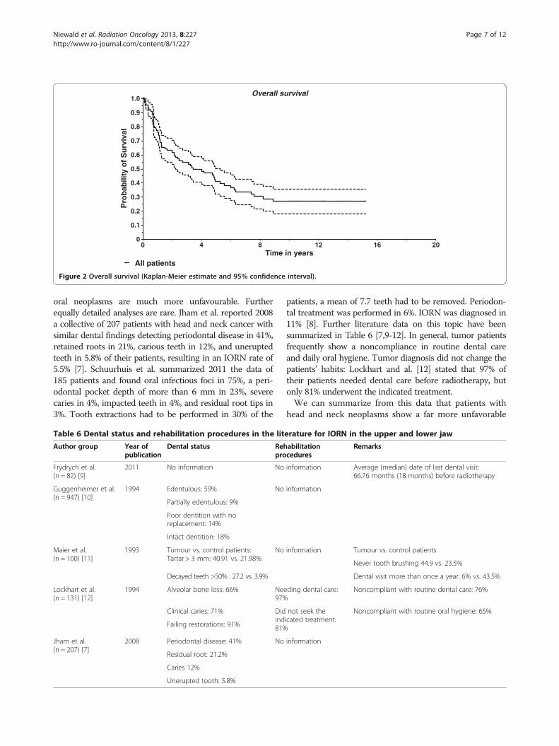
(34%), a progression in the regional lymph nodes in12/61 patients (20%), and distant metastases in 12/59patients (20%). No sufficient data to this point wereavailable from the remaining patients. The median sur-vival time was 3.1 years. The two-year survival rate was62% while the five-year survival rate amounted to 41%.The Kaplan-Meier estimate for overall survival isdepicted in Figure 2. As to acute side effects occurringduring or shortly after radiotherapy, oral mucositis gradeII WHO was found in 47 patients (54%), grades III andIV in further 5 patients (6%, n = 87). Sialadenosis (dry-ness of mouth) as a typical long-term side effect ofradiotherapy was found in 72 patients (82%; grade 1

Table 5 Prognostic factors for the occurrence of IORN inthe lower jaw
Univariate analysis
Teeth p= Remarks
Dental status before starting radiotherapy
Absent 0.093
Present 0.095 Trend
Carious 0.047 Significant
Destroyed 0.556
Loose 0.730
Root remainders 0.247
Devital 0.688
Roots – filled completely 0.555
Roots – filled incompletely 0.774
Apical periodontitis 0.956
Cysts 0.023 Significant
Retained 0.291
Conservative treatment possible 0.129
No conservative treatment possible 0.830
Filled 0.758
Not sufficiently filled teeth 0.517
Teeth with not sufficient crowns 0.897
Item
Chronic periodontitis with less to moderateattatchment loss
0.572
Localized
General
Chronic periodontitis with severe attatchment loss 0.548
Localized
General
Biofilm 0.188
None
Moderate
Intense
Dental calculus and subgingival concrements 0.188
None
Moderate
Intense
Partial prosthesis in lower jaw 0.5254
Complete prosthesis in lower jaw 0.9026
Dental treatment before radiotherapy
Endodontic treatment 0.474
Removal of root remainders 0.413
Tooth extraction 0.939
Conserving treatment 0.498
Cystectomy 0.261
Table 5 Prognostic factors for the occurrence of IORN inthe lower jaw (Continued)
Healthy teeth remaining after dental rehabilitation 0.085 Trend
Dental treatment after radiotherapy 0.768
Demographic and oncological data
Age 0.118
Karnofsky performance status 0.455
T-stage 0.784
N-stage O.797
Total dose 0.774
BED2 0.410
Daily fraction 0.170
Multivariate analysis
Carious teeth 0.1808
Present teeth 0.3154
Odontogenic cysts 0.0180 Significant
Healthy teeth remaining after dental treatment 0.1552
Niewald et al. Radiation Oncology 2013, 8:227 Page 6 of 12http://www.ro-journal.com/content/8/1/227
EORTC/RTOG: 22 patients (25%), grade 2: 38 patients(43%), grade 3: 12 patients (14%); n = 88).
DiscussionThe authors are well aware of the limitations of thisretrospective evaluation. In this nearly homogenous col-lective of patients with oral cavity cancer having under-gone radiotherapy +/− surgery, we have found completedata sets concerning dental status and restoration proce-dures of nearly all patients. The IORN data have beeninvestigated meticulously, but due to the knownincompliance of head and neck patients we cannot ex-clude that single events did not become known to theauthors.
Dental health statusThe comparison of our data concerning dental findings be-fore radiotherapy to those of the Forth German Trial ofOral Health (original name in German: IV. DeutscheMundgesundheitsstudie des Instituts der DeutschenZahnärzte DMS IV) resulted in notable differences [6].Summarizing the data of more than 4000 Germans beforedental treatment, in adults (33–44 years of age) on average14.5 teeth were found carious, in older people (> = 45 yearsof age) 22.1 teeth. These teeth were rehabilitated completelyin 95.6% and in 94.8%, respectively. A mean of 2.77 teeth inadults and of 14.2 teeth in older people were missing. 72%of the adults and 60.6% of the seniors were found to per-form sufficient mouth hygiene. All these values were im-proved compared to the results of a former trial in 1997.On the other hand, the frequency of periodontitis was ris-ing (moderate in 52.9% and intense in 39.8% of the popula-tion). Compared to those data our findings in patients with
0 4 8 12 16 200
0.1
0.2
0.3
0.4
0.5
0.6
0.7
0.8
0.9
1.0
Time in years
Pro
bab
ility
of
Su
rviv
al
Overall survival
All patients
Figure 2 Overall survival (Kaplan-Meier estimate and 95% confidence interval).
Niewald et al. Radiation Oncology 2013, 8:227 Page 7 of 12http://www.ro-journal.com/content/8/1/227
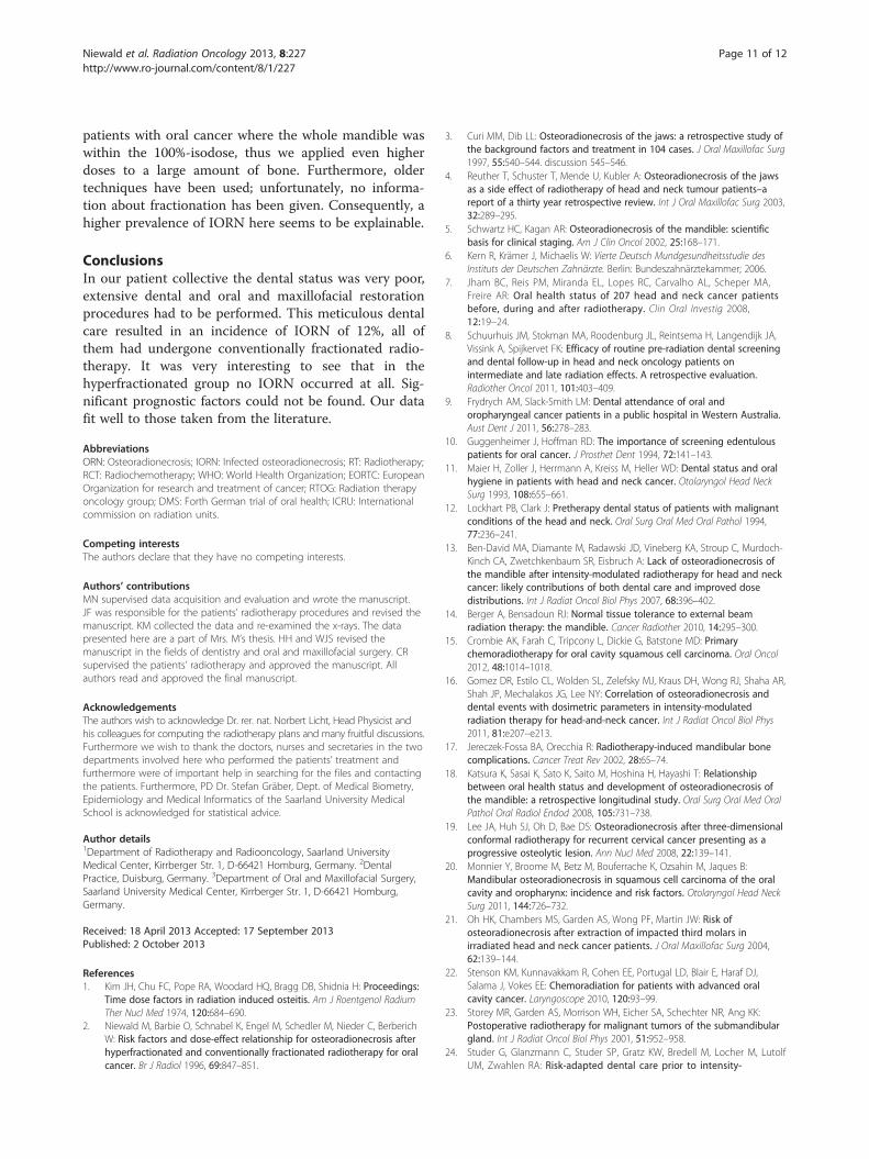
oral neoplasms are much more unfavourable. Furtherequally detailed analyses are rare. Jham et al. reported 2008a collective of 207 patients with head and neck cancer withsimilar dental findings detecting periodontal disease in 41%,retained roots in 21%, carious teeth in 12%, and uneruptedteeth in 5.8% of their patients, resulting in an IORN rate of5.5% [7]. Schuurhuis et al. summarized 2011 the data of185 patients and found oral infectious foci in 75%, a peri-odontal pocket depth of more than 6 mm in 23%, severecaries in 4%, impacted teeth in 4%, and residual root tips in3%. Tooth extractions had to be performed in 30% of the
Table 6 Dental status and rehabilitation procedures in the lit
Author group Year ofpublication
Dental status Rehpro
Frydrych et al.(n = 82) [9]
2011 No information No
Guggenheimer et al.(n = 947) [10]
1994 Edentulous: 59% No
Partially edentulous: 9%
Poor dentition with noreplacement: 14%
Intact dentition: 18%
Maier et al.(n = 100) [11]
1993 Tumour vs. control patients:Tartar > 3 mm: 40.91 vs. 21.98%
No
Decayed teeth >50% : 27.2 vs. 3.9%
Lockhart et al.(n = 131) [12]
1994 Alveolar bone loss: 66% Nee97%
Clinical caries: 71% Didind81%Failing restorations: 91%
Jham et al.(n = 207) [7]
2008 Periodontal disease: 41% No
Residual root: 21.2%
Caries 12%
Unerupted tooth: 5.8%
patients, a mean of 7.7 teeth had to be removed. Periodon-tal treatment was performed in 6%. IORN was diagnosed in11% [8]. Further literature data on this topic have beensummarized in Table 6 [7,9-12]. In general, tumor patientsfrequently show a noncompliance in routine dental careand daily oral hygiene. Tumor diagnosis did not change thepatients’ habits: Lockhart and al. [12] stated that 97% oftheir patients needed dental care before radiotherapy, butonly 81% underwent the indicated treatment.We can summarize from this data that patients with
head and neck neoplasms show a far more unfavorable
erature for IORN in the upper and lower jaw
abilitationcedures
Remarks
information Average (median) date of last dental visit:66.76 months (18 months) before radiotherapy
information
information Tumour vs. control patients
Never tooth brushing 44.9 vs. 23.5%
Dental visit more than once a year: 6% vs. 43.5%
ding dental care: Noncompliant with routine dental care: 76%
not seek theicated treatment:
Noncompliant with routine oral hygiene: 65%
information

Table 7 Incidence of IORN of the upper and lower jaw in the literature
Author group Year of publication Incidence Remarks
Ben-David et al. [13] (n = 176) 2007 0 Multiple tumour localizations
Primary treatment (no surgery)
IMRT
108/176 radiochemotherapy
Berger et al. [14] 2010 1-5% Literature survey
Crombie et al. [15] (n = 54) 2012 36% 53/54 radiochemotherapy
Gomez et al. [16] (n = 168) 2011 1.2% Multiple tumour localizations
IMRT
Gomez et al. [29] (n = 35) 2009 5% IMRT
Jerecek-Fosså et al. [17] 2002 0.4-56% Literature survey
Jham et al. [7] (n = 207) 2008 5.5% Head and neck cancer
Katsura et al. [18] (n = 39) 2008 15%
Lee et al. [19] (n = 189) 2008 6.6% Oral cavity and oropharynx
Monnier et al. [20] (n = 73) 2011 40% Oral cavity and oropharynx
Oh et al. [21] (n = 81) 2004 4.9%
Reuther et al. [5] (n = 830) 2003 8.2% Oral cavity and oropharynx
Stenson et al. [22] (n = 27) 2010 18.4% Surgery, adjuvant radiochemotherapy
Storey et al. [23] (n = 83) 2001 6% Malignant submandibular tumours
Studer et al. [24] (n = 304) 2011 Grade 2 EORTC: 1.6% Oral cavity and oropharynx
Conventional dental care vs. risk-adapted dental care
IMRT
Thiel [25] 1989 4-35% Literature survey
Thorn et al. [26] (n = 80) 2000 74%/3 years Multiple tumour localizations
Tsai et al. [27] (n = 402) 2013 7.5% Oropharyngeal cancer, median time to IORN 8 months
Turner et al. [28] (n = 333) 1996 5.9%
Niewald et al. Radiation Oncology 2013, 8:227 Page 8 of 12http://www.ro-journal.com/content/8/1/227
denta health status than a healthy population does; ourfindings are well within the range of the data taken fromthe literature seen above.
Frequency of IORNThe incidence of IORN varies widely (0 – 74%) asdepicted in Table 7 whereas the majority of data are in arange of 5-10%. However, the comparison of these valuesto each other and to our results is very difficult due to adifferent definition or staging of IORN, different tumorlocalizations, therapy schedules, radiation techniquesand dosages. Our results fit well within the range of datataken from the literature [5,7,13-29]. One of the datasets in the literature most comparable to our dataset hasbeen published by Lee et al. [19] who experienced com-parable IORN frequencies in a collective of patients hav-ing been operated on mainly.
Risk factors for the development of IORNNumerous prognostic factors for the development of IORNhave been tested and published. A selection of these is
summarized in Table 8 [3,4,14,17,18,20,27,28,30-39]. Thelocalization of the primary tumour in the oral cavity withits microbial colonization and the abundant involvement ofthe mandibular bone with its unique blood supply probablypromotes IORN. Unfavourable dental status, periodontaldisease and irritation of the gingiva by pressure sore trig-gered by dental prosthesis are important as well as dentalextractions before and especially after radiotherapy and doalso promote an infestation of the upper jaw.Radiation dose should not exceed 60 – 66Gy to the
mandibular bone whenever possible, the target volumewithin the bone should be limited. Some authorsregard hyperfractionation a risk factor for IORN whereasIntensity modulated radiotherapy (IMRT) has been foundadvantageous compared to conventional 3D-planned radio-therapy. Additional factors may be chemotherapy, higherbody mass index and the use of steroids.We found odontogenic cysts and carious teeth to
be significant prognostic factors univariately andodontogenic cysts multivariately. This rather unusualfinding may result from the low number of patients in

Table 8 Risk factors for IORN of the upper and lower jaw in the literature
Author group Year of publication Risk factor(s) Remarks
Ahmed et al. [30] 2009 Intensity modulated radiotherapy (IMRT) advantageous comparedto conventional radiotherapy
Berger et al. [14] 2010 Total dose >66Gy Literature survey
Bhide et al. [31] 2012 Total dose > 60Gy Literature survey
Volume of mandible within the treatment field. Trauma relatedORN after lower doses
IMRT
Chopra et al. [32] 2011 White ethnicity
Secondary infection
Advanced age
Stage IV
Total dose
Post-RT dental extractions
Lack of pre-RT dental extractions
Goldwasser et al. [33] 2007 Higher body mass index Multivariate analysis
Use of steroids
Radiation dose >66Gy
Jerecek-Fosså et al. [17] 2002 Total dose Literature survey, only part ofthe factions mentioned in the
paper cited hereBrachytherapy dose
Dose per fraction
Interval between fractions
Volume of the horizontal ramus of the mandible irradiated with ahigh dose
Dental status
Bad oral hygiene
Dental extractions after radiotherapy
Katsura et al. [18] 2008 Oral health status after radiotherapy
Periodontal pocket depth
Dental plaque
Alveolar bone loss level
Radiographic periodontal status
Lee et al. [19] 2009 Univariate: Mandibular surgery Multivariate analysis:Mandibular surgery
Co-60 BED >106.2Gy
Lozza et al. [35] 1997 Dose rate Brachytherapy exclusively
Reference volume
Curi et al. [3] 1997 Oral cancer
Invasion of bone
Tumour surgery
Total radiation dose
Dose rate/day
Mode of radiation delivery
Dental status
Time from radiation therapy until the onset of ORN
Niewald et al. Radiation Oncology 2013, 8:227 Page 9 of 12http://www.ro-journal.com/content/8/1/227

Table 8 Risk factors for IORN of the upper and lower jaw in the literature (Continued)
Monnier et al. [20] 2011 Oral cavity tumours Multivariate analysis: bonesurgery
Bone invasion
Surgery prior to radiotherapy
Bone surgery
Nabil et al. [36] 2012 Hyperfractionation Literature survey
Reduced risk after accelerated radiotherapy with reduced dose
Reuther et al. [4] 2003 Advanced tumours
Segmental resection of the mandible
Tooth extractions (pre/post RT)
Pre-surgical radiotherapy worse than post-surgical radiotherapy
Støre and Boysen [37] 2000 Tumour localization in tongue and floor of mouth
trauma
Thiel et al. [38] 1989 Caries
Periondontosis
Periapical pathology
Injury
Irritation by prostheses
Dental extractions before and after radiotherapy
Bone surgery because of remaining or recurrent tumours
Thorn et al. [39] 2000 Removal of teeth
Surgery
Injury from prosthesis
Spontaneous breakdowns
Tsai et al. [27] 2013 Total dose
Dental status
Smokers
Alcohol
Larger tumours
Turner et al. [24] 1996 Bone involvement
Synchronous Methotrexate
Scattered dose from elective
neck treatment
Increasing dose
Increasing target volumes for doses <55Gy
Dental extractions
Niewald et al. Radiation Oncology 2013, 8:227 Page 10 of 12http://www.ro-journal.com/content/8/1/227
the IORN group and the fact that for some variables thedata were incomplete.Apart from this analysis, hyperfractionation seemed to
have a protective effect whereas this could not be examinedfurther due to the small number of events. In our ancientpublication on this topic [2] (a very detailed comparison iscurrently under preparation) we experienced a very highfrequency of IORN after hyperfractionated radiotherapywhich may have been caused by to high total doses on theone hand and a too short interfraction interval (time inter-val between the two daily fractions) on the other hand.
Both factors have been taken into account here, conse-quently the results were improved markedly.An important paper has been published by Tsai et al.
in 2013 [27]. They reviewed the records of patients withsmall oropharyngeal cancers having undergone radio-therapy or radiochemotherapy. The overall prevalence ofIORN was 7.5%, higher doses, use of nicotine and alco-hol, dental status as well as more advanced tumors werefound significant risk factors for the development ofIORT. In contrast to this paper our patients’ primarysituation seems more unfavorable: we only examined
Niewald et al. Radiation Oncology 2013, 8:227 Page 11 of 12http://www.ro-journal.com/content/8/1/227
patients with oral cancer where the whole mandible waswithin the 100%-isodose, thus we applied even higherdoses to a large amount of bone. Furthermore, oldertechniques have been used; unfortunately, no informa-tion about fractionation has been given. Consequently, ahigher prevalence of IORN here seems to be explainable.
ConclusionsIn our patient collective the dental status was very poor,extensive dental and oral and maxillofacial restorationprocedures had to be performed. This meticulous dentalcare resulted in an incidence of IORN of 12%, all ofthem had undergone conventionally fractionated radio-therapy. It was very interesting to see that in thehyperfractionated group no IORN occurred at all. Sig-nificant prognostic factors could not be found. Our datafit well to those taken from the literature.
AbbreviationsORN: Osteoradionecrosis; IORN: Infected osteoradionecrosis; RT: Radiotherapy;RCT: Radiochemotherapy; WHO: World Health Organization; EORTC: EuropeanOrganization for research and treatment of cancer; RTOG: Radiation therapyoncology group; DMS: Forth German trial of oral health; ICRU: Internationalcommission on radiation units.
Competing interestsThe authors declare that they have no competing interests.
Authors’ contributionsMN supervised data acquisition and evaluation and wrote the manuscript.JF was responsible for the patients’ radiotherapy procedures and revised themanuscript. KM collected the data and re-examined the x-rays. The datapresented here are a part of Mrs. M’s thesis. HH and WJS revised themanuscript in the fields of dentistry and oral and maxillofacial surgery. CRsupervised the patients’ radiotherapy and approved the manuscript. Allauthors read and approved the final manuscript.
AcknowledgementsThe authors wish to acknowledge Dr. rer. nat. Norbert Licht, Head Physicist andhis colleagues for computing the radiotherapy plans and many fruitful discussions.Furthermore we wish to thank the doctors, nurses and secretaries in the twodepartments involved here who performed the patients’ treatment andfurthermore were of important help in searching for the files and contactingthe patients. Furthermore, PD Dr. Stefan Gräber, Dept. of Medical Biometry,Epidemiology and Medical Informatics of the Saarland University MedicalSchool is acknowledged for statistical advice.
Author details1Department of Radiotherapy and Radiooncology, Saarland UniversityMedical Center, Kirrberger Str. 1, D-66421 Homburg, Germany. 2DentalPractice, Duisburg, Germany. 3Department of Oral and Maxillofacial Surgery,Saarland University Medical Center, Kirrberger Str. 1, D-66421 Homburg,Germany.
Received: 18 April 2013 Accepted: 17 September 2013Published: 2 October 2013
References1. Kim JH, Chu FC, Pope RA, Woodard HQ, Bragg DB, Shidnia H: Proceedings:
Time dose factors in radiation induced osteitis. Am J Roentgenol RadiumTher Nucl Med 1974, 120:684–690.
2. Niewald M, Barbie O, Schnabel K, Engel M, Schedler M, Nieder C, BerberichW: Risk factors and dose-effect relationship for osteoradionecrosis afterhyperfractionated and conventionally fractionated radiotherapy for oralcancer. Br J Radiol 1996, 69:847–851.
3. Curi MM, Dib LL: Osteoradionecrosis of the jaws: a retrospective study ofthe background factors and treatment in 104 cases. J Oral Maxillofac Surg1997, 55:540–544. discussion 545–546.
4. Reuther T, Schuster T, Mende U, Kubler A: Osteoradionecrosis of the jawsas a side effect of radiotherapy of head and neck tumour patients–areport of a thirty year retrospective review. Int J Oral Maxillofac Surg 2003,32:289–295.
5. Schwartz HC, Kagan AR: Osteoradionecrosis of the mandible: scientificbasis for clinical staging. Am J Clin Oncol 2002, 25:168–171.
6. Kern R, Krämer J, Michaelis W: Vierte Deutsch Mundgesundheitsstudie desInstituts der Deutschen Zahnärzte. Berlin: Bundeszahnärztekammer; 2006.
7. Jham BC, Reis PM, Miranda EL, Lopes RC, Carvalho AL, Scheper MA,Freire AR: Oral health status of 207 head and neck cancer patientsbefore, during and after radiotherapy. Clin Oral Investig 2008,12:19–24.
8. Schuurhuis JM, Stokman MA, Roodenburg JL, Reintsema H, Langendijk JA,Vissink A, Spijkervet FK: Efficacy of routine pre-radiation dental screeningand dental follow-up in head and neck oncology patients onintermediate and late radiation effects. A retrospective evaluation.Radiother Oncol 2011, 101:403–409.
9. Frydrych AM, Slack-Smith LM: Dental attendance of oral andoropharyngeal cancer patients in a public hospital in Western Australia.Aust Dent J 2011, 56:278–283.
10. Guggenheimer J, Hoffman RD: The importance of screening edentulouspatients for oral cancer. J Prosthet Dent 1994, 72:141–143.
11. Maier H, Zoller J, Herrmann A, Kreiss M, Heller WD: Dental status and oralhygiene in patients with head and neck cancer. Otolaryngol Head NeckSurg 1993, 108:655–661.
12. Lockhart PB, Clark J: Pretherapy dental status of patients with malignantconditions of the head and neck. Oral Surg Oral Med Oral Pathol 1994,77:236–241.
13. Ben-David MA, Diamante M, Radawski JD, Vineberg KA, Stroup C, Murdoch-Kinch CA, Zwetchkenbaum SR, Eisbruch A: Lack of osteoradionecrosis ofthe mandible after intensity-modulated radiotherapy for head and neckcancer: likely contributions of both dental care and improved dosedistributions. Int J Radiat Oncol Biol Phys 2007, 68:396–402.
14. Berger A, Bensadoun RJ: Normal tissue tolerance to external beamradiation therapy: the mandible. Cancer Radiother 2010, 14:295–300.
15. Crombie AK, Farah C, Tripcony L, Dickie G, Batstone MD: Primarychemoradiotherapy for oral cavity squamous cell carcinoma. Oral Oncol2012, 48:1014–1018.
16. Gomez DR, Estilo CL, Wolden SL, Zelefsky MJ, Kraus DH, Wong RJ, Shaha AR,Shah JP, Mechalakos JG, Lee NY: Correlation of osteoradionecrosis anddental events with dosimetric parameters in intensity-modulatedradiation therapy for head-and-neck cancer. Int J Radiat Oncol Biol Phys2011, 81:e207–e213.
17. Jereczek-Fossa BA, Orecchia R: Radiotherapy-induced mandibular bonecomplications. Cancer Treat Rev 2002, 28:65–74.
18. Katsura K, Sasai K, Sato K, Saito M, Hoshina H, Hayashi T: Relationshipbetween oral health status and development of osteoradionecrosis ofthe mandible: a retrospective longitudinal study. Oral Surg Oral Med OralPathol Oral Radiol Endod 2008, 105:731–738.
19. Lee JA, Huh SJ, Oh D, Bae DS: Osteoradionecrosis after three-dimensionalconformal radiotherapy for recurrent cervical cancer presenting as aprogressive osteolytic lesion. Ann Nucl Med 2008, 22:139–141.
20. Monnier Y, Broome M, Betz M, Bouferrache K, Ozsahin M, Jaques B:Mandibular osteoradionecrosis in squamous cell carcinoma of the oralcavity and oropharynx: incidence and risk factors. Otolaryngol Head NeckSurg 2011, 144:726–732.
21. Oh HK, Chambers MS, Garden AS, Wong PF, Martin JW: Risk ofosteoradionecrosis after extraction of impacted third molars inirradiated head and neck cancer patients. J Oral Maxillofac Surg 2004,62:139–144.
22. Stenson KM, Kunnavakkam R, Cohen EE, Portugal LD, Blair E, Haraf DJ,Salama J, Vokes EE: Chemoradiation for patients with advanced oralcavity cancer. Laryngoscope 2010, 120:93–99.
23. Storey MR, Garden AS, Morrison WH, Eicher SA, Schechter NR, Ang KK:Postoperative radiotherapy for malignant tumors of the submandibulargland. Int J Radiat Oncol Biol Phys 2001, 51:952–958.
24. Studer G, Glanzmann C, Studer SP, Gratz KW, Bredell M, Locher M, LutolfUM, Zwahlen RA: Risk-adapted dental care prior to intensity-

Niewald et al. Radiation Oncology 2013, 8:227 Page 12 of 12http://www.ro-journal.com/content/8/1/227
modulated radiotherapy (IMRT). Schweiz Monatsschr Zahnmed 2011,121:216–229.
25. Thiel HJ: Osteoradionecrosis. I. Etiology, pathogenesis, clinical aspectsand risk factors. Radiobiol Radiother 1989, 30:397–413.
26. Thorn JJ, Kallehave F, Westergaard P, Hansen EH, Gottrup F: The effect ofhyperbaric oxygen on irradiated oral tissues: transmucosal oxygentension measurements. J Oral Maxillofac Surg 1997, 55:1103–1107.
27. Tsai CJ, Hofstede TM, Sturgis EM, Garden AS, Lindberg ME, Wei Q, Tucker SL,Dong L: Osteoradionecrosis and radiation dose to the mandible in patientswith oropharyngeal cancer. Int J Radiat Oncol Biol Phys 2013, 85:415–420.
28. Turner SL, Slevin NJ, Gupta NK, Swindell R: Radical external beamradiotherapy for 333 squamous carcinomas of the oral cavity–evaluationof late morbidity and a watch policy for the clinically negative neck.Radiother Oncol 1996, 41:21–29.
29. Gomez DR, Zhung JE, Gomez J, Chan K, Wu AJ, Wolden SL, Pfister DG,Shaha A, Shah JP, Kraus DH, et al: Intensity-modulated radiotherapy inpostoperative treatment of oral cavity cancers. Int J Radiat Oncol Biol Phys2009, 73:1096–1103.
30. Ahmed M, Hansen VN, Harrington KJ, Nutting CM: Reducing the risk ofxerostomia and mandibular osteoradionecrosis: the potential benefits ofintensity modulated radiotherapy in advanced oral cavity carcinoma.Med Dosim 2009, 34:217–224.
31. Bhide SA, Ahmed M, Newbold K, Harrington KJ, Nutting CM: The role ofintensity modulated radiotherapy in advanced oral cavity carcinoma.J Cancer Res Ther 2012, 8(Suppl 1):S67–S71.
32. Chopra S, Kamdar D, Ugur OE, Chen G, Peshek B, Marunick M, Kim H, LinHS, Jacobs J: Factors predictive of severity of osteoradionecrosis of themandible. Head Neck 2011, 33:1600–1605.
33. Goldwasser BA, Chuang S-K, Kabal LB, August M: Risk factor assessmentfor the development of osteoradionecrosis. J Oral Maxillofac Surg 2007,65:2311–2316.
34. Lee IJ, Koom WS, Lee CG, Kim YB, Yoo SW, Keum KC, Kim GE, Choi EC, ChaIH: Risk factors and dose-effect relationship for mandibularosteoradionecrosis in oral and oropharyngeal cancer patients. Int J RadiatOncol Biol Phys 2009, 75:1084–1091.
35. Lozza L, Cerrotta A, Gardani G, De Marie M, Di Russo A, Kenda R, Tana S,Valvo F, Zucali R: Analysis of risk factors for mandibular boneradionecrosis after exclusive low dose-rate brachytherapy for oralcancer. Radiother Oncol 1997, 44:143–147.
36. Nabil S, Samman N: Risk factors for osteoradionecrosis after head andneck radiation: a systematic review. Oral Surg Oral Med Oral Pathol OralRadiol 2012, 113:54–69.
37. Store G, Boysen M: Mandibular osteoradionecrosis: clinical behaviour anddiagnostic aspects. Clin Otolaryngol Allied Sci 2000, 25:378–384.
38. Thiel HJ: Osteoradionecrosis. II: Therapy and prevention. Radiobiol Radiother(Berl) 1989, 30:493–501.
39. Thorn JJ, Hansen HS, Specht L, Bastholt L: Osteoradionecrosis of the jaws:clinical characteristics and relation to the field of irradiation.J Oral Maxillofac Surg 2000, 58:1088–1093. discussion 1093–1085.
doi:10.1186/1748-717X-8-227Cite this article as: Niewald et al.: Dental status, dental rehabilitationprocedures, demographic and oncological data as potential risk factorsfor infected osteoradionecrosis of the lower jaw after radiotherapy fororal neoplasms: a retrospective evaluation. Radiation Oncology2013 8:227.
Submit your next manuscript to BioMed Centraland take full advantage of:
• Convenient online submission
• Thorough peer review
• No space constraints or color figure charges
• Immediate publication on acceptance
• Inclusion in PubMed, CAS, Scopus and Google Scholar
• Research which is freely available for redistribution
Submit your manuscript at www.biomedcentral.com/submit