department - dm5migu4zj3pb.cloudfront.netdm5migu4zj3pb.cloudfront.net/manuscripts/100000/... ·...
TRANSCRIPT

THE HEALING OF RICKETrS COINCIDENT WITH LOWSERUMINORGANIC PHOSPHORUS
By GENEVIEVE STEARNSAND JULIAN D. BOYD
(From the Department of Pediatrics, College of Medicine, State University of Iowa,Iowa City)
(Received for publication February 21, 1931)
Within the past year, roentgenological evidence of healing of ricketscoincident with a persistently low level of serum inorganic phosphorushas been observed in 2 children. These findings are of importance inrelation to the theories of bone formation.
The subjects of the study were 2 girls of 12 and 13 years of age,respectively, both of whom showed roentgenological, clinical andchemical evidence of active rickets. The serum calcium values ofboth children were within normal limits, the serum inorganic phos-phorus markedly depressed. The children were given dietary treat-ment and kept under observation for a period of several months.During this time roentgenological evidence of marked healing of thebone was observed, but the serum inorganic phosphorus of each childremained essentially at the same low level.
METHODS
Blood was drawn before breakfast and the serum separated from theclot as rapidly as possible. Calcium was determined by the methodof Kramer and Tisdall (1), allowing the serum to stand 24 hours afterthe addition of the oxalate. The method of Fiske and Subbarow (2)was used for inorganic phosphorus: protein was determined by micro-Kjeldahl (3). All possible precautions were taken in order to insureaccuracy in the determinations.
RESULTS AND DISCUSSION
Blood and metabolic studies of the 2 children were made at intervalsduring a period of several months. The values obtained for serum
591
LOWSERUMPHOSPHORUSIN HEALING RICKETS
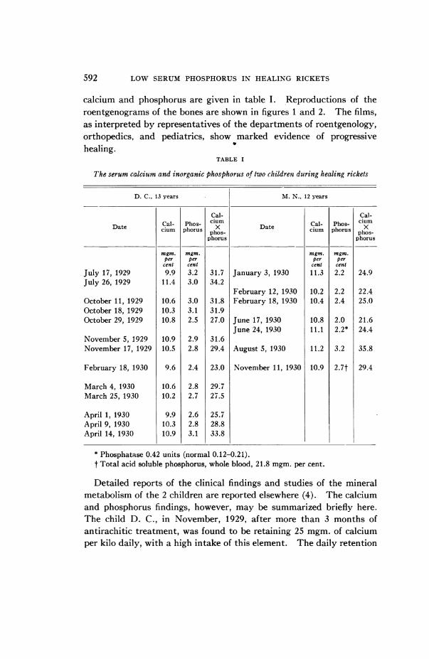
calcium and phosphorus are given in table I. Reproductions of theroentgenograms of the bones are shown in figures 1 and 2. The films,as interpreted by representatives of the departments of roentgenology,orthopedics, and pediatrics, show marked evidence of progressivehealing.
TABLE I
The serum calcium and inorganic phosphorus of two childrent during healing rickets
D. C., 13 years M. N., 12 years
Cal- Cal-
Date cal- Phos- x Date Cal- Phos- ciXmcium phorus phos- cium phorus phos-phorus phorus
mgm. mgm. mgm. mgm.per per per per
cent cent cent centJuly 17, 1929 9.9 3.2 31.7 January 3, 1930 11.3 2.2 24.9July 26, 1929 11.4 3.0 34.2
February 12, 1930 10.2 2.2 22.4October 11, 1929 10.6 3.0 31.8 February 18, 1930 10.4 2.4 25.0October 18, 1929 10.3 3.1 31.9October 29, 1929 10.8 2.5 27.0 June 17, 1930 10.8 2.0 21.6
June 24, 1930 11.1 2.2* 24.4November 5, 1929 10.9 2.9 31.6November 17, 1929 10.5 2.8 29.4 August 5, 1930 11.2 3.2 35.8
February 18, 1930 9.6 2.4 23.0 November 11, 1930 10.9 2.7t 29.4
March 4, 1930 10.6 2.8 29.7March 25, 1930 10.2 2.7 27.5
April 1, 1930 9.9 2.6 25.7April 9, 1930 10.3 2.8 28.8April 14, 1930 10.9 3.1 33.8
* Phosphatase 0.42 units (normal 0.12-0.21).t Total acid soluble phosphorus, whole blood, 21.8 mgm. per cent.
Detailed reports of the clinical findings and studies of the mineralmetabolism of the 2 children are reported elsewhere (4). The calciumand phosphorus findings, however, may be summarized briefly here.The child D. C., in November, 1929, after more than 3 months ofantirachitic treatment, was found to be retaining 25 mgm. of calciumper kilo daily, with a high intake of this element. The daily retention
592

GENEVIEVE STEARNSAND JULIAN D. BOYD
of phosphorus was still better, 30 mgm. per kilo. In March, 1930,after the continuation of the same treatment, the daily retentions ofcalcium and phosphorus were 30 and 21 mgm. per kilo respectively.The addition of 60 drops of viosterol daily increased these retentions
_I __ __c
FIG. 1. ROENTGENOGRAMSOF THE RIST OF D. C.a was taken July 24, 1929; b was taken October 4, 1929; c was taken March
31, 1930. During this time the child received antirachitic treatment.Metabolic observations indicated adequate retention of calcium and phos-phorus, yet the serum inorganic phosphorus was consistently low (2.5 to3 mgm. per 100 cc.) throughout the entire period.
to 39 and 29 mgm. per kilo respectively. The child M. N. had beenreceiving antirachitic treatment for about 5 weeks previous to thefirst study. The daily retentions of calcium and phosphorus firstobserved were 24 and 15 mgm. per kilo. Four months later, with no
change in diet, but with sunlight treatment in addition to the cod liveroil for one month previous to the observation period, the daily calciumretention had increased to 30 mgm. per kilo, the phosphorus retentionremaining at the same level as in the previous study. The retentionsof calcium and phosphorus observed in both children were thus wellabove the average normal retention as given by Sherman (5), and may
be considered adequate for the healing of rickets. Serum analyses forcalcium and inorganic phosphorus were made at intervals during theperiods of study and are reported in table I. Of the other bloodanalyses, the values obtained for carbon dioxide capacity, for non-
protein nitrogen and for chloride were always within normal limits.Determinations of pH were not made.
1 The viosterol used in these studies was supplied by Mlead Johnson andCompany.
593

594 LOWSERUMPHOSPHORUSIN HEALING RICKETS
0 cd
_ ~~~~~z0,
....C.
CL
|~~~~- Q -t.o
I_
O 4.:;:
Q 0
_>EE 9Ca ~~~~~~~~~w b.c.W~~~~~~~~~~~~~~~~-

GENEVIEVE STEARNSAND JULIAN D. BOYD
The daily retentions of calcium and phosphorus observed in eachchild during these studies were amply sufficient to provide the mineralsnecessary for calcification of bone. It is evident from the roent-genograms that such calcification took place. Nevertheless the seruminorganic phosphorus of each child remained approximately at thesame level, fluctuating between 2 and 3 mgm. per cent. The existenceof such values in cases of healing rickets is noteworthy because it hasbeen generally accepted heretofore that serum phosphorus values aslow as these are not compatible with healing rickets, but are indicativeof rachitic activity. Almost as striking is the fact that in the childD. C. the rickets was active during the late summer, and that despitethe low serum inorganic phosphorus, the period of marked healingoccurred during the late winter-a reversal of the usual seasonalactivity and healing of rickets.
A careful review of the literature has disclosed but two other reportsof healing rickets concomitant with low serum inorganic phosphorus,and in one of these reports the low phosphorus was but a transitoryfinding. Howland and Kramer (6) in their original series of obser-vations of serum calcium and inorganic phosphorus in infantile ricketslisted 2 children with "unmistakable evidence of healing" when theserum inorganic phosphorus values were 3.2 mgm. per cent. Theauthors made no special comment on these cases but concluded that aserum inorganic phosphorus of 3 mgm. per cent or less was indicativeof active rickets. Gargill and his collaborators (7) have recentlyreported a case of osteomalacia, or adult rickets, in a 38 year oldwoman, wherein definite increase in density of bone was observedduring a period of ten months. The serum inorganic phosphorus ofthe patient remained consistently low during the entire study, varyingfrom 2.16 to 2.64 mgm. per cent. Though this fact occasioned nocomment by the authors, we believe it to be of significance.
On the other hand, Hess, Weinstock, Rivkin, and Gross (8) haveobserved active rickets in infants whose serum calcium and inorganicphosphorus levels were quite normal. By proper manipulation of thediet, these authors were able to increase the serum inorganic phos-phorus of rachitic rats to normal values, and yet delay markedly thehealing of the rachitic lesions. These results were interpreted asindicative of the fact that both local and systemic factors were involvedin the production of rickets.
595

LOWSERUMPHOSPHORUSIN HEALING RICKETS
The occurrence of calcification of bone despite a low serum inorganicphosphorus is most readily explained by considering that the depositmight consist almost wholly of calcium carbonate, instead of thenormal calcium phosphate-carbonate complex. This explanation,however, is not satisfactory in the cases studied by us, in view of thelarge amounts of phosphorus retained by each child. The amount ofphosphorus utilized in the production of tissues other than bone maybe estimated roughly from the retention of nitrogen, the ratio ofN : P in the soft tissues being approximately 17: 1 (9). The maximumretention of nitrogen observed in either child, 87 mgm. per kilo, canaccount for only about 5 mgm. per kilo of the retained phosphorus. Adaily storage of from 10 to 24 mgm. of phosphorus per kilo bodyweight in tissues other than bone seems even more difficult to reconcilewith the maintenance of the low serum inorganic phosphorus thandoes its storage in bone.
If it may be granted that at least a part of the deposit must consistof calcium phosphate, the occurrence of this marked deposition ofminerals at the metaphyses, coincident with the maintenance of a lowserum inorganic phosphorus, must have a direct bearing upon thetheories of calcification of bone. Any theory, to be adequate, mustexplain the calcification which occurs under unusual conditions such asthose described here.
The modern theories of normal calcification of bone may be dividedinto 2 groups: those concerned primarily with conditions existing inserum, as related to the probable precipitation of insoluble calciumsalts in bone; and those concerned only with conditions at the site ofbone formation. In the first group, Shear and Kramer (10) havebrought forward evidence to prove that the calcium phosphatecompound of blood serum is the dicalcium, instead of the tricalciumsalt. According to these authors, the serum of children is normallysaturated, or nearly saturated, with dicalcium phosphate. Calcifica-tion of bone was obtained in vitro only when the bone was immersed insolutions containing sufficient calcium and phosphate ions so that theion product (Ca") X (HP04=) was greater than 2.5 X 10⁻⁶ an ionproduct corresponding to an empirical product of calcium X phos-phorus = 35. In serum, the calcium is not wholly ionized; thereforethe empirical product of serum calcium and phosphorus represents a
596

GENEVIEVE STEARNSAND JULIAN D. BOYD
greater value than the ion product (Ca") X (HPO4=). Becausecalcification did not take place in vitro when the empirical productcalcium X phosphorus was less than 35, these authors postulated thatcalcification in vivo would not occur until the empirical productreached a value of 35 or greater.
An examination of the empirical products calcium X phosphorus ofthe serum of the child D. C. (table I) reveals but 2 values approxi-mating 35 and one of these values was obtained when the rachiticprocess was most active. A product of 36 was obtained with thesecond child M. N., during the period of greatest healing. Othervalues for this product obtained during healing were as low as 22.According to the calculations of Shear and Kramer, the serums of bothchildren were undersaturated with dicalcium phosphate to a degreethat would preclude calcification entirely. Yet calcification occurredin both cases, as is shown in figures 1 and 2.
In 1925, Holt, La Mer, and Chown (11) studied the serum calciumand inorganic phosphorus relative to the solubility product of tri-calcium phosphate, concluding that normal serum is supersaturatedwith tricalcium phosphate to the extent of about 200 per cent, and thateven in active rickets the serum is still supersaturated with tricalciumphosphate. Holt (12) suggested that calcification is not entirelyarrested in active rickets, but that it is so retarded that bone growthexceeds it in activity, thus producing the inadequately calcified bonecharacteristic of rickets.
It might be argued, from this theory, that in these cases of laterickets, growth of bone was slow enough so that even though calci-fication was much decreased, it was still carried on sufficiently to makea noticeable difference in the density of the bone during the periodstudied. Such an explanation is unsatisfactory in many respects.The first roentgenograms are typical of active rickets. The child D. C.grew an inch in height during the 9 months observation period, so thegrowth of bone was not inordinately slow. The retentions of calciumand phosphorus observed in each child were of the order of healingrickets; the calcification which occurred was apparently of the typecharacteristic of healing, not of active rickets. In every respectexcepting the low serum inorganic phosphorus, the picture resemblesthe healing observed in infantile rickets when the serum phosphorus
39
597

LOWSERUMPHOSPHORUSIN HEALING RICKETS
has increased to normal, so that the idea of depressed calcificationdoes not seem applicable to these cases.
Our knowledge of the chemical composition of the minerals of bonehas been much clarified by the study of x-ray spectrograms of bone.Taylor and Sheard (13) and, more recently, Roseberry, Hastings, andMorse (14) have shown that bone has a crystal structure fundamentallythe same as that of other members of the apatite series. The latterauthors give the probable composition of bone as CaCO3: nCa3(PO4)2,where n is not less than 2 nor greater than 3. No evidence of thepresence of CaHPO4or CaCO3 in bone was found by either group ofinvestigators, although Roseberry and his colleagues studied newly-deposited, as well as old bone. In studies of the solubility relationshipsof bone salts, Taylor and Sheard (13) recommend that the solubilityproduct of podolite, 3Ca3(PO4)2: CaCO3, be used, rather than that ofbrushite, CaHPO4 2H20, or of tricalcium phosphate. We are notaware of any published studies comparing the relationships of serumcalcium and inorganic phosphorus with the solubility product ofpodolite.
A true concept of the physicochemical conditions at the point ofbone formation cannot be gained from examination of the serum alone.The concentration of calcium and phosphorus in other body fluidsdiffers from that of the serum (15), the differences being largelyaccounted for by the decreased content of protein, which, accordingto the Donnan equilibrium, necessitates a rearrangement of electro-lytes. Normally the amounts of both calcium and inorganic phos-phorus are lower in other body fluids than in serum. In the patientsherein reported, it wa,s thought possible that sufficient alterations in thephysicochemical system of the body fluids might have occurred tocompensate, in some measure at least, for the low serum inorganicphosphorus. Analysis of fluids at the site of bone formation was, ofcourse, impossible, but it was felt that any marked disturbance ofequilibrium would be reflected in all of the body fluids. With thispoint in mind, simultaneous samples of blood and cerebrospinal fluidof M. N. were taken during the period of greatest healing of the rachiticlesions. The analyses of protein, calcium, and inorganic phosphorus,together with those of a nonrachitic child of the same age, are given intable II. The serum protein of the rachitic child is somewhat de-
598

GENEVIEVE STEARNSAND JULIAN D. BOYD
TABLE II
Calcium, inorganic phosphorus, and protein in serum and cerebrospinal fluid of a
rachitic child and of a non-rachitic child of the same age
Fluid Calcium Phosphorus Protein
mgm. per cent mgm. per cent grams per cent
M. N., rachitic, June 17, 1930
Serum 10.8 2.0 6.6Cerebrospinal fluid 6.0 0.6 .011Ratio Serum: Cerebrospinal fluid 1.8 3.32 600.
K. K., non-rachitic, June 17, 1930
Serum 11.2 4.4 8.1Cerebrospinal fluid 6.4 1.4 .011Ratio Serum: Cerebrospinal fluid 1.75 3.14 735.
creased, the serum inorganic phosphorus markedly so. The phos-phorus of the spinal fluid is very low, but the ratio of serum phos-phorus: spinal fluid phosphorus is practically the same in the rachiticas in the nonrachitic child. Reasoning by analogy, it appears that theinorganic phosphorus content of the fluids at the metaphyses must alsobe abnormally low.
Two of the modern theories of bone calcification are concernedchiefly with the conditions in the ossifying cartilage. According to thetheory of Freudenberg and Gyorgy (16), calcification takes placethrough the formation of intermediary compounds with protein, acalcium-protein compoundc being formed first, which reacts withphosphate and carbonate ions to form a second complex. This lattercompound breaks down slowly, depositing calcium phosphate andcarbonate, and liberating the protein to combine with more calciumand begin the cycle again. The rate of formation of these intermediarycomplexes would, of course, vary with the amount of available calciumand phosphate ions; therefore this theory offers no explanation of thephenomenon of healing without coincident increase in serum phosphate.
Robison and his collaborators (17) discovered the presence inossifying cartilage of an enzyme capable of liydrolyzing certainphosphate esters. From their experimental findings, these investi-gators suggested that through the action of this enzyme, the concen-
599

LOWSERUMPHOSPHORUSIN HEALING RICKETS
tration of phosphate ion at the point of calcification could be increasedto such an extent that the solubility product of calcium phosphatewould be exceeded and precipitation take place. These authors alsodemonstrated that in rickets of rats the amount of bone phosphatase isnot decreased but is probably increased. The failure of calcificationin rickets, therefore, is probably not due to lack of enzyme, but tosome other factor, as a deficient amount of substrate.
Phosphate esters which can be hydrolyzed by the bone phosphataseare found in the red corpuscles, leucocytes, and to a slight extent inthe blood plasma (18). The amounts of these esters in other bodyfluids are not known. Small amounts of phosphatase having thesame properties as bone phosphatase and presumably identical with itare found in plasma (19). Kay (20) has recently published datashowing that in certain diseases of man wherein there is destruction ofbone, or lack of deposition of bone salts, the phosphatase content ofplasma is markedly increased. In 10 cases of infantile rickets,reported by him, the plasma phosphatase averaged 0.95 units (thenormal for this age group is 0.17-0.34 units); in one case of ado-lescent rickets the very high value of 2.4 units was observed. Theplasma phosphatase decreases when the child is given antirachiticdietary treatment (21). A study of the phosphatase content of theblood of our patients seemed desirable. Through the courtesy of Dr.Kay, an analysis of the phosphatase content of the plasma of M. N.was obtained. The child had been receiving antirachitic treatmentfor about 6 months and evidences of healing were very definite. Thevalue found, 0.42 units, is definitely above normal (0.12-0.20 units,normal adult range) but far below the values noted in Kay's cases ofuntreated rickets. If the amount of plasma phosphatase is indicativeof the amount available in bone, there should be sufficient enzymepresent for normal calcification.
In order to account for the calcification observed in these 2 childrenwith late rickets according to Robison's theory, it is necessary topostulate the hydrolysis of an unusually large amount of phosphateester to provide sufficient phosphate ion at the metaphyses for theprecipitation of calcium phosphate. Such an increased hydrolysis iswithin the realms of possibility, although the data obtained areinsufficient either to support or to deny such a conclusion. A single
600

GENEVIEVE STEARNSAND JULIAN D. BOYD
determination of total acid-soluble phosphorus of whole blood of M. N.indicated that the total ester phosphorus was within normal limits.Whether the fraction of phosphate ester which can be hydrolyzed bybone enzyme is normal or below normal has not been determined. Itis hoped that these studies may be continued.
SUMMARY
The clinical healing of late rickets coincident with a persistently lowlevel of serum inorganic phosphorus has been observed in 2 children.
The retention of calcium and phosphorus observed in each child wasconsidered amply sufficient for the building of bone.
The data herein reported do not offer evidence definitely substanti-ating any of the prevalent theories of bone calcification; moreover, it isdifficult to reconcile certain of these theories with the findings.
BIBLIOGRAPHY
1. Kramer, B., and Tisdall, F. F., J. Biol. Chem., 1921, xlvii, 475. ASimple Technique for the Determination of Calcium and Magnesiumin Small Amounts of Serum.
2. Fiske, C. H., and Subbarow, Y., J. Biol. Chem., 1925, lxvi, 375. TheColorimetric Determination of Phosphorus.
3. Hawk, P. B., and Bergeim, O., Practical Physiological Chemistry.Philadelphia, Blakiston's, 1926, 9th ed., p. 398.
4. Stearns, G., Oelke, M. J., and Boyd, J. D., Am. J. Dis. Child., 1931,xlii, 88. A Study of Mineral Metabolism in Late Rickets.
5. Sherman, H. C., and Hawley, E., J. Biol. Chem., 1922, liii, 375. Cal-cium and Phosphorus Metabolism in Childhood.
6. Howland, J., and Kramer, B., Am. J. Dis. Child., 1921, xxii, 105.Calcium and Phosphorus in the Serum in Relation to Rickets.
7. Gargill, S. L., Gilligan, D. R., and Blumgart, H. L., Arch. Int. Med.,1930, xlv, 879. Metabolism and Treatment of Osteomalacia.
8. Hess, A. F., Weinstock, M., Rivkin, H., and Gross, J., Proc. Soc. Exp.Biol. and Med., 1929-30, xxvii, 140. Observations Suggesting aLocal Factor in Pathogenesis and Healing of Rickets.
J. Biol. Chem., 1930, lxxxvii, 37. The Lack of Relationship be-tween the Development and Cure of Rickets and the InorganicPhosphorus Concentration of the Blood.
9. Schabad, J. A., Arch. f. Kinderheilk., 1910, lii, 68. Zur Bedeutungdes Kalkes in der Pathologie der Rachitis.
10. Shear, M. J., and Kramer, B., J. Biol. Chem., 1928, lxxix, 125. Com-position of Bone. III. Physicochemical Mechanism.
601

LOWSERUMPHOSPHORUSIN HEALING RICKETS
11. Holt, L. E., Jr., La Mer, V. K., and Chown, H. B., J. Biol. Chem., 1925,lxiv, 509, 567. Studies in Calcification. I. The Solubility Productof Secondary and Tertiary Calcium Phosphate under Various Con-ditions. II. Delayed Equilibrium between the Calcium Phosphatesand its Biological Significance.
12. Holt, L. E., Jr., J. Biol. Chem., 1925, lxiv, 579. Studies in Calcification.III. A Quantitative Study of the Equilibria Concerned with theCalcification of Bone.
13. Taylor, N. WV., and Sheard, C., J. Biol. Chem., 1929, lxxxi, 479. Micro-scopic and X-Ray Investigations on the Calcification of Tissue.
14. Roseberry, H. H., Hastings, A. B., and Morse, J. K., J. Biol. Chem.,1931, xc, 395. X-Ray Analysis of Bones and Teeth.
15. Arnold, R. M., and Mendel, L. B., J. Biol. Chem., 1927, lxxii, 189.Interrelationships between the Chemical Composition of the Bloodand the Lymph of the Dog.
Merritt, H. H., and Bauer, W., J. Biol. Chem., 1931, xc, 215. TheEquilibrium between Cerebrospinal Fluid and Blood Plasma. III.The Distribution of Calcium and Phosphorus between Cerebro-spinal Fluid and Blood Serum.
16. Freudenberg, E., and Gy6rgy, P., Biochem. Ztschr., 1920, cx, 299; 1921,cxv, 96; 1921, cxviii, 50; 1921, cxxi, 131, 142; 1921, cxxiv, 299.
UYber Kalkbindung durch Tierische Gewebe. I-VI.Gyorgy, P., Jahrb. f. Kinderheilk., 1923, cii, 145. (Uber Rachitis und
Tetanie.17. Robison, R., Biochem. J., 1923, xvii, 286. The Possible Significance of
Hexosephosphoric Esters in Ossification.Robison, R., and Soames, K. M., Biochem. J., 1924, xviii, 740. The
Phosphoric Esterase of Ossifying Cartilage.18. Kay, H. D., and Robison, R., Biochem. J., 1924, xviii, 755. The
Action of the Bone Enzyme on the Organic Phosphorus Compoundsin Blood.
Kay, H. D., Brit. J. Exp. Path., 1926, vii, 177. The Function of aPhosphatase in Bone Formation.
Kay, H. D., and Byrom, F. B., Brit. J. Exp. Path., 1927, viii, 240.Blood Phosphorus in Health and Disease. I. The Distribution ofPhosphorus in Human Blood in Health.
19. Martland, M., Hansman, F. S., and Robison, R., Biochem. J., 1924,xviii, 1152. The Phosphoric Esterase of Blood.
20. Kay, H. D., J. Biol. Chem., 1930, lxxxix, 249. Plasma Phosphatase.II. The Enzyme in Disease, Particularly in Bone Disease.
21. Kay, H. D., Personal Communication.
602