department of biological anthropology and anatomy date:
TRANSCRIPT

i
v
The Bent Hip and Bent Knee Gait and its Possible Role in the
Evolution of Modern Human Bipedalism
by
Megan Pruette Wilson
Department of Biological Anthropology and Anatomy Duke University
Date:_______________________ Approved:
___________________________
Daniel Schmitt, Supervisor
___________________________ Rich Kay
___________________________
Steve Churchill
___________________________ Ken Glander
___________________________
Ershela Sims
Thesis submitted in partial fulfillment of the requirements for the degree of Master of Science in the Department of Biological
Anthropology and Anatomy in the Graduate School of
Duke University
2010

i
v
ABSTRACT
The Bent Hip and Bent Knee Gait and its Possible Role in the
Evoluion of Modern Human Bipedalism
by
Megan Pruette Wilson
Department of Biological Anthropology and Anatomy Duke University
Date:_______________________ Approved:
___________________________
Daniel Schmitt, Supervisor
___________________________ Rich Kay
___________________________
Steve Churchill
___________________________ Ken Glander
___________________________
Ershela Sims
An abstract of a thesis submitted in partial fulfillment of the requirements for the degree of Master of
Science in the Department of Biological Anthropology and Anatomy in
the Graduate School of Duke University
2010

Copyright by Megan Pruette Wilson
2010

iv
Abstract The relatively stiff gait of modern humans minimizes the muscular work done to
move the lower limbs and the center of mass. Nonhuman primates, and perhaps our
earliest ancestors, use a form of bipedalism in which the hip and knee are held in a
flexed position. This thesis follows up on other studies examining loading and energetic
costs of these compliant walking gaits by examining the effects of increased hip and
knee flexion on kinetic, kinematic, and energy exchange variables. The bipedal gait of
twelve human subjects using normal and bent hip and bent knee gait were compared.
The subjects walked along force plates embedded in the ground while 3D kinematic data
was simultaneously gathered. The data was then processed using EvaRT, Orthotrak,
and Matlab to evaluate the variables used. During the bent hip and bent knee bipedal
locomotion subjects demonstrated lower peak vertical and parallel ground reaction
forces, much higher ankle flexion, less hip extension, and less energy recovery during a
full stride. These data provide novel insight into the nature and costs of locomotion in
bipedal primates and the earliest human ancestors.

v
Contents
Abstract ......................................................................................................................................... iv
List of Tables................................................................................................................................. vi
List of Figures .............................................................................................................................. vii
Acknowledgements ...................................................................................................................viii
1. Introduction ................................................................................................................................1
1.1 Experimental Proposal .....................................................................................................1
1.2 Expectations .......................................................................................................................9
2. Materials and Methods ...........................................................................................................11
3. Results .......................................................................................................................................15
3.1 Kinetic and Kinematic Data ...........................................................................................15
3.2 Energy Exchange .............................................................................................................21
4. Discussion .................................................................................................................................23
4.1 Kinetic and Kinematic Data ...........................................................................................23
4.2 Energy Exchange .............................................................................................................33
4.3 A Broader Perspective of the Evolution of Bipedalism .............................................33
4.4 Conclusions ......................................................................................................................36
References .....................................................................................................................................39
vi
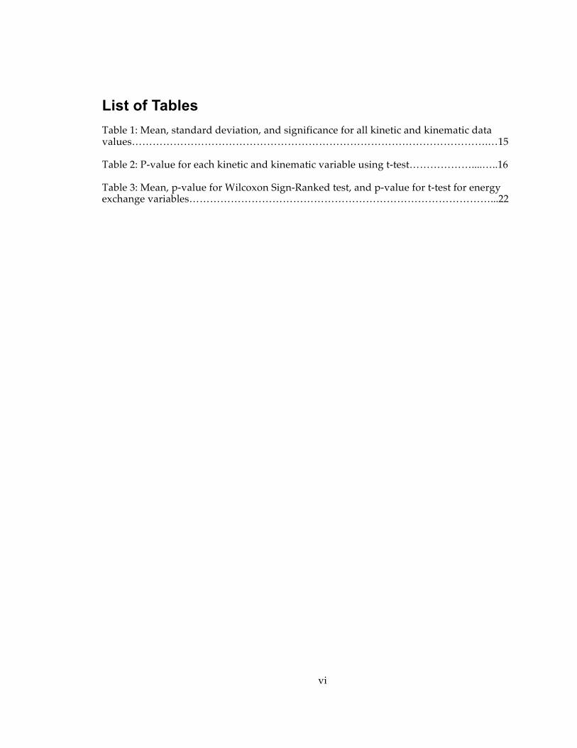
List of Tables Table 1: Mean, standard deviation, and significance for all kinetic and kinematic data values………………………………………………………………………………………….…15 Table 2: P-value for each kinetic and kinematic variable using t-test………………....…..16
Table 3: Mean, p-value for Wilcoxon Sign-Ranked test, and p-value for t-test for energy exchange variables……………………………………………………………………………...22

vii
List of Figures
Figure 1: A schematic representation of the inverted pendulum model of human walking and its effects on the potential energy (PE) and kinetic energy (KE)………….…2 Figure 2: Normal and bent hip and bent knee peak forces…………………………………18
Figure 3: Toe clearance over substrate………………………………………………………..19
Figure 4: Ankle flexion in normal and bent hip and bent knee trials……..…………...…..20
Figure 5: Hip flexion and extension…………………………………………………………..21
Figure 6: Change in vertical ground reaction force curve…………………………………..24
Figure 7: Effect of bipedal style on parallel forces………………………………...….……..26
Figure 8: Demonstration of shear force on tibiotalar joint………………………………….31
Figure 9: Summary of difference in normal and bent hip and bent knee style gaits….…37

viii
Acknowledgements I thank Roshna Wunderlich, Tracy Kivell, Laura Johnson, Michael Malinzak, Kari
Allen, Julie Horvath, Charlotte Miller, and Nicole Griffin for helpful discussions,
comments, and advise in preparation of this thesis. I also thank Robin Queen and Bryan
Gibson for assistance in data collection. I would especially like to thank Dan Schmitt
and the rest of my committee, Rich Kay, Steve Churchill, Ershela Sims, and Ken Glander,
for their guidance and helpful comments.

1
1. Introduction
1.1 Experimental Proposal
Upright, striding bipedalism distinguishes modern humans from all other extant
animals (Alexander 2004). Bipedal locomotion as practiced by modern humans involves
numerous specializations in the anatomy of the lower limbs such as long lower limbs,
adducted femora, and a stable rigid foot, which allows humans to support and balance
the weight of the upper body in an erect posture. In addition to stabilizing the body, the
highly derived anatomy of humans facilitates long strides with fully extended legs and
regulation of whole body oscillations, both of which may reduce metabolic costs of
locomotion (Cavagna and Kaneko 1977). Mapping the evolutionary pathway to
modern human bipedalism has proven challenging because of the scarcity of the fossil
record and the difficulty inherent in interpreting the life of the animal from a few
fragmented fossils. Laboratory-based studies on the biomechanics of bipedalism in
modern humans such as those conducted by Li and Colleagues (1996), Schmitt (2003),
and the one presented here can shine light on some of the factors that should be
considered when reconstructing the locomotor habits of an extinct animal and give clues
about the functional anatomy of modern human bipedalism.
The central questions in this project are, how has the mechanics and functional
anatomy of bipedal locomotion changed in the more than six million years over which it
evolved and how does this connection between mechanics and anatomy inform our
interpretation of bipedal locomotion in early hominins? In that context, it is worth
beginning by describing the mechanics of bipedalism in modern humans.

2
Figure 1: A schematic representation of the inverted pendulum model of human walking and its effects on the potential energy (PE) and kinetic energy (KE). In all section a = heel-strike, b = midstance, c = cotralateral heel-strike. A. The mass of the person is modeled as a single point concentrated around the natural center of mass (COM). The COM is supported by a massless stiff strut indicated by the red line running along the lower limb. As the step proceeds this mass-strut reaches its highest vertical point at midstance. B. Placing the point of contact at a single place the movement of the mass-strut system approximates an inverted pendulum. The
change in height from heel-strike to mid-stance is described as delta-h and approximates the change in the PE in the system. C. The fluctuations of the curves in
PE and KE due to change in height and forward motion. In a stiff pendular model like this one PE is highest when KE is lowest.
Human beings walk on stiff, straight legs (Inman 1944). As a result, as modern
humans walk, their center of mass rises and falls throughout the stride (figure 1a). At
heel strike the center of mass is at its lowest point. Then the body vaults over the
relatively stiff stance leg and the center of mass reaches its zenith during midstance

3
(Cavagna et al. 1977 ; Geyer et al. 2006; Lee and Farley 1998). The energetics of the
periodic fluctuation of the center of mass can be modeled with an inverted pendulum
(Cavagna et al. 1976). First described by Cavagna and colleagues in 1976, the pattern
involves storage and release of potential and kinetic energy in the manner indicated in
figure 1b,c. When the center of mass rises from heel strike to single support, kinetic
energy is transferred into gravitational potential energy. When the center of mass is
highest the available gravitational potential energy is at its maximum and available
kinetic energy is at its minimum. Once the stride goes through the highest point the
gravitational potential energy is converted into kinetic energy that can be used to drive
the center of mass forward and back up again because the contrateral leg remains stiff
and the horizontal kinetic energy drives the center of mass forward and up (Cavagna et
al. 1977 ; Cavagna et al. 1976). Without the conversion of gravitation potential energy
into kinetic energy muscles would have to do all the work of redirecting the center of
mass from generally downward to generally upward at the step-to-step transition,
although the mechanics of that redirection also induces some costs as well so that the
energy savings of pendular exchange are slightly reduced by the need to redirect the
center of mass at all (Adamczyk et al. 2006; Cavagna et al. 1976; Donelan et al. 2002;
Ruina et al. 2005). Thus, the ability to exchange kinetic and gravitational potential
energy can reduce the work required by muscles and could potentially lower metabolic
costs of locomotion. Although direct measures of energy exchange and metabolic costs
are limited, this relationship has been supported (Griffin et al. 2004a; Ortega and Farley
2005). Humans have been show to recover close to 70% of the potential energy as kinetic
energy when they are walking at moderate speeds (Cavagna et al. 1977 ; Cavagna et al.
1976; Griffin et al. 2004a), which means less muscular effort is required from the limbs to
move the center of mass forward. At slow and fast speeds that energy recovery is

4
reduced but never below 50%. The high level of exchange at moderate walking speeds
is only achieved once a child reaches 10 years old (Schepens et al. 2004) because of
required changes in skeletal mass and length as well as neuromuscular control.
The potential and kinetic energy of the center of mass is determined from ground
reaction forces which are a direct measure of acceleration of the COM which in turn can
be used to calculate COM velocity and displacemement (Cavagna et al. 1977 ). Ground
reaction forces reflect the style of locomotion (Alexander and Jayes 1978). The pendular
locomotion of modern humans yields a distinct ground reaction force curve different
from that of other primates (Kimura et al. 1979; Li et al. 1996; Schmitt 2003) that has two
peaks greater than the mass of the body separated by a shallow valley (Alexander and
Jayes 1978).
The use of the inverted pendulum style of locomotion is seen in many other
animals including dogs, horses, and large birds (Cavagna et al. 1977 ; Griffin et al.
2004a), but to a much lesser degree in our closest relatives, the primates (Kimura 1996;
Kimura et al. 1979; Schmitt 2003). Many extant primates, instead, use compliant bipedal
or quadrupedal locomotion, meaning they use a deeply yielding (large angular change
from touchdown to midsupport (Schmitt 1999)) elbow or hip and knee while moving
(Kimura 1996; Schmitt 1999; Vereecke et al. 2006). Yielding at the hip and knee allows
for a smoother gait with no up to down transitions but with less exchange of energy
(Alexander and Jayes 1978; Kimura 1996; Schmitt 2003). Therefore, compliant
locomotion has little to no exchange of kinetic and gravitational potential energy and
does not benefit from its potential efficiency.1 The shape of the ground reaction force
1 This is less clear for quadrupeds see Griffin TM, Wicker SJ, Hoyt DF, Garcia S, and Kram R. 2004b. Determinants of Walk-trot Transition in Horses. Durham: Duke University. 30 p. for discussion of pendular mechanics in dogs and see alternative perspective for Lemur catta in O'Neill M. 2009. Musculoskeletal design predicts locomotor cost: a test of the force production model across speed and gait in nonhuman primate.

5
curve during compliant locomotion (high values of joint yield) is substantially different
from the two-peaked curve seen in pendular locomotion (Kimura 1996; Li et al. 1996;
Schmitt 2003). When moving compliantly, animals have a ground reaction force curve
that is fairly flat and only barely reaches or just surpasses the body mass of the
individual (Alexander and Jayes 1978; Kimura 1996; Li et al. 1996; Schmitt 2003).
Although compliant locomotion during bipedalism in most examples and
quadrupedalism in at least Eulemur fulvus severely limits exchange of kinetic and
gravitational potential energy, there are thought to be benefits to this type of locomotion.
In arboreal primates, it has been argued that a compliant gait may lead to smoother,
easier movement on tree branches without bouncing (Schmitt 1999). An additional
potential benefit of the flat ground reaction force curve associated with compliant
locomotion is that it may reduce the transient impact force and resulting shock wave
(Schmitt 2003; Voloshin et al. 1981) that passes through the joints of the body during
each step (Schmitt 2003; Schmitt et al. 1996). A bipedal animal using compliant
locomotion would have a reduced opportunity to take advantage of energy exchange,
but it may benefit from longer stride lengths (due to large angular excursion of a limb
landing and pushing off in an extended position and yielding during stance) and faster
walking speeds (Schmitt 2003). Lastly, although oscillations of the COM allow energy
exchange they also require redirection of the COM (Ruina et al. 2005) that represents
another cost, although on the whole the energy exchange may be worth this additional
cost and there are mechanisms such as overlapping support phases and creation of
pseudoelastic redirections that can reduce redirection costs in a pendular model
American Journal of Physical Anthropology 138:282. and O’Neill and Schmitt, submitted in which L. catta can achieve high values of energy exchange as well as very low levels.
6
(Adamczyk et al. 2006; Ruina et al. 2005). Bishop et al. (2008) argued that the stealthy
locomotion of cats may benefit somewhat from reduced redirection costs. Therefore, the
potential benefits of compliant locomotion are broad and seem to vary in different
animals that are exposed to an assortment of environmental pressures. This variation
highlights the fact that to understand potential selective pressures on animal locomotion
it is important to take the animal’s whole lifestyle into consideration. Energy efficiency
is very important to cursorial animals, but may not be the most important consideration
in animals that do not move quickly or travel great distances (Bishop et al. 2008).
Therefore, when recreating locomotor scenarios for extinct animals with few fossils for
reference an expansive perspective of their environment and evolutionary pressures is
necessary to make a reasonable hypothesis.
The fact that primates, our closest living relatives, use relatively compliant
(yielding) gaits brings to question whether modern human bipedalism went through a
compliant locomotion phase (Schmitt 2003). The fossil record does little to answer this
question. Indicators of bipedalism such as an obturator externus groove, elongated
femoral neck, and large, vertical gluteal tuberosity are seen in the femur as early as 6
million years ago in Orrorin tugenensis (Pickford et al. 2002; Richmond and Jungers 2008).
When the morphology of the proximal femur of Orrorin tugenensis is compared to the
proximal femur of great apes, modern humans, fossil Homo, and early hominins using a
cluster analysis, it falls within a distinct early hominin cluster (Richmond and Jungers
2008). This cluster is characterized by a very wide iliac blade and long femoral neck,
which creates a greater gluteal muscle moment arm (Richmond and Jungers 2008). The
unique femoral morphology and associated mechanical peculiarities of early hominins
seems to have persisted for 4 million years from 6 million years ago in Orrorin to 2
million years ago in Australopithecines (Richmond and Jungers 2008) and are associated
7
with the origins of bipedalism. A full understanding of how this suite of anatomical
features could affect locomotion is crucial to reconstructing the evolution of bipedalism.
The most complete, early example of a biped is the recently described
Ardipithecus ramidus from 4.4 million years ago (Lovejoy et al. 2009b). The anatomy of A.
ramidus shows a combination of both bipedal and arboreal characteristics (Lovejoy et al.
2009b) The short, broad iliac spine and ankle morphology indicate that this animal was
able to walk upright and propel itself forward with its feet, but the placement of the
ischial surface and abduction of the big toe suggest that A. ramidus was also somewhat
arboreal (Lovejoy et al. 2009a; Lovejoy et al. 2009c). This important fossil find was only
recently described in a peer-reviewed publication and the interpretation of its functional
anatomy has not been discussed at the level of detail applied to Australopitheucs afarensis
fossils from 3.2 million years ago. These fossils also display a combination of arboreal
and bipedal characters (Crompton et al. 2008; Lovejoy 1988; Schmitt 2003; Stern 2000;
Stern and Susman 1983 ; Susman et al. 1984 ; Ward 2002), although how these features
are interpreted varies widely.
Since its discovery in 1974, the A. afarensis fossils have spurred a debate
concerning the origins of bipedalism. Features associated with bipedalism found in the
A. afarensis fossils include short, broad iliac blades and valgus knees (Lovejoy 1988; Stern
and Susman 1983 ; Susman et al. 1984 ). Anatomical features generally associated with
an arboreal lifestyle are the long upper limbs, relatively short hindlimbs, rod-shaped
pisiform, curved phalanges, and cranially-oriented glenoid fossa (Lovejoy 1988; Stern
and Susman 1983 ; Susman et al. 1984 ). This combination of characters brings to light
the question of whether the mere presence of bipedal traits indicate full, upright,
striding bipedalism like modern humans or whether the presence of arboreal traits may
indicate a lifestyle compromised between bipedalism and arborealism with a bent hip

8
and bent knee, compliant type of locomotion (Crompton et al. 2008; Lovejoy 1988;
Schmitt 2003; Stern 2000; Stern and Susman 1983 ; Susman et al. 1984 ; Ward 2002).
Some features that may specifically indicate a bent hip and bent knee type of locomotion
include the tibiotalar joint that appears well suited to withstand large anteriorly directed
shear forces from increased ankle flexion, orientation of the iliac blade more laterally,
reorientation of the hamstrings, the diminutive size of the anterior horn of the acetabular
surface indicating a more flexed position of the femur, and a flat sacrum (Stern 2000;
Stern and Susman 1983 ; Susman et al. 1984 ). Additionally, the joints of the lower limbs
of Australopithecus afarensis are loose and poorly stabilized. For instance, the hip joint is
small with small femoral ligaments, the lateral lip of the patellar groove is not well
developed compared to modern humans, and the sacrum does not have developed
upper lateral angles for sacroiliac ligaments (Stern 2000; Stern and Susman 1983 ;
Susman et al. 1984 ). These weak joints of the lower limbs may have been adequate for a
bent hip and bent knee gait and not a stiff-legged vaulting gait because a bent hip and
bent knee gait has a reduced peak vertical and transient impact force felt at the joints
(Schmitt 2003; Susman et al. 1984 ). Although these fossils have been extensively
analyzed, bringing life to partial fossils has proven very difficult and there has been no
consensus about the locomotion of A. afarensis.
Even as more fossils are discovered in the future, recreating the bipedalism of
our ancestors will always be a challenge. Bones can provide a broad framework for
interpreting locomotor ability, but without empirical data on living animals, one can
only speculate about the specific movements and behaviors of an animal. This study
attempts to understand the implications of compliant locomotion in a bipedal ancestor
to determine if this type of locomotion is consistent with the fossil evidence. In this
study, I compare, lower limb joint flexion angles, ground reaction forces, stride
9
parameters, toe clearance, and mechanical work variables in humans walking normally
and with a bent hip and bent knee. These data provide novel insight that should be
taken into consideration when reconstructing the wide spectrum of possibilities for the
bipedalism of early hominins.
1.2 Expectations In the study, locomotor variables were compared when humans walked with an
upright, striding form of bipedalism and then with a bent hip and bent knee form of
bipedalism. Knee yield and sacral displacement (the change in the height of L5-sacral
joint during the stride) were measured to confirm that participants were in fact using a
compliant (yielding) bent hip and bent knee gait. When walking with a yielding gait it
is expected that knee yield would be higher and sacral displacement would be lower
than when walking with a normal gait. It is predicted that when walking with a bent
hip and bent knee gait subjects will show increased flexion in the ankle, knee, and hip
during both stance and swing phase. In addition, subjects will show less hip extension
because the leg is not fully extended.
Based on previously published data (Li et al. 1996; Schmitt 2003) subjects
walking with a bent hip and knee should demonstrate lower vertical ground reaction
forces as well as lower mediolateral and fore-aft ground reaction forces. This decrease in
ground reaction force is predicted to keep moments about the ankle and knee at similar
levels as they are in normal walking.
The spatiotemporal variables examined are velocity, stride length, cadence,
double support time, and single support time. Schmitt (2003) reported increased
velocity, stride length, and single support time in subjects walking with a bent hip and

10
bent knee. This study anticipates similar results. In addition, cadence should be lower
and double support time longer when walking with a bent hip and bent knee gait
because this gait is laboring and cumbersome to use. The toe clearance is expected to be
lower when walking with a bent hip and bent knee gait because subjects do not have as
much space to clear the substrate when walking with a bent hip and bent knee.
Along with the kinetic and kinematic analysis of bent hip and bent knee gait an
analysis of the mechanical work used during both types of gaits was done. Variables
considered where change in potential energy, change in kinetic energy, percent of
recovery of potential energy, and congruence of the potential and kinetic energy curves.
A yielding form of locomotion is expected to have similar changes in potential and
kinetic energy, but the curves of the two should be aligned differently. The bent hip and
bent knee form of locomotion should show more congruence between the two energy
curves and less energy recovery between the two types of energy because it moderates
the oscillation of the center of mass compared to normal walking (Alexander and Jayes
1978; Schmitt 1999).
The examination of these variables provides a full comparison between the
compliant, bent hip and bent knee gait and the modern, striding gait. This allows for
conclusions about how the bent hip and bent knee gait affect the biomechanics of
locomotion and the costs and benefits of this type of locomotion. The skeletal anatomy,
locomotor needs of the animal, and the effects the locomotion has on the biomechanics
must be taken into consideration when reconstructing the behavior of an extinct animal
as each plays a role in an animal’s behavior.
11
2. Materials and Methods Twelve subjects (5 male, 7 female) ranging in age from 18 to 50 and weight from
46.7kg to 119.8kg participated in the study. Three-dimensional kinematic and force data
were collected as each subject walked with both a normal human bipedal gait and with a
bent hip and bent knee gait. The instructions for maintaining a bent hip and bent knee
gait were to walk as smoothly as possible without bobbing the head up and down. Each
subject experimented with these instructions and finally settled on the bent hip and bent
knee walking. The speed, step length, and degree of flexion were not controlled and
subjects moved as they felt comfortable in the bent hip and bent knee gait.
The three-dimensional kinematic data was collected using a motion analysis
system (Motion Analysis Inc, Santa Rosa, CA). In preparation for data collection,
reflective markers were placed bilaterally on the following anatomical landmarks:
acromion process, lateral epicondyle of humerus, wrist, anterior superior iliac spine,
greater trochanter of femur, posterior superior iliac spine, thigh, lateral knee, shank,
lateral malleolus, lateral calcaneus, lower calcaneus, upper calcaneus, and 2nd webspace
of the foot. A marker was also placed at the superior aspect of the L5-sacral interface
and asymmetrically on the back. During the static trials, additional markers were placed
on the medial femoral condyle and the medial malleolus for identification of joint
centers during collection of static trials. These markers were removed once the static
trial was performed. Kinematic data were collected at 60 Hz.
Each subject was asked to walk along a 30-meter walkway with embedded
1200Hz AMTI force plates (Advanced Medical Technologies Inc., Watertown, MA).
First, the participants walked regularly with a speed that they comfortably use in every
day life. They were asked to walk back and forth on the walkway until ten trials could
be captured when a full stride was completed from heel strike of one foot to its next heel
12
strike. This same procedure was repeated while walking with a bent hip and bent knee
style of locomotion. Subjects chose their own walking speed in both types of trials.
EvaRT (Motion Analysis Inc, Santa Rosa CA) software was used to track the
reflective markers and condition the data. The raw data was smoothed using a 4th order,
recursive Butterworth filter with a 6Hz cutoff frequency. Three trials of normal and bent
hip bent and knee walking in which all markers were identified and the subject had
clean contact with the force plate were averaged to yield kinematic and kinetic data. The
following twenty-one variables were examined: sacral displacement (the change in
height of the L5-sacral interface marker during one full stride), knee yield (change in
knee flexion angle from heel strike to midstance), cadence, double support time, single
support time, vertical ground reaction force, mediolateral ground reaction force, fore-aft
ground reaction force, ankle moment, ankle moment arm, knee moment, velocity, stride
length, ankle flexion angle during stance phase, ankle flexion angle during swing phase,
knee flexion angle during stance phase, knee flexion angle during swing phase, hip
flexion angle during stance phase, hip flexion angle during swing phase, hip extension
angle, and toe clearance (the distance from the substrate to the second webspace of the
foot). Data such as hip flexion, knee flexion, ankle flexion and toe clearance were found
using EvaRT. Spatiotemporal variables (velocity, stride frequency, stride length, and
support time) as well as ground reaction forces and moments were computed using
OrthoTrak (Motion Analysis Inc, Santa Rosa, CA). Stride length and sacral
displacement data were normalized to subject height; force data were normalized to
subject weight.
Energetic data was computed using a Matlab 7.0 (Mathworks, Nadick, MA)
script written specifically for this purpose by Kristen Bishop. Methods are derived from
Cavagna et. al. (1976, 1977), Ahn et. al. (2004), and Bishop et. al. (2008). The change in

13
potential energy (PE) and kinetic energy (KE) from midstance to midstance for one full
stride was calculated. Midstance is when the center of mass is at its lowest point. The
potential energy was calculated from the vertical component of the displacement of the
center of mass from the formula:
PE = mgh
where m is body mass, g is gravitational acceleration (9.8 ms-2), and h is the vertical
position of the center of mass relative to its starting position. The kinetic energy was
calculated using the formula:
KE = ½ mv2
where m is body mass and v is the resultant velocity vector. The total mechanical
energy (TME) is the addition of the potential energy and kinetic energy. The percent of
energy recovery was calculated with the formula:
% Recovery = ∑PE+∑KE-∑TME x100
∑PE+∑KE
The phase relationship of the potential and kinetic energy curves is a major factor
in the amount of energy that can be exchanged. A quantity called congruity is a
calculation that allows for a quantitative comparison in the phase relationship of the
potential and kinetic energy (Ahn et al. 2004). It is calculated by multiplying the slope of
the potential energy curve by the slope of the kinetic energy curve for each value
collected (Ahn et al. 2004; Bishop et al. 2008). Therefore, congruity is positive when the
potential and kinetic energy change in the same direction and negative when they
change in opposite directions. The reported congruity value is the percent of the stride
when the congruity is positive.
Although the sampling of individuals is random and independent, the variables
were not always normally distributed and did not demonstrate equal variance. This

14
could be attributed to a relatively small sample size. For this reason, both parametric
and non-parametric statistics were used to analyze the data (Sokal and Rohlf 1995) to
provide the widest range of statistical information. Because the paired data (comparison
of normal to compliant) from each individual was treated as a random sample and did
not have influence on the paired data from another individual the Wilcoxon’s Signed-
Rank test was used to analyze the data, with a p-value of .05 used as a measure of
significance (Sokal and Rohlf 1995). The Wilcoxon’s Signed-Rank test examines values
of the normal mean minus the bent hip and bent knee mean for every variable. These
differences are then compared for all the subjects to determine if the pattern of positive
or negative differences is significant. In addition to the Wilcoxon’s Signed-Ranked test,
T-tests where performed to compare the means of each subject during bent hip and bent
knee walking and the means of the whole sample. Before statistical testing, a priori
assumptions were made about the data based on previously reported and unreported
samples. This allowed the use of one-tailed tests. Because many comparisons between
the normal and bent hip and bent knee locomotion were made and the variables
collected could not be assumed to be independent, a conservative conversion of the data
was done to decrease type 1 error (Sokal and Rohlf 1995). The Dunn-Sidák method was
used to convert the data and lower the type 1 error in each test so that the overall
probably of making the type 1 error in the whole sample will be lowered (Sokal and
Rohlf 1995). Statistical Analysis was performed using JMP 7 (version 7.0.1 for
Macintosh, SAS Institute Inc., Cary, NC).
All materials and methods were approved by the Duke University IRB; protocol
number 2240.

15
3. Results
3.1 Kinetic and Kinematic Data
Table 1: Mean, standard deviation, and significanec for all kinetic and kinematic data values. P-values were calculated using the Wilcoxon Sign-Rank testing. An asterisk
indicates significance at p-value = .05. Bolded items are significant variables once the Dunn-Sidak method adjusted the p-value. 1. Knee Yield is the difference in knee angle from heel strike to midstance. 2. Toe clearance is the distance from the 2nd
webspace to the floor. 3. Ankle angle is the angle between the lateral knee, lateral malleolus and 2nd webspace. 4. Knee angle is the angle between greater trochanter, lateral knee, and lateral malleolus. 5. Hip angle is defined as angle between ASIS,
greater trochanter, and lateral knee.
Normal Walking Bent Hip and Bent
Knee Walking p value Variable Mean St. Dev. Mean St. Dev. Sacral Displacement 0.26 0.09 0.19 0.05 0.0024* Knee Yield1 22.1 12.5 31.4 9.9 0.0081* Velocity (cm/s) 141.8 28.7 139.4 27.6 0.3188 Stride Length (cm) 139.1 17.0 146.0 24.2 0.1392 Cadence (strides/min) 121.2 11.7 113.6 11.3 0.0337* Double Support Time (s) 10.3 1.6 11.3 2.9 0.3188 Single Support Time (s) 60.4 1.6 61.8 2.4 0.0737 Vertical GRF (% Body Weight) 1.19 0.14 1.08 0.11 0.0005* Mediolateral GRF (% Body Weight) 0.06 0.02 0.06 0.02 0.3386 Fore-aft GRF (%Body Weight) 0.22 0.03 0.18 0.0 0.0044* Toe Clearance (mm)2 122.2 31.1 109.8 24.3 0.0320* Ankle Moment (N*cm) 2.4 1.8 2.1 1.6 0.1018 Ankle Moment Arm (cm) 3.3 2.8 3.5 3.0 0.4548 Knee Moment (N*cm) 3.0 3.2 3.3 2.9 0.1902 Stance Ankle Flexion Angle3 92.3 4.1 112.3 5.7 0.0002* Swing Ankle Flexion Angle3 86.9 2.7 95.7 7.5 0.0012* Stance Knee Flexion Angle4 33.7 9.2 59.0 9.2 0.000* Swing Knee Flexion Angle4 65.1 5.4 82.5 6.8 0.0002* Stance Hip Flexion Angle5 76.1 11.6 96.2 12.7 0.0002* Swing Hip Flexion Angle5 75.9 11.2 96.9 13.0 0.0002* Hip Extension Angle5 52.3 11.1 61.7 10.7 0.0024*
Table 1 summarizes the mean and standard deviation for the normal and bent
hip and bent knee gait for each variable. The p-value (test statistic from the Wilcoxon’s
Signed-Rank test) is reported as well. The values in table 1 indicated with an asterisk are

16
significant at a p-value of .05 and the bolded variables are those that maintained a
statistically significant pattern of change within the population once the Dunn-Sidák
conversion method was used with an adjusted p-value to decrease type 1 error. There is
a statistically significant difference between the normal and bent hip and bent knee trials
in sacral displacement and knee yield, although the significance of the latter is reduced
by the Dunn-Sidak adjustment (table 1).
Table 2: P-value for each kinetic and kinematic variable using t-test. The variables indicated with an asterisk are considered significantly different with a p-value of .05. The bolded variables are significantly different between the bent hip and bent knee and normal walking when the p-value is adjusted using the Dunn-Sidak conversion.
Variable p-value Sacral Displacement 0.0020* Knee Yield 0.0083* Velocity (cm/s) 0.3526 Stride Length (cm) 0.1117 Cadence (strides/min) 0.0307* Double Support Time (s) 0.1740 Single Support Time (s) 0.0449* Vertical GRF 0.0004* Mediolateral GRF 0.3350 Fore-aft GRF 0.0031* Toe Clearance (mm) 0.0303* Ankle Moment 0.0992 Ankle Moment Arm (cm) 0.3526 Knee Moment 0.3987 Stance Ankle Flexion Angle 0.0000* Swing Ankle Flexion Angle 0.0006* Stance Knee Flexion Angle 0.0000* Swing Knee Flexion Angle 0.0000* Stance Hip Flexion Angle 0.0001* Swing Hip Flexion Angle 0.0001* Hip Extension Angle 0.0109*
Table 2 reports the test statistic for each variable for the T-test comparing means.
Again, significant differences are indicated with an asterick and are significant at a p-
value of .05. The bolded variables are those that maintained statistically significant
17
differences in mean once the Dunn-Sidak conversion method was used to adjust the p-
value to decrease type 1 error. This data is similar to the Wilcoxon’s Sign-Ranked test
data.
There are no statistically significant differences in speed, double support, contact
time, or stride length when comparing normal and bent hip and bent knee walking. The
cadence, however, was significantly different between normal and compliant walking,
but not once the Dunn-Sidák method was used to convert the p-value. The lack of
difference in spatiotemporal parameters allowed for a more straightforward comparison
of kinematic and kinetic parameters without having to adjust for the effect of speed and
other variables.
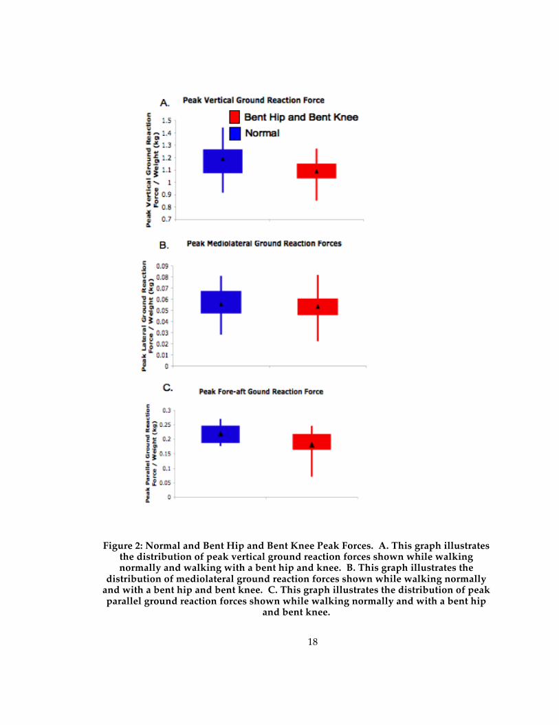
Figure 2a demonstrates that the peak vertical forces are significantly (see Table 1)
lower when walking with a bent hip and bent knee. On average, the peak vertical forces
decrease by 10% of body weight when subjects walked with a compliant gait. The fore-
aft forces are significantly different between trials when using the Wilcoxon’s Signed-
Rank test and Dunn-Sidak p-value conversion (figure 2c; Table 1). The fore-aft forces are
also considered significantly lower when the subjects walked with a bent hip and bent
knee gait compared to walking normally when using the T-tests to analyze the
difference in means. Also, 6 of the 12 individual comparisons of fore-aft forces are
significantly lower with a sample size of 3 for each individual. The difference in fore-
aft forces is on average 4% of body weight across gait types. There are no statistically
significant differences between the two samples in mediolateral forces.
18
Figure 2: Normal and Bent Hip and Bent Knee Peak Forces. A. This graph illustrates the distribution of peak vertical ground reaction forces shown while walking normally and walking with a bent hip and knee. B. This graph illustrates the
distribution of mediolateral ground reaction forces shown while walking normally and with a bent hip and bent knee. C. This graph illustrates the distribution of peak parallel ground reaction forces shown while walking normally and with a bent hip
and bent knee.

19
Figure 3 shows the difference between bent hip and bent knee and normal trials
in toe clearance. These differences are not statistically significant (Table 1, Table 2).
Along with the bent hip and bent knee trials having lower toe clearance, they also had a
much smaller standard deviation. There is no statistical difference between the ankle
moment, ankle moment arms or knee moments in normal and bent hip and bent knee
trials (Table 1, Table 2).
Figure 3: Toe clearance over substrate. This graph illustrates the distribution of maximum toe clearance for normal and bent hip and bent knee trials.
Ankle dorsiflexion angles increase significantly in swing and stance phase
during bent hip and bent knee trials compared to normal trials as seen in table 1 and
figure 4a. During stance, the ankle angle increases an average of 20 degrees and during
swing it increases an average of 8 degrees. In fact, the ankle was more flexed
throughout the whole stride when walking with a bent hip and bent knee gait (figure
4b). Knee flexion was also significantly higher in bent hip and bent knee locomotion
during swing phase (table 1). The knee angle increased by an average of 25 degrees
during stance phase and 17 degrees during swing phase.
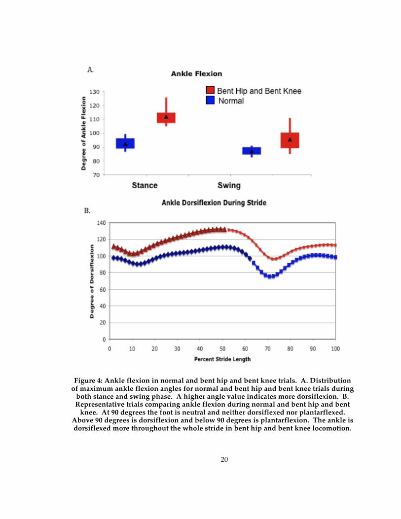
20
Figure 4: Ankle flexion in normal and bent hip and bent knee trials. A. Distribution of maximum ankle flexion angles for normal and bent hip and bent knee trials during
both stance and swing phase. A higher angle value indicates more dorsiflexion. B. Representative trials comparing ankle flexion during normal and bent hip and bent
knee. At 90 degrees the foot is neutral and neither dorsiflexed nor plantarflexed. Above 90 degrees is dorsiflexion and below 90 degrees is plantarflexion. The ankle is dorsiflexed more throughout the whole stride in bent hip and bent knee locomotion.

21
There is a statistically significant difference between hip flexion during both
stance and swing phase in the normal and bent hip and bent knee trials (table 1). Figure
5 shows the difference between the two. While walking with a bent hip and bent knee,
subjects increased their flexion by an average of 20 degrees during stance phase and 17
degrees during swing phase. Also, figure 4 shows the statistically significant (table 1)
difference in hip extension between normal and bent hip and bent knee trials. The
normal trials demonstrated much more hip extension with an average of 9 degrees more
hip extension than the bent hip and bent knee trials.
Figure 5: Hip flexion and extension. This graph shows the distribution of maximum and minimum hip angle values. Hip angles were measured as the angle between the
anterior superior iliac spine, greater trochanter, and lateral knee. A smaller angle value indicates more hip extension. A larger angle value indicates more hip flexion.
3.2 Energy Exchange The mean, test statistic for the Wilcoxon’s Signed-Rank test, and test statistic for
T-test are reported for each variable in table 3. Significant values with a p-value of .05
are indicated with an asterick while bolded values remain significant after the Dunn-
Sidak conversion of p-values. The change in kinetic energy, congruence, and percent of

22
energy recovery are significantly different in the bent hip and bent knee style of walking
compared to normal walking. The change in kinetic energy is not significantly different
between the two samples.
Table 3: Mean, p-value for the Wilcoxon Sign-Ranked test, and p-value for t-test for energy exchange variables. The significant p-values at p-value of .05 are indicated with an asterisk. The bolded values indicate significant differences between the normal and bent hip and bent knee walking once the p-value is
converted using the Dunn-Sidak method. 1. The delta KE variable represents the change in kinetic energy during one full stride from midstance to midstance. 2. The
delta PE variable represents the change in potential energy during one full stride from midstance to midstance.
Variable
Normal Walking Mean
Bent Hip and Bent Knee
Mean
Wilcoxon Sign-Ranked
p-value
T-test
p-value Delta KE1 19.747 28.212 0.0024* 0.0022* Delta PE2 22.858 17.087 0.0737 0.0870 Congruence 16.361 59.993 0.0005* 0.0000* Percent Recovery 62.701 25.566 0.0005* 0.0000*
The change in kinetic energy is an average of 8.5 Kilojoules lower in normal
walking than in bent hip and bent knee walking. The kinetic and potential energy
curves of bent hip and bent knee walking are an average of 38% more congruent, or
having similar slopes than the energy curves of normal walking. The difference in
congruence allows for the normal trials to demonstrate an average of 37% more energy
recovery during a stride than the bent hip and bent knee trials.

23
4. Discussion This study attempts to understand how walking with a bent hip and bent knee
gait affects the angles of the lower limb, stride parameters, ground reaction forces, toe
clearance, and energy exchange. Changes in these variables and the constraints of the
skeletal morphology should be considered when recreating the locomotor behavior of
extant bipedal hominins.
4.1 Kinetic and Kinematic Data The bent hip and bent knee mode of locomotion does not have an effect on most
spatiotemporal gait parameters; velocity, stride length, double support time, single
support time, and cadence all remain statistically equivalent between the two types of
locomotion. Previously, it has been reported that when subjects are asked to walk
quickly, subjects are significantly faster while using the bent hip and bent knee mode of
locomotion (Daegling and Schmitt 1999). It is probable that this study did not see these
differences in speed because we asked participants to walk as they felt comfortable and
did not control speed in any way. The bent hip and bent knee style of locomotion is not
a hindrance to spatiotemporal stride parameters such as speed and stride length.
There was a significant decrease of 10% of body weight in peak vertical ground
reaction forces when walking with a bent hip and bent knee, a value consistent with that
reported by Li et al. (1996) and Schmitt (2003). This difference indicates less of a peak
load experienced by the body during walking. Upright, striding bipedalism creates a
peak ground reaction force much larger than body weight (Alexander and Jayes 1978).
The modern human skeleton is able to sustain this peak load with heavily fortified joints
in the ankle, knee, and pelvis. The loosely stabilized joints of Austalopithecus afarensis
and other early hominins may reflect a relatively low peak load compared to full,

24
upright, striding bipedalism. Walking with a bent hip and bent knee may have been the
only mode of locomotion the joints of Australopitheucs afarensis would have been able to
handle on a daily basis, an argument articulated by Stern and Susman (1983) and
Schmitt (2003) and supported by the data here.
Figure 6: Change in vertical ground reaction force curve. The figure shows a representative vertical ground reaction force trace from subject 4 using the normal
and bent hip and bent knee style of bipedalism. The arrows indicate the change from the normal force trace to the bent hip and bent knee force trace. These are the
changes predicted in Stern et. al. (2004) with an increased change in the contact point of the foot.
Generally, a decreased peak vertical ground reaction force is attributed to a
longer support time (Schmitt 1999; Schmitt 2003), but in this case there is no difference in
support time between the two samples. Although, it should be noted that the difference
in single support phase closely approaches significance in the t-test comparing normal
and bent hip and bent knee means. In 2004, Stern and colleagues suggested an
alternative way to decrease the peak vertical ground reaction force by moving the
contact point during the stride. The authors created computer models to determine how

25
different parameters such as limb length, hip rotation, and movement of the contact
point affect the vertical ground reaction force curve. They found that when the contact
point moved during support phase the vertical ground reaction force maximum
lessened and minimum increased. In effect, the vertical ground reaction force curve lost
its two distinct peaks and became more of a plateau (Stern et al. 2004). The vertical
ground reaction force curves from the subjects walking with a bent hip and bent knee
show this pattern (Figure 6). When walking with a bent hip and bent knee, subjects may
have moved the point of pressure on their foot more substantially than they do when
walking normally to decrease the peak vertical ground reaction force without
significantly changing the support time. Moving the center of pressure on the foot
during stance phase shifts the ground reaction force vector forward and increases the
distance to the limb while walking normally (Carrier et al. 1994; Gruss 2007). This shift
is expected to have effects on the moments experienced at the joints. The long feet in
Australopithecus afarensis would have made it possible to move the point of pressure
even more during stance phase, thereby possibly lessening the peak vertical forces even
more than is seen in this study and further affecting the joint moments.

26
Figure 7: Effect of bipedal style on parallel forces. A. The distance from the point of pressure to the center of mass is highlighted in yellow. The full extension of the
lower limb during normal gait, especially during single support, creates a significant distance between the center of pressure on the foot and the center of mass. B.
During the bent hip and bent knee style of bipedalism the hip joint stays in a flexed position throughout the stride. The flexion of the lower limbs decreases the distance
from the point of pressure on the foot and the center of mass. This decreases the parallel forces experienced while using this type of locomotion.
The fore-aft (braking and propulsive) forces are also significantly different
between the bent hip and bent knee and normal trials. These forces decreased an
average of 4% of body weight when walking with a bent hip and bent knee. The
magnitude of braking and propulsive forces is influenced by the distance of the center of
mass from the point of pressure of the foot. When the point of pressure of the foot is
very close to the center of mass the force needed to brake or propulse the center of mass
is very low. Alternatively, when the point of pressure of the foot is far from the center of
mass the braking and propulsive forces required increase substantially. Lower fore-aft
forces may indicate the subject is more “balanced” around the center of mass (that is

27
there is a shorter horizontal distance between center of mass, pelvis, and point of contact
[figure 7]), and is therefore using a stable form of locomotion that may minimize braking
and propulsive forces with each step. Australopitehcus afarensis appears to have been
walking bipedally while maintaining skeletal adaptations for arboreal locomotion. Full,
striding bipedal locomotion as practiced by modern humans requires a strong,
reinforced skeleton to handle frequent large forces, while arboreal locomotion requires
loose, mobile joints to facilitate a wide range of limb motion. It is possible that the bent
hip and bent knee mode of locomotion allowed Australopithecus afarensis to move in a
stable manner, with the center of mass balanced closer to the center of pressure of the
foot while walking. This would decrease the required braking and propulsive forces.
Lower fore-aft and vertical forces may have further facilitated bipedal locomotion with a
mobile skeletal associated also with arboreal travel.
The ankle moments and ankle moment arms of the two gait styles are not
significantly different. Ankle moment arms are defined as a line from the center of
rotation (the ankle joint) perpendicular to the ground reaction force vector. The ankle
moment arm changes throughout stance phase of the stride with plantarflexion of the
foot as it moves from heel strike to toe off (Carrier et al. 1994; Gruss 2007; Stauffer et al.
1977; Stern and Susman 1983 ). Plantarflexion brings the center of rotation closer to the
point of pressure while it moves down the foot, thereby decreasing the distance from the
center of rotation to the ground reaction force vector. While walking with a bent hip
and bent knee subjects demonstrated much more dorsiflexion at the ankle. Although
this did not change the ankle moment arm significantly, the ankle moment arm average
is slightly higher when walking bipedally with a bent hip and bent knee because the
increased dorsiflexion of the ankle opposes the effect of the plantarflexion during toe off
and keeps the center of rotation further from the point of pressure. Even with larger

28
ankle moment arms, the ankle moments stay the same or a little lower in the bent hip
and bent knee trials. This is because the ankle moment is defined as the ankle moment
arm times the force vector. The lower forces seen while walking with a bent hip and
bent knee compensated for the increased ankle moment arm, so that both forms of
locomotion have similar ankle moments. Maintaining manageable ankle moments is
important, because much larger ankle moments would require stronger plantarflexors to
prevent the lower limb from collapsing. Balancing the slightly increased ankle moment
arm with the decreased ground reaction forces would be vital to maintain similar ankle
moments. It seems that the bent hip and bent knee mode of bipedal locomotion would
allow Australopithecus afarensis to balance these two variables and keep the ankle
moments at manageable levels.
Knee moments were the same when subjects walked with a normal gait and with
a bent hip and bent knee gait. As stated earlier, increasing the changes in the point of
pressure of the foot will move the force vector further from the center of mass (Carrier et
al. 1994; Gruss 2007). When the center of mass is in line with the joints, such as in erect,
striding bipedalism, this means that the moments could be increased at each joint as the
center of pressure moves away from the center of mass and increases the length of
moment arms from joints. This change in the relationship between the ground reaction
force vector and the center of rotation was diminished by the plantarflexion of the foot
during toe off. It seems that the yielding at the knee during stance phase helps to
maintain low moment arms from this joint. Although the point of pressure moves
further out during bent hip and bent knee walking, increased knee yield along with
lower vertical forces mitigates the effects of this movement and maintains the knee
moments at reasonable levels. In fact, walking with upright, striding bipedalism and the
long feet of early hominins would increase moment arms at the knee without the

29
compensation of knee yield. Larger knee moments would require more stabilization at
the joint, so preserving lower knee moments during bent hip and bent knee walking is
consistent with the skeletal anatomy of Australopithecus afarensis.
Toe clearance is the same when subjects walk with a bent hip and bent knee and
when they walk normally. Although, when walking with a bent hip and bent knee the
distribution of toe clearance values had a much lower standard deviation than when
walking normally. This may indicate that walking with a bent hip and bent knee comes
close to the boundary for the lowest attainable toe clearance while walking bipedally.
Australopithecus afarensis had particularly long toes, which may have required even more
flexion at the ankle, knee, and hip joint to maintain toe clearance. Increased flexion
would require additional muscular effort. It is possible that even with these
compensatory methods, the long, curved toes of Austalopithecus afarensis would have had
difficulty clearing the substrate and would have needed to curl their toes to gain
adequate toe clearance over the substrate. Increased lower limb flexion and possibly
curling the toes are behaviors that would be used in both bent hip and bent knee and
upright, striding bipedalism to mitigate the long, curved toes seen in early bipedal
hominins. The adjustments to maintain toe clearance while walking with a bent hip and
bent knee or while walking with an upright, striding form of bipedalism in
Austalopithecus afarensis would require more muscular effort to maintain. Although, this
is a potential energetic cost of bent hip and bent knee locomotion, it would be an
energetic cost added to upright, striding locomotion as well. It is possible that relatively
short feet and toes seen in modern humans allow for efficient clearance of the substrate
while walking bipedally.
When subjects walked with a bent hip and bent knee they showed increased
flexion at the hip and knee when compared to walking normally. This is consistent with
30
previous studies (Schmitt 2003). Subjects who walked with a bent hip and bent knee gait
also have more dorsiflexion at the ankle during both stance and swing phase than
subjects who walked normally. More muscular effort may be required to maintain the
deep dorsiflexion of the ankle throughout the stride than for normal walking. During
stance phase, more muscular effort would be needed to maintain stability in the ankle
when it is bearing body weight in a deeply flexed position as in bent hip and bent knee
walking. This problem is exacerbated for apes and early humans that had highly mobile
ankle joints (Stern and Susman 1983 ). Similarly, swing phase might also involve
increased muscular effort that would require more energy to maintain a dorsiflexed
ankle in order for the toe to clear the substrate. Maintaining increased dorsiflexion of
the ankle is an obvious area of energetic expenditure seen while walking with a bent hip
and bent knee mode of bipedal locomotion; upright, striding locomotion avoids this
energetic cost.

31
Figure 8: Demonstration of shear force on tibiotalar joint. A. Increased dorsiflexion at the ankle increases the anteriorly directed shear force at the tibiotalar joint. Changing
the direction of the Achille’s tendon force vector increases the anteriorly directed portion of the Achille’s tendon force vector. This vector is translated onto the
tibiotalar joint. B. Anteriorly tilted talar surface of the tibia in AL288-1 (from Stern and Susman, 1983). This is similar to the chimpanzee distal tibia and opposite form
the human distal tibia. Two other Australopithecus afarensis show the opposite orientation of this one and are oriented as the human distal tibia.
A deeply dorsiflexed ankle could also have an impact on the skeletal anatomy of
early hominins. Increasing dorsiflexion at the ankle increases the anteriorly directed
shear force at the tibiotalar joint (figure 8). The Achilles tendon force vector is the major
force vector on the posterior of the tibiotalar joint (Stauffer et al. 1977). This force vector
keeps the tibia from collapsing on the foot during stance phase. When the tibia is
32
directed more anteriorly and the foot is dorsiflexed as it is in bent hip and bent knee
locomotion the horizontal component of the Achilles tendon force vector increases. The
horizontal component can be translated onto the tibiotalar joint (Stauffer et al. 1977;
Stern and Susman 1983 ). Therefore, dorsiflexion of the foot increases the anteriorly
directed shear force at the tibiotalar joint. Increased shear at the tibiotalar joint would
need to be resisted by skeletal or cartilaginous features that strengthen the joint. The
anatomy of the tibiotalar joint in early hominins is not clear in this point. Stern and
Susman (1983) report an anteriorly inclined tibia in the AL288-1 (similar to that seen in
chimpanzees) but a posteriorly inclined tibia in AL333-6 and AL333-7 (similar to
humans). All three of these distal tibias belong to Australopithecus afarensis. It is possible
that the specimen that demonstrate posteriorly inclined tibias had other means of
resisting the anteriorly directed shear force such as strong anterior tibial tendons. If
early hominins walked with a bent hip and bent knee the anteriorly directed shear forces
would have been larger and there would need to be some resistant force in place to
prevent collapse of the tibia.
Lastly, subjects using the bent hip and bent knee style of locomotion
demonstrated significantly less hip extension than subjects walking normally. While
keeping the hip bent, subjects would not move their thigh as far backwards during toe
off as they do when they walk normally. With increased flexion at the knees and ankle,
decreased extension at the hip during toe off would be necessary to maintain low fore-
aft forces while walking with a bent hip and bent knee (Figure 6). Because our subjects
showed lower fore-aft forces during bent hip and knee locomotion, it is predicted that
they would also show less hip extension. Less extension at the hip joint during bent hip
and bent knee locomotion is consistent with predictions made from the Australopithecus
afarensis fossils. These fossils have a diminished anterior horn of the acetabulum, which

33
may have allowed for increased flexion and less extension at the hip joint. Decreasing
the hip extension while walking with a bent hip and bent knee keeps the center of mass
balanced over the point of pressure and further decreases the fore-aft forces.
4.2 Energy Exchange
With each step of normal, upright, striding bipedalism potential energy is
created as the center of mass lifts leading into midstance. This potential energy is then
converted into kinetic energy as the center of mass falls leading to toe off (Cavagna et al.
1976). The amount of energy recovery possible is determined by the change in potential
and kinetic energy during the stride and the phase relationship of the two curves
(Bishop et al. 2008). The change in kinetic energy was significantly different between the
two gait types, while the change in potential energy showed no difference. When these
potential and kinetic energy oscillations were compared the average congruence for bent
hip and bent knee walking was 59.9. This is significantly different than the average
congruence for upright, striding biepdalism, which was 16.3. Less congruence allows
for more energy recovery. Our normal walking trials showed an averaged percent
energy recovery of 62.7, which is consistent with previously reported results (Cavagna et
al. 1976). The bent hip and bent knee trials had an average percent energy recovery of
25.5. This is significantly lower and indicates that while using the bent hip and bent
knee style of locomotion there is little ability to recover potential energy to be used for
kinetic energy. Because the bent hip and bent knee style of locomotion cannot exchange
energy efficiently, muscular effort would be recruited to move the center of mass
forward. This is a potential area of cost in bent hip and bent knee locomotion.
4.3 A Broader Prospective of the Evolution of Bipedalism
34
The data obtained in this study about bent hip and bent knee bipedal locomotion
can be used to gain a full understanding of the evolution of the upright, striding form of
bipedalism of modern humans. Bipedalism is an inherently unstable form of
locomotion; balancing the body on two limbs instead of four provides less support and
more balance issues for the animal. The animal must obtain behavioral and skeletal
adaptations that allow it to move on two limbs without increasing their fore-aft or
mediolateral forces too much and compromising their balance. Many animals
demonstrate bipedal locomotion such as birds, kangaroos, and sometimes apes, but
none of these animals are able to maintain an erect posture and straighten the knee at
mid-stance to the degree found in humans (Alexander 2004). In birds that walk and run
bipedallly there is deep flexion at the hip and knee throughout the stride (Gatesy and
Biewener 1991). Kangaroos that bipedally hop also display a deeply flexed hip and
knee. This flexion helps to balance the center of mass on a larger, more stable base.
Most of a bipedal animal’s mass is placed anteriorly in the thorax and abdomen. A large
base assists in balancing the anterior and posterior masses on two limbs. In addition to
increased flexion at the joints, all other habitually bipedal animals have tails, which
serve to further balance the large anterior masses and the posterior mass on two limbs.
The adaptations that increase balance help to reduce the fore-aft forces. If these forces
were to become to high an animal would topple forward during single support phase,
unable to create a breaking force that would stop the large, natural movement of the
anterior mass forward and downward.
The erect, striding bipedalism of modern humans seems to break all the rules.
The limb is relatively stiff and straight throughout the stride, providing a small, unstable
base, and there is no tail to help balance the anterior forces. The anatomy of modern
humans is transformed to bring the anterior and posterior masses very close to the

35
center of mass. Instead of creating a large base support, modern humans appear instead
to bring mass closer to the center. Spinal curvature is one way this is accomplished. The
lordosis of the cervical and lumbar portions of the spine counteract the kyphosis in the
thoracic spine and help to bring the mass of the head, thorax, and abdomen more in line
with the strong, reinforced pelvis. This helps to reduce mass that is placed purely
anteriorly. In addition to bringing the anterior mass closer to the center, humans
increase their posterior mass with a large gluteal region. Humans have a realtively large
gluteus maximus that is pushed further posterior by the curvature of the sacrum
(Bramble and Lieberman 2004; Sigmon and Farlow 1986 ; Stern and Susman 1983 ). The
gluteal area that lies on the posterior surface of the pelvic bones is very muscular and
oftentimes very fatty. The additional mass on the posterior surface of the pelvic bones
form the gluteus maximus and associated fat creates more posterior mass and helps to
balance the anterior mass. Modern humans are unique in their ability to use an upright,
striding form of bipedalism because they bring the anterior mass close to the center with
spinal curvature and create posterior mass with the gluteal region.
Applying these ideas about minimizing the fore-aft forces is revealing when
considering the conception of obligate bipedalism. To maintain an erect, striding form
of bipedalism hominins would need the ability to balance the anterior and posterior
forces of the body on the pelvis. This problem would be exacerbated in early hominins
because of their large, barrel-like chests adding anterior mass. To keep the fore-aft
forces at a manageable level and to prevent toppling forward during single support,
hominins would require lordosis in the cervical and lumbar vertebrae along with a
massive, posteriorly positioned gluteal region. The sacrum of Australopithecus afarensis is
very flat (Stern and Susman 1983 ), therefore negating its ability to position mass more
posteriorly. It is possible that Australopithecus afarensis and other early bipedal hominins

36
still had massive gluteal regions, but this is difficult to determine given only fragmented
fossil evidence. If early bipedal hominis did not have significant lordosis or some way
to increase posterior mass, they would not have been able to use an erect form of
bipedalism. If this is the case, bent hip and bent knee bipedalism would be the only
option that would provide the needed stability and decrease the fore-aft forces.
Although bent hip and bent knee bipedalism has many costs associated with it, it may
be the only bipedal option available to an animal without the specialized anatomy of
modern humans.
4.4 Conclusions
The many changes seen when subjects walk with a bent hip and bent knee gait
are summarized in Figure 9. This type of locomotion lowers both the vertical and fore-
aft forces experienced. Lowering the peak vertical force is attributed not to increasing
the support time, but to increasing the changes in the point of pressure. While using
bent hip and bent knee locomotion, the subjects move the point of pressure in the foot
during stance phase slowly and fully, which allows for decreased peak pressures. Less
hip extension seen while walking with a bent hip and bent knee decreases the fore-aft
forces experienced by maintaining the center of mass balanced closely over the point of
pressure of the foot. The lower forces seen with bent hip and bent knee locomotion help
to mitigate the increased moment arms predicted with the Australopithecus afarensis
skeleton and maintain equal ankle and knee moments. The compensatory methods
required to clear the substrate would be required from both bent hip and bent knee and
normal bipedal locomotion. A potential area of large energetic costs in bent hip and
bent knee locomotion is the increased dorsiflexion throughout the stride. The loosely
stabilized joints seen in early bipedal hominins exacerbate these costs. An additional

37
area of cost in bent hip and bent knee locomotion is the inability to exchange potential
and kinetic energy efficiently.
Figure 9: Summary of differences in normal and bent hip and bent knee style gaits.
Several aspects of early hominin morphology may be interpreted as strong
indicators of arboreal locomotion such as long arms, loosely stabilized joints, and long,
curved phalanges. Whether these arboreal characteristics were actually used for
arboreal locomotion is irrelevant; they are present in the skeleton and would have affects
on the mode of terrestrial locomotion used. The bent hip and bent knee style of
bipedalism would 1.) lessen the forces on the body which is beneficial for a skeleton
with loosely stabilized joints, 2.) lessen hip extension, which is consistent with
acetabular anatomy, and 3.) increase changes in point of pressure of the foot, which

38
amplifies the benefits of decreased forces on the joints. Contrarily, the bent hip and bent
knee style of locomotion maintains increased dorsiflexion throughout the stride which
may require more muscular effort and therefore more energy to maintain while using
bent hip and bent knee locomotion. Also, the muscular effort required to create kinetic
energy during movement is very costly. The costs and benefits of this type of
locomotion should be considered in the context of the skeletal adaptations and
behavioral requirements of early bipedal hominins.
The costs and benefits of bent hip and bent knee locomotion cannot simply be
compared to determine if this was a likely mode of locomotion for early hominins. Each
species has a unique set of environmental factors that play a role their ultimate behavior.
The cost of increased energy requirements and benefits of compatibility with a skeleton
that has arboreal characteristics seen in bent hip and bent knee locomotion should be
considered when reconstructing the locomotor behavior of extinct hominin ancestors. It
is possible that although bent hip and bent knee locomotion is not the most efficient
mode of locomotion, it was the best that could be done with a morphology that
contained remnants of arboreal locomotion.
39
References
Adamczyk P, Collins S, and Kuo A. 2006. The advantages of a rolling foot in human walking. The Journal of Experimental Biology 209:3953-3963. Ahn A, Furrow, E., and Biewener A. 2004. Walking and running in the red-legged
running frog, Kassina maculata. Journal of Experimental Biology 207:399-410. Alexander RM. 2004. Bipedal animals, and their differences from humans. Journal of
Anatomy 204:321-330. Alexander RM, and Jayes AS. 1978. Vertical movements in walking and running. J Zool
Lond 185:27-40. Bishop K, Pai A, and Schmitt D. 2008. Whole body mechanics of stealthy walking in cats.
PLoS ONE 3(11). Bramble D, and Lieberman D. 2004. Endurance running and the evolution of Homo.
Nature 432:345-352. Carrier D, Heglund N, and Earls K. 1994. Variable gearing during locomotion in the
human musculoskeletal system. science 265(5172):651-653. Cavagna G, Heglund N, and Taylor R. 1977 Mechanical work in terrestrial locomotion:
two basic mechanisms for minimizing energy expenditure. . Am J Physiol 233:R243-261.
Cavagna GA, and Kaneko M. 1977. Mechanical work and efficiency in level walking and
running. J Physiol 268:467 - 481. Cavagna GA, Thys H, and Zamboni A. 1976. The sources of external work in level
walking and running. J Physiol 262:639 - 657. Crompton R, Vereecke E, and Thorpe S. 2008. Locomotion and posture from the
common hominoid ancestor to fully modern hominins, with special reference to the last common panin/hominin ancestor. Journal of Anatomy 212:501-543.
Daegling D, and Schmitt D. 1999. Bigfoot's screen test. Skeptical Inquirer May/June. Donelan JM, Kram R, and Kuo AD. 2002. Mechanical Work for Step-to-Step Transitions
is a Major Determinant of the Metabolic Cost of Human Walking. Gatesy SM, and Biewener AA. 1991. Bipedal locomotion: effects of speed, size, and limb
posture inbirds and humans. J Zool Lond 224:127-147. Geyer H, Seyfarth A, and Blickhan R. 2006. Compliant leg behaviour explains basic
dynamics of walking and running. Proceedings of the Royal Society B 273(2861-2867).
40
Griffin T, Main R, and Farley C. 2004a. Biomechanics of quadrupedal walking: how do
four-legged animals achieve inverted pendulum-like movements? The Journal of Experimental Biology 207:3545-3558.
Griffin TM, Wicker SJ, Hoyt DF, Garcia S, and Kram R. 2004b. Determinants of Walk-trot
Transition in Horses. Durham: Duke University. 30 p. Gruss L. 2007. Limb length and locomotor biomechanics in the genus Homo: an
experimental study. American Journal of Physical Anthropology 134:106-116. Inman. 1944. Human Walking. Human Locomotion. p 3-22. Kimura T. 1996. Centre of Gravity of the Body during the Ontogeny of Chimpanzee
Bipedal Walking. Folia Primatologia:1-10. Kimura T, Okada M, and Ishida H. 1979. Kinesiological Characteristics of Primate
Walking: Its Significance in Human Walking. In: M. Morbeck, Preuschoft H, and Gomberg N, editors. Environment, Behavior, Morphology: Dynamic Interactions in primates. New York:: Gustav Fischer.
Lee CR, and Farley CT. 1998. Determinants of the Center of Mass Trajectory in Human
Walking and Running. The Journal of Experimental Biology 201:2935-2944. Li Y, Crompton RH, Alexander RM, Gunther MM, and Wang W-J. 1996. Charactersitics
of Ground Reaction Forces in Normal and Chimpanzee-like Bipedal Walking by Humans. Folia Primatologia 66:137-159.
Lovejoy C, Latimer B, Suwa G, Asfaw B, and White T. 2009a. Combining prehension and
propulsion: the foot of ardipithecus ramidus. science 326(72). Lovejoy C, Suwa G, Simpson S, Matternes J, and White T. 2009b. The great divides:
Ardipithecus ramidus reveals the postcrania of our last common ancestors with african apes. Science 326(73):73-106.
Lovejoy C, Suwa G, Spurlock L, Asfaw B, and White T. 2009c. The pelvis and femur of
Ardipitheucs ramidus: the emergence of upright walking. science 326(71). Lovejoy CO. 1988 Evolution of Human Walking. Scientific American:118-125. O'Neill M. 2009. Musculoskeletal design predicts locomotor cost: a test of the force
production model across speed and gait in nonhuman primate. American Journal of Physical Anthropology 138:282.
Ortega J, and Farley C. 2005. Minimizing center of mass vertical movement increases
metabolic cost in walking. Journal of Applied Physiology 99:2099-2107. Pickford M, Senut B, Gommery D, and Treil J. 2002. Bipedalism in Orrorin tugenensis
revealed by its femora. Academie des sciences:191-203.
41
Richmond B, and Jungers W. 2008. Orrorin tugenensis femoral morphology and the evolution of hominin bipedalism. Science 319:1662-1665.
Ruina A, Bertram J, and Srinivasan M. 2005. Collisional model of the energetic cost of
support work qualitatively explains leg sequencing in walking and galloping, pseudo-elastic leg behavior in running and walk-to-run transition. Journal of Theoretical Biology 237:170-192.
Schepens B, Bastien G, Heglund N, and Willems P. 2004. Mechanical work and muscular
efficiency in walking children. Journal of Experimental Biology 207:587-596. Schmitt D. 1999. Compliant walking in primates. J Zool (Lond) 248:149-160. Schmitt D. 2003. Insights into the Evolution of Human Bipeadlism from Experimental
Studies of Humans and Other Primates. The Journal of Experimental Biology 206:1437-1448.
Schmitt D, Jr. JTS, and Larson SG. 1996. Compliant gait in humans: implications for
substrate rection forces during australopithecine bipedalism. Am j Phys Anthropol Suppl 22:209.
Sigmon B, and Farlow D. 1986 In: Swindler DR, and Erwin J, editors. Comparative
Primate Biology Vol 1: Systematics, Evolution, and Anatomy New York: : Liss Sokal RR, and Rohlf FJ. 1995. Biometry. New York: Freeman and Co. Stauffer R, Chao E, and Brewster R. 1977. Force and motion analysis of the normal,
diseased, and prosthetic ankle joint. Clinical Orthopaedic Related Research 127:189-196.
Stern J. 2000. Climbing to the top: a personal memoir of Australopithecus afarensis.
Evolutionary Anthropology:113-132. Stern J, Demes B, and Kerrigan D. 2004. Modeling human walking as an inverted
pendulum of varying length. Shaping primate evolution. Stern J, and Susman R. 1983 The locomotor anatomy of Australopithecus afarensis. . Am
J Phys Anthropol 60::279- 317. Susman R, Stern J, and Jungers W. 1984 Arboreality and Bipedality in the Hadar
Hominids. . Folia Primatol 43:113-156. Vereecke E, Aout K, and Aerts P. 2006. The dynamics of hylobatid bipedalism: evidence
for an energy-saving mechanism? Journal of Experimental Biology 209:2829-2838. Voloshin A, Wosk J, and Brull M. 1981. Force Wave Transmission Through the Human
Locomotor System. J of Biomechanical Engineering 103. Ward C. 2002. Interpreting the posture and locomotion of Australopithecus afarensis:
where do we stand? Yearbook of Physical Anthropology. 45:185-215.