description morphology in action - fritsma factor differential diagnosis iron deficiency anemia...
TRANSCRIPT

1
MORPHOLOGY IN ACTION Mini-case studies using morphology
Bernadette Rodak, MS, MT, SH(ASCP)
Professor emeritus Indiana University [email protected]
Description
Mini-case studies will be used to integrate patient presentation and laboratory test results to construct a working diagnosis. Emphasis will be on peripheral blood and bone marrow morphology. Interactive participation is expected.
OBJECTIVES
Correlate hemogram results with peripheral blood morphology. Correlate peripheral blood findings with expected bone marrow morphology. Using mini-cases, determine a working diagnosis and additional testing needed.
MINI-CASE ONE
PB x 500 PB x 1000
Differential Diagnosis
Iron deficiency anemia Thalassemia minor Lead poisoning Anemia of chronic inflammation
Laboratory Results
Hb - 6.1 g/dL (9.6 - 15.6) Hct - 22.4 vol% (34-48) MCV - 49 fL (76-92) MCHC - 27.2 g/dL (%) Retic - 4.5% (0.5-1.5%) Platelets - 676 x 109/L (150-450) RDW – 18.0% (11-14)

2
Tests to be ordered
Ferritin Serum Iron TIBC Reticulocyte count
Additional Lab Results
Total protein - 5.9 g/dL (6.0 - 7.6) Albumin - 3.5 g/dL (3.5 -4.7) Serum iron - 3 µg/dL ( 50-160) TIBC - 373 µg/dL (250-400) % Saturation - 1% Ferritin - <1ng/mL (10-106)
Patient history
18 month old who drank 64-80 oz of whole milk daily No vitamin supplements Mostly rice, bread, cereal, potatoes, very little meat
IRON DEFICIENCY ANEMIA
BM x1000
MINI-CASE 2
PB x 500 PB x 1000

3
Differential Diagnosis
Iron deficiency anemia Thalassemia minor Hemoglobinopathy (Liver disease)
Laboratory Results
WBC - 8.0 x 10⁹/L RBC - 5.74 x 10¹²/L Hb - 10.2 g/dL Hct - 35.5% RDW – 12.0% MCV – 62 fL MCHC – 28.7 g/dL
Tests to be ordered
Iron studies – Ferritin – Serum iron – TIBC
Hemoglobin electrophoresis
Additional Lab Results
Ferritin – Within Reference Interval (WRI) Serum iron – WRI TIBC – WRI Hb electrophoresis – Hb A2 increased – 5.0 % by column chromatography
Patient history
24 year old male medical student Sicilian ancestry Several siblings died from thalassemia major
THALASSEMIA MINOR
Thal minor Iron deficiency

4
QUESTIONS? MINI-CASE 3
Differential Diagnosis
Vitamin B12 deficiency Folate deficiency Myelodysplastic syndrome Liver disease
Laboratory Results
Hb - 1.8 g/dL (12.0 -15.0) Hct – 5.5% RBC – 0.45 x 10¹²/L MCV = 120 fL MCHC = 32.7 g/dL Plat - 8.0 x 10⁹/L (150-450) WBC – 1.6 x 10⁹/L “blast-like” cells seen on diff
What is the term for a reduction in all cell lines?
Pancytopenia
Tests to be ordered
Vitamin B12 Folate Liver function tests Bone marrow

5
Bone Marrow
BM x1000
Additional Lab Results Vitamin B12 – 23 pg/mL ↓ (200-850) Folic acid – decreased Bone marrow – F:C 0:100 – Marked megaloblastic changes
LD ->12,000U/L ↑(370-840) Protime-36.5 sec ↑(10.1-13.7) APTT – 82.5 sec ↑(25.8-39.8) Glucose – 17 mg/dL ↓ (65-200)
Patient history
9 year old biracial female Weight – 45 pounds Palpable liver and spleen; enlarged heart Born at home; no medical care Breast fed until 4 years old Strict vegan
MEGALOBLASTIC ANEMIA
Questions MINI-CASE 4

6
Differential Diagnosis
Aplastic anemia Fanconi anemia Megaloblastic anemia Myelodysplastic syndrome Paroxysmal nocturnal hemoglobinuria (PNH) Hemolytic anemia Leukemia
Laboratory results
Hb - 10.0 g/dL↓ (14.0-18.0) Hct - 30 % ↓ (40-54) RBC - 2.77 x 10¹²/L ↓ (4.6-6.0) MCV - 108 fL↑ (80-94) MCHC - 33.3 g/dL (32-36) WBC - 2.2 x 10⁹/L ↓ (4.5 – 11.5) PLT – 26.0 x 10⁹/L ↓ (150-450)
PANCYTOPENIA
Tests to be ordered Vitamin B12 Folate DAT Acidified serum Hb F Diepoxybutane induced breakage (for Fanconi) Chromosome analysis Bone marrow
Bone Marrow
Marked hypocellularity F:C 90:10 3% blasts with no evidence of leukemia
BM x 500
APLASTIC ANEMIA

7
MINI-CASE 5
PB x 500 PB x 1000
Differential Diagnosis
Hemolytic anemia – Inherited – Acquired
Laboratory Results
Hb - 6.7 g/dL (10.4-15.6) Hct - 18.2 % (35-51) MCV - 76 fL (78-102) MCHC – 36.8 g/dL (32-36) Retic - 16.3% (0.5-1.5) Platelet – 575 x 109/L (150-450)
Tests to be ordered
Direct Antiglobulin Test (DAT) Osmotic fragility
Additional Lab Results
DAT - negative Osmotic fragility - increased

8
Osmotic Fragility % NaCl % hemolysis Ref range
0.65 81 0-10
0.60 87 0-40
0.55 89 15-70
0.50 92 40-85
0.45 94 55-95
Osmotic Fragility
Patient History
Father had splenectomy for Hereditary Spherocytosis Patient received transfusions from directed donors until age 3½ Splenectomy for huge spleen and 4 accessory spleens.
HEREDITARY SPHEROCYTOSIS
QUESTIONS? MINI-CASE 6
PB x 500 PB x 1000

9
Differential Diagnosis
Microangiopathic Hemolytic Anemia (MAHA) – HUS – TTP – DIC
Severe burns
Laboratory Results
WBC - 23.0 x 10⁹/L (5.5-17.5) Hb - 6.2 g/dL (9.6-15.6) Hct - 18.3% (34-48) Plat – 32 x 109/L (150-450)
Tests to be ordered
DAT Prothrombin Time APTT D-dimer Cultures
Additional Lab results
DAT - negative PT – 12.8 sec (10.0-12.9) APTT – 32.5 sec (26-36) D-dimer – negative
Creatinine – 4.6 mg/dL (0.8-1.8)
Patient History
13 mo male – previously healthy GI prodrome Petechiae on chest/legs Edema in extremities Lethargic Anuric
MICROANGIOPATHIC HHEMOLYTIC ANEMIA (HUS)
PB x 500 PB x 1000

10
QUESTIONS?? MINI-CASE 7
Differential Diagnosis
Acute leukemia – Lymphoid – Myeloid
Laboratory Results
WBC – 43.3 x 10⁹/L Hb – 8.3 g/dL Hct – 24 % Platelets – 44.0 x 10⁹/L Blasts – 37% - Auer rods noted
Tests to be ordered
Bone marrow Cytochemistry Cytogenetics and molecular genetics Flow cytometry
Bone Marrow
Markedly hypercellular Blasts >90% of non-erythroid cells

11
Additional Lab Results Cytochemistry – MPO - positive – NSE – negative – PAS – diffuse positivity
Immunophenotype – CD 11+ – CD 13+ – CD 33+
Cytogenetics and molecular genetics – No abnormalities found
Patient History
32 year old female Three week history of fatigue, weakness, exertional dyspnea, palpitations, occasional chills and fever
ACUTE MYELOID LEUKEMIA- without maturation
PB x 1000 BM x 500
MINI-CASE 8
Differential Diagnosis
Acute Leukemia – Lymphoid – Myeloid
Laboratory Results
WBC - 3.2 x 10⁹/L Hb - 8.0 g/dL Hct - 24 vol% Platelets - 44.0 x 10⁹/L

12
Tests to be ordered
Cytochemistry panel Immunophenotyping Bone Marrow Cytogenetics
Cytochemistry
Myeloperoxidase - Nonspecific esterase ± Periodic Acid Schiff +
Immunophenotype positive martkers
CD 10 CD 19 CD 20 CD 23 CD 24 CD 38 CD 73
Clinical History
Six year old male Migrating bone pain Mild leukopenia Few reactive lymphocytes (no molecular studies available)
ACUTE LYMPHOID LEUKEMIA
PB x 1000 BM x 500
Compare last 2 cases

13
MINI-CASE 9 part one
PB x1000 PB x 500
MINI-CASE 9 part 2
BM x 1000 PB x 1000
Differential Diagnosis
Acute Myeloid Leukemia (M6), erythroid leukemia Myelodysplastic syndrome
Laboratory Results
WBC – 2.9 x 10⁹/L Hb – 6.1 g/dL MCV – 132 fL Platelets – 51.0 x 10⁹/L RDW – 20% No blasts
Tests to be ordered
Vitamin B12 Folate Bone marrow Cytogenetics

14
Additional Lab Results Vitamin B12 – 377 pg/mL (200-850) Folate – within reference range Bone marrow – Erythroid hyperplasia with marked dyspoiesis – Decreased granulopoiesis and megakaryopoiesis,
with dysplastic maturation – Blasts – 3% – Hemosiderin – 3+ with 50-60% ringed sideroblasts
Cytogenetics – duplication of 1q
Myelodysplastic Syndrome
Refractory Anemia with Ringed Sideroblasts (RARS)
Myelodysplastic Syndrome MDS
MINI-CASE 10
PB x 500 PB x 1000
Differential Diagnosis
Reactive lymphocytosis Lymphoproliferative disorder – Lymphoma – Leukemia

15
Laboratory Results
WBC – 118.0 x 10⁹/L Hb – 12.9 g/dL (12-15) Hct – 39 % Platelet – 237 x 109/L
Lymphocytes – 96% Smudge cells
Tests to be ordered
Immunophenotyping Cytogenetics
Additional Lab Results
Immunophenotyping – CD 5 – CD 19 – CD 20 – CD 22 – CD 23 – HLA-DR
Genetics – Trisomy 12
CHRONIC LYMPHOCYTIC LEUKEMIA
MINI-CASE 11
PB x 500 PB x 1000
Differential Diagnosis
Neutrophilic leukemoid reaction Chronic myelogenous leukemia (CML)

16
Laboratory Results
WBC – 400.0 x 10⁹/L* – *(selected diff data)
– Blasts -2 – Myelocytes- 22 – Bands – 22 – Eos – 7 – Baso - 6
Platelets – 600.0 x 10⁹/L Hb 12.0 g/dL (14-18)
Tests to be ordered
Leukocyte Alkaline Phosphatase (LAP) Bone marrow Cytogenetics and molecular genetics
Additional Lab Results
LAP – score of 3 – markedly reduced BM – M:E – 9:1 Cytogenetics – t(9;22) Molecular genetics – BCR/ABL identified
CHRONIC MYELOGENOUS LEUKEMIA
BM PB
Thanks for your participation!
(Tiarnan Butler-Ollry)
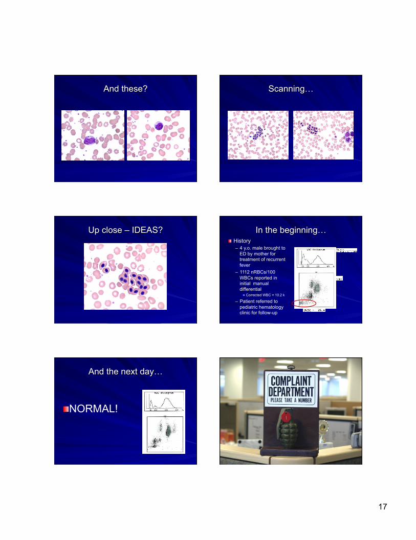
What can these be?
17
And these? Scanning…
Up close – IDEAS? In the beginning… History – 4 y.o. male brought to
ED by mother for treatment of recurrent fever
– 1112 nRBCs/100 WBCs reported in initial manual differential
Corrected WBC = 10.2 k
– Patient referred to pediatric hematology clinic for follow-up
And the next day…
NORMAL!

18
The investigation begins…
Sample labeled correctly QC okay No other samples in batch with similar abnormalities Reproduced results on original analyzer Made additional slides with new stain – same results Alternate staining method – same result Reproduced clinic results with analyzer used for ED sample All samples run on LH 750s
Microscopic Investigation
Here’s another picture… nRBC cytoplasm doesn’t appear to be the same color as surrounding cells No accompanying polychromatophilia nRBC shape is oval or disc-shaped
What you should see… Polychromatophilia Anisocytosis Various degrees of maturation
What is it really?
Total agreement
Avian nRBCs
In the end… Accidental contamination – No source
Intentional contamination – Disgruntled employee – Munchausen by Proxy

19
Münchausen by Proxy Aka Factitious Disorder by Proxy
Intentional production or feigning of physical or psychological signs or symptoms in another person who is under the individual’s care
Characteristics q Most at risk from 15 mos.
to 6 years q 98% of cases biological
mother is responsible q Symptoms are
inappropriate/incongruent q Child taken to many
health care providers - Our case study patient
was never admitted
q One parent, usually father, is absent during hospitalization
q Symptoms disappear when parent/caretaker is absentParent is overly attached
q Poor tolerance to treatment (vomiting, rash)
THIS IS CHILD ABUSE
Münchausen by Proxy ACKNOWLEDGEMENT Many figures in this presentation were taken from the following publications and used with permission: Carr JH and Rodak BF: Clinical Hematology Atlas, 3e.
Copyright Saunders Elsevier 2009. ISBN 978-1-4160-5039-1
Rodak BF, Fritsma GA, Doig K: Hematology Clinical
Principles and Applications, 3e. Copyright Saunders Elsevier 2007. ISBN 978-1-4160-3006-5
www.elsevierhealth.com
Ruler of the Rodak House Just following instinct
20
THE END