description of a novel species of torque teno sus virus ... · a tots els que hem compartit les...
TRANSCRIPT

Description of a novel species of Torque teno sus virus (TTSuV)
and first insights on immunization against TTSuVs in naturally
infected pigs
Memòria presentada per l’ Alexandra Jiménez Melsió per optar al grau de Doctora en el
programa de Bioquímica, Biologia Molecular i Biomedicina del Departament de
Bioquímica i Biologia Molecular de la Universitat Autònoma de Barcelona
Alexandra Jiménez Melsió Doctoranda
Bellaterra, setembre 2015
Dra. Tuija Kekarainen Directora
Dr. Joaquim Segalés i Coma Director
Dra. Anna Bassols Teixidó Tutora


La Dra. Tuija Kekarainen, investigadora del Centre de Recerca en Sanitat Animal (CReSA), i
el Dr. Joaquim Segales i Coma, professor titular del Departament de Sanitat i d’Anatomia
Animals de la Facultat de Veterinària de la Universitat Autònoma de Barcelona (UAB) i
investigador adscrit al CReSA, com a co-directors, i la Dra. Anna Bassols Teixidó,
professora titular del Departament de Bioquímica i Biologia Molecular de la UAB, com a
tutora acadèmica,
Certifiquen:
Que la memòria de tesi doctoral titulada: “Description of a novel species of Torque teno
sus virus (TTSuV) and first insights on immunization against TTSuVs in naturally infected
pigs “ presentada per l’Alexandra Jiménez Melsió s’ha realitzat sota la seva direcció en el
CReSA i n’autoritzen la seva presentació per tal de ser avaluada per la comissió
corresponent per l’obtenció del grau de Doctora del programa de Bioquímica, Biologia
Molecular i Biomedicina del Departament de Bioquímica i Biologia Molecular de la
Universitat Autònoma de Barcelona.
I per tal que consti, als efectes oportuns, signem el present certificat a Bellaterra, a 28 de
setembre del 2015.
Dra. Tuija Kekarainen Directora
Dr. Joaquim Segalés i Coma Director
Dra. Anna Bassols Teixidó Tutora


A la meva mare i el meu pare


Acknowledgements/ Agraïments
Aquestes alçades de la pel·lícula, l’escena final de la Tesi ha arribat. Sí, ha arribat el moment de
posar el punt i final però abans voldria donar les gràcies a totes les persones que m’han ajudar i
han estat el meu costat en aquesta etapa. No ser si sabre expressar tot el que passa pel meu cap
(m’estan passat fotogrames de tots aquests anys i molts sentiments) però...posaré música de
fonts per treure el ritme a les paraules.
Primer de tot vull agrair els meus directors, a la Tuija i en Quim per haver-me donat aquesta
oportunitat. Pel suport rebut en tot moment, sobretot en els moments més complicats i en
aquesta etapa final de l’escriptura de la memòria. Ha sigut tot un plaer haver-vos conegut. Tuija,
gràcies pels bons consells, per guiar-me i ensenyar-me com desenvolupar la feina al laboratori i
davant de l’ordinador, buscar els perquè o corregir els errors. Gràcies per guiar-me en aquesta
etapa i pels ànims. Quim, gràcies per trobar una estona per reunir-te amb mi quan ho he
necessitat. Per l’ajuda rebuda, i per tenir la porta oberta a venir i escoltar-me. Gràcies pels ànims
que sempre transmets.
Aquesta Tesi ha sigut possible gràcies els nostres col·laboradors holandesos. Gràcies a l’Erwin, per
donar-me l’oportunitat de fer una estada en l’empresa farmacèutica, per l’ajuda i l’aprenentatge
científics. Gràcies, he aprés a nivell científic i laboral però també he tingut la sort de conèixer
Holanda, un país petit amb una gent molt maca i acollidora. Al Paul, per l’ajuda prestada en el
laboratori i per transmetre la teva curiositat en els labs meetings. A la Denis, per les bones
estones compartides. Gràcies a l’equip i la gent del departament de virologia. M’heu acollit molt
bé i m’heu fet participats de les vostres activitats, barbacoes o la celebracions, com la de
Sinterklass (Les tulipes, han crescut!). Gràcies Christal per compartir uns minuts del dia en parla
de ciència i de la vida. I la resta del departament que m’heu saludat i heu compartit unes paraules
en algun moment. També he tingut la sort de conèixer a la gent del departament d’expressió
recombinant, moltes gràcies a tots per l’ajuda que m’heu donat en el moment que l’he necessitat.
En especial a la Karine i la Lissette . Per últim, esmentar a la Viviana i el Denis, amb qui hem
treballat i m’han ajudat a resoldre dubtes.
El CReSA ha sigut el lloc on he passat aquests últims 6 anys de la meva vida. La meva etapa del
doctorat va estar precedida per una etapa de tècnic. A tots els que heu format o formeu part
d’aquesta gran família científica que és el CReSA dir-vos que moltes gràcies. Me’n duc el millor
regal del tots, haver-vos conegut i haver compartit grans moments amb molts de vosaltres. És

cert, que sempre hi ha moments més difícil o moments més dolents però aquest se’n van i
queden els grans moments que he viscut amb vosaltres. Riures i bromes el laboratori o a la sala
de descans. Sempre he trobat ajuda quan l’he necessitat de part vostre.
En el CReSA vaig entra a formar part del group TTV, a tots els que vau formar-ne part (la Laura, el
Mario, la Lisa i el Martí), els que esteu acabant la vostre etapa doctoral (David) moltes gràcies. Em
vau acollir, ensenyar i explicar el que sabíeu de TTSuV o de com treballar el laboratori. A més,
durant aquests 6 anys, treballant el meu costat he tingut l’Anna Llorens, la tècnic del grup que ha
estat a l’inici i el final de la meva etapa en el centre. Anna, m’has ensenyat les normes del
laboratori, com treballar a cultiu cel·lular del CReSA. M’has ajudat en els moments de feina, m’has
ensenyat a titular i hem compartit bones estones el laboratori. Moltes gràcies Anna ja farem cotis
pel whatsap. I els estudiants que hem tingut en pràctica, en especial a la Silvia Pares, un plaer
haver-te tingut d’estudiant. Transmetre’ls el que sabia m’ha ajudat a formar-me.
Durant aquesta anys el CreSA, en el transcurs del meu doctorat he tingut la sort de compartir
bons moments i rialles amb molta gent. No voldria oblidar-me ningú així que un gràcies a tots en
que hem compartit algun moment del dia a dia en el CReSA. El laboratori de biologia molecular els
divendres era una festa, allà vam analitzar les cançons d’en Fito...gran moment del “garage a
pupilage” amb la Marta M., i la Rosa Valle. Rosa, hauràs de trobar una altra colegui d’
electroforesis pels divendres a la tarda. A les tècniques de bacter, ho sabeu, em mola molt el
vostre laboratori perquè sou molt divertides. Gràcies pels bons moments. Un dels grans moments
compartit aquests anys, ha sigut a la sala de descans quan coincidia amb la Mónica, Rosa V, la
Merche, Iván C. i la Chus. Si us plau, quin riure quan entraves i te’ls trobaves, això sí, vigila les
paraules, sempre poden tenir un altre sentit. Merche, gracies per ajudar-me, donar-me ànims I
recordar-te de mi quan estava d’estada. Trobaré a faltar els moments de gimnàs. Ei, que tenim un
torneig de pàdel que guanyar. I gràcies per donar-me el telèfon de la meva fisio, l’Elena. Gràcies
els moments de sessió m’han anat estupendo.
A l’Esme, Núria N., Cris R. sempre tant disposades ajudar i explicar-te les coses. A la Maite i la Zori,
per tenir caseïna en stock. A l’Eva per sempre tenir una rialla i disposada a ajudar. Marta V.
dispuesta siempre a solucionar cualquier tipo de problema y Iván M. gracias por preguntar como
va todo. A los dos, un placer compartir más de una vez la hora de comer y echarnos unes risas. A
l’equip d’estudis de camp (Sergio, Rosa i Diego), amb la vostra ajuda els porquets van anar
endavant. Gràcies per ensenyar-me, en aquest estudi vaig aprendre molt gràcies a vosaltres i la
vostra forma de treballa.

Gràcies a la Manoli, la Charo i la Mercedes. Manoli, no es que yo pise tu suelo mojado, es que tu
me persigues para mojar el suelo que yo piso. Charo, ya sabes que no es tu caso. Muchas gracias
chicas por ser tan divertida, alegráis el día a la gente y gracias por ayudar con el material y el
autoclave. Josep Maria, trobaré a faltar els teus crits, però gràcies per enviar el material sempre el
seu lloc.
Marta Lopez quin riure quan ens trobàvem a la fotocopiadora. Gràcies per animar-me anar a la
piscina. L’esport sempre va bé per desconnectar les neurones.
En aquesta anys, pel despatx de becaris han passat molts estudiants. Voldria donar les gracies, em
queda un molt bon record dels moments compartits amb els becaris que he vist formar-se mentre
jo tot just començava. En especial a l’Anna L., Júlia, Meri, Cris L., Laura, Joan T., Mario, Gerard, Jô,
Mar, Kate, Lisa. I en els despatx de becaris va haver un canvi generacional, podríem dir, i així
aquests últim anys els he compartit amb gent de tota arreu, molt alegra i divertida que m’heu
ajudat i animat els dies grisos, això de compartir misèries sempre va bé. Us voldria donar les
gràcies a tots. Bea G, ja saps que sempre que organitzis sopars ens tindràs el Guille, el Salva i a mi
per venir. Guille, gracias por los grandes momentos y por mi banda sonora, lo sabes que voy a
echar de menos el “chin chin chin chiniiin” jajajaja. Salva, ens vas abandonar el despatx, però et
vam perdonar ja que sempre ens vens a fer visites. Marco, echare de menos tus entradas
triunfantes en nuestro despacho. Karla, esta espiritualidad que desprendes relaja en los
momentos de estres. Juliana, hemos compartido largas horas en el despacho, muchas gracias por
todo y port tu caràcter y ser tant detallista; he conocido un poquito más Colombia y sus ritmos
gràcies a ti. Elisa, ara que estàs d’estada se’t troba a faltar, sobretot les galetes i magdalenes de la
cooperativa i aquesta alegria que desprens. Cristina S., como voy a echar de menos tu “me
encanta” y el “que fuerte tronca”, un plaer compartir aquests anys amb tu. Paula, la gallega,
gracias por compartir tus curiosidades musciales y por . Sara, Eli L., Fer, Marc, Bernardo i Max,
gràcies nois per compartir i per fer-me riure. Eli M. hem sigut companyes de taula i compartim la
passió pels animals, sobre tot pels rucs, gràcies pel teu caràcter tant divertit i simpàtic, no deixis
enganyar-te pel compañerito hahahahaha. Feng, gràcies per compartir bones estones i explicar-
me coses de Xina, sempre estàs disposat a donar un cop de mà, les nostres xerrades m’han ajudat
a pràctica el meu anglès. Oscar, vam ser companys de despatx una temporada, tot un plaer i
moltes gràcies per la teva ajuda informàtica, sempre estàs preparat per dedicar uns minuts a
solucionar els nostres problemes informàtics.

A tots els que hem compartit les reunions del Labmeeting dels dimarts. Els investigadors i
estudiants per escoltar la meva feina i per donar-me idees. A la Vicky i a la Flor en especial per
organitzar-ho. A la Marina, per demanar-me com van les coses i ajudar-me quan he necessitat
resoldre alguns dubtes. Fernando R., gracias por tus ánimos y soporte, por tener la puerta
siempre abierta para venir a hablar de ciéncia y guiarme cuando andaba perdida. En general,
voldria donar les gràcies a totes els investigadors que formen part del centre. Francesc i Sònia per
acceptar la feina de ser responsables en el meu seguiment de doctorat, gràcies els dos. Sònia per
l’ajuda en temes de seqüencies i de clonatges.
En aquest anys, he pogut conèixer a molta gent i fer bones amistats. Prova d’això són les
Monstrus, la Judith, Marta i Jô. Marta i Judith, us he pogut tenir des del principi fins el final en el
CReSA, meu ajudat sempre que heu pogut. Judith, sempre se’t pot demanar ajuda, gràcies per
estar quan se’t necessita. Marta, has posat ordre a les meves celles, ets la partner i sempre estàs
allà per quan se’t necessita. Ja saps, les “meriendas”. Jô, por compartir grandes momentos, i estar
allí. Gràcies a les tres per compartir grans moments, sopars i sabeu que el final d’aquest any
farem una gran festa i un gran viatja.
I parlant d’amistats, al llarg de la meva vida he tingut la sort de conèixer grans persones que
formen part de la meva vida. Ells m’han ajudat sempre i en especial en aquest etapa. Així que us
vull dedicar unes línies d’agraïment.
A les nenes…va rebel·larem el nom de guerra: Les Pitxurris. Moltes gràcies noies, Anna S., Cris P.,
Cris L., Gloria i Laura. Gràcies pels sopars, per les grans converses de whatsap, pels Piernites (per
cert, toca el de la temporada de tardo aviat), per estar allà sempre. Per compartir grans moments.
Gràcies Cris L, per haver-me ajudat en coses de escriptura per donar-me la teva opinió i consell,
per estar sempre disponible. Gloria, per tenir sempre un moment per preocupar-te per com estic i
com va, i això que ets una dóna enfeinada. Lau, els teus iconos de whatsap ho diuen tot, com dius
tu “ fas petar de riure”, gràcies Lau per preguntar i per preocupar-te. Cris P., per compartir amb
nosaltres les teves aventures, ajudaven a distreure la ment quan tocava fer feina a casa. Anna S.
ara farem més vinitos, moltes gràcies per demanar com vaig i oferir-me la teva ajuda. Els
respectius, Mariano, Raül i Hèctor sou collonuts els meschios, gràcies per fer-me riure. A la resta
dels de l’uni, cert que aquest últims anys hem estat més desapareguts uns dels altres però a la
que diem de fer un soparet o quelcom sempre trobem el moment. Així que gràcies, d’alguna o
altra manera sempre esteu presents Benet, Juancho, Joan i Norbert.

Piltrafillos S.A, sempre heu estat allà, visitant-me a Roma o a Holanda i on sigui. Tot i que aquest
últim any no ens hem vist gaire, heu de saber que heu estat present. Però, ja començaré a omplir
la tardo amb activitats de tot tipus i recuperem. Moltes gràcies Albert, Cristina, Txe, Raül, Núria
M. i Marc. Gràcies a tots, per sempre estar present i per les estones compartides, per la gran
amistat que ja fa anys que tenim. Per cert, Raül i Núria M, tot i que en el brainstorming vam
treure idees bones per la portada (em vau inspirar), això ho cobrareu amb unes línies més
d’agraïments, tal i com vam quedar. És broma, donar-vos les gràcies els dos perquè el dia del
concert dels Amics a Girona va ser una gran nit. I bé, la reunió de savis va ajudar en temes
d’edició. A més, en especial, et vull donar les gràcies Núria M. per l’ajuda, pel suport i per estar
sempre en tot moment. Fer-te un whatsap demanant un favor i el minut zero, ja me l’estàs fent.
Hem compartit grans moments i grans viatges...i els que queden. En breu, el del Nepal. Per cert,
parlant de Nepal, vull donar les gràcies a l’equip del viatja: Meri, Pilar, Núria M., Raül i Beth. Per
organitzar i per ajudar-me a desconnectar per una estona de la Tesi amb una bonica ruta pel
Montseny.
Gràcies a la Núria M., aquests últims dies, em van acollir a Girona una gran família financera. Em
van deixar fer feina d’edició en el seu despatx. No sabia que les lleis fiscals podrien ser tant
divertides fins ara. Gràcies Esther, Miquel i Toni, el canvi d’aires van ajudar a portar més coll avall
els últims dies.
Laia Freixa, és el teu torn cotorreta. Dir-te que moltes gràcies per estar sempre, per venir de visita
a Holanda. Per compartir grans estones el telèfon, no cal que digui gaire paraules ja que sempre
saps que vull dir. Estem molt compenetrades. T’he explicat els meus mals de caps i neguis i m’has
entès. Gràcies cotorreta. Per cert, haurem de recupera moltes Viaders.
A la Gemma i l’Àlex, pels bons berenars i sopars. Gemma gràcies pels teus consells i donar-me el
teu punt de vista. Per animar-me i les llargues estones compartides.
A la Julie i l’Imma, per organitzar sopars i dir-me que només vingués, i vosaltres us encarregàveu
del menjar. Pels moments de manta i sofà que m’han fet desconnectar de la feina. També gràcies
el Carlos i el Jonathan, els grans moments compartits tots junts.
Vicky Miller, thank you so much. Muchas gracias por las clases de inglés, practicando el speaking
te explicaba mi día o la semana de Trabajo. Eran una carga de energies las clases. Por ayudarme
con la redacción y todo el soporte dado. Muchas gracias, tu alegría y optimismo me han ayudado
mucho.

Per últim les persones més importants, la meva família. El meu pare i a la meva mare, per estar el
meu costat, per recolzar-me en les meves decisions, donar-me ànims. Pare, gràcies pels plats que
m’has cuinats aquesta última etapa de feina. Mare, moltes gràcies per estar sempre i preocupar-
te perquè tot surti bé. Els meus germans, a la Roser i a l’Agustí, sóc afortunada de tenir-vos,
gràcies pel suport i interessar-vos per la meva feina. A l’Adri, la cunyi, per donar-me ànims. Els
meus nebots, l’Ot i Teo. A l’Ot, el meu fillol, venir-te a veure o jugar amb tu han sigut una molt
bona teràpia. M’he rigut molt i m’has regalats moments molt bonics i dolços. Venir-te a veure,
jugar amb tu i preguntar-me: “Tieta, la feina com va?”. Aquí tot l’estrès de la feina se’n va anar.
Teo, el nou vingut de la família, gràcies per les teves rialles d’aquest últims mesos. Els meus tiets,
en especial a la meva padrina, la Tia Dolors, moltes gràcies per fer Skyps amb mi, per animar-me i
pregunta com vaig. Els meus cosins, en especial a la Sara i a la Bet, per les trucades, les visites i
per escoltar-me quan us explicava tota l’evolució. Bet, per ajudar-me a resoldre dubtes en la
meva vista a Londres. La Mia i l’Anna, la “tieta i l’avia postissa”, simplement molt agraïda a les
dues, sempre esteu pendents del que fem i de com ens van les coses.
I voldria fer una agraïments a les persones que ara ja no estant aquí però han ajudat i donat molta
força per continuar els meus somnis. Gràcies Matí per les teves paraules.
Per últim dir-vos que, i perdoneu si em repeteixo, però el que sento és gratitud i durant aquests
anys me sentit molt estimada per tots vosaltres, per tots els que heu estat el meu costat d’una
forma o d’una altra, dir-vos GRÀCIES. I per acabar, “fotem una festa, muntem un sidral” perquè
avui és la fi de aquesta aventura doctoral però el començament d’una nova etapa. Una etapa
incerta però plena de bons records i dolços del meu pas pel CReSA. I no és un adéu sinó és un fins
aviat.
“Petons, mitjons i americanes “ dels Els amics de les arts

I
Contents
Summary ................................................................................................................ III
Resum ..................................................................................................................... V
List of abbreviations .............................................................................................. VII
1. Introduction ........................................................................................................ 1
1.1. History and discovery of Torque teno virus ................................................ 3
1.2. Taxonomy and classification of anelloviruses ............................................ 4
1.3. Molecular characteristics of anelloviruses ................................................. 8
1.4. Epidemiology ............................................................................................ 10
1.5. Pathogenesis and immunology ................................................................ 12
1.6. TTSuV disease association . ....................................................................... 15
1.7. Prevention and control of TTSuV ............................................................. 16
2. Objectives ......................................................................................................... 19
3. Study I .............................................................................................................. 23
Discovery of a novel Torque teno sus virus species: genetic characterization,
epidemiological assessment, and disease association
3.1. Introduction .............................................................................................. 25
3.2. Material and methods .............................................................................. 26
3.3. Results ...................................................................................................... 31
3.4. Discussion ................................................................................................. 39
4. Study II ............................................................................................................. 43
Expression of and pig immunization with Torque teno sus virus proteins
4.1. Introduction .............................................................................................. 45
4.2. Materials and methods ............................................................................ 47
4.3. Results ...................................................................................................... 55
4.4. Discussion ................................................................................................. 67

II
5. Study III ............................................................................................................. 84
Vaccination of pigs reduces Torque teno sus virus viremia during natural infection
5.1. Introduction .............................................................................................. 73
5.2. Materials and methods ............................................................................ 74
5.3. Results ...................................................................................................... 79
5.4. Discussion ................................................................................................. 85
6. General discussion ............................................................................................ 84
7. Conclusions ..................................................................................................... 104
8. References ...................................................................................................... 105

III
Summary
Anelloviruses are a highly diverse group of circular single-stranded DNA viruses infecting
vertebrates. Torque teno sus viruses (TTSuV) are ubiquitous pig-infecting anelloviruses.
Three different viral species have been described, namely TTSuV1a and 1b within the
genus Iotatorquevirus and TTSuVk2a, within the genus Kappatorquevirus. These viruses
are genetically very distinct (>56% sequence diversity) but share similar genome
organization and expression strategy. TTSuV infection in pigs is distributed worldwide,
and is characterized by a persistent viremia. TTSuV themselves are considered non-
pathogenic; however, it is believed that these viruses play a role in co-infection with
other economically important viral porcine infections. Apparently, TTSuV infection can
influence the development or may contribute to the pathogenesis of various porcine
diseases during co-infection. The real impact of TTSuV on the pig health, if any, is still
under debate.
The present Thesis aimed to characterize a novel TTSuV species and to explore possible
ways of vaccination against TTSuVs.
The first study describes the discovery, genetic characterization and epidemiology of a
novel TTSuV species, named Torque teno sus virus k2b (TTSuVk2b). According to
phylogenetic analysis, this new virus belongs to the Kappatorquevirus genus, belonging to
the same genus as TTSuVk2a. Quantitative PCR techniques based on SybrGreen
technology were developed; one for quantification of total TTSuV load (TTSuV broad-
spectrum qPCR) and others for quantification of each TTSuV species separately. These
techniques were used for epidemiological studies and assess the geographical
distribution. Moreover, prevalence and viral DNA load were determined in porcine
circovirus type 2-systemic disease (PCV2-SD)-affected animals and healthy counterparts,
since previous studies have associated another kappatorquevirus species, to the disease.
The epidemiological study revealed that TTSuVk2b is worldwide distributed, although less
abundant and displaying lower viral DNA titres in serum than TTSuV1 and TTSuVk2a.
TTSuVk2b was associated with PCV2-systemic disease (PCV2-SD), which revealed that the
two kappatorquevirus species are both genetically and biologically related.

IV
The second study contained two objectives. On one hand, TTSuV proteins were expressed
in a baculovirus-based platform; on the other hand, the impact of a multivalent
experimental vaccine was evaluated in a natural TTSuV infection model of pigs. The ORF1,
ORF1-A, ORF2 and ORF3 recombinant proteins of all four known TTSuVs were successfully
expressed in a baculovirus expression system. In additional, the multivalent experimental
vaccine containing ORF1 and ORF3 proteins was administered by intramuscular and
intradermal routes using two different vaccination schedules (twice or three times).
Seroconversion and viral titres in blood were measured from 3 weeks until 15 weeks of
age, using the indirect ELISA based on baculoviruses proteins and species-specific qPCRs,
respectively. This study showed that vaccination induced anti-TTSuV antibodies; however
the multivalent vaccine was not able to control viremia during TTSuV natural infection.
Finally, in the third study, the immunization against TTSuVk2a during natural infection
was evaluated using a different approach. The immunizations consisted of a combination
of DNA and protein to increase the possibilities of activating both cellular and humoral
immune responses. Quantitative PCR techniques were used to detect and quantify the
viremia levels of each TTSuV species, while the induction of specific antibodies was
monitored by indirect ELISA. The vaccinated group showed a seroconversion and a
significant reduction of the TTSuVk2a viral loads compared to the control group. This
study demonstrated for the first time that TTSuV viremia can be controlled by a combined
DNA and protein immunization.
Overall, the present Thesis contributes to increase the knowledge on TTSuV by means of
describing a novel species, which may be involved in disease progression in co-infection
with other pig pathogens. Moreover, TTSuV infection can be controlled by the
administration of a combined DNA/protein immunization while a multivalent protein
based vaccine was not efficient.

V
Resum
Els anellovirus són un grup de virus de cadena senzilla d’ADN amb una elevada diversitat
genètica i que infecten a vertebrats. Els Torque teno sus virus (TTSuV) son ubics i és
l’espècie d’anellovirus específica que infecta els porcs. S’han descrit 3 espècies víriques
diferents, anomenades TTSuV1a i TTSuV1b dins del gènere Iotatorquevirus i TTSuVk2a,
dins el gènere Kappatorquevirus. Aquests virus son genèticament molt diferents (>56% de
diversitat de seqüencia nucleotídica) però comparteixen la mateixa organització del
genoma i estratègia d’expressió. L ‘infecció de TTSuV en porcs es caracteritza per una
virèmia persistent. Els TTSuVs no es consideren patogènics, però es creu que juguen un
paper en la co-infecció amb altres agents infecciosos que causen malalties porcines
importants. L'impacte real dels TTSuVs sobre la salut dels porcs, si n’hi ha , segueix sent
objecte de debat.
L'objectiu principal d'aquesta Tesi fou caracteritzar la variabilitat de TTSuV i explorar el
desenvolupament de possibles prototipus vacunals enfront els TTSuVs.
El primer estudi descriu la caracterització genètica d'una nova espècie de TTSuV,
anomenada Torque teno sus virus k2b (TTSuVk2b). Segons l'anàlisi filogenètica, aquest
nou virus pertany al gènere Kappatorquevirus, el mateix al qual pertany TTSuVk2a. Es van
desenvolupar tècniques quantitatives de PCR basades en la tecnologia de SybrGreen pels
estudis epidemiològics i per l’estudi de la distribució geogràfica. En concret, es va
desenvolupar una tècnica que quantificava la càrrega total de TTSuV (TTSuV broad-
spectrum qPCR) i tres més que quantificaven la càrrega viral específica de cada espècie de
TTSuV. D'altra banda, es va estudiar la prevalença i la càrrega d'ADN vírica en animals
afectats amb per circovirosi porcina (CP). L'estudi epidemiològic va revelar que TTSuVk2b
es distribueix a tot el món, encara que és menys abundant que TTSuV1 i TTSuVK2a.
TTSuVk2b es va associar amb la CP, una malaltia porcina d'importància econòmica molt
significativa.
El segon estudi contenia dos objectius: d'una banda, l'expressió de les proteïnes TTSuVs;
per l'altra banda, l’avaluació de l'impacte d’una vacuna experimental multivalent enfront

VI
les 4 espècies de TTSuVs conegudes, utilitzant com a model l’infecció natural de TTSuV
en porcs. Les proteïnes recombinants ORF1, ORF1-A, ORF2 i ORF3 dels quatre TTSuVs
coneguts es van expressar amb èxit mitjançant un sistema d'expressió de baculovirus. Per
altra part, la vacuna experimental multivalent (formada per les proteïnes dels gens ORF1 i
ORF3) va ser administrada per via intramuscular i intradèrmica utilitzant dos esquemes de
vacunació diferents (dues o tres dosis). La seroconversió i els títols vírics en sang es van
mesurar a partir de 3 fins a 15 setmanes d'edat, utilitzant respectivament l'ELISA indirecte
basat en proteïnes del virus expressades en baculovirus i tècniques de PCR quantitativa
per detectar i quantificar els nivells de virèmia de cada espècie TTSuV, respectivament.
Aquest estudi va mostrar que la vacuna multivalent induïa anticossos anti-TTSuV, però
aquest prototipus vacunal no era capaç de controlar la virèmia durant l’infecció natural
de TTSuV.
Finalment, en el tercer estudi es va avaluar l’immunització enfront TTSuVk2a durant
l’infecció natural en porcs. L’immunització va consistir en una combinació d'ADN i
proteïnes víriques per augmentar les possibilitats d'activació de respostes immunitàries
tant cel·lulars com humorals. Es van utilitzar tècniques de PCR quantitativa per detectar i
quantificar els nivells de virèmia de cada espècie de TTSuV, mentre que l’inducció
d'anticossos específics va ser monitoritzada mitjançant ELISA indirecte. El grup vacunat va
mostrar una seroconversió i una reducció significativa de les càrregues víriques de
TTSuVk2a en comparació amb el grup control. Aquest estudi va demostrar per primera
vegada que la virèmia per TTSuV pot ser controlada a través de l’ús d’una combinació que
inclou ADN i proteïnes recombinant.
En general, aquesta Tesi Doctoral contribueix a augmentar el coneixement dels TTSuVs
mitjançant la descripció d’una nova espècie vírica, la qual pot estar associada al
desenvolupament de malalties del porc, com s’ha indicat per TTSuVk2a. A més, l’infecció
per TTSuV pot ser controlada mitjançant l'administració d'una vacuna d'ADN i proteïnes
recombinats del virus, mentre que la vacuna basada en múltiples proteïnes (multivalent)
no va ser eficient.

VII
List of abbreviations
A adenine
aa amino acid
ASFV African swine fever virus
BEI binary ethylenimine
BFDV Beak and feather disease virus
BSA bovine serum albumin
C Cytosine
CAV Chicken anaemia virus
CMV Cytomegalovirus
DNA deoxyribonucleic acid
DENV Dengue virus
Dpi days post infection
ds double stranded
dNTP deoxynucleotide triphosphate
EDTA ethylenediaminetetraacetic acid
ELISA enzyme linked immunosorbent assay
FIPV Feline infectious peritonitis virus
FITC fluorescein isothiocyanate
G Guanine
GFP green fluorescent protein
HCV Hepatitis C virus
HEPE N-(2-Hydroxyethyl)-piperazine-N′-(2-ethanesulfonic acid)
HPV Human papilloma virus
ICTV International Committee for the Taxonomy of Viruses
ID intradermal
IFA immunofluorescence assay
IM intramuscular
IPTG isopropyl ß-D-1-thiogalactopyranoide
Ig immunoglobulin

VIII
Kb kilobase
KDa kilodalton
LUX light upon extension
LOD imit of deteccion
M Molar
mAb monoclonal antibody
min minutes
MOI multiplicity of infection
mM milimolar
mRNA messenger ribonucleic acid
MW molecular weight
NLS nuclear localization signal
OD optical density
ORF open reading frame
P P-value statistics
PASC pairwise sequence comparison
PBMC peripheral blood mononuclear cells
PBS phosphate-buffered saline
PCR polymerase chain reaction
PCV Porcine circovirus
PCV1 Porcine circovirus type 1
PCV2 Porcine circovirus type 2
PCVD porcine circovirus diseases
PCV2-SD porcine circovirus type 2-systemic disease
PDNS porcine dermatitis and nephropathy syndrome
PFA paraformaldehyde
PI pairwise identity
PRRSV Porcine reproductive and respiratory syndrome virus
qPCR real time quantitative PCR
R2 correlation coefficient
RCA rolling-circle amplification

IX
RCR rolling-circle replication
RDA representational difference assay
Rpm revolution per minute
s sedimentation coefficient
sec seconds
ss single stranded
SD standard deviation
SDS-PAGE Sodium Dodecyl Sulfate-polyacrylamide gel electrophoresis
Sf Spodoptera frugiperda
SISPA sequence-independent single primer amplification
SPSS Statistical package for social sciences
T Thymine
TBS-T Tris-buffered saline –Tween 20
TMB 3,3’,5,5’-tetramethylbenzidine
TTV Torque teno virus
TTSuV Torque teno sus virus
U enzyme unit
UTR untranslated region
Vol volume
VLP virus like particles


1. Introduction


Introduction
3
1.1. History and discovery of Torque teno virus
The first identification of the Torque teno virus (TTV) was in 1997, in the plasma of a
Japanese patient with post-transfusion hepatitis of unknown aetiology (Nishizawa et al.,
1997). Later studies demonstrated that TTV was the first circular single-stranded DNA
(ssDNA) virus found to infect humans (Miyata et al., 1999; Mushahwar et al., 1999). The
novel virus was called ‘TT’ virus (TTV) in reference to the initials of the affected patient.
The International Committee on Taxonomy of Viruses (ICTV), in charge of the official
taxonomic nomenclature, proposed a new meaning for ‘TT’, namely “Torque” and
“Teno”, deriving from the Latin terms meaning “necklace” and “thin”, respectively,
reflecting the organisational arrangement of the TTV genome (Todd et al., 2005).
A number of TTV-like viruses has also been identified in several vertebrates including pets
(dogs and cats), farm animals (pigs, chicken, cow and sheep), wild animals (wild boar,
tupaia, sea lions, sea turtle and rodents) and primates (chimpanzee, Japanese macaque,
cotton-top tamarin and douroucouli) (Brassard et al., 2008; Inami et al., 2000; Leary et al.,
1999; Martínez et al., 2006; Ng et al., 2009a; Ng et al., 2009b; Nishiyama et al., 2014;
Okamoto et al., 2000a; Okamoto et al., 2001; Okamoto et al., 2000b; Okamoto et al.,
2002). These viruses, included nowadays in the family Anelloviridae, are considered
species-specific (Okamoto et al., 2000a). The initially designated swine TTV was
discovered in 1999 (Leary et al., 1999) and subsequently named as Torque teno sus virus
(TTSuV) by the ICTV.
Most of the current research is done in human infecting anelloviruses, but swine could be
an excellent model for the research due to its similarity to human metabolism and
physiology. Therefore, the use of the TTSuV model might contribute to understand better
the infection in humans as well (Kekarainen & Segalés, 2009). During the last years the
knowledge about TTSuVs has significantly increased and it has become more important
especially due to the co-infection with other pig pathogens as Porcine circovirus type 2
(PCV2), an economically important viral agent of swine (Kekarainen & Segalés, 2009).
Introduction
4
1.2. Taxonomy and classification of anelloviruses
After discovery, TTVs were initially proposed to be classified in the group of viruses with
ssDNA genomes within the Circoviridae family (Miyata et al., 1999; Takahashi et al.,
2000). Circoviridae family includes a number of small DNA viruses infecting several
domesticated animals and bird species, such as Porcine circovirus type 1 (PCV1) and type
2 (PCV2), Chicken anemia virus (CAV) and Beak and feather disease virus (BFDV).
Although, the genome organization of TTV is similar to CAV, subsequent studies showed
distinct biophysical and molecular characteristics exhibited by CAV that suggested their
taxonomic grouping into a new genus or family different from Circoviridae family
(Mushahwar et al., 1999). At the end of 2001, the ICTV created the floating genus
Anellovirus, unattached to any viral family. The term “Anello”, deriving from Latin and
meaning “the ring”, was related to the circular nature of their genome. The description of
the genus Anellovirus was effective in 2004 and corresponded to the first step in the
official classification (Biagini et al., 2005). Finally, in 2009, based on the molecular and
structural characteristics of TTVs, a new viral family named Anelloviridae was created
(Biagini, 2009).
Anelloviridae family (Figure 1.1) includes a number of viruses that are genetically highly
variable, but share a common genomic organization (Biagini, 2009). However, these
analyses also show different genome length and a great variability in the sequence
identity. The taxonomic classification is currently based on the analysis of a genomic
region corresponding to the entire open reading frame 1 (ORF1), which is representative
of the whole genome identity as demonstrated by distance matrix comparison (Biagini,
2009). Actually, anelloviruses are divided in eleven genera depending on the host species,
(http://www.ictvonline.org/virusTaxonomy.asp). TTSuVs are separated in two distinct
genera namely Iotatorquevirus and Kappatorquevirus (http://www.ictvonline.org/virus
Taxonomy.asp?taxnode_id=20141494). Iotatorquevirus genus includes two genetically
distinct viral species Torque teno sus virus 1a (TTSuV 1a) (AB076061) and Torque teno sus
virus 1b (TTSuV1b) (AY823990). Kappatorquevirus includes only one species, namely
Torque teno sus virus k2a, TTSuVk2 (AY823991) (Niel et al., 2005).

Introduction
5
Figure 1.1 Taxonomical classification of anelloviruses according to ICTV (www.ictvonline.org).
1.2.1. Genetic variability
The extensive divergence described for anelloviruses has been attributed to different
mechanisms of genomic variation, being mainly the genomic mutation, insertions-
deletions, and transversions and transitions in the translated regions (Biagini et al., 1999;
Cortey et al., 2011; de Villiers et al., 2011; Nishizawa et al., 1999). Pairwise sequence
comparison (PASC) is a useful method that is used to plot the frequency distribution of
pairwise nucleotide sequence identity percentage from all available genomic sequences
of viruses in the same family (Bao et al., 2008). Based on the phylogenetic and PASC
analysis of anallovirus ORF1 nucleotide sequences, ICTV nowadays recommends to use
the cut-off values for sequence divergence to distinguish different genera (>56%) and
species (>35%). Is to be noted that ICTV is not responsible for classification and
nomenclature of virus taxa below the rank of species and this task remains to research
groups.

Introduction
6
TTSuVs are genetically very distinct (>56% sequence diversity), but share similar genome
organization and gene expression strategy (Martínez-Guinó et al., 2011). Comparison of
the sequences available and TTSuV genomes identified from Spain revealed that the
number of variable nucleotide positions among genomes is lower in the untranslated
region (UTR) and larger in the translated area (Cortey et al., 2011). TTSuV1 was shown to
contain more variability than TTSuVk2 (Cortey et al., 2011).
TTSuV classification is not as accurate compared to that of human TTVs, since the number
of complete TTSuV genomes available in the Genbank is not that high. However, the full-
length genomes of TTSuVs have been increasing in the last years, and type and subtype
definitions have been proposed for TTSuV species (Cortey et al., 2011, 2012; Huang et al.,
2010b). Based on this, TTSuVs could be further classified to variants (>95% PI), subtypes
(85-95 % PI), types (67-85% PI), species (55-67%), and genus (35-55%) based on several
tentative pairwise identity (PI) thresholds (Huang et al., 2010b). According to the
suggested PI limits, each TTSuV species could be further divided from 3 to 4 types and
subtypes. However, considering the high genetic divergence but lack of biological data of
TTSuVs, classification beyond the species level should be postponed (Cortey et al., 2011;
Huang et al., 2010b; Li et al., 2013; Liu et al., 2013)
1.2.2. Discovering novel anelloviruses
The prototype anellovirus, the human TTV, was discovered using the representational
difference assay (RDA) (Nishizawa et al., 1997). The novel DNA was detected amplifying a
specific nucleotide sequence of 500 bp (named N22), which showed low homology to
DNA sequences found in databases at that time. Following studies to detect novel
anelloviruses were based on the detection of DNA by using primer specific for the short,
highly conserved sequence in the non-coding region of the known TTV (Okamoto et al.,
2001; Okamoto et al., 2002). Therefore, a number of new anelloviruses were discovered
by means of PCR and sequencing, but this methodology allows only detection of close
relatives of known viruses (Delwart, 2007).

Introduction
7
New methodologies have been subsequently developed for the discovery of new viruses
without prior knowledge on their genomic sequences such as virus-particle purification
and shotgun sequencing, included within metagenomics packages (Delwart, 2007). Viral
metagenomics allow identifying novel viruses from uncultured environments or clinical
samples. Viruses have to be purified through filtrate processing and their nucleic acids
randomly amplified prior to subcloning and sequencing. With this technology viruses have
been characterized from samples with several origins like seawater, near shore
sediments, mammal faeces, serum, plasma and respiratory secretions. Also, the viral
metagenomic approach is useful in the case of viruses that cannot be cultivated in the
laboratory or viruses unrelated to already known ones (Delwart, 2007; Edwards &
Rohwer, 2005). Viral metagenomics has been also used to identify other pathogens as
well as ssDNA viruses (Ng et al., 2009a) and anelloviruses (Ng et al., 2009b). A sequence-
independent single primer amplification (SISPA) method has been also used in human
plasma to identify two novel TTV species with a highly divergent circular sequence (Jones
et al., 2005). These methods, due to their simplicity, relative speed and lack of bias in
identifying particular groups of viruses, have advantages to detect novel agents highly
divergent from those already known (Delwart, 2007). The method is based on converting
the target sequence to a double-stranded DNA, and use endonuclease restriction
followed by non-specific linker ligation and PCR amplification (Allander et al., 2001; Reyes
& Kim, 1991)
The rolling-circle amplification (RCA) based sequence-independent approach has been
also applied to identify TTVs (Niel et al., 2005). RCA is a relatively novel technique based
on the property of circular DNA molecules to replicate using the rolling circle mechanism
(Dean et al., 2001). The method utilizes the bacteriophage phi29 DNA polymerase, with
strong strand-displacing capability, high processivity and proofreading activity (Esteban et
al., 1993). The amplification is done using random hexamer primers, without need of
prior knowledge of the target nucleotide sequence. Indeed, nine anelloviruses have been
detected in human plasma and cat saliva by means of a combination of RCA and SISPA
(Biagini et al., 2007) and virus sequences from pig and human sera have been detected by
using a combination of RCA with a single-step-inverse-PCR (Macera et al., 2011).

Introduction
8
1.3. Molecular characteristics of anelloviruses
1.3.1. Genomic organization
Anelloviruses are small, non-enveloped viruses of 30-32 nm in diameter and icosahedral
symmetry (Mushahwar et al., 1999). The human TTV (prototype of Anelloviridae family)
virions have a buoyant density of 1.31-1.33 g/cm3 in cesium chloride for TTVs detected in
serum and 1.22-1.35 g/cm3 for TTVs from faeces (Mushahwar et al., 1999; Okamoto et al.,
1998). The genome, of negative polarity, may have different size depending on the host
species, ranging between 2.1 (cat) to 3.9 kilobases (kb) (human), being approximately 2.8
kb for TTSuV (Miyata et al., 1999; Mushahwar et al., 1999; Nishizawa et al., 1997;
Okamoto et al., 2002; Okamoto et al., 1999; Peng et al., 2002).
TTSuVs have a similar genomic organization with human TTVs, but share less than 45% nt
sequence identity (Niel et al., 2005; Okamoto et al., 2002). As all anlleoviruses, the TTSuV
genome can be divided into an UTR and a coding region (Okamoto et al., 2002) (Figure
1.3). The UTR in TTSuV is characterized as a short stretch of sequence with high GC
content. It contains the most conserved region of the genome including the promoter and
enhancer elements important for the virus replication and transcription (Kamada et al.,
2004; Suzuki et al., 2004).
1.3.2. Gene transcription and protein translation
Despite of high divergence among TTV sequences, the splice sites and motifs between
various sequences are well-conserved, which led to propose similar protein structure and
functions (Peng et al., 2002).
The TTSuV genome contains (Figure 1.2) two major ORFs; ORF1 and ORF2. Several
analyses of TTSuV nucleotide sequences reveal the existence of additional mRNAs, all as
result of splicing events. ORF3 mRNA has its 5’end identical to ORF2 and 3’ end results
after splicing event partially overlapping ORF1 encoding region. On the other hand,

Introduction
9
alternative splicing of ORF1 results in several mRNAs (Huang et al., 2012b; Martínez-
Guinó et al., 2011). Indeed, the predominant transcripts detected in vitro and in vivo of
ORF1 are spliced and the full-length ORF1 transcript has not been detected so far (Huang
et al., 2012b; Martínez-Guinó et al., 2011). Splicing of TTSuV1 ORF1 generates two
isoforms (ORF1-A and B) of different sizes although the reading frame and the encoded
amino acid (aa) sequence do not change (Martínez-Guinó et al., 2011). In contrast,
splicing of TTSuVk2a ORF1 results in three protein isoforms (ORF1-A, -B and -C) and,
depending on the splicing site used, the aa composition between them varies (Huang et
al., 2012b; Martínez-Guinó et al., 2011). TTSuVk2 ORF3 generates three protein isoforms,
two of which altered the reading frame and TTSuV1 ORF3 only produces one isoform
(Huang et al., 2012b; Lu et al., 2013; Martínez-Guinó et al., 2011).
Figure 1.2 Schematic view of the TTSuV genome. UTR and encoding regions are shown. ORFs encode different TTSuV proteins
The ORF1 gene encodes the largest TTSuV protein with a variable length ranging from
638-650 aa in TTSuV1 and 625-628 aa in TTSuVk2). By analogy with CAV, PCV2 and human
TTV, ORF1 encodes a predicted viral capsid domain at the N-terminal half of the protein
and a putative replication-associated domain in its C-terminal half (Miyata et al., 1999;
Mushahwar et al., 1999). ORF1 possesses a high number of arginine residues at the N-

Introduction
10
terminus and rolling-circle replication (RCR) motifs, which were differently present in
TTSuV species, but highly conserved within species (Huang et al., 2010b). ORF2 smallest
predicted gene, encode a non-structural protein and encodes protein of 73–74 aa in
TTSuV1 and 69 aa in TTSuV2. It has been suggested that the ORF2 protein is involved in
viral replication (Hijikata et al., 1999; Huang et al., 2010b). ORF2 protein conserved
protein-tyrosine phosphatase (PTPase-like motife) at the N-terminal, typically found in
anelloviruses (Huang et al., 2010b). At present, the role of the ORF3 protein is unknown
and is believed to encode non-structural protein that shares its N-terminus with ORF2
(Biagini et al., 2001; Cortey et al., 2011).
1.3.3. Replication mechanism
The exact mechanism of TTV replication is not fully understood. Based on similarities to
other ssDNA-genome viruses, it is assumed that TTV uses the RCR mechanism
(Mushahwar et al., 1999). It is believed that TTSuVs and human TTVs use the cellular
polymerase and replication machinery from the host, like the majority of small DNA
viruses (Kakkola et al., 2007). Due to the lack of a cell culture system to propagate
anelloviruses, little is known regarding the molecular biology and pathogenesis of
anelloviruses. Moreover, attempts to replicate both human TTV and TTSuV in cell cultures
(free from TTVs) have not been successful (Huang et al., 2012b; Kakkola et al., 2007). To
date, there is no available in vitro efficient replication model for anelloviruses.
1.4. Epidemiology
The prevalence and detection of TTSuVs DNA have been mainly based on species-specific
nested PCR (Kekarainen et al., 2006) and direct PCR (Brassard et al., 2008; Segalés et al.,
2009) techniques detecting the UTR. Recently, quantitative (q) PCR techniques have been
developed using different pairs of primers and probes (Brassard et al., 2010; Gallei et al.,
2010; Huang et al., 2010a; Lee et al., 2010; Nieto et al., 2011; Xiao et al., 2012).

Introduction
11
Detection and quantification of anelloviruses by molecular systems are constantly
improving. The results obtained in different laboratories may be a source of confusion
and results may not be comparable due to the differences on the applied techniques,
since the molecular tools are highly dependent on the DNA fragment subjected to
amplification and primers used in each laboratory. Therefore, effective diagnostic tools
would also help to progress in the study of the TTSuV infections.
TTSuV DNA was detected in swine for the first time in 1999 (Leary et al., 1999). However,
a retrospective study from Segalés et al. (2009) revealed evidence of TTSuV infections in
pigs as early as 1985 in Spain. Nowadays it is known that TTSuVs are ubiquitous in
domestic pigs and wild boar (Kekarainen & Segalés, 2009) and co-infections of TTSuV
species are often detected (Blois et al., 2014; Gallei et al., 2010; Huang et al., 2010b). It is
believed that anelloviruses are transmitted between members of the family Suidae
(domestic pig, wild boar and bush pigs), since TTSuVs found in these hosts are highly
genetically identical to each other (Blomström et al., 2012; Leary et al., 1999; Martínez et
al., 2006).
TTSuV infection is distributed worldwide. They have been found in swine serum from
many different countries as Canada, China, France, Italy, Korea, Spain, Thailand and the
United Sates with prevalence rates ranging from 24% to 100% (Bigarré et al., 2005; Gallei
et al., 2010; Kekarainen et al., 2006; Martelli et al., 2006; McKeown et al., 2004; Taira et
al., 2009). Globalization and international trading of live animals is considered to play a
crucial role in the worldwide propagation of TTSuV in pig population (Cortey et al., 2012).
TTSuV infections occur early in life, as they have been found in foetuses (Martinez-Guino
et al., 2010; Xiao et al., 2012). In addition, viral prevalence increases with age and it looks
like that a high proportion of animals get persistently infected (Aramouni et al., 2010;
Blois et al., 2014; Nieto et al., 2011; Sibila et al., 2009a; Sibila et al., 2009b; Taira et al.,
2009; Xiao et al., 2012). TTSuV1 and TTSuVk2 seem to display similar dynamics of
infection, with a prevalence of viraemic piglets increasing progressively over time, and
with a maximum rate of TTSuV1 detection at 11 weeks of age and at 15 weeks of age for
TTSuVk2 (Nieto et al., 2011; Sibila et al., 2009a).

Introduction
12
Several investigations have noted a higher prevalence of TTSuV1 DNA compared to that
of TTSuVk2 DNA in different countries (Gallei et al., 2010; Martínez-Guinó et al., 2009;
Perez et al., 2011; Sibila et al., 2009a; Sibila et al., 2009b). In contrast, one study from the
Czech Republic (Jarosova et al., 2011) and another from China (Li et al., 2013) detected
higher TTSuVk2 prevalences Since the outcome of a given study is highly dependent on
the PCR method used, it is expectable that considerable discrepancies and variations have
occurred.
TTSuV DNAs in tissues have also been detected by a semi-quantitative method.
Prevalence increased over age, from foetal period until 15 weeks of age, and then
maintained until slaughter age (Aramouni et al., 2010). TTSuVs have been also found in
semen, colostrum, nasal cavity and faeces (Kekarainen et al., 2007; Martínez-Guinó et al.,
2009; Sibila et al., 2009b), indicating the occurrence of both vertical and horizontal
transmission (Kekarainen & Segalés, 2012a).
Moreover, commercial pigs vaccines, human drugs with components of pig origin, trypsin
derived from pigs used for cell culturing, as well as in cell lineages derived from different
species have been found contaminated with TTSuV DNA (Kekarainen et al., 2009; Kulcsar
et al., 2010; Teixeira et al., 2011)
In summary, TTSuVs natural infections are characterized for being ubiquitous, increasing
viral prevalence with age, and establishing long-lasting to persistent infections, such as it
has been observed in human TTVs (Maggi et al., 2001; Moen et al., 2003).
1.5. Pathogenesis and immunology
To date, the pathogenic potential of anelloviruses is still controversial and they are
considered non-pathogenic by themselves. Anelloviruses are characterized by high
prevalence in the general human and pig populations that are apparently healthy
(Kekarainen & Segalés, 2012b; Okamoto, 2009a). So far, there are no clinical
manifestations that can be directly attributed to anellovirus infections. Experimental

Introduction
13
studies of gnotobiotic pigs demonstrated mild lesions after their inoculation with TTSuV1-
containing tissue homogenate (Krakowka and Ellis, 2008) or TTSuVk2-containing tissue
homogenate (Mei et al., 2011). In another study, mild pathological lesions in brain, kidney
and liver were observed in colostrum-deprived (CD) pigs after inoculation with an
infectious TTSuVk2 clone (Huang et al., 2012b). Inoculated gnotobiotic pigs did not show
any clinical sign during the experiments. However, it is believed that TTSuVs might
influence the development or even modulate some diseases and play a pathogenic role
during co-infection with other pathogens (Kekarainen & Segalés, 2012b; Okamoto,
2009a).
The immunology of anelloviruses is poorly understood (Maggi & Bendinelli, 2009) and
little is known about the interaction that anelloviruses establish with their specific host. It
is assumed that these viruses generate a persistent and life-long infection in the human
and pig populations. Moreover, the high genetic variability of anelloviruses has been
considered as an efficient mechanism of immune evasion to establish persistent
infections (Jelcic et al., 2004). It is possible that only certain genotypes/
genogroups/species of human TTVs could be more pathogenic than others, as it is well
known for human papillomaviruses (Okamoto, 2009a).
Recently serodiagnostic tools have permitted to detect the humoral immune response
produced by natural TTSuV infections and provided new knowledge about TTSuV.
Western blot and indirect ELISA assay based on the recombinant C-terminal portion of
ORF1 antigen have been described for TTSuV1a and 1b and TTSuVk2 (Huang et al., 2011;
Huang et al., 2012a). Also, an indirect ELISA using peptides based on antigenic regions in
the C-terminal part of ORF1 (Jarosova & Celer, 2013) has been described to detect
antibodies against TTSuVs.
In conventional healthy pigs the antibody levels during natural TTSuV infection increase
with age; specific antibodies can be detected TTSuV in all age categories (Huang et al.,
2011; Jarosova & Celer, 2013; Nieto et al., 2015). TTSuV-specific antibodies usually start
to appear when the animals are around 10 weeks of age and peak around week 20.
TTSuV1- and TTSuVk2-specific antibodies have been detected at 4 weeks and decreased
afterwards (Jarosova & Celer, 2013). The authors of this work suggested that the antibody

Introduction
14
detection at 4 weeks of age could be residual maternally derived antibodies, which could
be normally present in weaned piglets for several weeks. The levels of anti-TTSuVk2
antibodies were low or absent in young animals and in the foetuses in a study based on
the recombinant C-terminal portion of ORF1 antigen (Xiao et al., 2012).
Recently, an ELISA assay using the ORF1-A (splicing variant) against TTSuV1 and TTSuVk2a
has been developed (Nieto et al., 2015). All tested pigs in this study developed antibodies
against TTSuV1 and TTSuVk2 along their productive life (from nursery to slaughter), which
fits with previous studies using truncated ORF1 protein (Huang et al., 2011; Huang et al.,
2012a; Xiao et al., 2012) or peptides (Jarosova & Celer, 2013).
Huang and co-workers analysed TTSuV1 (Huang et al., 2012a) and TTSuVk2 (Huang et al.,
2011) antibodies in a two-point longitudinal study of 10 conventional pigs and showed
that the antibodies increased with the age. They also found that higher level of ORF1
antibodies correlated with low levels of viral DNA in serum (Jarosova & Celer, 2013). The
decreased viremia in adult animals could be due to gradual virus neutralization by virus-
specific antibodies together with the activation of cell-mediated immunity (Huang et al.,
2011; Jarosova & Celer, 2013). However, these antibodies may not be able to completely
clear TTSuVs, which might explain the persistence of the virus (Jarosova & Celer, 2013;
Nieto et al., 2015). Considering that TTSuV infection has been detected in foetuses, in the
second third of gestation, before the immunocompetence age, it would not be surprising
that TTSuV may be immunologically tolerated by the host immune system (Aramouni et
al., 2010).
Moreover, antigenic regions have been identified at the C-terminus portion of ORF1
protein (the putative capsid), so it is a theoretically appropriate target for development of
serodiagnostic assays (Huang et al., 2011; Huang et al., 2012a; Jarosova & Celer, 2013).
Additionally, a most recent study identified new epitopes on the ORF1 protein of TTSuV1
using monoclonal antibodies (mAbs) from immunized mice (Liu J, 2013). Although the
presence of immune-dominant epitope has been revealed in the ORF1 of TTSuVs further
studies are required to clarify if this region induces the production of neutralizing
antibodies.

Introduction
15
Anti-TTSuV antibodies are not recognizing other anelloviruses (aa identity about 20%).
TTSuVs from different genus (aa identity about 25%) do not cross-react as well (Huang et
al., 2012a). However, cross-reactive antibodies can be detected against species within the
same genus, like TTSuV1a and TTSuV1b (aa identity about 50%) (Huang et al., 2012a).
1.6. TTSuV disease association.
Anellovirus infections are believed to influence the development or modulate disease
outcome during co-infection with other pathogens. Human TTVs have been linked to a
number of inflammatory diseases, liver pathologies and neoplasia (Okamoto, 2009a).
Regarding TTSuV, it is believed that it might play a collaborative role in co-infection
scenarios, as a potential disease triggering factor or, alternatively, as opportunistic
pathogen (Kekarainen & Segales, 2012).
Several reports have associated both TTSuVs species with clinical disease manifestations
in pigs affected by porcine circovirus diseases (PCVD) (Aramouni et al., 2011; Kekarainen
et al., 2006; Nieto et al., 2011). TTSuV is often present in pigs with PCV2- PCV2-systemic
disease (SD), the most significant PCVD (Aramouni et al., 2011; Blomström et al., 2010;
Kekarainen et al., 2006; Nieto et al., 2011). In fact, higher prevalence and load of TTSuVk2
have been detected in pig sera with PCV2-SD compared to healthy animals; such
difference was not observed in the case of TTSuV1 (Aramouni et al., 2011; Kekarainen et
al., 2006; Nieto et al., 2011). It has been speculated that TTSuVk2 is up-regulated in the
framework of immunosuppression associated with PCV2-SD (Kekarainen et al., 2010).
Furthermore, PCV2-SD affected pigs have been found with high TTSuVk2 DNA loads but
significantly lower TTSuVk2 antibody levels than non-affected animals (Huang et al.,
2011). However, the association of TTSuVs with this disease is not entirely clear, since
some preliminary studies of virus load in PCV2-SD affected and non-affected pigs
suggested no association (Lee et al., 2010; Taira et al., 2009).
PDNS is considered an immune-complex disease in which PCV2 is suspected to be the
associated antigen (Segalés et al., 2005). TTSuVs are often present in pigs with PDNS; it

Introduction
16
has been observed that TTSuVk2 load in serum was higher in PDNS affected pigs than in
healthy pigs, while no differences were observed in the case of TTSuV1 (Aramouni et al.,
2011). On the contrary, concomitant inoculation of PRRSV and a PRRSV-negative tissue
homogenate containing TTSuV1 were considered to contribute to the experimental
induction of a porcine dermatitis and nephropathy syndrome (PDNS)-like condition in
gnotobiotic pigs (Krakowka et al., 2008). However, the appropriate examination of tissues
of such experimentally inoculated animals did not confirm the classical form of PDNS.
Also, a study revealed that natural TTSuV1 infection could reduce the immune response
of the host induced by PRRSV vaccine and may aggravate clinical signs associated to PRRS
(Zhang et al., 2012). Again, this interpretation is highly speculative and difficult to
demonstrate, since it is not really known if TTSuV infection suppresses the immune
responses of the pig.
Another study found association between TTSuVs and porcine respiratory disease
complex (PRDC). It was found that TTSuV1, and not TTSuVk2, was highly prevalent in pigs
affected by this clinical multi-aetiological and multifactorial condition (Rammohan et al.,
2012). The authors of the study suggested the hypothesis of role of TTSuV as co-factor in
PRDC development.
The fact of not getting a cell culture system to propagate anellovirus complicates the
research on pathogenicity and TTSuV disease associations. Moreover, in TTSuV
investigations, their extreme ubiquity further complicates the studies since it is almost
impossible to select uninfected animals.
1.7. Prevention and control of TTSuV
Nowadays, the impact of TTSuV infections and their consequence in the health of the pig
have not yet been established. To study the characteristics and the pathogenicity of
TTSuV or their relation with other pig pathogens would be crucial to prevent or control
the TTSuV infection.

Introduction
17
Animal viral diseases can be controlled and prevented using vaccines. Many factors can
influence whether or not a vaccine will be efficient. One of them is the composition of the
vaccine to stimulate the humoral and cellular responses. Currently, several classes and
types of vaccines are used to prevent and control viral pathogens, such as DNA vaccines,
recombinant vaccines and classical inactivated (killed) or modified live (attenuated)
products. Vaccine studies should cover several aspects such as administration routes or
the efficient vaccine dose, and to select the proper strategy to induce the protective
immune responses in the host to prevent the infection.
Although TTSuVs are currently considered non-pathogenic, it is hard to assume that a
viral infection would not have any consequences to its host. Indeed, it has been shown
that a similar viruses, such as PCV2, is ubiquitous in the swine population and is necessary
but not sufficient for the onset of PCV2-SD (Segalés et al., 2005).
For this particular virus, PCV2, its capsid protein (encoded by ORF2) is the principal
immunogenic protein (Blanchard et al., 2003; Mahé et al., 2000) and consequently has
been used to develop vaccines and serodiagnostic assays for tracking PCV2-specific
immune responses (Beach & Meng, 2012). Currently, efficient commercial vaccines
against PCV2 are available to the pig market worldwide (Opriessnig et al., 2007), which
are efficient in reducing the serum viral titres, preventing the disease and improving the
performance of the farm (Beach & Meng, 2012). Current vaccines, however, do not
completely prevent infection or spread of PCV2 (Beach & Meng, 2012). Moreover, PCV2
infection causes clinical signs and subclinical effects (PCVD) in the pig population. The
decreased average daily weight gain without any evidence of clinical signs has been
associated to PCV2 infection in healthy animals with relatively high virus load in serum
(Segalés, 2012). In fact, even in PCV2 subclinical infection scenarios PCV2 vaccination is
able to improve productive parameters (average daily gain, percentage of runts, body
condition and carcass weight) in pig farms (Young et al., 2011). Therefore, a likely
scenario should not be discarded for TTSuVs.

Introduction
18

2. Objectives


Objectives
21
The first anelloviruses were detected in humans and subsequent studies demonstrated
that these viruses are able to infect several vertebrate species, including farm animals,
pets, and wild animals including non-human primates. Currently, new anelloviruses are
constantly found by applying novel discovery methods. TTSuVs are the anelloviruses able
to infect swine, and represent an excellent model to study the infection by anelloviruses.
TTSuV infection is characterized by high viral loads and persistent infections that
apparently do not have evident clinical consequences to its host. However, it is difficult to
believe that such long lasting infection would not have consequences to such infected
host. Furthermore, there is increasing interest in their association in co-infection with
economically important pig pathogens. As known in other single-stranded DNA viruses,
subclinical infections can also negatively affect pig production; if such would be the case
for TTSuVs, still remains unsolved. It is also known that anelloviruses display high genetic
variability, which may account as a mechanism to counteract the immune system
responses from the host.
With the main objective to gain further insights in TTSuV biology, this PhD Thesis aimed to
characterize TTSuV variability and explore possible ways of vaccination against TTSuVs.
Therefore, the specific objectives of this Thesis were:
1. To identify, characterize and study disease association of a novel pig anellovirus
using molecular and epidemiological studies.
2. To evaluate the impact of pig immunization against all known TTSuV species using
a cocktail of ORF1 and ORF3 recombinant proteins expressed in a baculovirus system.
3. To evaluate the impact of pig immunization against TTSuVk2a in a natural
infection model using a combination of DNA and proteins.


3. Study I
Discovery of a novel Torque teno sus virus species: genetic
characterization, epidemiological assessment, and disease
association
Published in Journal of General Virology (2012) 93:2682-2691


Study I
25
3.1. Introduction
Anelloviruses are a highly diverse group of circular ssDNA viruses infecting vertebrates.
Torque teno sus virus 1 (TTSuV1, genus Iotatorquevirus) and k2 (TTSuVk2, genus
Kappatorquevirus) are ubiquitous pig-infecting anelloviruses (Kekarainen & Segales,
2012). These viruses are genetically very distinct (>56% sequence diversity) but share
similar genome organization and expression strategy (Martínez-Guinó et al., 2011).
Indeed, torque teno virus (TTV) strains are considered species and genera if ORF1
nucleotide sequences differ more than 35% and 56%, respectively (Biagini, 2009; Biagini
et al., 2011; Biagini et al., 2005). TTSuVs are widespread and transmitted by vertical and
horizontal routes (Martinez-Guino et al., 2010; Martínez-Guinó et al., 2009; Pozzuto et al.,
2009). Lately, TTSuVk2a has been associated with PCV2-SD (Aramouni et al., 2011;
Blomström et al., 2010; Kekarainen et al., 2006; Nieto et al., 2011), an economically
important multifactorial disease in pig production (Segalés et al., 2005).
After the first anellovirus was described in 1997 in human, novel anelloviruses have been
detected by using primers specific for the short, highly conserved sequences in the non-
coding region of the genome (Okamoto et al., 2001; Okamoto et al., 2002). Recently, the
RCA-based sequence-independent approach has been applied to identify TTVs (Biagini et
al., 2007; Macera et al., 2011; Niel et al., 2005).
In this study, a novel TTSuV species, tentatively named Torque teno sus virus k2b
(TTSuVk2b), originally found in pig sera by applying the RCA technique is described.
Molecular and epidemiological studies were performed to further characterize this novel
pig virus. For such purpose, the full-length genomic sequences of three TTSuVk2b strains
were obtained. Furthermore, qPCR techniques were developed to detect and quantify all
TTSuVs species together and separately. For epidemiological studies, the geographic
distribution and viral loads of TTSuVk2b were determined and its association to PCV2-SD
assessed.

Study I
26
3.2. Material and methods
3.2.1. Samples
Commercial porcine sera were used to identify a novel TTSuV species applying the RCA
technique. These sera were purchased from Hyclone (New Zealand origin, FSD29672),
Kraeber (German origin, 9071172-G50, 9050957-G50, SS02/06-1, SS03/06) and Sigma
(USA origin, 026k8453, 064K8451), which are pools from a large number of pigs.
For a preliminary epidemiological assessment, 224 pig serum samples were collected
from commercial farms located in 17 different countries: Greece, the Netherlands, Brazil,
Vietnam, China, Philippines, Lithuania, Mexico, Thailand, Russia, Belarus, Ukraine,
Venezuela, South Africa, Mozambique, Canada and Spain. Furthermore, sera from 34
PCV2-SD affected pigs and 29 age-matched healthy pigs aged between 11 and 21 weeks
from a previously published study (Aramouni et al., 2011; Nieto et al., 2011) were also
tested. It has to be noted that in this study the 29 healthy pig sera were also used as the
Spanish sera in the epidemiological study and are therefore the same.
3.2.2. DNA Extraction
DNA extraction from individual pig serum samples were performed on the MagNA Pure
96 Instrument using the MagNA Pure 96 DNA and Viral Nucleic Acid Small Volume Kit
(Roche Diagnostic). This procedure was performed automatically using the Viral NA
Universal SV 2.0 protocol. Serum from PCV2-SD affected pigs, as well as pig serum from
Spain and Mozambique were processed as described previously Aramouni et al., 2011.
The commercial porcine sera were processed manually by the QIAamp® MinElute® Virus
Spin Kit according to the manufacturer’s instructions (Qiagen).
Study I
27
3.2.3. Rolling circle amplification
The RCA-based sequence-independent approach was to identify a novel TTSuV species.
For RCA, 10 μl of extracted viral DNA was denaturated for 5 min at 95°C. After cooling on
ice, 10 μl of a mixture was added containing 10 U phi29 DNA Polymerase (New England
Biolabs), 150 pmol exo-resistant random primers (Fermentas), 48 nmol dNTP (HT
Biotechnology LTD), 40 ng BSA and 2 μl of 10x phi29 DNA Polymerase Reaction Buffer
(New England Biolabs). Amplification was done for 18h at 30°C followed by 10 min at 65°C
to inactivate phi29 DNA polymerase. Subsequently, 5 μl of the amplification product was
digested with 10 U BamHI or EcoRI. Restriction digests were separated by 0.8 % agarose
gel electrophoresis. Restriction fragments of approximately 2.9 kb were purified from gel
and inserted into the BamHI or EcoRI sites of vector pSC-A-amp/kan (Agilent). Inserts
were sequenced using the Big Dye Terminator v1.1 Cycle Sequencing Kit (Applied
Biosystems).
3.2.4. Sequence and phylogenetic analyses
The phylogenetic analyses of the obtained nucleotide sequences [(GenBank accession
numbers: JQ406844 (TTSuVk2b-38E05), JQ406845 (TTSuVk2b-38E19), and JQ406846
(TTSuVk2b-38E23)] were done using MEGA version 5 (Tamura et al., 2011) including
TTSuV1 and TTSuVk2a sequences obtained from GenBank (Cortey et al., 2011; Huang et
al., 2010b). The alignments were performed using a ClustalW multiple alignment tool
with a gap creation penalty of 10 and a gap extension penalty of 5. Phylogenies were
inferred from p-distance matrices using the neighbour-joining method (Saitou & Nei,
1987). Statistical significance of the branching was estimated using bootstrap with 1000
replications and from this a consensus phylogenetic tree was built. Pairwise sequences
comparison (PASC; http://www.ncbi.nlm.nih.gov/sutils/pasc/) was performed using
available anellovirus sequences from GenBank (Bao et al., 2008)
The annotation of the TTSuVk2b genomes was based on the available animal TTV
genomes already characterized (Okamoto, 2009b; Okamoto et al., 2001). For gene

Study I
28
predictions, the Open Reading Frame Finder program (http://www.ncbi.nlm.nih.
gov/projects/gorf/) was used. The NetGene2 program (http://www.cbs.dtu.dk/services
/NetGene2/) (Brunak et al., 1991) was used to predict splice sites in TTSuVk2b genes and
PSORT II (http://psort.hgc.jp) (Nakai & Horton, 1999) was used for protein localization
predictions as described previously (Martínez-Guinó et al., 2011). The promoter region
was predicted using the Neural Network Promoter Prediction program version 2.2
(http://www.fruitfly.org/seq_tools) with a score cut-off of 0.80.
3.2.5. q PCR specific for TTSuV1, TTSuVk2a, and TTSuVk2b
Species-specific qPCRs were developed to detect TTSuV1 (detecting both species a and b),
TTSuVk2a, and TTSuVk2b in porcine serum. The sequence of the species-specific primers
and their location in the genome are listed in Table 3.1. All primers were tested to be
species specific by BLAST analysis and challenging each primer set with using full-length
TTSuV clones of each species as a template in qPCR (data not shown). The 25l PCR
reaction contained 12.5 l of iQ-SYBR Green Supermix (Bio-rad), 10 pmol forward primer,
10 pmol reverse primer and 2 l of DNA. The qPCR was performed on a CFX 96 Real-Time
System (Bio-rad) and the program consisted of an initial denaturation step of 5 min at
95C, followed by 40 cycles of 30 sec at 95C, 15 sec at 56C (for TTSuV1 and TTSuVk2a)
or 62C (for TTSuVk2b and broad spectrum TTSuV qPCR), and 30 sec at 68C. The
specificity of each PCR was determined by melting curve analysis: After an extra extension
step of 7 min at 68C, the temperature was raised from 70C to 95C with 0.5C
increments. Fluorescence was measured during each extension step and melting curve
analysis. As standard curve a dilution series of plasmids TTV001, TTV008, or 38E05 was
used. The qPCR amplification efficiency was 94.6% for TTSuV1, 93.0% for TTSuVk2a, and
90.2% for TTSuVk2b; the inter-assay variation was 2.3% for TTSuV1, 1.7% for TTSuVk2a,
and 1.8% for TTSuVk2b; the limit of detection (LOD) was 10 copies reaction-1 for each
qPCR, which corresponded to 1250 copies ml serum-1.
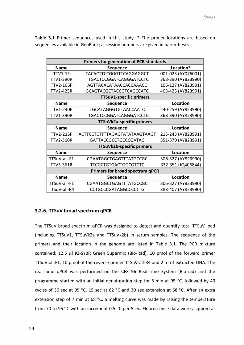
Study I
29
Table 3.1 Primer sequences used in this study. * The primer locations are based on
sequences available in GenBank; accession numbers are given in parentheses.
Primers for generation of PCR standards
Name Sequence Location* TTV1-1F
TACACTTCCGGGTTCAGGAGGCT 001-023 (AY076001)
TTV1-390R TTGACTCCGGATCAGGGATCCTC 368-390 (AY823990) TTV2-106F
AGTTACACATAACCACCAAACC 106-127 (AY823991)
TTV2-425R GCAGTACGCTACCGTCAGCCATC 403-425 (AY823991) TTSuV1-specific primers
Name Sequence Location
TTV1-240F TGCATAGGGTGTAACCAATC 240-259 (AY823990) TTV1-390R TTGACTCCGGATCAGGGATCCTC 368-390 (AY823990)
TTSuVk2a-specific primers
Name Sequence Location
TTV2-215F ACTTCCTCTTTTAGAGTATATAAGTAAGT 215-243 (AY823991) TTV2-360R GATTACCGCCTGCCCGATAG 351-370 (AY823991)
TTSuVk2b-specific primers
Name Sequence Location
TTSuV-all-F1 CGAATGGCTGAGTTTATGCCGC 306-327 (AY823990) TTV3-361R TTCGCTGTGACTGGCGTCTC 332-351 (JQ406844)
Primers for broad spectrum qPCR
Name Sequence Location
TTSuV-all-F1 CGAATGGCTGAGTTTATGCCGC 306-327 (AY823990) TTSuV-all-R4 CCTGCCCGATAGGCCCCTTG 388-407 (AY823990)
3.2.6. TTSuV broad spectrum qPCR
The TTSuV broad spectrum qPCR was designed to detect and quantify total TTSuV load
(including TTSuV1, TTSuVk2a and TTSuVk2b) in serum samples. The sequence of the
primers and their location in the genome are listed in Table 3.1. The PCR mixture
contained: 12.5 l iQ-SYBR Green Supermix (Bio-Rad), 10 pmol of the forward primer
TTSuV-all-F1, 10 pmol of the reverse primer TTSuV-all-R4 and 2 l of extracted DNA. The
real time qPCR was performed on the CFX 96 Real-Time System (Bio-rad) and the
programme started with an initial denaturation step for 5 min at 95 C, followed by 40
cycles of 30 sec at 95 C, 15 sec at 62 C and 30 sec extension at 68 C. After an extra
extension step of 7 min at 68 C, a melting curve was made by raising the temperature
from 70 to 95 C with an increment 0.5 C per 5sec. Fluorescence data were acquired at

Study I
30
each extension step and during the melt curve. As standard curve a dilution series of
plasmid TTV008 was used. The qPCR amplification efficiency was 102.8%; the inter-assay
variation was 1.6%; the LOD was 10 copies reaction-1, which corresponded to 1250 copies
ml serum-1.
3.2.7. Generation of PCR standard curves and viral DNA load calculation
The TTSuV1 PCR standard, TTV001, was generated by PCR amplification of a 390 bp
sequence from DNA isolated from Kreaber porcine serum (9071172-G50) using primers
TTV1-1F and TTV1-390R (Table 3.1). For the generation of the TTSuVk2a and broad
spectrum TTSuV qPCR standard, TTV008, a 320 bp PCR product was obtained from DNA
isolated from Hyclone porcine serum (FSD29672) using primers TTV2-106F and TTV2-
425R (Table 3.1). Both PCR products were cloned in vector pSC-A-amp/kan following the
instruction manual of the StrataClone PCR cloning kit (Stratagene). A 10-fold dilution
series of plasmids TTV001 and TTV008 was used to generate PCR standard curves
corresponding to 1x10⁷ - 1x101 copies of DNA per reaction. To calculate the amount of
TTSuVk2b DNA in sera, a 10-fold dilution series was used corresponding to 3.3x10⁵ -
3.3x100 copies reaction-1 of plasmid 38E05 that contained the full-length sequence of
clone 38E05 in vector pSC-A-amp/kan.
Finally, the average log10 DNA copies ml-1 of serum was used to compare data.
3.2.8. Statistical analyses
The correlation test was used to compare the prevalence of viruses. Normality of data
was assessed using the Shapiro-Wilk test. In order to assess differences of viral loads
between healthy and PCV2-SD groups for each TTSuV, Student’s t-test was used for
normally distributed samples (TTSuV1 and TTSuVk2b) and Mann–Whitney U test was
used for non-parametric data (TTSuVk2a). All statistical analysis were made using SPSS
Statistics for Windows (SPSS Inc). Significance was set at P<0.05 for all tests.

Study I
31
3.3. Results
3.3.1. A novel pig-infecting Anellovirus identified using RCA technique
Commercial sera are generally pools of serum from several pigs and therefore a useful
source for detecting viruses. To determine which porcine Anelloviruses were present in
commercial sera, the RCA technique with random primers was applied on extracted
serum DNA. Amplicons of approximately 2.9 kb corresponding to the size of the full-
length TTSuV genome were cloned resulting in 21 clones of which 11 contained a full-
length TTSuV genome and 10 a partial TTSuV sequence of 2.6-2.7 kb in length.
All clones were sequenced and compared with the sequence data available in GenBank to
annotate the genomes. Eleven clones had an insertion that was closely related to TTSuV1.
In Table 1 these clones were further divided in four different subtypes (Cortey et al.,
2011). Five clones were annotated as TTSuVk2 subtypes. Five clones contained similar
inserts that were identified as TTSuV sequences, but were considerably different from
either TTSuV1 or TTSuVk2 sequences, suggesting that a novel TTSuV species was
identified (Table 3.2).
Table 3.2 Characterization of 21 TTSuV clones derived from Hyclone Porcine Serum
Subtype
Total 1a 1b 1c 1d 2a 2b
TTSuV1 (complete genome) 3 - - 1 2 - - TTSuV1 (partial genome) 8 1 7 - - - -
TTSuVk2 (complete genome) 5 - - - - 5 - TTSuVk2 (complete genome) 3 - - - - - 3
TTSuVk2 (partial genome) 2 - - - - - 2
3.3.2. Novel TTSuV sequences represented a new anellovirus species
The International Committee on Taxonomy of Viruses (ICTV) has classified TTVs into the
family Anelloviridae based on the DNA sequence of the largest open reading frame, ORF1
(Biagini, 2009; Biagini et al., 2011; Biagini et al., 2005). According to the ICTV the ORF1

Study I
32
nucleotide sequence divergence cut-off values to distinguish species is >35% and >56%
for genera. To further classify the novel sequences, phylogenetic and pairwise sequence
comparison (PASC) analyses were performed. The mean group distance of the ORF1
sequences with TTSuV1 isolates was 63.1 (61.9-65.6%) and 41.7% (40.26-43.59%) with
TTSuVk2, while the phylogenetic distance between TTSuV1 and TTSuVk2 species is 59.5%
(56.6-62%) (Figure 3.1). Phylogenetic tree constructions using nucleotide sequence of full-
length genomes resulted in similar tree topology as obtained with ORF1. Therefore, the
unclassified viral sequences were considered to represent a novel pig anellovirus species
within the genus Kappatorquevirus. The novel virus was provisionally designated as
TTSuVk2b. Logically, and following the naming of Iotatorqueviruses, TTSuVk2 should be
renamed as TTSuVk2a.
Next, the three obtained full length TTSuVk2b sequences, 38E05, 38E19, and 38E23, were
annotated and the deduced genomic features are presented in Table 3.2. Genomes 38E19
and 38E23 were 99% identical on the nucleotide sequence level, whereas genome 38E05
was only 95-96% identical to them. The TTSuVk2a genomes contained the characteristic
GC-rich region and conserved sequences in the non-coding region In addition, ORF1 and
its splice variant ORF1/1, ORF2 and ORF3 were identified. The deduced aa sequences of
ORF1 contained a typical arginine-rich N-terminus and nuclear localization signals. Rolling-
circle replication motif III located at aa position 473 (YxxK) (Huang et al., 2010b; Martínez-
Guinó et al., 2011; Müeller et al., 2008; Mushahwar et al., 1999) while motif II (HxQ)
(Huang et al., 2010b; Mankertz et al., 2004) could not be identified. This domain has only
been detected in TTSuVk2a, but not in TTSuV1 (Huang et al., 2010b; Martínez-Guinó et
al., 2011). Motif I (FTL) described in human TTV studies (Müeller et al., 2008) was not
detected in any TTSuV species. TTSuVk2b-ORF2 contained a protein-tyrosine phosphatase
motif (aa position 21-41) typically found in anelloviruses (Martínez-Guinó et al., 2011;
Müeller et al., 2008). The TTSuVk2b3 ORF1, ORF1/1 and ORF3 proteins were predicted to
be nuclear with more than 90% reliability, all of them containing bipartite, pat4 and pat7
nuclear localization signals (NLS). No NLS were detected in ORF2, which was predicted to
be cytoplasmic with the reliability of 94%. The predicted promoter region (5’-
CTTTTTAAGTATATAAGTAAGTGCACTGGCGAATGGCTGAGTTTATGCCG-3’) was highly

Study I
33
conserved between TTSuVk2b and TTSuVk2a. The splicing site for ORF1/1 and ORF3 was
similar to the one described in TTSuVk2a ORF1 (Martínez-Guinó et al., 2011). However,
other splice sites mapped for TTSuVk2a in transfection experiments (Martínez-Guinó et
al., 2011) could not be annotated for TTSuVk2b.
Figure.3.1. Neighbour-joining phylogenetic tree based on the percentage identity of
TTSuV ORF1 sequence available in GenBank and from this study. Confidence bootstrap
values higher than 50 % are shown at the nodes.

Study I
34
Table 3.3. Summary of the TTSuV genomic features. Genome and ORF lengths in base
pairs are shown. Data for TTSuV1 and k2 were taken from previous studies (Cortey et al.,
2011;Martínez-Guinó et al., 2011).
3.3.3. TTSuV species from different origins are found in commercial porcine sera
of different origin
Quantitative broad spectrum and species specific qPCR techniques were applied to screen
seven commercial sera originating from either USA, New Zealand or Germany for the
presence of TTSuVs (Table 3.3). All tested commercial sera, which are pools from a large
number of pigs, contained each of the three viral species. The amount of total TTSuV
varied between 5.93 and 6.65 log10 DNA copies ml-1 serum. In each batch a similar pattern
of prevalence of the three TTSuVs was found and the prevalence of TTSuVk2b was much
lower than that of the other two viruses. This can be confidently concluded since the
three species-specific qPCRs (TTSuV1, TTSuVk2a and k2b) had the same limit of detection
and very similar amplification efficiencies (see 3.2 section). The relative percentage of
TTSuVk2b detected was 0.7-2.1%, while those of TTSuV1 and TTSuVk2a were 18.0-55.3%
and 42.8-81.4%, respectively (Table 3.3)
Species
TTSuV1 TTSuVk2a TTSuVk2b
Genomic length 2863-2913 2735-2803 2899-2901
ORF1 nt (aa) 1914-1950 (638-650) 1875-1884 (625-628) 1890-1893 (629-630)
ORF1/1 nt (aa) 550 (181) 535 (176) 577 (188) ORF2 nt (aa) 219-222 (73-74) 207 (69) 243 (81) ORF3 nt (aa) 663-696 (221-232) 600-609 (200-203) 673 (224)

Study I
35
Table 3.4. TTSuV loads (log10 DNA copies ml-1 serum) in commercial batches of porcine
serum determined by the broad spectrum and TTSuV-specific qPCRs. The percentage of
TTSuV species is indicated between brackets.
Porcine serum Broad spectrum TTSuV1 TTSuVk2a TTSuVk2b
Hyclone 0000066666 6.48 5.84 (20.8) 6.41 (77.2) 4.84 (2.1) Sigma 026k8453 6.39 5.81 (24.6) 6.28 (73.6) 4.69 (1.9) Sigma 064k8451 6.08 5.98 (55.3) 5.87 (42.8) 4.52 (1.9) Kreaber 9050957-G50 6.65 5.92 (20.1) 6.52 (79.3) 4.45 (0.7) Kreaber 9071172-G50 6.47 5.66 (18.0) 6.32 (81.4) 4.23 (1.7) Kreaber SS02/06-1 6.09 5.63 (27.2) 6.04 (71.1) 4.41 (1.7) Kreaber SS03/06 5.93 5.70 (42.6) 5.81 (55.2) 4.40 (2.1)
Average 6.30 5.79 (26.3) 6.18 (72.2) 4.50 (1.4)
3.3.4. Global prevalence and viral DNA loads of TTSuV species in conventional
pigs
Once the presence of TTSuVk2b3 in commercial pools of porcine sera was confirmed we
aimed to determine the global distribution of it and its relation to the already well
characterized TTSuV1 and k2a (Cortey et al., 2012; McKeown et al., 2004). For this a total
of 244 sera samples collected from farms located in 17 different countries were analysed
by the different qPCRs.
In general, the total prevalence of TTSuV was 92.6 (226/244) and 74.6 % for TTSuV1
(182/244), 61.1 % for TTSuVk2a (149/244), and 41.0 % for TTSuVk2b (100/244) (Table
3.5). While TTSuV1 could be detected in sera from all countries analysed, TTSuVk2a was
not found in the samples originating from Brazil and Vietnam, and TTSuVk2b was not
present in sera from Brazil, Ukraine and Vietnam (Figure.3.2 and Table 3.5). TTSuVk2a and
TTSuVk2b global prevalences were correlated (P<0.05), while the prevalence of TTSuV1
did not correlate with that of the other two.

Study I
36
Figure 3.2. TTSuV loads and prevalences in pig sera collected from pig farms from several countries. TTSuV1, TTSuVk2a and TTSuVk2b log10 DNA copies ml-1 serum and
error bars represent SD. Total number of analysed sera is shown after the name of the country and number of positive samples in the corresponding bar

Study I
37
There was in general a very good correlation between the total viral loads determined by
the broad spectrum qPCR and the sum of the viral loads determined by the TTSuVq-
TTSuVk2a- and TTSuVk2b-specific qPCRs: of 224 samples analysed, only 21 showed a
greater than fivefold difference (data not shown). In three cases the broad spectrum
qPCR detected TTSuV, while the specific qPCRs failed to detect TTSuV species. It was
attempted by RCA to isolate TTSuV from the corresponding three sera, but this was
unsuccessful probably due to the low levels of TTSuV in these sera.
Table 3.5 TTSuV prevalence (positives samples/total number of samples) in pig sera from
17 different countries determined by qPCR. Percentage of the positive sera is shown in
parenthesis.
Country TTSuV1 TTSuVk2a TTSuVk2b Borad spectrum
Belarus 6/8 (75.0) 4/8 (50.0) 8/8 (100) 8/8 (100)
Brazil 3/8 (37.5) 0/8 (0.0) 0/8 (0.0) 3/8 (0.0)
Canada 25/30 (83.3) 30/30 (100) 25/30 (83.3) 30/30 (83.3)
Greece 38/40 (95.0) 28/40 (70.0) 23/40 (57.5) 40/40 (57.5)
China 7/8 (87.5) 7/8 (87.5) 6/8 (75.0) 8/8 (75.0)
Lithuania 8/8 (100) 5/8 (62.5) 3/8 (37.5) 8/8 (37.5)
Mexico 6/8 (75.0) 3/8 (37.5) 1/8 (12.5) 5/8 (12.5)
Mozambique 3/22 (13.6) 22/22 (100) 9/22 (40.9) 22/22 (40.9)
Netherlands 11/20 (55.0) 4/20 (20.0) 1/20 (5.0) 18/20 (5.0)
Philippines 7/8 (87.5) 1/8 (12.5) 2/8 (25.0) 7/8 (25.0)
Russia 6/8 (75.0) 1/8 (12.5) 1/8 (12.5) 5/8 (12.5)
South Africa 13/15 (86.6) 15/15 (100) 7/15 (46.6) 15/15 (46.6)
Spain 25/29 (86.2) 13/29 (44.8) 10/29 (34.4) 27/29 (34.4)
Thailand 3/8 (37.5) 7/8 (87.5) 2/8 (25.0) 8/8 (25.0)
Ukraine 8/8 (100) 2/8 (25.0) 0/8 (0.0) 7/8 (0.0)
Venezuela 6/8 (75.0) 5/8 (62.5) 2/8 (25.0) 8/8 (25.0)
Vietnam 7/8 (87.5) 2/8 (25.0) 0/8(0.0) 7/8(0.0)
Total 182/244 (74.6) 149/244 (61.1) 100/244 (41.0) 226/244 (92.6)

Study I
38
Viral DNA loads were in the range of 3.12 to 7.90 (average 5.79) log10 DNA copies ml-1
serum for TTSuV1, 3.18 to 8.41 (average 4.58) log10 DNA copies ml-1 serum for TTSuVk2a,
and 3.13 to 6.87 (average 4.14) log10 DNA copies ml-1 serum for TTSuVk2b in conventional
pigs. There was no correlation of viral loads among the three TTSuV species. The average
TTSuV1 and TTSuVk2b loads in sera of individual pigs were similar to the loads observed
in commercial sera (Table.3.3). However, the average TTSuVk2a loads in individual sera
were approximately 1.5 log10 lower than that in commercial sera.
3.3.5. High TTSuVk2b loads in serum of PCV2-SD-affected pigs
TTSuVk2a has been associated in previous studies with porcine circovirus associated
diseases (Huang et al., 2011) and especially to PCV2-SD occurrence (Aramouni et al.,
2011; Nieto et al., 2011). Considering the molecular similarity of TTSuVk2a with
TTSuVk2b, and the correlation of their prevalence in geographical samples, the possible
association of TTSuVk2b with PCV2-SD was explored. TTSuV1, k2a and k2b viral DNA loads
were measured by the three TTSuV-specific qPCRs in 34 serum samples of PCV2-SD-
affected pigs and 29 healthy control animals. The average TTSuVk2a and TTSuVk2b loads
were significantly higher (P<0.05) in PCV2-SD-affected pigs than in healthy animals (Figure
3.2). Such difference was not observed for TTSuV1.
Figure 3.3. TTSuV1 (a), TTSuVk2a (b) and TTSuVk2b (c) log 10 mean viral DNA loads in
healthy and PCV2-SD-affected animals P-values are shown.

Study I
39
3.4. Discussion
Three species were so far known to infect pigs before the present study, namely TTSuV1a
and 1b within the genus Iotatorquevirus and TTSuVk2a within the genus
Kappatorquevirus. Here, a novel anellovirus was identified in porcine sera, which,
according to the degree of divergence with already known TTSuVs is sufficient to warrant
classification of this virus as a new species in the genus Kappatorquevirus. Considering the
current taxonomic organization of anelloviruses, this novel virus species was tentatively
named TTSuVk2b.
Although there have been attempts to discover novel pig anelloviruses (Macera et al.,
2011), TTSuVk2b was not detected before. Primers used in published TTSuV1- or
TTSuVk2a-specific PCRs (Brassard et al., 2010; Gallei et al., 2010; Huang et al., 2010a;
Nieto et al., 2011) are unlikely to amplify TTSuVk2b given the mismatch of these primers
with TTSuVk2b sequences. Furthermore, sequencing of these amplicons is necessary to
identify possible new species.
Different qPCR techniques were developed in this study. The wide spectrum qPCR was
shown to be very useful in determining the overall TTSuV load (TTSuV1, TTSuVk2a and
TTSuVk2b) in pig serum. This technique could be useful for diagnostic purposes and, when
necessary, the species-specific qPCRs can be useful to determine the loads of each TTSuV
species.
Bioinformatics analysis of full length TTSuVk2b sequences showed that these viruses are
rather distinct from, already known pig anelloviruses. However, a high similarity with
TTSuVk2a was observed on genomic regions like splice sites and promoter elements
(Martínez-Guinó et al., 2011). These predictions may not reflect the reality, sine a short
intron of 91 bp could not be predicted from any genome of TTSuVk2 but has been shown
to exist in genomes of TTSUVk2a by in vitro transfection experiments(Huang et al., 2012b;
Martínez-Guinó et al., 2011), and therefore further experiments are necessary to
characterize TTSuVk2b transcription and protein expression strategies. The relatedness
to TTSuVk2a was supported by phylogenetic analysis that grouped TTSuVk2b closer to
TTSuVk2a than TTSuV1. Considering the current criteria by the ICTV, TTSuVk2b would be

Study I
40
the second species in the genus Kappatorquevirus
In the epidemiological study, TTSuVk2b infection was confirmed in pigs from most of the
countries. However, for some countries where TTSuVk2b appeared to be absent the
number of serum samples analysed was low, so in order to conclude the presence of
TTSuVk2b in these countries, more samples and farms should be analysed. There is also a
possibility that the TTSuVk2b-specific primers do not detect all TTSuVk2b variants, since
the primer binding sites all TTSuVk2b variants, since the primer binding sites were
determined based only on the five TTSuVk2b sequences that were obtained in this study
(see Table 3.2). Therefore, more viral genomic sequences should be obtained to confirm
that the annealing sites of the used primers are highly conserved. Considering the global
trading and its influence on the distribution of TTSuVs (Cortey et al., 2012), it is likely that
each TTSuV species would be found in any given country.
Overall, it was seen for each TTSuV that the higher the prevalence the higher the viral
load in serum. It is well documented that TTSuV1 and TTSuVk2 viral loads and prevalences
increase with the age of the animals (Aramouni et al., 2010; Nieto et al., 2011; Xiao et al.,
2012). This is probably due to the efficient viral transmission by vertical and horizontal
routes and the persistent nature of TTSuV infection (Martínez-Guinó et al., 2009; Pozzuto
et al., 2009) in pig and wild boar (Martínez et al., 2006). Therefore, the observed
differences between countries in TTSuV prevalence and viral loads may reflect the age of
the tested animals, which was not known in this study.
Due to the genetic similarity between TTSuVk2a and TTSuVk2b and the reported
association of TTSuVk2a, but not TTSuV1, with PCV2-SD (Aramouni et al., 2011;
Kekarainen et al., 2006; Nieto et al., 2011), it was also assessed whether TTSuVk2b was
also associated with this disease. Indeed, both TTSuVk2a and TTSuVk2b DNA loads were
significantly higher in PCV2-SD-affected animals compared to healthy counterparts. No
significant difference was seen in the case of TTSuV1, which confirmed previous findings.
It has to be mentioned that the serum samples of PCV2-SD and healthy animals tested in
this study were the same as analysed before with different qPCR methods (Aramouni et
al., 2011; Kekarainen et al., 2006; Nieto et al., 2011). However, in this study the samples

Study I
41
were analysed with a SYBR Green-based qPCR technique, while in the earlier studies Light
Upon Extension (LUX) qPCRs were used. With both assays association of TTSuVk2a loads
with PCV2-SD was confirmed, but the viral loads were quantified about 10-fold higher
with the LUX assay. This is not surprising considering that the techniques use different
quantification methods and primer pairs.
With the description here of a novel TTSuV species, provisionally named TTSuVk2b, this
group of pig-infecting viruses has become even more intriguing. These viruses are
ubiquitous in pig and it has been demonstrated that the three species, TTSuV1, TTSuVk2a,
and -TTSuVk2b, are often found with high loads in serum of the same animal. This may be
due to lack of cross-protection between species. Indeed, antigenic cross-reactivity has
been demonstrated to exist between different genotypes (aa identity about 50%) but are
lacking between species (aa identity about 25%) (Huang et al., 2012a). The aa identity of
ORF1 between TTSuVk2a and TTSuVk2b is about 58.3% and therefore cross-reactivity is
likely to exist between these species. To get further insight into the possible cross
protection, the infection dynamics of and antibody development against TTSuVk2a and
TTSuVk2b should be investigated using longitudinal serum samples.
Since TTSuVs are known to cause persistent infections, important questions are raised
about the role of these viruses in modulating the immune status of the animals. Although
considered non-pathogenic by themselves, TTSuV is often present in pigs with diseases
like PCV2-SD, PRDS (Rammohan et al., 2012), and PDNS (Aramouni et al., 2011; Krakowka
et al., 2008). Studies are undertaken in our laboratory to further address the disease
association of TTSuV.

42

4. Study II
Expression of and pig immunization with Torque teno sus virus
proteins


Study II
45
4.1. Introduction
Anelloviruses are species-specific and the ones infecting swine are classified into four
species, namely Torque teno sus virus 1a and 1b (TTSuV1a, TTSuV1b; genus
Iotatorquevirus), and TTSuVk2a and TTSuVk2b (described study I) (genus
Kappatorquevirus). These genera are genetically very distinct (>56% sequence diversity),
but share similar genome organization and gene expression strategy (Martínez-Guinó et
al., 2011). TTSuV infection in pigs is distributed worldwide and characterized by a
persistent viremia with high prevalence of TTSuV1 and TTSuVk2a species (Sibila et al.,
2009a) while prevalence of TTSuVk2b is lower (according study I). TTSuVs have been
found in serum, plasma, faeces, semen, colostrum and nasal cavity, suggesting the
transmission can be both vertical and horizontal (Kekarainen & Segalés, 2012a). A single
pig can be co-infected with different TTSuV species or subtypes such as confirmed in
study I and previous reported (Gallei et al., 2010; Huang et al., 2010b)
The length of the TTSuV genome is approximately 2.8 kb (Okamoto et al., 2002),
consisting of at least four putative open reading frames (ORF), namely ORF1, ORF1-A (also
known as ORF1/1), ORF2, and ORF3 (also known as ORF2/2) (Huang et al., 2012b; Lu et
al., 2013; Martínez-Guinó et al., 2011). ORF1 gene encodes the largest protein (~75 kDa)
with a predicted viral capsid domain in the N-terminal half of the protein and a putative
replication-associated domain in its C-terminal half. ORF2 gene encodes a non-structural
protein, ORF2 protein, assumed to be involved in viral replication (Hijikata et al., 1999;
Huang et al., 2012b). ORF3 can only be translated after splicing of a mRNA precursor
(Huang et al., 2012b; Martínez-Guinó et al., 2011). The ORF3 protein (~25 kDa) shares its
N-terminus with the ORF2 protein (~8 kDa) and its role is unknown at present. The
predominant transcripts (i.e. mRNA) detected in vitro and in vivo of ORF1 gene are spliced
and full-length ORF1 transcripts have not been detected so far (Huang et al., 2012b;
Martínez-Guinó et al., 2011). Splicing of TTSuV1 ORF1 gene results in two protein
isoforms (ORF1-A and -B) of different sizes (~20 and ~15 kDa, respectively) (Martínez-
Guinó et al., 2011). In contrast, splicing of TTSuVk2a ORF1 gene results in three protein
isoforms (ORF1-A, -B, and -C) of different sizes (~20, ~15 and ~11 kDa, respectively); the

Study II
46
aa composition between them varies, depending on the splicing site used (Huang et al.,
2012b; Martínez-Guinó et al., 2011).
TTSuVs persistently infect a high proportion of animals that are apparently healthy (Sibila
et al., 2009b). TTSuV infection by itself does not cause immediate disease but it is
believed that TTSuVs influence the development of some economically important viral
diseases or even affect their outcome. Evidence for TTSuV disease association has been
accumulated, especially with regard to Porcine circovirus type 2 (PCV2) infection and its
associated conditions, namely PCVDs (Aramouni et al., 2011; Ellis et al., 2008; Kekarainen
et al., 2006; Nieto et al., 2011).
Although TTSuVs are currently considered non-pathogenic, it is hard to believe that a viral
infection characterized by high viral loads and virus persistence would not have any
consequence to its host. Indeed, farm performance is improved when PCV2 (also a single
stranded circular DNA virus) infection is controlled by vaccination even in farms without
disease, displaying only a subclinical infection (Beach & Meng, 2012).
This study had two main objectives. On one hand, the four TTSuV proteins for all known
TTSuV species (i.e. TTSuV1a, 1b, k2a, and k2b) were expressed in a baculovirus system.
On the other hand, a multivalent vaccine prototype was designed against currently
circulating TTSuV species to be used to control viremia in naturally infected pigs. For this
latter purpose, the vaccine was designed containing baculovirus expressed ORF1 and
ORF3 proteins of the four known TTSuV species. Subsequently, the vaccine prototype
efficacy was evaluated by immunizing pigs; humoral immunity and viremia during natural
TTSuV infection were monitored by ELISA and species-specific quantitative PCRs (qPCR),
respectively. In addition, specific antibodies against the multivalent vaccine prototype
where followed by indirect ELISA.

Study II
47
4.2. Material and methods
4.2.1. TTSuV protein selection for expression in a baculovirus expression system
A selection of available ORF1, ORF1-A, ORF2 and ORF3 sequences of the four TTSuV
species were downloaded from GenBank and aligned using MEGA version 5 (Tamura et
al., 2011). Representative sequences from each of the 4 TTSuV groups were selected
(Table 4.1), which conserve a nucleotide sequence of each gene with the majority of the
sequence analysed for each species. Clones containing each gene in the pFastBac/CT-
TOPO vector were ordered from GenScript. All genes contained a Strep-tag coding
sequence at their 3’end with the exception of the ORF1 constructs of TTSuV1b and
TTSuVk2a, which were already available in our laboratory with a 6xHis-tag sequence. As a
control in expression experiments, a GFP gene containing a Strep-tag at its 3’ end in
pFastBac/CT-TOPO was ordered from GenScript (Table 4.1).
4.2.2. Production of TTSuV baculovirus stocks
Baculovirus production was done according to the manufacturer’s instructions
(Invitrogen). Briefly, the pFastBac constructs were transformed into Escherichia (E.) coli
DH10BacΔcc (Kaba et al., 2004). PCR was used to confirm the correctness of recombinant
Bacmids. Bacmid DNA was subsequently isolated from E.coli cultures and used to
transfect Spodoptera frugiperda (Sf9) insect cells using Cellfectin II reagent (Invitrogen).
The cultures were incubated at 27 °C for 5 days, and supernatants were harvested as the
first passage (P1) viral stocks. P1 stocks were used to infect insect cell cultures at a
multiplicity of infection (MOI) of 0.1 to obtain high virus titre stocks (P2). To quantify the
viral stock titre, TTSuV baculovirus stocks were added at 10-fold serial dilutions to Sf9
cells in 96-well microtitre plates and after 6 days incubation at 27°C, an
immunofluorescence assay (IFA) was performed to detect the baculoviruses.

Study II
48
Table 4.1 Overview of the generated Bacmids, the size of the TTSuV insert and
corresponding proteins when expressed in Sf9 cells. The size of the PCR fragment to
confirm the correct insertion of the TTSuV gene into the Bacmid is also indicated. Control
bacmids, dccGFP and dcc_empty, or also listed. GenBank accession numbers for each
TTSuV are indicated.
Name construct
Accession numbers
Gene PCR product (DNA
Bacmid) Insert (nt)
Protein (aa)
TTSu
V1
a dccTTSuV1a_ORF1 GU570202 ORF1 4291 1991 73
dccTTSuV1a_ORF1-1 GU570202 ORF1-1 2896 596 22
dccTTSuV1a_ORF2 GU570202 ORF2 2563 263 10
dccTTSuV1a_ORF3 GU570202 ORF3 3040 740 27
TTSu
V1b
dccTTSuV1b_ORF1 AY823990 ORF1 4213 1913 70
dccTTSuV1b_ORF1-1 AY823990 ORF1-1 2893 593 22
dccTTSuV1b_ORF2 AY823990 ORF2 2563 263 10
dccTTSuV1b_ORF3 AY823990 ORF3 3031 731 27
TTSu
Vk2
a dccTTSuVk2a_ORF1 AY823991 ORF1 4174 1874 69
dccTTSuVk2a_ORF1-1 GU570205 ORF1-1 2890 590 22
dccTTSuVk2a_ORF2 GU570205 ORF2 2551 251 9
dccTTSuVk2a_ORF3 GU570205 ORF3 2953 653 24
TTSu
Vk2
b dccTTSuVk2b_ORF1 JQ406845 ORF1 4237 1937 71
dccTTSuVk2b_ORF1-1 JQ406845 ORF1-1 2911 611 22
dccTTSuVk2b_ORF2 JQ406845 ORF2 2587 287 11
dccTTSuVk2b_ORF3 JQ406845 ORF3 3019 719 26
dcc_GFP GFP 3064 764 25
dcc_empty - 2300 - -
For IFA procedure, Sf9 cells were fixed with 4 % paraformaldehyde for 10 min at room
temperature. Next, cells were washed three times with 1x phosphate buffered saline (1x
PBS) and permeabilized with 0.5 % Triton X-100 in 1x PBS for 15 min at room temperature
and finally washed with 1x PBS for 5 min. Then, the cells were incubated at 37ºC for 1 h
with anti-Strep (Qiagen) or anti-His (in house; INT-HIS-O1-O3) monoclonal antibodies
diluted 1:1000 in 1x PBS. Then, the cells were washed three times with sterile water and
incubated with a FITC-conjugated goat anti-mouse IgG (whole molecule) antibody (Sigma)
diluted 1:100 in 1x PBS with Evans Blue at 37ºC for 1 h. Finally, cells were covered with 1x
PBS/glycerol (1:1) before observing with a fluorescence microscope at a wavelength of
488 nm.

Study II
49
4.2.3. Expression of TTSuV proteins in insect cells
ORF1, ORF1-A, ORF2, and ORF3 baculovirus stocks were used to infect Sf9 insect cells at a
MOI of 0.1 to produce TTSuV recombinant proteins. In addition, a GFP and an empty
baculovirus were used as controls. The cultures were incubated at 27ºC and harvested at
4 days post infection (dpi) and then centrifuged at 1,000xg for 5 min at 4ºC. The resulting
supernatants and cell pellets were analysed by Sodium Dodecyl Sulfate-polyacrylamide
gel electrophoresis (SDS-PAGE) and Western blotting. For this, samples were diluted 1:1
in 2x Laemmli sample buffer (Bio-Rad), heated to 95°C for 5 min and loaded onto 4-15%
precasted polyacrylamide gels (Criterion TGX, Bio-Rad) followed by SDS-PAGE. Coomassie
blue dye (Bio-Rad) was used to stain the proteins present in the SDS-PAGE gels.
Alternatively, the gels were not stained and proteins were transferred to polyvinylidene
fluoride (PVDF) membranes (Bio-Rad), which were blocked in 1% non-fat milk in TBS-T
(Tris-buffered saline, 0.1% Tween20) blocking buffer. PVDF membranes were then
incubated with anti-Strep (Qiagen) or anti-His (in house; INT-HIS-O1-O3) antibody, both
diluted to 1:1000 in blocking buffer at room temperature for 1 h. Next, membranes were
incubated with a horseradish peroxidase conjugated goat anti-mouse antibody (Nordic) at
a 1:1000 dilution in blocking buffer at room temperature for 1 h. Peroxidase (Vector
Laboratories) was used as substrate and colour development was stopped by rinsing the
membranes with water and air drying.
4.2.4. Purification of the recombinant ORF1, ORF2, ORF1-A and ORF3 proteins
Pelleted baculovirus-infected insect cells were used to prepare crude cellular lysates.
Solubility tests were performed to find the optimal lysis buffer for each expressed
protein. All lysis buffers were supplemented with a protease inhibitor cocktail (Complete,
EDTA-free; Roche). The optimal lysis buffer for the ORF1 protein was Buffer 5O, pH 8 (50
mM Na2HPO4, 600 mM NaCl, 0.05% Tween 20, 1 U/µl Benzonase). For the ORF2 protein it
was Buffer NP-T, pH8 (50 mM NaH2PO4, 300 mM NaCl, 0.05% Tween 20, 1 U/µl
Benzonase) and finally for the ORF1-A and ORF3 proteins it was Buffer 4O, pH 8 (50 mM
Na2HPO4, 300 mM NaCl, 10% glycerol, 1 U/µl Benzonase). After lysis, suspensions were
sonicated three times on ice at 1 min cycles with 10 s intervals at 20% amplitude. The

Study II
50
sonicated lysates were centrifuged at 16,000xg at 4ºC for 10 min. Resulting supernatants
were used to purify the proteins of interest.
Strep- and His-tagged recombinant TTSuV ORF1 proteins were purified using the
sedimentation coefficient (s) by Beckman Coulter ultracentrifugation. The
ultracentrifugation was done using the SW41Ti rotor with a K factor of 123.6 at maximum
speed (41,000 rpm) at 4ºC. In total, 3 steps were used and the step(s) that yielded the
best protein yield in the pellet and highest purity was/were selected: s200: 37min, s100:
1h 14min, and s50: 2h 28min. Equilibration buffer (250 mM MgCl2 and 50 mM HEPES) was
used to adjust volume up to 12 ml.
Strep-tagged recombinant TTSuV ORF2 proteins were purified using Strep-Tactin
Magnetic beads (Qiagen) according to the manufacture’s protocol using washing buffer
Buffer NP-T, pH 8 (without protease inhibitor and Benzonase) and elution buffer NPB-T
(50 mM NaH2PO4, 300 mM NaCl, 10 mM biotin and 0.05% Tween 20) at pH 8. Strep-
tagged recombinant TTSuV ORF1-A and ORF3 proteins were purified using Strep-Tactin
Magnetic beads (Qiagen) according to the recommended protocol using washing buffer
4O, pH 8 (without protease inhibitor and Benzonase) and elution buffer Buffer 4-B, pH 8
(50 mM Na2HPO4, 300 mM NaCl, 10 mM biotin, 10% glycerol). Purified proteins were
analysed by SDS-PAGE and Western blotting as described above.
4.2.5. Pig immunization with multivalent TTSuV vaccine
A multivalent vaccine prototype was designed including His- and Strep-tagged
recombinant TTSuV ORF1 and Strep-tagged ORF3 proteins produced as described above.
However, Strep-Tactin Superflow Plus (Qiagen) was used to purify the Strep-tagged ORF3
protein following the recommended protocol (with the same changes to protocol as
above: Buffer 4O, pH8 used in the wash step and Buffer 4O-B, pH8 used in the elution
step). Recombinant baculoviruses present in the purified protein preparations were
inactivated by binary ethylenimine (BEI) inactivation using a treatment of 30 mM BEI for
the ORF1 preparation and 5 mM for the ORF3 preparation. Protein concentrations were

Study II
51
estimated by SDS-PAGeS and comparison of band intensities to that of a Bovine Serum
Albumin (BSA) standard. Preparation of diluted BSA standards was done following the
manufacturer Pierce™ BCA Protein Assay Kit (Life Technologies). To prepare the
multivalent vaccine, ORF1 and ORF3 purified proteins of each TTSuV species were pooled
and mixed 1:1 with an oil-in-water adjuvant (X-Solve, MSD Animal Health). Intramuscular
(IM) and intradermal (ID) vaccines contained in total 20 µg of protein per dose. The
volume per injection for IM administration was 2 ml and 0.2 ml for ID administration. The
first administration was given in the right side of the neck, alternating left and right sides
in subsequent immunizations.
Figure 4.1. Experimental design of the animal trial. The administration route,
intramuscular (IM) or intradermal (ID), and the vaccination schedule (twice or three
times) are shown. Control group did not receive the multivalent vaccine. Date of serum
collection is also indicated.

Study II
52
An overview of the experimental design of the animal trial is seen in Figure 4.1. Seventy-
five (75) 4 day-old piglets (Large White x Landrace x Duroc) were selected from a
conventional Dutch farm, randomized and divided into five groups (n=15 per group). The
farm was selected based on the prevalence of TTSuV species (Figure 4.2) determined by
species-specific qPCRs performed on sera from piglets and sows (described in study I).
Pigs were immunized using the developed multivalent vaccine prototype. Intramuscular
(IM; groups 1 and 2) and intradermal (ID; groups 3 and 4) routes of administration were
tested based on the vaccination schedule shown in Figure 4.1. Thus, animals in groups 1
(IMx3) and 3 (IDx3) were vaccinated three times at 3, 5, and 7 weeks of age. Groups 2
(IMx2) and 4 (IDx2) received the multivalent vaccine twice, the first administration at 4
days and the second at 3 weeks of age. In addition, unvaccinated pigs were used as
control animals (group 5). Animals from different groups were housed together
throughout the study. Treatment, housing and husbandry conditions conformed to the
guidelines of the European Union for animal welfare and the study was performed
according to Good Experimental Practices. Serum samples were collected when the
animals were 3 weeks of age and every two weeks thereafter until the pigs reached 15
weeks of age. During the study, 3 animals died, being 1 animal from group 2 at 12 weeks
of age and two animals from group 4, one at 3 and the other at 12 weeks of age.
Figure 4.2. Dynamics of TTSuV infection in the selected farm where the animal trial was
carried out. TTSuV species were determined by species-specific qPCRs performed on sera
from piglets at different weeks of age and sows. The figure shows the percentage of
positive sample of TTSuV1 and TTSuVk2a. In total, five animals per each group of age
were tested.

Study II
53
4.2.6. Determination of viral DNA load
TTSuV DNA was extracted from 200 µl of sera using the MagNA Pure 96 DNA and Viral
Nucleic Acid Small Volume Kit (Roche Diagnostic) and using the Viral NA Universal SV 2.0
protocol. Next, TTSuV1 (detecting both species a and b), TTSuVk2a and TTSuVk2b DNA
loads were determined by species-specific qPCRs as described in the study I.
4.2.7. Antibody detection by ELISA and Western Blot analysis.
Seroconversion of animals was tested by ELISA using purified ORF1 or ORF3 TTSuV1a and
k2a baculovirus proteins (the same ones included in the vaccine) as antigens. In addition,
purified ORF1-A or ORF2 TTSuV1a and k2a proteins generated by the E.coli expression
system (Nieto et al., 2015) were used as antigens in a second ELISA also to detect TTSuV-
specific antibodies.
For both ELISA methods (based on baculovirus and E. coli proteins), low binding 96-well
microtitre plates were coated with 50 µl per well containing 680 ng/ml protein in
carbonate-bicarbonate buffer pH 9.6. After overnight incubation at 4ºC, plates were
washed 3 times with TBS-T and then blocked with blocking buffer (1x phosphate-buffered
saline, 1% casein, 0.05% Tween 20). After incubation at 37°C for 1 h, each well was
washed 3 times with TBS-T. Serum samples were diluted 1:200 in blocking buffer and 50
µl was transferred to the corresponding well. After incubation at 37ºC for 2 h, plates were
washed 3 times with TBS-T. Then 50 µl per well the diluted HRP-conjugated anti-swine
whole IgG (1:40,000 in blocking buffer; Sigma) was added and plates were incubated at
37ºC for 1 h. Finally, 50 µl of TMB substrate (3,3,5,5-Tetramethylbenzidine liquid,
supersensitive, Sigma) was added and the plates were incubated in the dark at room
temperature until the OD650 of the negative controls reached about 0.05. At this time-
point the reaction was stopped by adding 50 µl/well of 2M H2SO4, and the OD450 of each
sample was determined. All serum samples were run in duplicate. Each plate contained
one positive and one negative control serum.

Study II
54
The cut-off value of the ELISA based on baculovirus proteins was calculated as the mean
OD at 450 nm of the serum samples from control animals plus three times the standard
deviation, and the positive cut-off value was set at 0.2 for ORF3 and 0.6 for ORF1. For the
ELISA based on E. coli proteins, the cut-off value was calculated as the mean OD at 450
nm of the negative sample from each ELISA plate plus three times the standard deviation.
The relative optical density was calculated by dividing the mean OD of a sample by the
mean OD of the negative control, and the positive cut-off value was set at 2.
A Western blot assay was used to confirm the seroconversion results obtained by ELISA
based on baculovirus proteins. For this, purified TTSuV1a or TTSuVk2a ORF1 and ORF3
proteins were run under denaturing conditions in a NuPageNovex 4-12% Bis-Tris
polyacrylamide Gel (Invitrogen) and subsequently transferred to Hybond ECL
nitrocellulose membranes (GE Healthcare). The membranes were blocked with blocking
buffer (1x phosphate-buffered saline, 2% casein, 0.05% Tween 20) and incubated with
porcine sera diluted 1:100 in blocking buffer for 1 h at room temperature, followed by
addition of anti-swine whole IgG (Sigma) at a 1:150,000 dilution in blocking buffer for 1 h
at room temperature. Captured antibodies were detected by Western Lightning ultra
(PerkinElmer) and visualized by using a FluoroChem HD2 chemiluminescent workstation
(Alpha Innotech). An Strep-Tactin® horseradish peroxidase (HRP) conjugate (IBA, Solution
for life) monoclonal antibody (Invitrogen) was used as positive control to detect TTSuV1a
or TTSuVk2a ORF1 and ORF3 proteins.
4.2.8. Statistical analyses
Statistical analysis was done with IBM SPSS Statistics software, version 20. Normal
distribution was assessed using the Shapiro-Wilk test. In order to assess differences in
viral loads between groups, the student’s t-test was used for normally distributed
samples and Mann-Whitney U test was used for non-parametric data. Correlations
between antibody levels (represented by OD values) and log10 viral titres were
determined by the Pearson's correlation coefficients. P-values <0.05 were considered
statistically significant.

Study II
55
4.3. Results
4.3.1. Recombinant baculovirus construction and TTSuV protein expression and
distribution in transfected insect cells
In order to express TTSuV proteins using the baculovirus system, recombinant
baculoviruses were generated containing the ORF1, ORF1-A, ORF2 and ORF3 genes from
each TTSuV species. For that, the corresponding pFastBac vectors were transformed into
E.coli DH10BacΔcc to generate Bacmid DNA. The presence of the ORF1, ORF1-A, ORF2
and ORF3 genes in the Bacmid DNA was verified by PCR (Figure. 4.3, panel A). The
expected size of each gene is shown in Table 4.1. Bacmid DNA was subsequently
transfected into Sf9 insect cells to yield recombinant baculoviruses. The
immunofluorescence assay (IFA) demonstrated that transfected Sf9 cells expressed TTSuV
proteins as demonstrated in Figure 4.2, panel B. The ORF1 and ORF1-A proteins were
located in the cell nucleus (Figure 4.3a and b, panel B) while the ORF2 protein was located
in the cytoplasm (Figure 4.3c, panel B). The ORF3 protein was detected in both cytoplasm
and nucleus (Figure 4.3d, panel B).
4.3.2. Successful expression of and purification of TTSuV proteins from infected
insect cells.
Sf9 cells were infected using the respective TTSuV baculovirus stocks (dccBacORF1,
dccBaccORF1-A, dccBaccORF2 and dccBacORF3 for each of the four TTSuV species) and
SDS-PAGE analysis revealed the successful expression of four TTSuV proteins in the cell
fraction (data no shown). The ORF2 protein appeared also to be present in the cell culture
supernatant (Figure 4.4). While the predicted molecular weights (MW) of the ORF1-A and
ORF3 proteins for all TTSuVs are ~22 kDa and ~26 kDa, respectively (Table 4.1), the
observed MWs were approximately ~8kDa larger (Figure 4.3 a and b). The ORF1 and ORF2
proteins were observed approximately at the predicted size (~72 kDa and ~10 kDa,
respectively), except the ORF2 protein of TTSuV1a that was ~5 kDa larger than predicted.
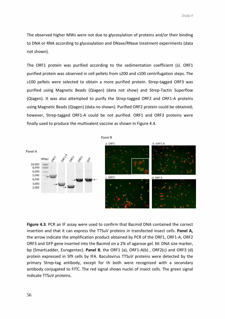
Study II
56
The observed higher MWs were not due to glycosylation of proteins and/or their binding
to DNA or RNA according to glycosylation and DNase/RNase treatment experiments (data
not shown).
The ORF1 protein was purified according to the sedimentation coefficient (s). ORF1
purified protein was observed in cell pellets from s200 and s100 centrifugation steps. The
s100 pellets were selected to obtain a more purified protein. Strep-tagged ORF3 was
purified using Magnetic Beads (Qiagen) (data not show) and Strep-Tactin Superflow
(Qiagen). It was also attempted to purify the Strep-tagged ORF2 and ORF1-A proteins
using Magnetic Beads (Qiagen) (data no shown). Purified ORF2 protein could be obtained;
however, Strep-tagged ORF1-A could be not purified. ORF1 and ORF3 proteins were
finally used to produce the multivalent vaccine as shown in Figure 4.4.
Figure 4.3. PCR an IF assay were used to confirm that Bacmid DNA contained the correct
insertion and that it can express the TTSuV proteins in transfected insect cells. Panel A,
the arrow indicate the amplification product obtained by PCR of the ORF1, ORF1-A, ORF2
ORF3 and GFP gene inserted into the Bacmid on a 2% of agarose gel. M: DNA size marker,
bp (SmartLadder, Eurogentec). Panel B, the ORF1 (a), ORF1-A(b) , ORF2(c) and ORF3 (d)
protein expressed in Sf9 cells by IFA. Baculovirus TTSuV proteins were detected by the
primary Strep-tag antibody, except for th both were recognized with a secondary
antibody conjugated to FITC. The red signal shows nuclei of insect cells. The green signal
indicate TTSuV proteins.

Study II
57
Figure 4.4 Analysis of protein
expression (ORF1, ORF1-A,
ORF2 and ORF3) of four TTSuV
species by the baculovirus
expression system. Western
blot (a, b) analysis of protein
that is released to the
supernatant (sup) and protein
that is intracellular (cell, pellet
cell). Black arrow heads
indicate the TTSuV proteins,
expressed in the cell pellet.
GFP and empty baculoviruses
were used as positive and
negative expression controls,
respectively
Figure 4.5 Analysis of protein expression (ORF1, ORF1-A, ORF2 and ORF3) of four TTSuV
species by the baculovirus expression system. Western blot (a, b) analysis of protein that
is released to the supernatant (sup) and protein that is intracellular (cell, pellet cell). Black
arrow heads indicate the TTSuV proteins, expressed in the cell pellet. GFP and empty
baculoviruses were used as positive and negative expression controls, respectively
Study II
58
4.3.3. Vaccination did not lower the TTSuV loads
The vaccination against TTSuVs was done using a multivalent vaccine applied either
intramuscularly (IM; groups 1 and 2) or intradermally (ID; groups 3 and 4) following the
schedule shown in Figure 4.1. Viral loads in vaccinated and control groups throughout the
experiment were monitored by qPCR.
TTSuV1 was detected in 7% (1/15) of control group (group 5) animals at 3 weeks of age
and reached 53% (8/15) at 15 weeks of age (Figure 4.5a, panel A). Similar prevalence was
observed in vaccinated groups, reaching to 60% (9/15) at the end of the study (Figure
4.5a, panel A). The average TTSuV1 load in control group was below 5 log10 mean DNA
copies ml-1 throughout the study, similar to those of the ID inoculated groups; however,
group 4 pigs (IDx2) vaccinated first time at 4 days of age showed a higher but statistically
not significant, viral load at 12 weeks of age (Figure 4.6a, panel B). Although IM
inoculated groups (groups 1 and 2) had viral loads above 5 log10 mean DNA copies ml-1 at
7 weeks of age, being 5.16 log10 (group 1) and 5.45 log10 (group 2) mean DNA copies ml-1,
it was followed by a viral load reduction (4.78 log10 and 4.50 log10 mean DNA copies ml-1,
respectively) until the end of the study (Figure 4.6a, panel B).
ID and IM inoculated groups showed non-significant higher TTSuVk2a prevalence
compared to the control group throughout the study, reaching 80-100% at 15 weeks of
age (Figure. 4.6b, panel A). TTSuVk2a loads observed in groups 2 (IMx2) and 4 (IDx2),
vaccinated twice, were non-significance higher than the control group and the other
vaccinated groups, being 5 log10 mean DNA copies ml-1 at 12 weeks of age. Finally, the
TTSuVk2b prevalence reached to 40-43% for IM groups (groups 1 and 2) and 33-38% for
ID groups (groups 3 and 4) compared to 33% for the control group (Figure 4.6c, panel A).
The average TTSuVk2b loads animals were below 4.5 log10 mean DNA copies ml-1. In fact,
the viral titre in the control group was non-significantly lower than those of the
vaccinated groups (Figure. 4.6c, panel B).

Study II
59
Figure 4.6 TTSuV1 (a), TTSuVk2a (b) and TTSuVk2b (c) viral load dynamics in
intramuscularly inoculated groups (G1 and G2), intradermally inoculated groups (G3 and
G4) and control group (G5), as determined by qPCR. The triangle indicates vaccination at
4 days (4d) and 3 weeks (w) of animals of groups 2 and 4. The asterisk indicates
vaccination at 3w, 5w and 7w of animals of groups 1 and 3. IM, intramuscular group. ID,
intradermal group. X2 indicates twice vaccinated and X3 three times. Panel A. Percentage
of positive TTSuV1 (a), TTSuVk2a (b) and TTSuVk2b (c) in vaccinated groups (1, 2, 3 and 4)
and control group (group 5) by qPCR. Panel B, TTSuV1 (a), TTSuVk2a (b) and TTSuVk2b (c)
viral load dynamics in vaccinated groups (1, 2, 3 and 4) and control group (group 5).
Average viral loads and standard deviations at different sampling times are represented
in log10 scale.

Study II
60
4.3.4. Seroconversion and specific antibodies against TTSuVs were detected
From the previous section it can be concluded that no differences in viremia (measured
by qPCR) were observed between vaccinated and control pigs. For that reason, ELISAs
based on baculovirus proteins were used to see if vaccinated animals produced specific
antibodies against the multivalent vaccine prototype that included ORF1 and ORF3
recombinant TTSuVs proteins.
All pigs (15/15) from group 1 (IMx3) seroconverted against the ORF1 protein of TTSuV1a
by 7 weeks of age (28 days after the first vaccination) and remained positive until 15
weeks of age (Figure 4.7a). A total of 93% (14/15) of pigs from group 2 (IMx2)
seroconverted against the ORF1 protein of TTSuV1a by 5 weeks of age (31 days after the
first vaccination), but reached 100% (15/15) by 7 weeks of age. Group 1 (IMx3) animals
produced higher antibody titres than group 2 (IMx2). However, antibody titres in group 2
(IMx2) started to decrease from 9 weeks onwards (Figure 4.7 a). Similar results were
observed when TTSuVk2a anti-ORF1 antibodies were measured (Figure 4.8 a). Regarding
ORF3 protein, group 1 (IMx3) reached 80% (12/15) seroconversion at 12 weeks of age for
TTSuV1a and 87% (13/15) at 9 weeks of age for TTSuVk2a. However, in group 2, 20%
(3/15) of pigs seroconverted by 9 weeks of age against TTSuV1a (Figure 4.7 c) and 14%
(3/14) by 12 weeks for TTSuVk2a (Figure 4.8 c).
In pigs vaccinated ID (groups 3 and 4), the level of TTSuVk2a anti-ORF1 antibodies was
comparable to those of pigs immunized intramuscularly (groups 1 and 2) (Figure 4.8b).
However, antibodies against ORF1 TTSuV1a were lower (Figure 4.7b). Group 3 (IDx3) pigs
reached 93% (14/15) seroconversion at 7 weeks of age and remained two weeks later (9
weeks of age). Group 4 (IDx2) pigs reached the maximum percentage of seropositivity
against the TTSuV1a ORF1 protein by 5 weeks of age (71%, 10/14). Antibody prevalence
against the ORF3 protein in group 3 reached 87% (13/15) for TTSuV1a and 60% (9/15) for
TTSuVk2a at 9 weeks of age. None of the animals in group 4 (IDx2) seroconverted against
the ORF3 protein.

Study II
61
Figure 4.7. Specific antibodies
against multivalent vaccine
detected by ELISA based on
baculovirus expressed proteins.
Antibody dynamics against
ORF1 (a and b) and ORF3 (c and
d) proteins at different ages is
displayed in both panels for
TTSuVk2a. Panels display
intramuscular (groups 1 and 2)
(a and c) and intradermal
groups (group 3 and 4) (b, d).
The triangle 4 days (4d) and 3
weeks (w) indicate vaccination
of animals of group 2 and 4.
Asterisk drawn at 3w, 5w and
7w indicate vaccination of
animals of group 1 and 3.
Values were obtained by
reading the ELISA plates at OD
450 nm. The horizontal dotted
line shows the cut-off value at
0.6 for ORF1 and 0.2 for ORF3.
The antibody dynamics within
each group is shown with the
average of value (AVG).

Study II
62
Figure 4.8 Specific antibodies
against multivalent vaccine
detected by ELISA based on
baculovirus expressed proteins.
Antibody dynamics against ORF1
(a and b) and ORF3 (c and d)
proteins at different ages is
displayed in both panels for
TTSuVk2a. Panels display
intramuscular (groups 1 and 2)
(a and c) and intradermal groups
(group 3 and 4) (b, d). The
triangle 4 days (4d) and 3 weeks
(w) indicate vaccination of
animals of group 2 and 4.
Asterisk drawn at 3w, 5w and
7w indicate vaccination of
animals of group 1 and 3. Values
were obtained by reading the
ELISA plates at OD 450 nm. The
horizontal dotted line shows the
cut-off value at 0.6 for ORF1 and
0.2 for ORF3. The antibody
dynamics within each group is
shown with the average of value
(AVG).

Study II
63
Western blot analyses were performed to confirm that antibodies detected by ELISA were
specifically recognizing vaccine antigens. Indeed, it was confirmed that serum from
immunized animals contained antibodies, which specifically detected a 70 kDa and
30kDa sized band corresponding to the ORF1 and ORF3 proteins, respectively (Figure
4.9).
Figure 4.9 Specific antibodies against ORF1 and ORF3 in pigs by Western blot assay. As a
positive control, ORF1 TTSuV1a (a), ORF1 TTSuVk2a (b), ORF3 TTSuV1a (d) and ORF3
TTSuVk2a (e) purified proteins were detected with an anti-Strep monoclonal antibody
(line 2). Sera from vaccinated group (lanes 3 to 5), with value higher than 0.2 for ORF3
and 0.6 for ORF1 and four control sera from group 5 (lanes 2 to 5) were analysed.
Furthermore, to determine the TTSuV specificity of serum antibodies, all serum samples
were tested by an ELISA based on E. coli expressed ORF1-A (this protein shares its N-
terminal parts with that of the ORF1 protein) and ORF2 (this protein is identical to the N-
terminus of the ORF3 protein) as antigens (Figure 4.10 and 4.11). Using this method anti-
TTSuVs antibodies were detected against ORF1-A in 60% (9/15) of animals of group 1
(IMx3) at 7 weeks of age (Figure 4.10). In group 4 (IDx2), 3 animals had antibodies against
TTSuV1 ORF1-A already at 3 weeks of age, being at higher level against TTSuVk2a (Figure
4.11b). In contrast, just a few animals in the control group had low levels of antibodies at
3, 7, 9, and 12 weeks of age.
Taken together, vaccinated animals developed an antibody response against the ORF1
and ORF3 proteins upon vaccination. In general, pigs that received early vaccination,
group 2 (IMx2) and group 4 (IDx2), showed the maximum seroconversion rate by 5 weeks

Study II
64
of age, while groups that received the first vaccination at 3 weeks old, group 1 (IMx3) and
3 (IDx3), reached the peak of antibodies at 9 weeks of age.

Study II
65
Figure 4.10. TTSuV antibodies in
vaccinated and control pigs were
analysed by ELISA based on E.coli
expressed ORF1-A and ORF2
TTSuV1a proteins. Serum samples
taken from intramuscular groups
(group1 and 2) (a and c) and
intradermal groups (group3 and 4)
(b, d) at different age were tested.
The triangle drawn at 4 days (4d) and
3 weeks (w) indicate vaccination of
animals of group 2 and 4. Asterisk
drawn at 3w, 5w and 7w indicate
vaccination of animals of group 1
and 3. The values are presented as
the ratio of the mean sample OD
over the mean negative control OD.
Horizontal dotted line shows the cut-
off value at 2.

Study II
66
Figure 4.11. TTSuV antibodies
in vaccinated and control pigs
were analysed by ELISA based
on E.coli expressed ORF1-A
and ORF2 TTSuVk2a proteins.
Serum samples taken from
intramuscular groups (group1
and 2) (a and c) and
intradermal groups (group3
and 4) (b, d) at different age
were tested. The triangle
drawn at 4 days (4d) and 3
weeks (w) indicate vaccination
of animals of group 2 and 4.
Asterisk drawn at 3w, 5w and
7w indicate vaccination of
animals of group 1 and 3. The
values are presented as the
ratio of the mean sample OD
over the mean negative
control OD. Horizontal dotted
line shows the cut-off value at
2

Study II
67
4.4. Discussion
This study describes, firstly, the production of four proteins (i.e. the ORF1, ORF1-A, ORF2,
and ORF3 proteins) for the currently known TTSuV species (i.e. TTSuV1a, TTSuV1b,
TTSuVk2a, and TTSuVk2b) using the baculovirus expression system. Secondly, a
multivalent vaccine prototype containing recombinant ORF1 and ORF3 proteins of the
four known TTSuV species was tested during natural TTSuV infection by monitoring viral
genome load in blood and seroconversion by ELISA.
To our knowledge, putative capsid protein (ORF1 protein) has never been successfully
expressed as a full-length protein using either bacterial, insect cell or mammalian
expression systems (Kakkola et al., 2008; Kakkola et al., 2002), although at transcriptional
level ORF1 mRNA has been detected (Kakkola et al., 2008; Kakkola et al., 2002; Martínez-
Guinó et al., 2011). However, the ORF1-A, ORF2, and ORF3 proteins of both human TTV
and TTSuVs have already been expressed in mammalian cells, insect cells or bacterial cells
(Huang et al., 2011; Huang et al., 2012b; Kakkola et al., 2008; Martínez-Guinó et al.,
2011). In this study, the four open reading frames (ORF1, ORF1-A, ORF2 and ORF3) of
TTSuV1a, 1b, TTSuVk2a, and k2b were expressed successfully using Sf9 insect cells. To
improve protein expression in the present study, all genes included in the baculovirus
expression vectors were codon optimized to the polyhedrin gene of baculoviruses. In
addition, perhaps the short affinity-tags (Strep- or His-tag) used here may have facilitated
the expression of the full-length proteins. The GST- and GST-6XHis-tags used in other
studies have a size of 26 kDa while Strep- (eight aa sequence) or His-tags (polyhistidine
residues) are approximately 1 kDa.
ORF1-A and ORF3 glycosylation or excessive binding to DNA or RNA were discarded as
reasons for higher MW observed in SDS-PAGE analysis. Such differences in the observed
and expected protein sizes have already been reported for the ORF3 (also known as
ORF2/2) protein expressed in mammalian and bacterial cells (Lu et al., 2013). In general,
ORF1, ORF1-A, ORF2 and ORF3 proteins for the four TTSuVs had optimal expression.

Study II
68
Subsequently, different purifying methods were carried out to obtain the TTSuV proteins.
Unfortunately, the ORF1-A isoform could not be efficiently purified and one reason could
be the folding of the protein making the Strep-tag inaccessible for the anti-Strep
microbeads used in the purification protocol. Affinity purification could also not be
applied for the ORF1 protein and the putative viral capsid protein could be successfully
purified using an ultracentrifugation method. The usefulness of this method may indicate
that the expressed ORF1 protein formed virus like particles (VLPs). In the future, electron
microscopy should be used to proof that the protocol used in this study would result in
TTSuV VLPs.
ORF1 protein was considered a good protein to be included in the vaccine prototype since
ORF1 protein is the putative capsid protein containing known antigenic domains (Huang
et al., 2011; Huang et al., 2012a; Jarosova & Celer, 2013). ORF3 protein was also
considered ideal to be included in the vaccine prototype, since it covered also the aa
sequence of ORF2 protein (the N-terminal region of ORF3 is identical to almost the entire
ORF2 protein). Therefore, the multivalent vaccine prototype containing ORF1 and ORF3
proteins of currently circulating TTSuV species was considered for testing by means of
two administration routes (intramuscular and intradermal) and two vaccination schedules
(two or three administrations).
The effect of immunization was evaluated by monitoring viral genome load in blood.
Prevalence and load of TTSuVs are known to increase with the age of the animals and
maximum prevalence in healthy animals is reached at about 3 months of age (Martínez-
Guinó et al., 2009; Nieto et al., 2011; Sibila et al., 2009a). Indeed, such increments on
prevalence and TTSuV loads were also observed in this study and non-significant
differences in viral loads were observed between control and vaccinated animals.
Notably, TTSuV1 loads in serum of animals from all groups were unexpectedly low. This
may be due to low infection pressure in the farm used in this study
Since no differences were observed in viremia between vaccinated and control groups,
the vaccine ability to induce antibodies was evaluated. For that, the ORF1 and ORF3
proteins from TTSuV1a and TTSuVk2a were selected as representing species of each

Study II
69
genera of TTSuVs, as antibody cross-reactivity has been demonstrated to exist even
between different TTSuV types (aa identity about 50%), but not between TTSuV from
different species (aa identity about 25%) (Huang et al., 2012a). Indeed, vaccinated
animals generated antibodies against antigens included in the vaccine. Since baculovirus
ELISA assays used the same antigens included in the vaccine, we further wanted to assay
the specificity of antibodies using antigens produced in the E. coli expression system.
Indeed, specific antibodies against TTSuVs were detected in vaccinated animals.
Noteworthy, this study was based on natural TTSuV infection and therefore antibodies
could develop also in control group animals. However, only 4 pigs in the control group
generated detectable levels of TTSuV antibodies at 3, 7, 9 and 12 weeks of age. It is
known that TTSuV-specific antibodies appear around 10 weeks of age and peak when
animals reach around 20 weeks of age (Jarosova & Celer, 2013). Differences between the
present and published results may be related with the immunological methods used in
antibody detection or different infection dynamics of the analysed farms as already
mentioned above. However, the present study showed that vaccination resulted in the
development of humoral immunity against antigens used in the vaccine and, therefore,
validated the success of seroconversion elicited by vaccination. It has to be noted that
further optimization of the used ELISA techniques should be done in the case of their
utilization in studies aimed to study humoral immune responses in non-vaccination
scenarios.
In some cases, the vaccinated animals had higher, but non-significant, TTSuV loads than
non-vaccinated control animals. It could be that vaccination activated humoral immune
responses resulting in non-neutralizing antibodies; those might enhance the viral
infection through interactions of complement system and antibody receptors, creating
antibody-virus complexes, up-regulating TTSuV replication (Boonnak et al., 2008; Willey
et al., 2011). Indeed, exacerbation has been described with other experimental vaccines,
like in the case of an as African swine fever virus (ASFV) vaccine prototype. Specifically,
one experimental vaccine study observed a good response after immunization of pigs;
however, it did not confer protection against ASFV lethal challenge and a viremia
exacerbation was observed in vaccinated pigs (Argilaguet et al., 2011). However, little is

Study II
70
known about the interaction that TTVs establish with the host immune system and more
work is needed to study this phenomenon.
Although vaccinated animals did mount humoral responses against TTSuVs, they did not
have any effect on the virus loads in blood. Although the used vaccine prototype did
include predicted antigenic epitopes, the lack of a virus neutralization assay makes
impossible to conclude if these epitopes evoked neutralizing antibodies (Huang et al.,
2011; Jarosova & Celer, 2013). Furthermore, both cellular and humoral immune
responses are believed to be necessary in controlling viral infections. In the present study,
it could be that the vaccine protein only stimulated the humoral response, which may not
be sufficient to control TTSuV viremia. Thus, it may be needed to improve the overall
immune responses by using alternative prime-boost strategies, which are effective in
generating high levels of T-cell memory (Ramshaw & Ramsay, 2000). Several studies have
demonstrated the efficient use of priming with DNA, followed by a different agent like
recombinant virus or protein as a ‘boost’ dose to obtain enhanced protective efficacy (Lu
et al., 2009; Woodland, 2004). In further studies, the multivalent vaccine should be used
in combination with a DNA vaccine to see if such approach would improve the outcome.
It is known that anelloviruses are genetically variable and their infection is composed of
different variants. For that reason, to control their infection, our goal was to design a
multivalent vaccine to induce early immunity against all 4 known, currently circulating
TTSuV species. The representative strains used to prepare the multivalent vaccine may
not elicit a robust neutralising antibody response to TTSuVs and produce antibodies of
the appropriate specificity. Another reason for the failure of our prototype vaccine is that
TTSuVs may have developed multiple mechanisms to evade the neutralizing antibody
response. This could indeed explain our observation, because during natural infection and
in older pigs, antibodies and TTSuV virus co-exist.
In conclusion, the multivalent vaccine prototype developed in this work, based on
baculovirus expressed TTSuV ORF1 and ORF3 proteins, was not able to control viremia
during TTSuV natural infection. Since the immunized animals seroconverted, it is
suggested that generated antibodies were not sufficient to control the viremia.


84
5. Study III
Vaccination of pigs reduces Torque teno sus virus viremia
during natural infection
Published in Vaccine (2015) 33: 3497-503

85

Study III
73
5.1. Introduction
Currently, four species have been found to infect swine, namely Torque teno sus virus
(TTSuV) 1a and 1b (TTSuV1a, TTSuV1b; genus Iotatorquevirus), TTSuVk2a and TTSuVk2b
(genus Kappatorquevirus). TTSuV infection in pigs is distributed worldwide and
characterized by a persistent viremia with high prevalence of TTSuV1 and TTSuVk2a
species (Sibila et al., 2009a), while prevalence of TTSuVk2b is lower (according the study I)
For the most prevalent TTSuVs, circulating levels of up to 106 DNA copies per ml of sera
for TTSuV1 and up to 107 for TTSuVk2a have been detected (Nieto et al., 2011).
The TTSuV genome contains four open reading frames (ORF), ORF1, ORF1/1 (also known
as ORF1-A), ORF2, and ORF3 (also known as ORF2/2). ORF3 can only be translated after
splicing of the mRNA precursor, and splicing of the ORF1 mRNA transcript results in
different protein isoforms (Huang et al., 2012b; Martínez-Guinó et al., 2011), as is also
indicated in human TTV (Müeller et al., 2008; Qiu et al., 2005). The predominant
transcripts detected in vitro and in vivo of ORF1 are spliced and full-length ORF1
transcripts have not been detected (Huang et al., 2012b; Martínez-Guinó et al., 2011).
Splicing of TTSuVk2a ORF1results in three protein isoforms (ORF1-A, -B and -C) and the aa
composition between them varies, depending on the splicing site used (Huang et al.,
2012b; Martínez-Guinó et al., 2011).
ORF1 encodes the largest TTSuV protein with a predicted viral capsid domain in the N-
terminal half of the protein and a putative replication-associated domain in its C-terminal
half (Maggi & Bendinelli, 2009). It has been suggested that the ORF2 protein is involved in
viral replication (Hijikata et al., 1999; Huang et al., 2010b). At present, the role of the
ORF3 protein is unknown.
The pathogenic potential of anelloviruses is still controversial. TTSuVs persistently infect a
high proportion of animals that are apparently healthy (Sibila et al., 2009b). Thus,
infection by itself does not cause immediate disease and therefore TTSuV by itself is not
considered pathogenic. However, it is believed that TTSuVs can influence the

Study III
74
development of some diseases or even affect their outcome (Kekarainen & Segales,
2012).
In the present study, the impact of pig immunization against TTSuVk2a was evaluated by
following the specific seroconversion and the viral load dynamics during natural infection.
Our strategy was to use a combination of DNA and protein immunizations. Quantitative
PCR techniques were used to detect and quantify the viremia levels of each TTSuV species
separately and together while the induction of specific antibodies where followed by
indirect ELISA. This showed that vaccination against TTSuVk2a resulted in a specific
antibody induction and in a significant reduction of the TTSuVk2a loads, while DNA levels
of the other TTSuV species were not affected. Further studies are needed to elucidate the
intrinsic mechanisms involved in protection.
5.2. Materials and methods
5.2.1. Experimental design of the animal trial
Thirty six (36) male piglets (Landrace x Piétrain genetic origin) that were tested TTSuVk2a
negative by PCR (Segalés et al., 2009) were selected for this study. The 3-week old
animals were randomly distributed over group A, immunized animals (n=18), and group B,
control animals (n=18), but taking into account the piglets’ weight, sow, and PCR
positivity to TTSuV1 (3 animals tested PCR positive for TTSuV1 at the starting time point)
(Figure 5.1). Throughout the study animals from both groups were housed together. Pigs
in Group A were DNA immunized twice (when 3 and 5 weeks old) followed by a protein
boost (at 7 weeks of age). Pig weight was recorded at 3, 7 and 15 weeks of age and serum
samples were collected every two weeks until pigs reached 15 weeks of age. Treatments,
housing and husbandry conditions conformed to the European Union Guide lines for
animal welfare and the study was performed according to Good Experimental Practices.
At the end of the experiment (when the animals were 15-weeks old), after final blood

Study III
75
sampling and weighing, all animals were euthanized using an intravenous (jugular vein)
lethal dose of sodium pentobarbital.
The assays to detect antibodies against Porcine circovirus 2, Porcine reproductive and
respiratory syndrome virus (PRRSV), Mycoplasma hyopneumoniae (MH) and
Actinobacillus pleuropneumoniae (APP) were performed by the R&D Service Laboratory at
MSD Animal Health, Boxmeer (The Netherlands), according to standard procedures.
Figure 5.1. Overview of experiment design. The animal groups, age of
animals at each point immunizations and samples taken is shown
5.2.2. Preparation and application of DNA vaccine
Constructs, based on the pcDNA3.1 plasmid, containing the three ORF1 isoform proteins
to cover as much of the antigenic regions variability as possible, being TTSuVk2a ORF1
splicing variants ORF1-A, ORF1-B, and ORF1-C, as well as ORF2 genes under the control of
the cytomegalovirus (CMV) promoter were used for the first two immunizations. Plasmid
DNA purification was carried out using the Endofree Plasmid Purification Maxi Kit

Study III
76
(QIAGEN) following the manufacturer’s instructions. Each animal from group A received
400 µg of pooled plasmids (i.e. 100 µg per plasmid) in both the first and second
immunization. Control animals in group B received the empty pcDNA3.1 plasmid. The
DNA in a total volume of 1.5 ml of saline solution (0.9% w/v of NaCl) was administered
intramuscularly in the neck, alternating right and left side in subsequent immunizations.
5.2.3. Preparation and application of protein vaccine
TTSuVk2a ORF1-A and ORF2 genes were cloned into E. coli expression plasmid pET24a in
frame with a 6xHistidine tag (Novagen). For protein production, E. coli Rosetta 2 (DE3)
cells were used. The pET24a-transformed bacteria were cultured in 5 ml of Luria broth
(LB) medium with kanamycin (30 mg/ml) and chloramphenicol (34 mg/ml) for 8 h at 37ºC.
This starter culture was subsequently inoculated into 1 l of LB containing kanamycin (30
mg/ml) and chloramphenicol (34 mg/ml) and grown until the optical density reading at
600 nm (OD600) reached 0.6. At this point, protein expression was induced by adding 1
mM isopropyl ß-D-1-thiogalactopyranoside (IPTG) for 4 h. Cell pellets were collected by
centrifugation at 4,000x g at 4ºC for 30 min and stored at -80ºC until use. Protein
purification was done using His SpinTrap columns (GE Healthcare Life Science) according
to the recommended protocol. The ORF1-A protein was purified under denaturing
conditions using 8 M urea and the ORF2 protein under native conditions. Purified proteins
were further dialysed 4 times with PBS using Dialysis cassettes with 20 kDA molecular
weight cut-off (Pierce). Protein preparations were analysed for purity by gel
electrophoresis followed by coomassie staining and for identity by Western blot analysis
using an anti-His antibody. Protein concentrations were determined using a BCAtm Protein
Assay Kit (Pierce) according to the manufacturer’s instructions. Purified ORF1-A and ORF2
proteins were pooled and 27 µg/animal mixed 1:1 with an oil-in-water adjuvant (XSolve;
MSD Animal Health) was used in the 3rd immunization. The eighteen pigs in control group
B were each injected with saline solution mixed with adjuvant (1:1).

Study III
77
5.2.4. Detection of antibodies against TTSuVk2a ORF1-A and ORF2 by ELISA
Seroconversion of animals was tested by ELISA using either purified ORF1-A or ORF2
proteins as antigen. Low binding 96-well microtitre plates were coated with 50 µl per well
containing 680 ng/ml protein in carbonate-bicarbonate buffer pH 9.6. After overnight
incubation at 4ºC, plates were washed 3 times with TBS-T (Tris-buffered saline, 0.1%
Tween20) and then blocked with blocking buffer (1x phosphate-buffered saline, 1%
casein, 0.05% Tween20). After incubation at 37°C for 1 h, each well was washed 3 times
with TBS-T. Serum samples were diluted 1:200 in blocking buffer and 50 µl was
transferred to the corresponding well. After incubation at 37ºC for 2h, plates were
washed 3 times with TBS-T. Then 50µl per well the diluted HRP-conjugated anti-swine
whole IgG (1:40,000 in blocking buffer; Sigma) was added and plates were incubated at
37ºC for 1h. Finally, 50 µl of TMB substrate (3,3,5,5-Tetramethylbenzidine liquid,
supersensitive, Sigma) was added and the plates were incubated in the dark at room
temperature until the OD650 of the negative controls reached about 0.05. At this time-
point the reaction was stopped by adding 50 µl/well of 2 M H2SO4 and the OD450 of each
sample was determined. All serum samples were run in duplicate. Each plate contained
one positive and one negative control serum. The ELISA cut-off value was calculated as
the mean OD at 450nm of the negative control sample from each ELISA plate performed
plus three times the standard deviation. The relative optical density was calculated by
dividing the mean OD of a sample by the mean OD of the negative control, and the
positive cut-off value was set at 2. To confirm the ELISA results, twelve samples were
analysed by western blotting.
5.2.5. Detection of antibodies against TTSuVk2a ORF1-A and ORF2 by Western
blot analysis
Western blot assays were used to confirm the seroconversion results obtained by ELISA.
For this, purified TTSuVk2a ORF1-A and ORF2 proteins were run under denaturing
conditions in a NuPageNovex 4-12% Bis-Tris polyacrylamide Gel (Invitrogen) and
subsequently transferred to a Hybond ECL nitrocellulose membranes (GE Healthcare). The

Study III
78
membranes were blocked with blocking buffer (1x phosphate-buffered saline, 2% casein,
0.05% Tween20) and incubated with porcine sera diluted 1:100 in blocking buffer for 1h
at room temperature, followed by addition of anti-swine whole IgG (Sigma) at a
1:150,000 dilution in blocking buffer for 1h at room temperature. Captured antibodies
were detected by ECL Advance Western Blotting Detection Kit (Amersham Biosciences)
and visualized by using a FluoroChem HD2 chemiluminescent workstation (Alpha
Innotech). An anti-His (C-term) monoclonal antibody (Invitrogen) was used as positive
control to detect TTSuVk2a ORF1-A and ORF2.
5.2.6. Detection and quantification of TTSuV in serum by PCR
DNA was extracted from 200 µl of pig serum using a Nucleospin® Blood DNA extraction kit
(Macherey-Nagel GmbH & Co KG Düren, Germany) according to the manufacturer’s
instructions. DNA was eluted in 100 µl of 5 mM Tris/HCl, pH 8.5.
A standard PCR based on the untranslated region (UTR) was used for screening and
selection of animals for the trial. The standard PCR was also used to pre-screen all serum
samples and only positive ones were further analysed by quantitative PCR. Presence or
absence of TTSuV1 (both genotypes TTSuV1a and 1b) and TTSuVk2a in serum samples
was assessed as described (Segalés et al., 2009). Amplification products were run on a
1.8% TAE-agarose gel followed by ethidium bromide staining. The limit of detection (LOD)
of the standard PCRs is 400 molecules per ml of serum.
Quantitative PCR for detection of TTSuV1 (types a and b) and TTSuVk2a was performed
according to a published method based on primers that bind to the UTR of the genome
(Nieto et al., 2011). The inter-assay variation was below 1.21% for TTSuV1 and 3.12% for
TTSuVk2a. The amplification efficiency was 87.35% for TTSuV1 and 94.15% for TTSuVk2a.
Total TTSuV (detection of all known TTSuVs) and TTSuVk2b quantifications were done
according to the study I methods based on primers that bind to the UTR of the genome
The LOD of the qPCR assays is 2500 viral genomes per 1 ml of serum (3.4 LOG10
molecules/ml).

Study III
79
5.2.7. Statistical analyses
Statistical analyses were done using IBM SPSS Statistics for Windows Version 20.0. Normal
distribution was assessed using the Shapiro-Wilk test. In order to assess differences in
viral loads between group A and B student’s t-test was used for normally distributed
samples and Mann-Whitney U test was used for non-parametric data. The Chi-square test
was applied to evaluate the proportion of positive and negative results in PCR positive
and negative samples. P-values <0.05 were considered statistically significant.
5.3. Results
5.3.1. Immunization resulted in seroconversion against TTSuVk2a
In this study 18 animals in group A were vaccinated against TTSuVk2a, while group B
animals received a placebo. An ELISA was used to test if animals produced specific
antibodies for TTSuVk2a ORF1-A or ORF2. Until 9 weeks of age, none of the animals had
detectable amounts of antibodies. The number of animals in group A having antibodies
against ORF1-A increased from 9 weeks onwards being 17% (3/18) at 9 weeks, 44% (8/18)
at 12 weeks and 56% (10/18) at 15 weeks (Figure. 5.2a). A similar tendency was seen for
antibodies against ORF2 (Figure. 5.2c); 56% (10/18) seroconverted at 9 weeks of age and
94% (17/18) from week 12 onwards. None of the animals in group B seroconverted to
ORF2 (Figure 5.2d), while 3 pigs at week 15 had low levels of antibodies against ORF1-A
(Figure 5.2b).
Detection of ORF1-A and ORF2 antibodies in sera by Western blot analysis was performed
to confirm the ELISA results and to check the specificity of the serum antibodies. For this,
serum samples from immunized animals (OD ratio in ELISA greater than 2) were used to
detect TTSuVk2a ORF1-A and ORF2 proteins in comparison with serum sample from
control animals. Serum obtained from group A pigs at 12 and 15 weeks of age specifically
detected a ~30 kDa size protein, which corresponds to the size of the ORF1-A protein

Study III
80
(Figure 5.3a), and a ~12 kDa size protein that corresponds to the size of the ORF2 protein
(Figure. 5.3b). In contrast, serum from control animals had no detectable antibodies
against TTSuVk2a ORF1-A and ORF2 proteins (Figure. 5.3).
Taken together, the TTSuVk2a-immunized animals developed an antibody response
against the ORF1-A and ORF2 proteins used for vaccination.
Figure 5.2 Seroconversion of animals against ORF1-A (a, b) and ORF2 (c, d) proteins
measured by ELISA. Serum samples taken from group A animals (a, c) and group B animals
(b, d) at different ages were tested. Values are presented as the ratio of the mean sample
OD over the mean negative control OD. Horizontal dotted line shows the cut-off value at
2. Circles drawn at 3w and 5w indicate DNA immunization and the square at 7w indicates
protein or PBS immunization

Study III
81
Figure 5.3. The presence of antibodies against ORF1-A and ORF2 in pigs determined by
Western blot. As a positive control, ORF1-A (a) and ORF2 (b) purified proteins were
detected with an anti-His (C-term) monoclonal antibody (lane 1). Sera from group A
animals taken at 12 weeks of age (lanes 2 to 4) and 15 weeks of age (lanes 5 and 6), and
five control sera from group B animals taken at 12 (lanes 7 to 9) and 15 weeks of age
(lanes 10 and 11) were analysed.
5.3.2. Immunization led to delayed onset of TTSuVk2a infection
TTSuVk2a-positive animals were first detected by PCR at 3 weeks of age and reaching
100% (36/36) at week 12 in both groups (Figure 5.4a). This prevalence was observed in
group B until the end of the study, while in group A the prevalence decreased to 83%
(15/18). Furthermore, onset of TTSuVk2a infection was delayed in immunized animals
(group A) in comparison to control group B. A few animals were already infected by
TTSuV1 at 2 weeks of age and the prevalence reached 100% (18/18) in group A and 83%
(15/18) in group B by the end of the study. There was no statistically significant difference
in prevalence between the groups at any time.
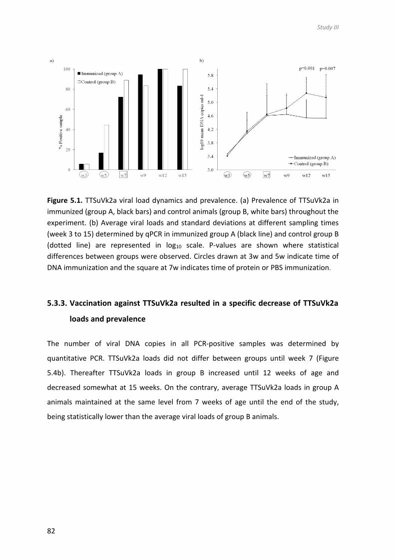
Study III
82
Figure 5.1. TTSuVk2a viral load dynamics and prevalence. (a) Prevalence of TTSuVk2a in
immunized (group A, black bars) and control animals (group B, white bars) throughout the
experiment. (b) Average viral loads and standard deviations at different sampling times
(week 3 to 15) determined by qPCR in immunized group A (black line) and control group B
(dotted line) are represented in log10 scale. P-values are shown where statistical
differences between groups were observed. Circles drawn at 3w and 5w indicate time of
DNA immunization and the square at 7w indicates time of protein or PBS immunization.
5.3.3. Vaccination against TTSuVk2a resulted in a specific decrease of TTSuVk2a
loads and prevalence
The number of viral DNA copies in all PCR-positive samples was determined by
quantitative PCR. TTSuVk2a loads did not differ between groups until week 7 (Figure
5.4b). Thereafter TTSuVk2a loads in group B increased until 12 weeks of age and
decreased somewhat at 15 weeks. On the contrary, average TTSuVk2a loads in group A
animals maintained at the same level from 7 weeks of age until the end of the study,
being statistically lower than the average viral loads of group B animals.

Study III
83
During the study period only four animals in group A reached TTSuVk2a loads higher than
5log10, and at 15 weeks of age all but three animals had viral titres below 5log10 and 3
animals had cleared the viremia to below the detection limit of the qPCR (Figure 5.5). In
comparison, all animals in group B were viremic at 15 weeks of age, with 9 animals having
TTSuVk2a viral titres well above 5log10. The delayed increase of TTSuVk2a viremia from 3
to 7 weeks in group A as compared to group B is again clearly observed if TTSuVk2a titres
in individual animals are presented (Figure 5.4).
Figure 5.5. TTSuVk2a viral loads in individual animals in immunized (group A) (a) and
control group (group B) (b) at different sampling times. PCR negative samples were given
value 0. The dotted line represents the qPCR detection limit. The continuous line is drawn
at 5log10. Circles drawn at 3w and 5w indicate time of DNA immunization and the square
at 7w indicates time of protein or PBS immunization.
To analyse if immunization was specifically affecting TTSuVk2a viremia, prevalence and
viral loads of other TTSuV species were determined. Upon analysing the other TTSuV
species (i.e. TTSuV1 and TTSuVk2b) no significant differences in the viral load or
prevalence between groups were observed (Figure 5.5a and b). However, total TTSuV
viral loads in group A reached its maximum at week 9, thereafter declining, while in group
B a maximum was reached at week 12. At this age of the animals, the average total TTSuV
loads in the immunized group was statistically lower than that in the unimmunized group

Study III
84
(Figure 5.6c). Therefore, reduction in TTSuVk2a viremia was also reflected in the total
TTSuV qPCR results.
Average daily weight gain or the health status of animals was not different between the
groups. None of the animals in the study developed antibodies against PCV2 and MH.
Antibodies against PRRSV and APP were detected in immunized animals, being 22%
(4/18) and 17% (3/18) respectively, and this was similar in control group animals, being
17% (3/18) for PRRSV and 11% (2/18) for APP.
Taken together, the onset of TTSuVk2a infection was delayed and lower TTSuVk2a titres
were observed in animals immunized against TTSuVk2a and this immunization was
specific, as no other TTSuV species were affected.
Figure 5.6. TTSuV1 (a), TTSuVk2b (b) and total TTSuV (c) viral load dynamics in immunized
group (black line) and control group (dotted line) determined by qPCR. Average viral loads
and standard deviations at different sampling times are represented in log10 scale. P-
values are shown where statistical differences between groups were observed.
Prevalence at different weeks of age is shown below the graphs and is expressed as the
number of qPCR positive serum samples per group (on a total of 18) and the percentage
positive is shown in parentheses. Circles drawn at 3w and 5w indicate time of DNA
immunization and the square at 7w indicates time of protein or PBS immunization.

Study III
85
5.4. Discussion
This study describes for the first time the impact of successful pig immunization against
TTSuVk2a during natural infection. Immunization was started at an early age and applied
a total of three times at 14-day intervals. A combination of DNA and protein
immunizations was chosen to increase the possibilities of activating both cellular and
humoral immune responses. Pigs were housed in a local conventional farm with high
hygiene levels until the age of 2.5 weeks, and thereafter kept under the same conditions
as in conventional farms with the two study groups mixed. Thus, the results reflect a
situation in which animals can infect each other as would occur in conventional farms.
It was found that TTSuVk2a viral loads were significantly lower in the immunized group
than in the control group at the end of the study. Such differences in viremia were not
observed for TTSuV1 or TTSuVk2b. It is known that the prevalence and titer of TTSuV
increase with the age of the animals and maximum prevalence in healthy animals is
normally reached at about 3 months of age (Sibila et al., 2009b), which was also the case
in this study. However, a delayed increase in TTSuVk2a prevalence was observed in
immunized animals (group A), especially apparent in 5-weeks old animals, two weeks
after the first DNA immunization. At that time, animals did not seroconvert and therefore
it may be that the first DNA immunization activated a cellular immune response, which
may have limited the infection. However, the role of cellular immune responses could not
be proven in this study due to for example the lack of peripheral blood mononuclear cell
fractions. However, DNA vaccines are known to stimulate immune responses in several
animal infection models and in fact DNA vaccines participate in different molecular
pathways involved in the immune system such as T cell responses (Bolhassani & Yazdi,
2009; Rodriguez & Whitton, 2000).
Not all immunized animals cleared TTSuVk2a infection and/or at the end of the study had
relatively high viral titres. The rationale behind this reality could be explained by many
different reasons including pig genetic differences or haplotypes. Also, it is known that
anelloviruses infect the host as quasispecies, meaning that infection is composed of
different, closely related genomic variants of the virus (Cortey et al., 2012; Huang et al.,

Study III
86
2010a; Ng et al., 2009a) . Therefore, one plausible explanation is that the strain used to
prepare the DNA/protein used for immunization may not work against all TTSuVk2a
strains potentially infecting the pigs in this study. Despite the fact that antibody cross-
reactivity has been demonstrated to exist even between different TTSuV genotypes (aa
identity about 50%), but not between TTSuV from different species (aa identity about
25%) (Huang et al., 2012a), our results did not corroborate these observations. In our
study, the generated antibodies were not cross-reacting with different genotypes (ORF1
protein is 58% identical between TTSuVk2a and TTSuVk2b (described in study I) or with
different species (ORF1 protein is about 25% identical between TTSuVk2a and TTSuV1
(Huang et al., 2010b)). Cross-protection would be a desirable feature of a vaccine that
should reduce total TTSuV loads.
In our study, the antibodies against TTSuVk2a ORF1-A and ORF2 proteins were detected
in serum in TTSuVk2a-vaccinated animals at 9 weeks of age until the end of the study (15
weeks of age). When antibodies are efficiently neutralizing viruses from circulation, it is
expected that an increase in antibody titers is followed by a decrease in viral serum titers.
However, in our study the antibody titers did not correlate with virus serum titers,
indicating that they are not efficient at controlling the viral infection, perhaps explaining
why during natural infection and in older pigs, antibodies and TTSuV co-exist.
In conventional healthy pigs the antibody levels during natural TTSuV infection increase
with age. It has been observed in a recent study that TTSuV-specific antibodies started to
appear when the animals were 10 weeks of age and peaked at week 20, using peptides
based on antigenic regions in the C-terminal part of ORF1 (Jarosova & Celer, 2013). In
fact, the highest TTSuVk2a seroprevalence has been detected in adult pigs, while being
lower or absent in young animals (Huang et al., 2011; Xiao et al., 2012). In our study, only
a few unimmunized animals generated detectable levels of ORF1-A antibodies at the end
of the study. Since our experiment ended when pigs reached 15 weeks of age, it is
impossible to say how the natural antibody response would have evolved in older
animals. On the other hand, the difference in antibody detection between the vaccinated
and non-vaccinated groups evidenced the success of the used immunization method to
induce early humoral responses against TTSuVk2a.

Study III
87
Anelloviruses are considered non-pathogenic by themselves but it is believed that TTSuV
play an important role during co-infection with other pig pathogens. TTSuV is often
present in pigs with diseases like PCV2-SD (Aramouni et al., 2011; Blomström et al., 2010;
Kekarainen et al., 2006; Nieto et al., 2011) and porcine respiratory disease complex
(Rammohan et al., 2012). In fact, TTSuV1 combined with porcine reproductive and
respiratory syndrome virus (PRRSV) was considered to contribute to the experimental
induction of a porcine dermatitis and nephropathy syndrome (PDNS)-like condition
(Krakowka et al., 2008). Furthermore, a previous study revealed that natural infection
with TTSuV suppresses the host immune response and aggravates porcine reproductive
and respiratory syndrome (PRRS) in pigs (Zhang et al., 2012) . In our study a few animals
were detected PRRSV positive by ELISA in both the immunized and control groups.
Excluding the PRRSV positive samples from the analysis did not change the results with
PRRSV positive samples included (data not shown).
Conversely, both the delay in TTSuVk2a infection and the TTSuVk2a virus load showed
significant reductions directly after DNA immunization and in the absence of specific
antibodies, indicating that specific T-cell responses might be involved in the observed
partial protection. At this point, it is difficult to say how the infection would have evolved
during the study if only DNA vaccines were used.
While the average viral titer of TTSuVk2a in group A never reached 5log10, group B
averages were well above 5log10 for the two last sampling points. The viral titers in group
B are in accordance with earlier data showing that TTSuVk2a viral serum titers in healthy
animals older than 2 months are higher than 5 log10 (Nieto et al., 2011). Even though the
TTSuVk2a viremia was lowered by immunization, this was not reflected in the average
daily weight gain. It is to be seen if immunization against all TTSuV variants would lower
the total TTSuV loads in such a way that a gain in average daily weight can be obtained.
In summary, it can be concluded that a combination of DNA and protein immunization
can control TTSuVk2a viremia, but clearly further studies are needed to improve vaccine
efficacy, to know the mechanism behind this, and to assess the impact for pig producers.

Study III
88


6. General discussion


Discussion
91
Novel anelloviruses have been described in several vertebrate species since the first
description in humans in 1997. The first anelloviruses were discovered by PCR methods
and the improvements in molecular techniques developed in the last years have been
used for investigating anellovirus variability and discovering diverse novel anelloviruses in
several species as wild rodents (Nishiyama et al., 2014) or horses (Li et al., 2015). All
anelloviruses share certain characteristics such as high genetic diversity, conserved UTR
region, similar genome organization and similar gene expression strategy. This occurs in
the case of TTSuVs, which infect pigs and wild boar. Since TTSuVs discovery in 1999, a
significant research focus has been placed on them and their infections, variability,
sequencing of full-length genomes and diagnostic methods.
Study I of this PhD thesis included the description of a new TTSuV species. After such
discovery, up to four pig infecting viral species are known, namely TTSuV1a and TTSuV1b
(genus Iotatorquevirus), and TTSuVk2a and TTSuVk2b (genus Kappatorquevirus). The
phylogenetic analysis carried out in the study I is currently being used in the taxonomic
proposal made by the ICTV to generate the genus Kappatorquevirus with its
corresponding species. Furthermore, the study I also describes the presence of TTSuVk2b
in commercial pig sera. Interestingly, TTSuVk2b was not previously detected although
there had been attempts to discover novel pig anelloviruses (Macera et al., 2011). Before
the results of the present PhD thesis, the primers used in published TTSuV1- or TTSuVk2a-
specific PCRs (Brassard et al., 2010; Gallei et al., 2010; Huang et al., 2010a; Nieto et al.,
2011) were not able to amplify TTSuVk2b given the mismatches of these described
primers with TTSuVk2b sequences. The phylogenetic analysis used in the study I grouped
TTSuVk2b close to TTSuVk2a and clearly apart from TTSuV1, further supporting the
existence of a novel Kappatorquevirus genus. The different qPCR techniques, broad
spectrum and species-specific qPCR, developed in the study I were used to determine the
epidemiology of this novel anellovirus as well as its potential association with pig
diseases, mainly with PCV2-SD.
TTSuV species existing so far before the knowledge of the novel one are widespread and
ubiquitous in domestic pig and wild boar (Kekarainen & Segalés, 2012a). Epidemiological
studies carried out in study I by species-specific qPCR confirmed that the novel species

Discussion
92
was detected in almost all 17 countries scrutinized, but showed lower viral load
compared to other TTSuV species. However, in three countries where TTSuVk2b was
apparently absent, the number of serum samples analysed was probably low; therefore,
more samples and farms should be analysed to rule out the presence of TTSuVk2b in
these countries. Also, more viral genomic sequences should be obtained to confirm that
the annealing sites of the used primers are highly conserved, thus guaranteeing the
detection of all species variants. A recent study detected and confirmed the lower viral
load of TTSuVk2b compared to other TTSuVs in Italian pig herds (Blois et al., 2014).
TTSuVs are currently considered non-pathogenic. TTSuV infection by itself does not cause
immediate disease, but it is believed that TTSuVs influence the development of some
economically important viral diseases or even affect their outcome. Several studies have
associated TTSuVs to a number of pig diseases, especially with PCVDs. These conditions
have been grouped because of the underlying aetiological agent, PCV2. PCVDs represent
some of the most significantly economic diseases in pig production worldwide.
The epidemiological study carried out in the first study of this PhD Thesis showed that
both TTSuVk2a and TTSuVk2b DNA loads were significantly higher in PCV2-SD-affected
pigs, compared to healthy counterparts, while no significant differences were observed in
the case of TTSuV1. This confirms previous findings about the association of TTSuVk2a but
not TTSuV1 with PCV2-SD (Aramouni et al., 2011; Kekarainen et al., 2006; Nieto et al.,
2011). This result suggests that the species included in the Kappatorquevirus genus
(TTSuVk2a and k2b) could be a factor of clinical aggravation in co-infection with other
pathogens. Perhaps only certain types or subtypes of TTSuV may be particularly
pathogenic or potentially associated with some pig diseases. Moreover, TTSuV1
combined with PRRSV has been considered to contribute to the experimental induction of
a porcine dermatitis and nephropathy syndrome (PDNS)-like condition (Krakowka et al.,
2008). Indeed, in the complex association of TTSuVs with potential pig diseases, it should
be taken into account not only the TTSuV infection, but also the specific health status of
each animal, possible co-infecting pathogens and the immune status of the host. Thus,
nowadays the disease association of TTSuVs is under debate. In fact, the idea that some
species are assumed to be more virulent than others has been suggested for human TTVs

Discussion
93
as well as possible implications of co-infections with other pathogens (Okamoto, 2009a).
As an example, among other ssDNA viruses such as porcine circoviruses, PCV2 is
described a pathogenic and causing PCVDs, while PCV1 is considered non-pathogenic. In
fact, PCV2 and PCV1 share only about 76% of nucleotide sequence identity and have
similar genomic organizations (Hamel et al., 1998; Meehan et al., 1998; Morozov et al.,
1998). Furthermore, PCV2 is ubiquitous in the swine population, like TTSuV, and it is
necessary but not sufficient for the onset of PCV2-SD (Segalés et al., 2005).
In addition, in study I, serum samples of PCV2-SD-affected and healthy pigs were used to
study disease association with the novel TTSuVk2b. These samples were used in the past
by means of different qPCR methods to detect the so far existing TTSuVs (Aramouni et al.,
2011; Nieto et al., 2011). Indeed, the literature shows methods like PCR or qPCR that
have been developed by several laboratories using different technologies, methods
and/or primer pairs. This may lead to disagreements on the results and even
misinterpretation of them. For that, it would be important to compare the currently used
methods for TTSuV detection and quantification. Thus, in the study I, a ring-trial to
estimate the reliability for TTSuV detection was done in two different laboratories. The
applied qPCRs in study I were based on SYBR Green-based and Light Upon Extension (LUX)
qPCR techniques. Both assays detected TTSuVk2a with good concordance, but the LUX
assay quantified about 10-fold higher loads compared to the SYBR Green technique.
These results should not be considered surprising since they use different quantification
methods as well as primer pairs. In the LUX qPCR, one fluorogenic primer was used and
labelled with a fluorophore close to the 3’-end that was quenched by the hairpin
structure of the primer. On the formation of the PCR product, the fluorescence increased
due to extension of the hairpin structure. On the contrary, the SYBR Green-based qPCR
technology was based on a double-stranded DNA binding dye and unlabelled primers. The
primers used in each technique hybridized at different regions of the viral genome and
were likely to cause differences in detection rates between technologies, especially
considering the high nucleotide variability of TTSuVs (Cortey et al., 2011; Huang et al.,
2010b). It should be added, as well, that the samples were not tested contemporaneously

Discussion
94
with both techniques (1-year difference), so, it could not be ruled out that the quality of
them was in a better shape when the LUX assay was performed.
For studies II and III of this PhD Thesis, several proteins were produced in different
platforms, such as E. coli and baculovirus expression systems. At the beginning of the
studies, it was already known that the TTSuV genome contains four ORFs, ORF1, ORF1/1
(also known as ORF1-A), ORF2, and ORF3 (also known as ORF2/2) (Huang et al., 2012b; Lu
et al., 2013; Martínez-Guinó et al., 2011). ORF3 can only be translated after splicing of the
mRNA precursor, and splicing of the ORF1 mRNA transcript results in different protein
isoforms (Huang et al., 2012b; Martínez-Guinó et al., 2011), as also observed in human
TTVs (Müeller et al., 2008; Qiu et al., 2005).
Even if TTSuVs are currently considered non-pathogenic, it is hard to assume that a viral
infection would not have consequences to its host. As another example, farm
performance is improved when PCV2 (also a single stranded circular DNA virus) infection
is controlled by vaccination even in farms without disease, displaying only a subclinical
infection (Beach & Meng, 2012). Currently, commercial vaccines against PCV2 are
efficient in reducing the serum viral load, preventing the disease and improving the
performance of the farm. Nevertheless, the prevention and control against TTSuV
infections have not yet been established, but it was hypothesized the possibility that the
subclinical infection by TTSuVs had relevant consequences for the pig production. In
consequence, two experimental (study II and III) vaccines against TTSuVs were developed
and tested in this PhD Thesis.
The second study evaluated the impact of a multivalent vaccine in naturally TTSuV
infected pigs. It is known that anelloviruses are genetically variable and different variants
may cause infection. In fact, multiple infections of TTSuV with different genotypes or
subtypes in a single pig have been reported (Blois et al., 2014; Gallei et al., 2010; Huang et
al., 2010b), as observed in Study I. Also, it is known that anelloviruses infect the host as
quasispecies, meaning that infection is composed of different, closely related genomic
variants of the virus (Cortey et al., 2012; Huang et al., 2010b; Ng et al., 2009a). For that
reason, the multivalent vaccine was a priori a good tool to cover the maximum of the

Discussion
95
TTSuV diversity. The development and use of multivalent vaccine strategies have been
successfully used in human papilloma virus (HPV), also a genetically diverse virus (IARC,
2007). The development of a multivalent vaccine against HPV types 6, 11, 16 and 18
afforded protection against two diseases (cervical and genitals warts) and has
demonstrated that this 4-valent vaccine was able to broaden the reach of immune
response against four circulating strains of HPV (Garland et al., 2007; Group., 2007;
Muñoz et al., 2009).
The multivalent vaccine prototype (including ORF1 and ORF3 proteins) against the four
known TTSuV species was not able to control viremia during TTSuV natural infection
despite of seroconversion of vaccinated animals. This result suggested that generated
antibodies were not able to neutralize the virus sufficiently to control viremia. It could be
that the protein vaccine only stimulated the humoral response, which may not be enough
to control TTSuV viremia.
In study II, recombinant ORF1 and ORF3 proteins from TTSuV1 (1a and 1b) and TTSuVk2
(k2a and k2b) were considered good vaccine candidates a priori. On one hand, ORF1 is
the putative capsid protein containing known antigenic domains (Huang et al., 2011;
Jarosova & Celer, 2013). On the other hand, ORF3 N-terminal region is identical with
ORF2 and, therefore, ORF3 protein covers aa sequence of the two different viral proteins
encoded by these genes. Although the vaccine prototype did include predicted antigenic
epitopes, the lack of a reliable virus neutralization assay did not allow concluding if they
evoked neutralizing antibodies (Huang et al., 2011; Jarosova & Celer, 2013). It may
happen that the specific strains used to prepare the multivalent vaccine did not elicit a
robust neutralising antibody response to TTSuVs and, in consequence, did not produce
antibodies of the appropriate specificity; it has to be reminded that there is a significant
genetic variability even within the same species of TTSuV. Representative sequences from
each of the 4 TTSuV groups were selected of each gene. Despite of that, maybe the TTSuV
strains circulating in the pigs of this study were distinct from the vaccine strains.
Therefore, it could be that viruses closest to the antigens in the vaccines would have been
working better. On the other hand, it cannot be ruled out that the formulation of a
multivalent prototype vaccine may cause interference between serotypes; maybe the

Discussion
96
response against the dominant serotypes was low or even absent, thus not causing an
efficient reduction of viremia.
Specifically, the ORF1 full-length capsid protein of four known TTSuVs was expressed
using a baculovirus expression system for the first time. Other research groups have used
bacterial, insect cell or mammalian expression systems. However, expression of entire
full-length ORF1 protein had not been successful before (Kakkola et al., 2008; Kakkola et
al., 2002), although ORF1 mRNA has been detected at transcriptional level (Huang et al.,
2012b; Kakkola et al., 2008; Kakkola et al., 2002; Martínez-Guinó et al., 2011). Indeed, in
the study II, TTSuV proteins were first codon optimized and expressed with a short
affinity-tag (Strep or His) in their C-terminal end. This design may have improved the
baculovirus expression and/or stability of the proteins, resulting in detectable levels of
ORF1.
Although not significant, vaccinated animals with the multivalent vaccine prototype had
higher TTSuV loads than non-vaccinated control animals. It could be that vaccination
activated humoral immune responses resulting in non-neutralizing antibodies, which
might enhance the viral infection through interactions with the complement system and
its receptors, creating antibody-virus complexes that might up-regulate TTSuV replication
(Boonnak et al., 2008; Willey et al., 2011). Indeed, viremia exacerbation has been
described with other experimental vaccines and viral infections, such as Dengue virus
(DENV), Feline Infectious Peritonitis virus (FIPV) or African swine fever virus (ASFV)
(Argilaguet et al., 2011; Vennema et al., 1990; Whitehead et al., 2007). However, little is
known about the interaction that TTVs establish with the host immune system and more
work should be needed to study this phenomenon.
Since the seroconversion of vaccinated animals against the multivalent vaccine antigens
could not control the TTSuV infection, a second animal trial (study III) was designed to
apply a different strategy. In the new approach, a DNA prime-boost design was used to
enhance the immune response combined with a protein third boost. Several studies have
developed DNA vaccine strategies against different pathogens to improve the immune
response. Thus, the overall immune responses using the prime-boost strategy might be

Discussion
97
improved, and may be effective at generating high levels of T- memory cells (Ramshaw &
Ramsay, 2000). Furthermore, the second animal trial included a vaccine against only one
TTSuV species to compare the behaviour of a vaccine against a single species in front of a
multivalent vaccine (study II). The TTSuVk2a was the species selected because of its
documented relationship of co-infection with other viral pathogens. In the second
vaccination approach (study III), the strategy was to get wider coverage of the antigenic
regions of the ORF1 gene in order to increase the antigenic region. To cover as much of
the antigenic region variability as possible, the 3 isoforms from ORF1 (A, B and C) were
used in the new approach (study III) in front of the full length ORF1 protein in the
multivalent vaccine prototype (study II). These isoforms shared the N-terminus region;
however, the C-terminus varied depending on the splicing site, being the isoforms
different in aa composition, thus increasing the variability. In fact, the C-terminus of ORF1
protein has been identified as the main antigenic region (Huang et al., 2012a; Jarosova &
Celer, 2013). On the other hand, ORF2 (DNA/protein vaccine) was used in study III in
contrast with ORF3 (used in the multivalent vaccine prototype, study II).
The third study describes the immunization against TTSuVk2a during natural infection of
pigs using a combination of viral DNA and protein. It was found that TTSuVk2a loads were
significantly lower in the immunized group than in the control group at the end of the
study. In fact, a number of animals of the immunized group cleared the TTSuVk2a
viremia, which was not the case in the control group at the end of the study (15 weeks of
age). Moreover, a delay in TTSuVk2a natural infection was observed after DNA
immunization (used as prime boost to stimulate the cellular response). Taking into
account that such situation occurred in absence of specific antibodies, it may be
indicative of specific T-cell responses involved in the observed partial virological
protection. In fact, the DNA vaccine was used as prime-boost in study III to improve the
immune response and exert their effect in different molecular pathways involved in the
immune system such as T cell responses (Bolhassani & Yazdi, 2009; Rodriguez & Whitton,
2000). The third immunization with a cocktail of proteins (ORF1-A isoform and ORF2 with
adjuvant) was used to induce humoral responses against TTSuVk2a. Several studies have
demonstrated that priming with DNA combined with a subsequent immunization with a

Discussion
98
different product like recombinant viruses or proteins allows obtaining enhanced
protective efficacy (Lu et al., 2009; Woodland, 2004).
Overall, the results of study III, where TTSuVk2a infection was controlled by combination
of DNA and protein vaccination, suggest that both cellular and humoral immune
responses should be necessary to control TTSuV infection. It would have been very
interesting to monitor experimentally vaccinated pigs by means of humoral and cellular
immune responses, since both are believed to be necessary in controlling viral infections.
Vaccine development studies have been shown the importance to enhance both humoral
and cellular immune response, such as for Hepatitis C virus (HCV). Several reports have
shown that an effective T-cell immune response is necessary for HCV clearance, while the
neutralizing antibodies play an important role in eradicating HCV (Esumi et al., 1999;
Mancini et al., 2009 ; Owsianka et al., 2008; Puig et al., 2004; Sun et al., 2015).
It is known that TTSuV-specific antibodies increase with age in conventional healthy pigs
during natural infection. Also, in a recent study, it has been observed that TTSuV-specific
antibodies started to appear when the animals were 10 weeks of age and peaked at week
20. The same study detected antibodies at 4 weeks of age (probably residual maternal
antibodies) and these decreased until 10 weeks of age (Jarosova & Celer, 2013). Another
recent study corroborated these previous studies (Nieto et al., 2015) .
Studies II and III were based on natural TTSuV infection and, therefore, antibodies
developed also in control group animals. However, few animals of control group
generated detectable levels in both studies (II and III). In study II, only 4 pigs in the control
group generated detectable antibody levels at 3, 7, 9 and 12 weeks of age. In study III,
only three non-immunized animals generated detectable levels of antibodies at the end
of the study (15 week of age). Since studies II and III ended when pigs reached 15 weeks
of age, it is impossible to know the natural antibody response would have evolved at
older ages. Importantly, the difference in antibody detection between vaccinated and
non-vaccinated groups evidenced the success of the used immunization methods in both
studies to induce early humoral responses against TTSuVs.
In study III, the DNA/protein combination controlled viremia, but maybe not against all
potentially existing TTSuVk2a variants. In fact, not all immunized animals cleared

Discussion
99
TTSuVk2a infection and, at the end of the study, some of them had relatively high viral
loads. Moreover, as observed in study III where the animals were vaccinated only against
TTSuVk2a, difference in viral load of other TTSuVs (TTSuV1 or TTSuVk2b) was not
observed. Moreover, antibody cross-reactivity has been demonstrated to exist even
between TTSuV1a and 1b (aa identity about 50%), but not between TTSuV1 and TTSuVk2a
(ORF1 protein is about 25% identical between TTSuVk2a and TTSuV1) (Huang et al.,
2012a). In study III, the generated antibodies were not cross-reacting between TTSuVk2a
and k2b (ORF1 protein is 58% identical between TTSuVk2a and TTSuVk2b, as shown in
study I). As discussed above, the high degree of genetic variability of TTSuV and the
potential divergent TTSuV strains present during infection may explain why the vaccine
prototype did not completely protect against TTSuVk2a. Regarding the genetic variability,
cross-protection would be a desirable feature and, indeed, a multivalent vaccine seemed
a good strategy a priori to reduce TTSuVs infection. Also, the use of isoforms from ORF1
genes to increase antigenic variability to cover antigenic regions was initially
hypothesized as a good strategy. Anyway, practical results indicated that the multivalent
vaccine was not efficient; maybe the multivalent vaccine did not activate the implicated
mechanisms in protection against TTSuV infection, which is currently unknown.
As summary of the presented three studies, this Thesis contributes to increase the
knowledge of TTSuVs by describing a novel species, which may be involved as a co-
pathogen with other infectious agents. On the other hand, to our knowledge, two vaccine
studies on TTSuVs were developed for the first time to provide novel information about
viral infection control. In fact, TTSuVk2a infection was controlled by the administration of
a DNA and protein combined vaccine strategy, while a multivalent protein-based vaccine
was not efficient despite it induced specific antibodies. Further studies are needed to
improve vaccine efficacy, to elucidate the protective mechanism behind, and to assess
the impact of TTSuVs for pig producers. Maybe the multivalent vaccine should be used in
combination with a DNA vaccine to see if such approach would improve the outcome.

Discussion
100


7. Conclusions


Conclusions
103
1. A novel TTSuV species, named TTSuVk2b, was identified. According to the
phylogenetic analysis, it belongs to the Kappatorquevirus genus together with the
already known TTSuVk2a.
2. TTSuVk2a and TTSuVk2b are not only genetically related, but also their viral loads in
serum were higher in PCV2-SD affected animals compared with healthy pen mates.
3. TTSuV proteins, namely ORF1, ORF1-A, ORF2 and ORF3, of the four known TTSuVs
species were successfully expressed in a baculovirus system.
4. A multivalent vaccine prototype based on baculovirus expressed TTSuV proteins
(ORF1 and ORF3 proteins) was not able to control viremia during TTSuV natural
infection. This lack of efficacy occurred despite that immunized animals
seroconverted against the virus. In consequence, the generated humoral immune
response was not sufficient to control the viremia.
5. A combination of DNA and protein immunization was shown to control TTSuVk2a
viremia during TTSuV natural infection. The onset of viremia was delayed and viral
loads were lower in immunized animals. The vaccine prototype was specifically
controlling TTSuVk2a infection and did not affect other TTSuV species.

Conclusions
104

8. References


References
107
Allander, T., Emerson, S. U., Engle, R. E., Purcell, R. H. & Bukh, J. (2001). A virus discovery
method incorporating DNase treatment and its application to the identification of
two bovine parvovirus species. Proc Natl Acad Sci U S A 98, 11609-11614.
Aramouni, M., Segalés, J., Cortey, M. & Kekarainen, T. (2010). Age-related tissue
distribution of swine Torque teno sus virus 1 and 2. Vet Microbiol 146, 350-353.
Aramouni, M., Segalés, J., Sibila, M., Martin-Valls, G. E., Nieto, D. & Kekarainen, T.
(2011). Torque teno sus virus 1 and 2 viral loads in postweaning multisystemic
wasting syndrome (PMWS) and porcine dermatitis and nephropathy syndrome
(PDNS) affected pigs. Vet Microbiol 153, 377-381.
Argilaguet, J. M., Perez-Martin, E., Gallardo, C., Salguero, F. J., Borrego, B., Lacasta, A.,
Accensi, F., Diaz, I., Nofrarias, M., Pujols, J., Blanco, E., Perez-Filgueira, M.,
Escribano, J. M. & Rodriguez, F. (2011). Enhancing DNA immunization by targeting
ASFV antigens to SLA-II bearing cells. Vaccine 29, 5379-5385.
Bao, Y., Kapustin, Y. & Tatusova, T. (2008). Virus Classification by Pairwise Sequence
Comparison (PASC). In Encyclopedia of Virology 3rd edn, pp. 342-348. Edited by B.
W. J. Mahy & v. R. M.H.V. Oxford, UK: Elsevier.
Beach, N. M. & Meng, X. J. (2012). Efficacy and future prospects of commercially
available and experimental vaccines against porcine circovirus type 2 (PCV2). Virus
Res 164, 33-42.
Biagini, P. (2009). Classification of TTV and related viruses (anelloviruses). Curr Top
Microbiol Immunol 331, 21-33.
Biagini, P., Bendinelli, M., Hino, S., Kakkola, L., Mankertz, A., Niel, C., Okamoto, H.,
Radial, S., Teo, S. G. & Todd, D. (2011). Anelloviridae: Virus Taxonomy,9th Report
of the ICTV.
Biagini, P., Gallian, P., Attoui, H., Cantaloube, J. F., de Micco, P. & de Lamballerie, X.
(1999). Determination and phylogenetic analysis of partial sequences from TT
virus isolates. J Gen Virol 80 419-424.
Biagini, P., Gallian, P., Attoui, H., Touinssi, M., Cantaloube, J.-F., de Micco, P. & de
Lamballerie, X. (2001). Genetic analysis of full-length genomes and subgenomic
sequences of TT virus-like mini virus human isolates. J Gen Virol 82, 379-383.
Biagini, P., Todd, D., Bendinelli, M., Hino, S., Mankertz, A., Mishiro, S., Niel, C.,
Okamoto, H., Raidal, S., Ritchie, B. W. & Teo, G. C. (2005). Anellovirus. In Virus

References
108
Taxonomy, VIIIth Report of the International Committee for the Taxonomy of
Viruses. Edited by C. M. Fauquet, M. A. Mayo, J. Maniloff, U. Desselberger & L.A.
Ball. London: Elsevier/Academic Press.
Biagini, P., Uch, R., Belhouchet, M., Attoui, H., Cantaloube, J. F., Brisbarre, N. & de
Micco, P. (2007). Circular genomes related to anelloviruses identified in human
and animal samples by using a combined rolling-circle amplification/sequence-
independent single primer amplification approach. J Gen Virol 88, 2696-2701
Bigarré, L., Beven, V., de Boisseson, C., Grasland, B., Rose, N., Biagini, P. & Jestin, A.
(2005). Pig anelloviruses are highly prevalent in swine herds in France. J Gen Virol
86, 631-635.
Blanchard, P., Mahé, D., Cariole, t. R., Keranflec'h, A., Baudouard, M. A., Cordioli, P.,
Albina, E. & Jestin, A. (2003). Protection of swine against post-weaning
multisystemic wasting syndrome (PMWS) by porcine circovirus type 2 (PCV2)
proteins. Vaccine 21, 4565-4575.
Blois, S., Mallus, F., Liciardi, M., Pilo, C., Camboni, T., Macera, L., Maggi, F. & Manzin, A.
(2014). High prevalence of co-infection with multiple Torque teno sus virus
species in Italian pig herds. PLoS One 9, e113720.
Blomström, A.-L., Belák, S., Fossum, C., Fuxler, L., Wallgren, P. & Berg, M. (2010).
Studies of porcine circovirus type 2, porcine boca-like virus and torque teno virus
indicate the presence of multiple viral infections in postweaning multisystemic
wasting syndrome pigs. Virus Res 152, 59-64.
Blomström, A. L., Stahl, K., Masembe, C., Okoth, E., Okurut, A. R., Atmnedi, P., Kemp, S.,
Bishop, R., Belak, S. & Berg, M. (2012). Viral metagenomic analysis of bushpigs
(Potamochoerus larvatus) in Uganda identifies novel variants of Porcine
parvovirus 4 and Torque teno sus virus 1 and 2. Virol J 9, 192.
Bolhassani, A. & Yazdi, S. R. ( 2009). DNA immunization as an efficient strategy for
vaccination. Avicenna J Med Biotechnol 1, 71-88.
Boonnak, K., Slike, B. M., Burgess, T. H., Mason, R. M., Wu, S. J., Sun, P., Porter, K.,
Rudiman, I. F., Yuwono, D., Puthavathana, P. & Marovich, M. A. (2008). Role of
dendritic cells in antibody-dependent enhancement of dengue virus infection. J
Virol 82, 3939-3951.

References
109
Brassard, J., Gagne, M. J., Houde, A., Poitras, E. & Ward, P. (2010). Development of a
real-time TaqMan PCR assay for the detection of porcine and bovine Torque teno
virus. J Appl Microbiol 108, 2191-2198.
Brassard, J., Gagne, M. J., Lamoureux, L., Inglis, G. D., Leblanc, D. & Houde, A. (2008).
Molecular detection of bovine and porcine Torque teno virus in plasma and feces.
Vet Microbiol 126, 271-276.
Brunak, S., Engelbrecht, J. & Knudsen, S. (1991). Prediction of human mRNA donor and
acceptor sites from the DNA sequence. J Mol Biol 220, 49-65.
Christensen, J. K., Eugen-Olsen, J., M, S. L., Ullum, H., Gjedde, S. B., Pedersen, B. K.,
Nielsen, J. O. & Krogsgaard, K. (2000). Prevalence and prognostic significance of
infection with TT virus in patients infected with human immunodeficiency virus. J
Infect Dis 181, 1796-1799.
Cortey, M., Macera, L., Segales, J. & Kekarainen, T. (2011). Genetic variability and
phylogeny of Torque teno sus virus 1 (TTSuV1) and 2 (TTSuV2) based on complete
genomes. Vet Microbiol 148, 125-131.
Cortey, M., Pileri, E., Segalés, J. & Kekarainen, T. (2012). Globalisation and global trade
influence molecular viral population genetics of Torque Teno Sus Viruses 1 and 2
in pigs. Vet Microbiol 156, 81-87.
de Villiers, E. M., Borkosky, S. S., Kimmel, R., Gunst, K. & Fei, J. W. (2011). The diversity
of Torque teno viruses: In vitro replication leads to the formation of additional
replication competent subviral molecules. J Virol 85, 7284-7295.
Dean, F. B., Nelson, J. R., Giesler, T. L. & Lasken, R. S. (2001). Rapid amplification of
plasmid and phage DNA using Phi 29 DNA polymerase and multiply-primed rolling
circle amplification. Genome Res 111095-1099.
Delwart, E. (2007). Viral metagenomics. Rev Med Virol 17, 115–131.
Edwards, R. A. & Rohwer, F. (2005). Viral metagenomics. Nat Rev Microbiol 3, 504-510.
Ellis, J. A., Allan, G. & Krakowka, S. (2008). Effect of coinfection with genogroup 1 porcine
torque teno virus on porcine circovirus type 2-associated postweaning
multisystemic wasting syndrome in gnotobiotic pigs. Am J Vet Res 69, 1608-1614.

References
110
Esteban, J. A., Salas, M. & Blanco, L. (1993). Fidelity of phi 29 DNA polymerase.
Comparison between proteinprimed initiation and DNA polymerization. J Biol
Chem 268.
Esumi, M., Rikihisa, T., Nishimura, S., Goto, J., Mizuno, K., Zhou, Y. & Shikata, T. (1999).
Experimental vaccine activities of recombinant E1 and E2 glycoproteins and
hypervariable region 1 peptides of hepatitis C virus in chimpanzees. Arch Virol
144, 973-980.
Fenaux, M., Opriessnig, T., Halbur, P. G., Elvinger, F. & Meng, X. J. (2004). Two amino
acid mutations in the capsid protein of type 2 porcine circovirus (PCV2) enhanced
PCV2 replication in vitro and attenuated the virus in vivo. J Virol 78, 13440-13446.
Fraile, L., Sibila, M., Nofrarías, M., López-Jimenez, R., Huerta, E., Llorens, A., López-
Soria, S., Pérez, D. & Segalés, J. (2012). Effect of sow and piglet porcine circovirus
type 2 (PCV2) vaccination on piglet mortality, viraemia, antibody titre and
production parameters. Vet Microbiol 161, 229-234.
Gallei, A., Pesch, S., Esking, W. S., Keller, C. & Ohlinger, V. F. (2010). Porcine Torque teno
virus: determination of viral genomic loads by genogroup-specific multiplex rt-
PCR, detection of frequent multiple infections with genogroups 1 or 2, and
establishment of viral full-length sequences. Vet Microbiol 143, 202-212.
Garland, S. M., Hernandez-Avila, M., Wheeler, C. M., Perez, G., Harper, D. M., Leodolter,
S., Tang, G. W., Ferris, D. G., Steben, M., Bryan, J., Taddeo, F. J., Railkar, R., Esser,
M. T., Sings, H. L., Nelson, M., Boslego, J., Sattler, C., Barr, E., Koutsky, L. A. &
Females United to Unilaterally Reduce Endo/Ectocervical Disease, I. I. (2007).
Quadrivalent vaccine against human papillomavirus to prevent anogenital
diseases. N Engl J Med 356, 1928-1943.
Group., F. I. S. (2007). Quadrivalent vaccine against human papillomavirus to prevent
high-grade cervical lesions. N Engl J Med 356, 1915-1927.
Hamel, A. L., Lin, L. L. & Nayar, G. P. (1998). Nucleotide sequence of porcine circovirus
associated with postweaning multisystemic wasting syndrome in pigs. J Virol 72,
5262-5267.
Hijikata, M., Iwata, K., Ohta, Y., Nakao, K., Matsumoto, M., Matsumoto, H., Kanai, K.,
Baba, K., Samokhvalov, E. I. & Mishiro, S. (1999). Genotypes of TT virus (TTV)
compared between liver disease patients and healthy individuals using a new PCR
system capable of differentiating 1a and 1b types from others. Arch Virol 144,
2345-2354.
References
111
Huang, Y. W., Dryman, B. A., Harrall, K. K., Vaughn, E. M., Roof, M. B. & Meng, X. J.
(2010a). Development of SYBR green-based real-time PCR and duplex nested PCR
assays for quantitation and differential detection of species- or type-specific
porcine Torque teno viruses. J Virol Methods 170, 140-146.
Huang, Y. W., Harrall, K. K., Dryman, B. A., Beach, N. M., Kenney, S. P., Opriessnig, T.,
Vaughn, E. M., Roof, M. B. & Meng, X. J. (2011). Expression of the putative ORF1
capsid protein of porcine Torque teno sus virus 2 (TTSuV2) and development of
Western blot and ELISA serodiagnostic assays: Correlation between TTSuV2 viral
load and IgG antibody level in pigs. Virus Res 158, 79-88.
Huang, Y. W., Harrall, K. K., Dryman, B. A., Opriessnig, T., Vaughn, E. M., Roof, M. B. &
Meng, X. J. (2012a). Serological profile of torque teno sus virus species 1 (TTSuV1)
in pigs and antigenic relationships between two TTSuV1 genotypes (1a and 1b),
between two species (TTSuV1 and -2), and between porcine and human
anelloviruses. J Virol 86, 10628-10639.
Huang, Y. W., Ni, Y. Y., Dryman, B. A. & Meng, X. J. (2010b). Multiple infection of porcine
Torque teno virus in a single pig and characterization of the full-length genomic
sequences of four U.S. prototype PTTV strains: implication for genotyping of PTTV.
Virology 396, 289-297.
Huang, Y. W., Patterson, A. R., Opriessnig, T., Dryman, B. A., Gallei, A., Harrall, K. K.,
Vaughn, E. M., Roof, M. B. & Meng, X. J. (2012b). Rescue of a porcine anellovirus
(torque teno sus virus 2) from cloned genomic DNA in pigs. J Virol 86, 6042-6054.
IARC (2007). Human papillomaviruses. IARC Monogr Eval Carcinog Risks Hum 90, 1-636.
Inami, T., Obara, T., Moriyama, M., Arakawa, Y. & Abe, K. (2000). Full-Length nucleotide
sequence of a simian TT virus isolate obtained from a chimpanzee: evidence for a
new TT virus-like species. Virology 277, 330-335.
Jarosova, V. & Celer, V. (2013). Preliminary epitope mapping of Torque teno sus virus 1
and 2 putative capsid protein and serological detection of infection in pigs. J Gen
Virol 94, 1351-1356.
Jarosova V, Pogranichniy. R., Celer V. (2011). Prevalence and age distribution of porcine
torque teno sus virus (TTSuV) in the Czech Republic. Folia Microbiol (Praha) 56, 90-
94.
Jelcic, I., Hotz-Wagenblatt, A., Hunziker, A., Zur Hausen, H. & de Villiers, E. M. (2004).
Isolation of multiple TT virus genotypes from spleen biopsy tissue from a

References
112
Hodgkin's disease patient: genome reorganization and diversity in the
hypervariable region. J Virol 78, 7498-7507.
Jones, M. S., Kapoor, A., Lukashov, V. V., Simmonds, P., Hecht, F. & Delwart, E. (2005).
New DNA Viruses Identified in Patients with Acute Viral Infection Syndrome. J Virol
79, 8230-8236.
Kaba, S. A., Salcedo, A. M., Wafula, P. O., Vlak, J. M. & van Oers, M. M. (2004).
Development of a chitinase and v-cathepsin negative bacmid for improved
integrity of secreted recombinant proteins. J Virol Methods 122, 113-118.
Kakkola, L., Bondén, H., Hedman, L., Kivi, N., Moisala, S., Julin, J., Ylä-Liedenpohja, J.,
Miettinen, S., Kantola, K., Hedman, K. & Söderlund-Venermo, M. (2008).
Expression of all six human Torque teno virus (TTV) proteins in bacteria and in
insect cells, and analysis of their IgG responses. Virology 382, 182-189.
Kakkola, L., Hedman, K., Vanrobaeys, H., Hedman, L. & Soderlund-Venermo, M. (2002).
Cloning and sequencing of TT virus genotype 6 and expression of antigenic open
reading frame 2 proteins. J Gen Virol 83, 979-990.
Kakkola, L., Tommiska, J., Boele, L. C. L., Miettinen, S., Blom, T., Kekarainen, T., Qiu, J.,
Pintel, D., Hoeben, R. C., Hedman, K. & Söderlund-Venermo, M. (2007).
Construction and biological activity of a full-length molecular clone of human
Torque teno virus (TTV) genotype 6. FEBS Journal 274, 4719-4730.
Kamada, K., Kamahora, T., Kabat, P. & Hino, S. (2004). Transcriptional regulation of TT
virus: promoter and enhancer regions in the 1.2-kb noncoding region. Virology
321, 341-348.
Kekarainen, T., López-Soria, S. & Segalés, J. (2007). Detection of swine Torque teno virus
genogroups 1 and 2 in boar sera and semen. Theriogenology 68, 966-971.
Kekarainen, T., Martínez-Guinó, L. & Segalés, J. (2009). Swine torque teno virus
detection in pig commercial vaccines, enzymes for laboratory use and human
drugs containing components of porcine origin. J Gen Virol 90, 648-653.
Kekarainen, T., McCullough, K., Fort, M., Fossum, C., Segalés, J. & Allan, G. M. (2010).
Immune responses and vaccine-induced immunity against Porcine circovirus type
2. Vet Immunol Immunopathol 136, 185–193.
Kekarainen, T. & Segales, J. (2012). Torque teno sus virus in pigs: an emerging pathogen?
Transbound Emerg Dis 59 Suppl 1, 103-108.

References
113
Kekarainen, T. & Segalés, J. (2009). Torque teno virus infection in the pig and its potential
role as a model of human infection. Vet J 180, 163-168.
Kekarainen, T. & Segalés, J. (2012a). Porcine Anelloviruses. In Transbound Emerg Dis.
Kekarainen, T. & Segalés, J. (2012b). Torque teno sus virus in pigs: an emerging
pathogen? Transbound Emerg Dis 59 Suppl 1, 103-108.
Kekarainen, T., Sibila, M. & Segalés, J. (2006). Prevalence of swine Torque teno virus in
post-weaning multisystemic wasting syndrome (PMWS)-affected and non-PMWS-
affected pigs in Spain. J Gen Virol 87, 833-837.
Krakowka, S., Hartunian, C., Hamberg, A., Shoup, D., Rings, M., Zhang, Y., Allan, G. &
Ellis, J. A. (2008). Evaluation of induction of porcine dermatitis and nephropathy
syndrome in gnotobiotic pigs with negative results for porcine circovirus type 2.
Am J Vet Res 69, 1615-1622.
Kulcsar, G., Farsang, A. & Soos, T. (2010). Testing for viral contaminants of veterinary
vaccines in Hungary. Biologicals 38,346-9.
Leary, T. P., Erker, J. C., Chalmers, M. L., Desai, S. M. & Mushahwar, I. K. (1999).
Improved detection systems for TT virus reveal high prevalence in humans, non-
human primates and farm animals. J Gen Virol 80, 2115-2120.
Lee, S.-s., Sunyoung, S., Jung, H., Shin, J. & Lyoo, Y. S. (2010). Quantitative detection of
porcine Torque teno virus in Porcine circovirus-2-negative and Porcine circovirus-
associated disease-affected pigs. J Vet Diagn Invest 22, 261-264.
Li, K., Wang, L. Q., Wu, Y. Y., Chao, A. J., Lu, Q. W., Wei, Z. Y., Cui, B. A. & Chen, H. Y.
(2013). Molecular detection and genomic characterization of Torque teno sus
virus 1 and 2 from domestic pigs in central China. Virus Genes 46, 479-486.
Li, L., Giannitti, F., Low, J., Keyes, C., Ullmann, L. S., Deng, X., Aleman, M., Pesavento, P.
A., Pusterla, N. & Delwart, E. (2015). Exploring the virome of diseased horses. J
Gen Virol 96, 2721-2733.
Liu, J., Guo, L., Zhang, L., Wei, Y., Huang, L., Wu, H. & Liu, C. (2013). Three new emerging
subgroups of Torque teno sus viruses (TTSuVs) and co-infection of TTSuVs with
porcine circovirus type 2 in China. Virol J 10, 189.

References
114
Liu J, Z. L., Huang L, Wei Y, Wu H, Liu C. (2013). Identification of two new antigen
epitopes on the putative capsid protein encoded by torque teno sus virus type 1
ORF1. Vet Microbiol 167, 372-382.
Lu, Y., Ouyang, K., Fang, J., Zhang, H., Wu, G., Ma, Y., Zhang, Y., Hu, X., Jin, L., Cao, R.,
Fan, H., Li, T. & Liu, J. (2009). Improved efficacy of DNA vaccination against
prostate carcinoma by boosting with recombinant protein vaccine and by
introduction of a novel adjuvant epitope. Vaccine 27, 5411-5418.
Lu, Y., Yu, H., Nie, X., Li, Y., Zhang, L. & Lu, C. (2013). Nonstructural proteins of Torque
teno sus virus 2 from O2AUG: prediction to experimental validation. Virus Res 178,
272-280.
Macera, L., Cortey, M., Maggi, F., Segalés, J. & Kekarainen, T. (2011). A novel rolling
circle amplification assay to detect members of the family Anelloviridae in pigs
and humans. Virus Research 160, 424-427.
Madsen CD, E.-O. J., Kirk O, Parner J, Kaae Christensen J, Brasholt MS, Ole Nielsen J,
Krogsgaard K. (2002). TTV viral load as a marker for immune reconstitution after
initiation of HAART in HIV-infected patients. HIV Clin Trials 3(4), 287-295.
Maggi, F. & Bendinelli, M. (2009). Immunobiology of the Torque teno viruses and other
anelloviruses. Curr Top Microbiol Immunol 331, 65-90.
Maggi, F., Pistello, M., Vatteroni, M., Presciuttini, S., Marchi, S., Isola, P., Fornai, C.,
Fagnani, S., Andreoli, E., Antonelli, G. & Bendinelli, M. (2001). Dynamics of
Persistent TT Virus Infection, as Determined in Patients Treated with Alpha
Interferon for Concomitant Hepatitis C Virus Infection. J Virol 75, 11999-12004.
Mahé, D., Blanchard, P., Truong, C., Arnauld, C., Le Cann, P., Cariolet, R., Madec, F.,
Albina, E. & Jestin, A. (2000). Differential recognition of ORF2 protein from type 1
and type 2 porcine circoviruses and identification of immunorelevant epitopes. J
Gen Virol 81, 1815-1824.
Mancini, N., Diotti, R., Perotti, M., Sautto, G., Clementi, N., Nitti, G., Patel, A., Ball, J.,
Clementi, M. & Burioni. R (2009 ). Hepatitis C virus (HCV) infection may elicit
neutralizing antibodies targeting epitopes conserved in all viral genotypes. PLoS
One 4, e8254.
Mankertz, A., Caliskan, R., Hattermann, K., Hillenbrand, B., Kurzendoerfer, P., Müeller,
B., Schmitt, C., Steinfeldt, T. & Finsterbusch, T. (2004). Molecular biology of

References
115
Porcine circovirus: analyses of gene expression and viral replication. Vet Microbiol
98, 81-88.
Martelli, F., Caprioli, A., Di Bartolo, I., Cibin, V., Pezzotti, G., Ruggeri, F. M. & Ostanello,
F. (2006). Detection of swine Torque Teno virus in Italian pig herds. J Vet Med B
Infect Dis Vet Public Health. 53, 234-238.
Martínez-Guinó, L., Ballester, M., Segalés, J. & Kekarainen, T. (2011). Expression profile
and subcellular localization of Torque teno sus virus proteins. J Gen Virol 92, 2446-
2457.
Martinez-Guino, L., Kekarainen, T., Maldonado, J., Aramouni, M., Llorens, A. & Segales,
J. (2010). Torque teno sus virus (TTV) detection in aborted and slaughterhouse
collected foetuses. Theriogenology 74, 277-281.
Martínez-Guinó, L., Kekarainen, T. & Segalés, J. (2009). Evidence of Torque teno virus
(TTV) vertical transmission in swine. Theriogenology 71, 1390-1395.
Martínez, L., Kekarainen, T., Sibila, M., Ruiz-Fons, F., Vidal, D., Gortazar, C. & Segalés, J.
(2006). Torque teno virus (TTV) is highly prevalent in the European wild boar (Sus
scrofa). Vet Microbiol 118, 223-229.
McKeown, N. E., Fenaux, M., Halbur, P. G. & Meng, X. J. (2004). Molecular
characterization of porcine TT virus, an orphan virus, in pigs from six different
countries. Vet Microbiol 104, 113-117.
Meehan, B. M., McNeilly, F., Todd, D., Kennedy, S., Jewhurst, V. A., Ellis, J. A., Hassard,
L. E., Clark, E. G., Haines, D. M. & Allan, G. M. (1998). Characterization of novel
circovirus DNAs associated with wasting syndromes in pigs. J Gen Virol 79 ( Pt 9),
2171-2179.
Mei, M., Zhu, L., Wang, Y., Xu, Z., Zhao, L., Peng, X., Wu, Y. i., Li, S. & Guo, W. (2011).
Histopathological investigation in porcine infected with torque teno sus virus type
2 by inoculation. Virol J 8, 545.
Miyata, H., Tsunoda, H., Kazi, A., Yamada, A., Khan, M. A., Murakami, J., Kamahora, T.,
Shiraki, K. & Hino, S. (1999). Identification of a Novel GC-Rich 113-Nucleotide
Region To Complete the Circular, Single-Stranded DNA Genome of TT Virus, the
First Human Circovirus. J Virol 73, 3582-3586.

References
116
Moen, E. M., Sagedal, S., Bjøro, K., Degré, M., Opstad, P. K. & Grinde, B. (2003). Effect of
immune modulation on TT virus (TTV) and TTV-like-mini-virus (TLMV) viremia. J
Med Virol 70, 177-182.
Morozov, I., Sirinarumitr, T., Sorden, S. D., Halbur, P. G., Morgan, M. K., Yoon, K. J. &
Paul, P. S. (1998). Detection of a novel strain of porcine circovirus in pigs with
postweaning multisystemic wasting syndrome. J Clin Microbiol 36, 2535-2541.
Müeller, B., Maerz, A., Doberstein, K., Finsterbusch, T. & Mankertz, A. (2008). Gene
expression of the human Torque Teno Virus isolate P/1C1. Virology 381, 36-45.
Muñoz, N., Manalastas, R., Jr., Pitisuttithum, P., Tresukosol, D., Monsonego, J., Ault, K.,
Clavel, C., Luna, J., Myers, E., Hood, S., Bautista, O., Bryan, J., Taddeo, F. J., Esser,
M. T., Vuocolo, S., Haupt, R. M., Barr, E. & Saah, A. (2009). Safety,
immunogenicity, and efficacy of quadrivalent human papillomavirus (types 6, 11,
16, 18) recombinant vaccine in women aged 24-45 years: a randomised, double-
blind trial. Lancet 373, 1949-1957.
Mushahwar, I. K., Erker, J. C., Muerhoff, A. S., Leary, T. P., Simons, J. N., Birkenmeyer, L.
G., Chalmers, M. L., Pilot-Matias, T. J. & Dexai, S. M. (1999). Molecular and
biophysical characterization of TT virus: Evidence for a new virus family infecting
humans. Proc Natl Acad Sci USA. 96, 3177-3182.
Nakai, K. & Horton, P. (1999). PSORT: a program for detecting sorting signals in proteins
and predicting their subcellular localization. Trends Biochem Sci 24, 34-36.
Ng, T. F. F., Manire, C., Borrowman, K., Langer, T., Ehrhart, L. & Breitbart, M. (2009a).
Discovery of a novel single-stranded DNA virus from a sea turtle Fibropapilloma by
using viral metagenomics. J Virol 83, 2500-2509.
Ng, T. F. F., Suedmeyer, W. K., Wheeler, E., Gulland, F. & Breitbart, M. (2009b). Novel
anellovirus discovered from a mortality event of captive California sea lions. J Gen
Virol 90, 1256-1261.
Niel, C., Diniz-Mendes, L. & Devalle, S. (2005). Rolling-circle amplification of Torque teno
virus (TTV) complete genomes from human and swine sera and identification of a
novel swine TTV genogroup. J Gen Virol 86, 1343-1347.
Nieto, D., Aramouni, M., Grau-Roma, L., Segalés, J. & Kekarainen, T. (2011). Dynamics of
Torque teno sus virus 1 (TTSuV1) and 2 (TTSuV2) DNA loads in serum of healthy
and postweaning multisystemic wasting syndrome (PMWS) affected pigs. Vet
Microbiol 152, 284-290.

References
117
Nieto, D., Martínez-Guinó, L., Jiménez-Melsió, A., Segalés, J. & Kekarainen, T. (2015).
Development of an indirect ELISA assay for the detection of IgG antibodies against
the ORF1 of Torque teno sus viruses 1 and 2 in conventional pigs. Vet Microbiol
180, 22-7.
Nishiyama, S., Dutia, B. M., Stewart, J. P., Meredith, A. L., Shaw, D. J., Simmonds, P. &
Sharp, C. P. (2014). Identification of novel anelloviruses with broad diversity in UK
rodents. J Gen Virol 95, 1544-1553.
Nishizawa, T., Okamoto, H., Konishi, K., Yoshizawa, H., Miyakawa, Y. & Mayumi, M.
(1997). A novel DNA virus (TTV) associated with elevated transaminase levels in
posttransfusion hepatitis of unknown etiology. Biochem Biophys Res Commun.
241, 92-97.
Nishizawa, T., Okamoto, H., Tsuda, F., Aikawa, T., Sugai, Y., Konishi, K., Akahane, Y.,
Ukita, M., Tanaka, T., Miyakawa, Y. & Mayumi, M. (1999). Quasispecies of TT
virus (TTV) with sequence divergence in hypervariable regions of the capsid
protein in chronic TTV infection. J Virol 73, 9604-9608.
Okamoto, H. (2009a). History of discoveries and pathogenicity of TT viruses. Curr Top
Microbiol Immunol 331, 1-20.
Okamoto, H. (2009b). TT viruses in animals. Curr Top Microbiol Immunol 331, 35-52.
Okamoto, H., Akahane, Y., Ukita, M., Fukuda, M., Tsuda, Y., Miyakawa, Y. & Mayumi,
M. (1998). Fecal excretion of a nonenveloped DNA virus (TTV) associated with
posttransfusion non-A-G hepatitis. J Med Virol 56, 128-132.
Okamoto, H., Fukuda, M., Tawara, A., Nishizawa, T., Itoh, Y., Hayasaka, I., Tsuda, F.,
Tanaka, T., Miyakawa, Y. & Mayumi, M. (2000a). Species-Specific TT Viruses and
Cross-Species Infection in Nonhuman Primates. J Virol 74, 1132-1139.
Okamoto, H., Nishizawa, T., Takahashi, M., Tawara, A., Peng, Y., Kishimoto, J. & Wang,
Y. (2001). Genomic and evolutionary characterization of TT virus (TTV) in tupaias
and comparison with species-specific TTVs in humans and non-human primates. J
Gen Virol 82, 2041-2050.
Okamoto, H., Nishizawa, T., Tawara, A., Peng, Y., Takahashi, M., Kishimoto, J., Tanaka,
T., Miyakawa, Y. & Mayumi, M. (2000b). Species-specific TT Viruses in humans
and nonhuman primates and their phylogenetic relatedness. Virology 277, 368-
378.

References
118
Okamoto, H., Takahashi, M., Nishizawa, T., Tawara, A., Fukai, K., Muramatsu, U., Naito,
Y. & Yoshikawa, A. (2002). Genomic characterization of TT viruses (TTVs) in pigs,
cats and dogs and their relatedness with species-specific TTVs in primates and
tupaias. J Gen Virol 83, 1291-1297.
Okamoto, H., Takahashi, M., Nishizawa, T., Ukita, M., Fukuda, M., Tsuda, F., Miyakawa,
Y. & Mayumi, M. (1999). Marked genomic heterogeneity and frequent mixed
infection of TT Virus demonstrated by PCR with primers from coding and
noncoding regions. Virology 259, 428-436.
Opriessnig, T., Meng, X. & Halbur, P. G. (2007). Porcine circovirus type 2–associated
disease: Update on current terminology, clinical manifestations, pathogenesis,
diagnosis, and intervention strategies. J Vet Diagn Invest 19 591–615
Owsianka, A., Tarr, A., Keck, Z., Li, T., Witteveldt, J., Adair, R., Foung, S., Ball, J. & Patel,
A. (2008). Broadly neutralizing human monoclonal antibodies to the hepatitis C
virus E2 glycoprotein. J Gen Virol 89, 653-659.
Peng, Y. H., Nishizawa, T., Takahashi, M., Ishikawa, T., Yoshikawa, A. & Okamoto, H.
(2002). Analysis of the entire genomes of thirteen TT virus variants classifiable into
the fourth and fifth genetic groups, isolated from viremic infants. Arch Virol 147,
21-41.
Pérez-Martín, E., Gomez-Sebastian, S., Argilaguet, J. M., Sibila, M., Fort, M., Nofrarias,
M., Kurtz, S., Escribano, J. M., Segales, J. & Rodriguez, F. (2010). Immunity
conferred by an experimental vaccine based on the recombinant PCV2 Cap protein
expressed in Trichoplusia ni-larvae. Vaccine 28, 2340-2349.
Perez, L. J., Arce, H. D., Frias, M. T., Perera, C. L., Ganges, L. & Nuñez, J. I. (2011).
Molecular detection of Torque teno sus virus in lymphoid tissues in concomitant
infections with other porcine viral pathogens. Res Vet Sci 91, e154-7.
Pozzuto, T., Mueller, B., Meehan, B., Ringler, S. S., McIntosh, K. A., Ellis, J. A., Mankertz,
A. & Krakowka, S. (2009). In utero transmission of porcine torque teno viruses.
Vet Microbiol 137, 375-379.
Puig, M., Major, M., Mihalik, K. & Feinstone, S. (2004). Immunization of chimpanzees
with an envelope protein-based vaccine enhances specific humoral and cellular
immune responses that delay hepatitis C virus infection. Vaccine 22, 991-1000.

References
119
Qiu, J., Kakkola, L., Cheng, F., Ye, C., Soderlund-Venermo, M., Hedman, K. & Pintel, D. J.
(2005). Human circovirus TT Virus genotype 6 expresses six proteins following
transfection of a full-length clone. J Virol 79, 6505-6510.
Rammohan, L., Xue, L., Wang, C., Chittick, W., Ganesan, S. & Ramamoorthy, S. (2012).
Increased prevalence of torque teno viruses in porcine respiratory disease
complex affected pigs. Vet Microbiol 157, 61-68.
Ramshaw, I. A. & Ramsay, A. J. (2000). The prime-boost strategy: exciting prospects for
improved vaccination. Immunol Today 21, 163-165.
Reyes, G. & Kim, J. (1991). Sequence-independent, singleprimer amplification (SISPA) of
complex DNA populations. Mol Cell Probes 5, 473–481.
Rodriguez, F. & Whitton, J. L. (2000). Enhancing DNA immunization. Virology 268, 233-
238.
Saitou, N. & Nei, M. (1987). The neighbor-joining method: a new method for
reconstructing phylogenetic trees. Mol Biol Evol 4,406-25.
Segalés, J. (2012). Porcine circovirus type 2 (PCV2) infections: clinical signs, pathology and
laboratory diagnosis. Virus Res 164, 10-19.
Segalés, J. (2015). Best practice and future challenges for vaccination against porcine
circovirus type 2. Expert Rev Vaccines 14, 473-487.
Segalés, J., Allan, G. M. & Domingo, M. (2005). Porcine circovirus diseases. Anim Health
Res Rev 6, 119-142.
Segalés, J., Martínez-Guinó, L., Cortey, M., Navarro, N., Huerta, E., Sibila, M., Pujols, J. &
Kekarainen, T. (2009). Retrospective study on swine Torque teno virus
genogroups 1 and 2 infection from 1985 to 2005 in Spain. Vet Microbiol 134, 199-
207.
Sibila, M., Martínez-Guinó, L., Huerta, E., Llorens, A., Mora, M., Grau-Roma, L.,
Kekarainen, T. & Segalés, J. (2009a). Swine torque teno virus (TTV) infection and
excretion dynamics in conventional pig farms. Vet Microbiol 139, 213-218.
Sibila, M., Martínez-Guinó, L., Huerta, E., Mora, M., Grau-Roma, L., Kekarainen, T. &
Segalés, J. (2009b). Torque teno virus (TTV) infection in sows and suckling piglets.
Vet Microbiol 137, 354-358.

References
120
Sun, W., Li, Q., Zhu, D., Feng, J., Zhuang, Z., Sun, X., Xiao, G. & Duan, Y. (2015).
Enhancement of immune response to a hepatitis C virus E2 DNA vaccine by an
immunoglobulin Fc fusion tag. J Med Virol, doi: 10.1002/jmv.24277
Suzuki, T., Suzuki, R., Li, J., Hijikata, M., Matsuda, M., Li, T.-C., Matsuura, Y., Mishiro, S.
& Miyamura, T. (2004). Identification of Basal Promoter and Enhancer Elements in
an Untranslated Region of the TT Virus Genome. J Virol 78, 10820-10824.
Taira, O., Ogawa, H., Nagao, A., Tuchiya, K., Nunoya, T. & Ueda, S. (2009). Prevalence of
swine Torque teno virus genogroups 1 and 2 in Japanese swine with suspected
post-weaning multisystemic wasting syndrome and porcine respiratory disease
complex. Vet Microbiol 139, 347-350.
Takahashi, K., Iwasa, Y., Hijikata, M. & Mishiro, S. (2000). Identification of a new human
DNA virus (TTV-like mini virus, TLMV) intermediately related to TT virus and
chicken anemia virus. Arch Virol 145, 979-993.
Tamura, K., Peterson, D., Peterson, N., Stecher, G., Nei, M. & Kumar, S. (2011). MEGA5:
Molecular Evolutionary Genetics Analysis Using Maximum Likelihood, Evolutionary
Distance, and Maximum Parsimony Methods. Mol Biol Evol 28, 2731-9
Teixeira, T. F., Dezen, D., Cibulski, S. P., Varela, A. P., Holz, C. L., Franco, A. C. & Roehe,
P. M. (2011). Torque teno sus virus (TTSuV) in cell cultures and trypsin. PLoS One
6, e17501.
Thom, K. & Petrik, J. (2007). Progression towards AIDS leads to increased torque teno
virus and torque teno minivirus titers in tissues of HIV infected individuals. J Med
Virol 79, 1-7.
Todd, D., Bendinelli, M., Biagini, P., Hino, S., Mankertz, A., Mishiro, S., Niel, C.,
Okamoto, H., Raidal, S., Ritchie, B. W. & Teo, C. C. (2005). Virus Taxonomy VIIIth
Report of the International Committee on Taxonomy of Viruses: Academic Press.
Touinssi, M., Gallian, P., Biagini, P., Attoui, H., Vialettes, B., Berland, Y., Tamalet, C.,
Dhiver, C., Ravaux, I., De Micco, P. & De Lamballerie, X. (2001). TT virus infection:
prevalence of elevated viraemia and arguments for the immune control of viral
load. J Clin Virol 21, 135-141.
Vennema, H., de Groot, R., Harbour, D., Dalderup, M., Gruffydd-Jones, T., Horzinek, M.
& Spaan, W. (1990). Early death after feline infectious peritonitis virus challenge
due to recombinant vaccinia virus immunization. J Virol 64, 1407-1409.

References
121
Wang, X., Jiang, P., Li, Y., Jiang, W. & Dong, X. (2007). Protection of pigs against post-
weaning multisystemic wasting syndrome by a recombinant adenovirus expressing
the capsid protein of porcine circovirus type 2. Vet Microbiol 121, 215-224.
Whitehead, S., Blaney, J., Durbin, A. & Murphy, B. (2007). Prospects for a dengue virus
vaccine. Nat Rev Microbiol 5, 518-528.
Willey, S., Aasa-Chapman, M. M., O'Farrell, S., Pellegrino, P., Williams, I., Weiss, R. A. &
Neil, S. J. (2011). Extensive complement-dependent enhancement of HIV-1 by
autologous non-neutralising antibodies at early stages of infection. Retrovirology
8, 16.
Woodland, D. L. (2004). Jump-starting the immune system: prime-boosting comes of age.
Trends Immunol 25, 98-104.
Xiao, C. T., Giménez-Lirola, L., Huang, Y. W., Meng, X. J., Halbur, P. G. & Opriessnig, T.
(2012). The prevalence of Torque teno sus virus (TTSuV) is common and increases
with the age of growing pigs in the United States. J Virol Methods 183, 40-44
Yang, K., Li, W., Niu, H., Yan, W., Liu, X., Wang, Y., Cheng, S., Ku, X. & He, Q. (2012).
Efficacy of single dose of an inactivated porcine circovirus type 2 (PCV2) whole-
virus vaccine with oil adjuvant in piglets. Acta Vet Scand 54, 67.
Young, M., Cunningham, G. & Sanford, S. (2011). Circovirus vaccination in pigs with
subclinical porcine circovirus type 2 infection complicated by ileitis. J Swine Health
Prod. 19, 175–180.
Zhang, Z., Wang, Y., Fan, H. & Lu, C. (2012). Natural infection with torque teno sus virus 1
(TTSuV1) suppresses the immune response to porcine reproductive and
respiratory syndrome virus (PRRSV) vaccination. Arch Virol 157, 927-933.