description of the larva of lophodiplosis trifida , an australian gall...
TRANSCRIPT

BioOne sees sustainable scholarly publishing as an inherently collaborative enterprise connecting authors, nonprofit publishers,academic institutions, research libraries, and research funders in the common goal of maximizing access to critical research.
Description of the Larva of Lophodiplosis trifida, an AustralianGall Midge (Diptera: Cecidomyiidae) and Biocontrol Agent ofPaperbark in Florida, USAAuthor(s): Raymond J. Gagné, Susan A. Wright, Matthew F. Purcell, Bradley T.Brown, Paul D. Pratt and Ted D. CenterSource: Florida Entomologist, 92(4):593-597. 2009.Published By: Florida Entomological SocietyDOI: http://dx.doi.org/10.1653/024.092.0410URL: http://www.bioone.org/doi/full/10.1653/024.092.0410
BioOne (www.bioone.org) is a nonprofit, online aggregation of core research in thebiological, ecological, and environmental sciences. BioOne provides a sustainable onlineplatform for over 170 journals and books published by nonprofit societies, associations,museums, institutions, and presses.
Your use of this PDF, the BioOne Web site, and all posted and associated contentindicates your acceptance of BioOne’s Terms of Use, available at www.bioone.org/page/terms_of_use.
Usage of BioOne content is strictly limited to personal, educational, and non-commercialuse. Commercial inquiries or rights and permissions requests should be directed to theindividual publisher as copyright holder.
Gagné et al: Description of Lophodiplosis trifida Larva 593
DESCRIPTION OF THE LARVA OF LOPHODIPLOSIS TRIFIDA, ANAUSTRALIAN GALL MIDGE (DIPTERA: CECIDOMYIIDAE) AND
BIOCONTROL AGENT OF PAPERBARK IN FLORIDA, USA
RAYMOND J. GAGNÉ1, SUSAN A. WRIGHT2, MATTHEW F. PURCELL3, BRADLEY T. BROWN3, PAUL D. PRATT4
AND TED D. CENTER4
1Systematic Entomology Laboratory, PSI, Agricultural Research Service, U.S. Department of Agriculture,c/o Smithsonian Institution MRC-168, P.O. Box 37012, Washington, DC 20013-7012, USA
E-mail: [email protected]
2Invasive Plant Research Laboratory, Agricultural Research Service, U.S. Department of Agriculture,P.O. Box 147100, Gainesville, FL 32614-7100, USA
E-mail: [email protected]
3Australian Biological Control Laboratory, Agricultural Research Service, U.S. Department of Agriculture,CSIRO Entomology, 120 Meiers Road, Indooroopilly, Queensland, Australia 4068
E-mail: [email protected]; [email protected]
4Invasive Plant Research Laboratory, Agricultural Research Service, U.S. Department of Agriculture,Ft. Lauderdale, FL, 33314, USA
E-mail: [email protected]; [email protected]
ABSTRACT
Lophodiplosis trifida Gagné, an Australian gall midge on paperbark, Melaleuca quinque-nervia (Myrtaceae), is a recent release in southern Florida for the biological control of thathost. The larval stage is described for the first time and compared to that of other Lopho-diplosis species. Photos of galls and illustrations of larvae are provided. Second and third in-stars of L. trifida are unusual among Cecidomyiidae for the lack of setae on most papillae.
Key Words: Melaleuca, biological control, bud gall, cecidomyiid
RESUMEN
Lophodiplosis trifida Gagné, una mosquita de agalla sobre Melaleuca quinquenervia (Myr-taceae) de origen Australiano, fue recién liberada en el sur del estado de Florida para el con-trol biológico de este hospedero. Se describe el estadio larval por primera vez y se comparacontra larvas de otras especies de Lophodiplosis. Se provee fotos de las agallas e ilustracio-nes de las larvas. El segundo y tercer estadio de L. trifida no son comunes entre los Cecido-myiidae por la falta de setas sobre la mayoría de las papilae.
Lophodiplosis trifida Gagné has been success-fully introduced into southern Florida to aid in thecontrol of paperbark, Melaleuca quinquenervia(Myrtaceae) (P. D. Pratt, unpublished). This insectwas initially described as one of 5 Lophodiplosisspecies reared from Melaleuca spp. in the vicinity ofTownsville, Qld., Australia (Gagné et al. 1997). Nofurther species has been added to Lophodiplosisand, except for L. trifida in Florida, the genus is stillknown only from eastern Australia. The other 4 spe-cies, Lophodiplosis bidentata Gagné, Lophodiplosiscornuata Gagné, Lophodiplosis indentata Gagné,and Lophodiplosis denticulata Gagné, were takenfrom separate and distinctive galls of paperbark.Lophodiplosis trifida was initially thought to be aninquiline in galls of 3 of the other species because itwas reared in association with them but not associ-ated with any particular distinctive gall of its own(Gagné et al. 1997). Subsequent observations in thevicinity of Brisbane, Qld., showed that L. trifidaforms separate, often very prominent galls (Figs. 1-
4) in young shoots (Purcell et al. 2007). In the earliersituation in Townsville, stem galls of L. trifida gallswere evidently inconspicuous, perhaps containingonly 1 or 2 larvae, and had presumably beenmasked by galls of the other Lophodiplosis speciesand associated plant tissue in rearing cages. Thelarval stage of L. trifida was not described initially,unlike those of 3 of the other species of Lophodiplo-sis (that of L. denticulata remains undescribed).Now that L. trifida has taken on biological controlimportance, we take this opportunity to describe theinstars for future identification.
MATERIALS AND METHODS
Larvae of known L. trifida, as determined fromadults and pupae reared in quarantine in Ft. Lau-derdale, FL, were removed from paperbark shootgalls in various stages of growth. Specimens werethe progeny, many generations removed, of materialoriginally sent from Indooroopilly, Qld., Australia.

594 Florida Entomologist 92(4) December 2009
Figs. 1-4. Shoots of paperbark galled by Lophodiplosis trifida. 1, Galled young, soft shoot. 2, Longitudinal sectionof same largely filled with spherical larval cells containing first instars. 3, Older, woody gall with exit holes, the ar-rows indicating pupal exuviae still attached. 4, Same, longitudinal section, the topmost cell with pupa still inside.

Gagné et al: Description of Lophodiplosis trifida Larva 595
Specimens were placed on slides for viewing in ac-cordance with the method outlined in Gagné (1989).Other specimens were critical-point dried andplaced on SEM stubs. Terminology for larval mor-phology follows that in Gagné (1989).
Description of larva of L. trifidaThird Instar (Figs. 5-12)
Length, 1.3-2.0 mm (n = 10). Body cylindrical,spindleform. Head capsule hemispherical, with
Figs. 5-9. Lophodiplosis trifida, third instar. 5, Entire larva (dorsolateral). 6, Anterior segments (anteroventral).7, First thoracic segment with details of spatula and papillae (ventral). 8, Posterior segments showing anus at cen-ter of terminal segment (lateroventral). 9, Detail of eighth and terminal segments (laterodorsal). Abbreviations: lp= triplets of lateral papillae; spat = spatula; spir = spiracle; tp = terminal papillae.
596 Florida Entomologist 92(4) December 2009
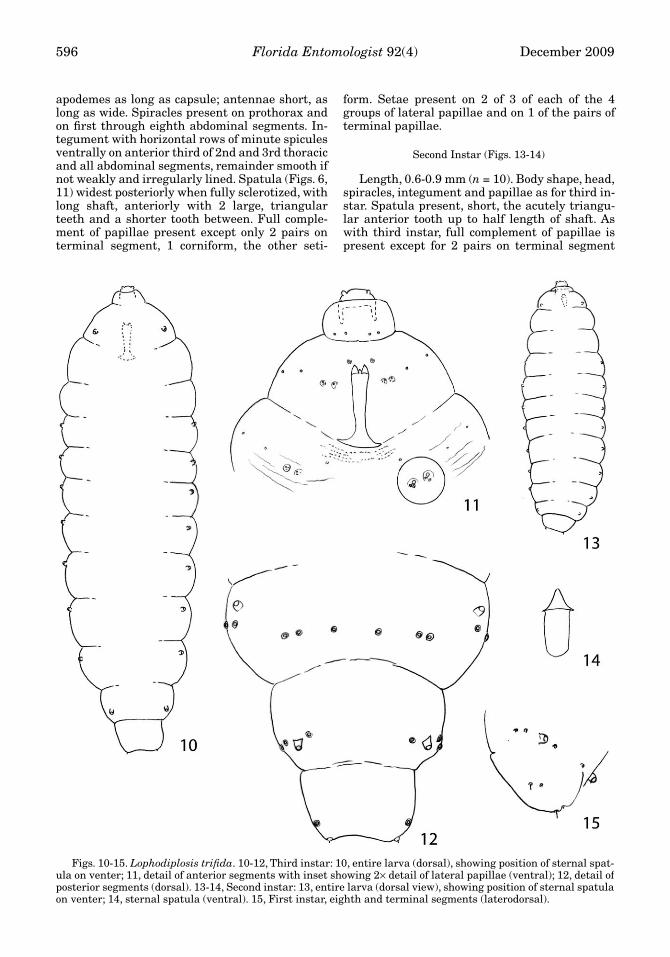
apodemes as long as capsule; antennae short, aslong as wide. Spiracles present on prothorax andon first through eighth abdominal segments. In-tegument with horizontal rows of minute spiculesventrally on anterior third of 2nd and 3rd thoracicand all abdominal segments, remainder smooth ifnot weakly and irregularly lined. Spatula (Figs. 6,11) widest posteriorly when fully sclerotized, withlong shaft, anteriorly with 2 large, triangularteeth and a shorter tooth between. Full comple-ment of papillae present except only 2 pairs onterminal segment, 1 corniform, the other seti-
form. Setae present on 2 of 3 of each of the 4groups of lateral papillae and on 1 of the pairs ofterminal papillae.
Second Instar (Figs. 13-14)
Length, 0.6-0.9 mm (n = 10). Body shape, head,spiracles, integument and papillae as for third in-star. Spatula present, short, the acutely triangu-lar anterior tooth up to half length of shaft. Aswith third instar, full complement of papillae ispresent except for 2 pairs on terminal segment
Figs. 10-15. Lophodiplosis trifida. 10-12, Third instar: 10, entire larva (dorsal), showing position of sternal spat-ula on venter; 11, detail of anterior segments with inset showing 2× detail of lateral papillae (ventral); 12, detail ofposterior segments (dorsal). 13-14, Second instar: 13, entire larva (dorsal view), showing position of sternal spatulaon venter; 14, sternal spatula (ventral). 15, First instar, eighth and terminal segments (laterodorsal).
Gagné et al: Description of Lophodiplosis trifida Larva 597
and setae present on only 2 of 3 of each of the 4groups of lateral papillae and on 1 pair of termi-nal papillae.
First Instar (Fig. 15)
Length, 0.3-0.5 mm (n = 10). Body shape, head,and integument as for second and third instars.Spiracles present only on prothorax and eighthabdominal segment. Spatula absent. Presence ofpapillae as for second and third instars exceptthat pleural and dorsal papillae and the non-set-ose pair of terminal papillae of second and thirdinstars all with minute seta.
Comments on Larval Stage of L. trifida
The first and second instars of this species areunusual among Cecidomyiidae for the almostcomplete absence of papillar setae. Setae occur ononly 2 papillae in each of the 4 sets of lateral trip-lets and on 2 terminal papillae. The first instar, onthe other hand, has tiny setae, no longer than thediameter of their papillae, on all pleural and dor-sal papillae, in addition to the setae already men-tioned for the third and second instars. The onlyother example known of the lack of papillar setaegenerally is the genus Caryomyia, a North Amer-ican genus, in which most of species completelylack papillar setae, even those on the lateral andterminal papillae (Gagné 2008). The integumentis remarkably smooth in L. trifida, with the an-teroventral horizontal bands of spicules the onlysculpturing.
DISCUSSION
The 5 species originally placed in Lophodiplo-sis come from simple to complex galls of Mela-leuca spp. (Gagné et al. 1997). These species arean eclectic mix that, besides having a host genusin common, share the distinguishing pupal char-acter of vertexal extensions that are evidentlyused in cutting through gall tissue prior to adultemergence. Many gall midges have variouslyshaped prominences on the head for that purpose,but they are almost always extensions of the an-tennal bases, not of the vertex. Larvae of Lopho-diplosis are diverse, but this is not unusual ingenera with a variety of gall shapes (Gagné 2008).
Larvae of L. trifida are unique in Lophodiplosisfor the lack of setae on most papillae, their long,narrow, tridentate spatula in the third instar, andmostly smooth integument. The other species ofLophodiplosis for which larvae are known, L. bi-dentata Gagné, L. cornuata Gagné, and L. inden-tata Gagné, have short setae on most papillae, ashort, wide, bidentate spatula, and a rugose in-tegument. The type species, L. indentata, and L.trifida both have only 4 papillae on the terminalsegment, but all 4 papillae have setae on L. inden-tata while only 2 have setae on L. trifida. Bothspecies have a spatula in the second instar. In gallmidges, a second instar spatula occurs only insome gall-making genera where it seems to havearisen de novo in each of the genera where it ap-pears (Gagné 2008).
ACKNOWLEDGMENTS
We thank P. Malikul for making the slide prepara-tions, Scott D. Whittaker, SEM Laboratory Manager,Smithsonian Institution for assistance with the scan-ning electron microscope, Diana Marquez for electroni-cally arranging the photos and drawings onto plates,and James A. Lollis and Elizabeth D. Mattison for assis-tance in collecting galls and larvae, and the South Flor-ida Water Management District for financial support tothis project. We are grateful also to Keith M. Harris,Woking, Surrey, United Kingdom, David A. Nickle andAllen L. Norrbom, Systematic Entomology Laboratory,and 2 anonymous reviewers for critical comments onthe manuscript.
REFERENCES CITED
GAGNÉ, R. J. 1989. The Plant-Feeding Gall Midges ofNorth America. Cornell University Press, Ithaca,New York. xiii & 355 pp. and 4 pls.
GAGNÉ, R. J. 2008. The gall midges (Diptera: Cecidomyi-idae) of hickories (Juglandaceae: Carya). Mem.American Entomol. Soc. 48: 1-147.
GAGNÉ, R. J., BALCIUNAS, J. K., AND BURROWS, D. W.1997. Six new species of gall midges (Diptera: Cecid-omyiidae) from Melaleuca (Myrtaceae) in Australia.Proc. Entomol. Soc. Washington 99: 312-334.
PURCELL, M. F., WINEWRITER (SIC! FOR WINERITER),AND S. A., BROWN, B. T. 2007. Lophodiplosis trifidaGagné (Diptera: Cecidomyiidae), a stem-gallingmidge with potential as a biological control agent ofMelaleuca quinquenervia (Myrtaceae). AustralianEntomol. 34: 123-125.