descriptions of four new species of the … shamshev & grootaert: descriptions of four new...
TRANSCRIPT
45
THE RAFFLES BULLETIN OF ZOOLOGY 2004
THE RAFFLES BULLETIN OF ZOOLOGY 2004 52(1): 45-58© National University of Singapore
DESCRIPTIONS OF FOUR NEW SPECIES OF THE GENUS MICROPHORELLA BECKER(DIPTERA: EMPIDOIDEA, MICROPHORIDAE, PARATHALASSIINI)
FROM SOUTHEAST ASIA AND NEW GUINEA, WITH NOTESON THE RELATIONSHIPS WITHIN THE GENUS
Igor V. ShamshevPermanent address: All-Russian Institute of Plant Protection, shosse Podbel’skogo 3, 188620, St.Petersburg –
Pushkin, Russia. Temporary at Royal Belgian Institute of Natural Sciences, BrusselsEmail: [email protected]
Patrick GrootaertDepartment of Entomology, Royal Belgian Institute of Natural Sciences, Rue Vautier 29, B-1000, Brussels, Belgium
Email: [email protected]
ABSTRACT. – Four new species of the genus Microphorella Becker, M. malaysiana, new species (Thailand,Singapore, Indonesia), M. papuana, new species (Papua New Guinea), M. bira, new species (Sulawesi), andM. satunensis, new species (Thailand) are described from the coasts of Southeast Asia and Papua New Guinea.The phylogenetic relationships of the new species within the genus are discussed. A preliminary analysisleads to the conclusion that the new species form a distinctive group within the genus based primarily onthe ecological data and characters of the male terminalia.
KEY WORDS. – Diptera, Empidoidea, Parathalassiini, Microphorella, new species, phylogeny, Oriental region,Thailand, Singapore, Indonesia, Papua New Guinea.
INTRODUCTION
The genus Microphorella Becker (with M. praecox Loew asthe type species) includes very small greyish flies inhabitingriver banks, wet stones and other places near fresh-waterreservoirs. Microphorella belongs to a problematicassemblage of genera, which are known nowadays as the tribeParathalassiini. This group is considered very closely relatedto the Dolichopodidae and is well represented in Cretaceousamber (Hennig, 1971; Chvála, 1988; Cumming & Brooks,20002). Chvála (1988) has revised the Palaearctic species ofMicrophorella. The genus comprises currently 9 speciesknown from a few localities of Europe (3 species), NorthAmerica (5 species), and Australia (1 species) (Melander,1928; Colless, 1963; Chvála, 1983, 1988). Our paper includesthe descriptions of four new Microphorella species. Theywere collected together with many other Empidoidea on thecoasts of Thailand, Singapore, Sulawesi and Papua NewGuinea.
MATERIAL AND METHODS
This study is based on Diptera materials housed in theEntomology Department of the Royal Belgian Institute of
Natural Sciences, Brussels. All are conserved in alcohol(RBINS, Brussels), except for some voucher specimens thatare on pin at the Zoological Reference Collection of theRaffles Museum of Biodiversity Research, NationalUniversity of Singapore (ZRC).
The flies were collected by sweeping or in white pan trapsand transferred to 75% ethanol. Additionally, pinnedspecimens of M. curtipes (Becker) [5 males, 1 female, France,Corse, VI, R.M.H.N.Belg.: 5.392, Coll. J. Villeneuve,Sciodromia curtipes Beck., det. Becker, Microphorellacurtipes (Beck.), det. M. Chvála, 1982] were also examined.Terms used for adult structures primarily follows those ofMcAlpine (1981), although the terminology for the antennais taken from Stuckenberg (1999). Homologies for the maleterminalia follow Sinclair (2000). To facilitate observations,some parts of the body were macerated in hot 10% KOH or85% lactic acid (terminalia) and immersed in glycerine.Drawings of morphological features were made with a cameralucida attached to a compound microscope. In describing thehypopygium, “dorsal” and “ventral” refer to morphologicalposition prior to genital rotation and flexion. Figures showingthe male genitalia in lateral view are oriented as they appearon the intact specimen (rotated and lateroflexed to the right),with the morphologically ventral surface up and dorsal surface

46
Shamshev & Grootaert: Descriptions of four new species of the genus Microphorella
down. Due to inconspicuous setation of this very small fliesthe term “bristle” is mainly used for differentiated large setaeon the head, mesonotum, and legs bearing a particular nameor one of a series with a particular name (e.g., notopleuralbristle, dorsocentral bristle, ocellar bristle).
List of abbreviations:
cerc = cercus, cib = cibarium, clyp = clypeus, clyp rdg =clypeal ridge, cst sp = costal spinule, epand = epandrium,hypd = hypandrium, hyphar = hypopharynx, lbl = labellum,lbr = labrum, ph = phallus, plp = palpus, pgt = postgonite,psdtrch = pseudotrachea, S = sternite, stp = stipes, sur =surstylus, T = tergite.
KEY TO THE SPECIES OF MICROPHORELLAFROM SOUTHEAST ASIA AND AUSTRALIA
1. Male ........................................................................................ 2– Female (unknown in M. satunensis, new species) ................ 62. 6 or 5 pairs of dorsocentral bristles ...................................... 3– 4 pairs of dorsocentral bristles .............................................. 53. Fore tarsus thickened (Fig. 6), entirely dark brown. Scutum with
6 pairs of dorsocentral bristles. Abdominal sternites 5 and 6 withequally small median posteromarginal processes (Figs. 11, 12)………..…….........................…. M. malaysiana, new species
– Fore tarsus slender. Scutum with 5 pairs of dorsocentral bristles................................................................................................. 4
4. Fore tarsus with tarsomeres 1-2 yellow, tarsomere 3 brownishyellow and tarsomeres 4-5 brownish. Halter pale. Abdominalsegments 5 and 6 with equally large processes (Figs. 36, 37)....................................................... M. satunensis, new species
– Legs entirely black. Halter black. Abdominal segments lackingprocesses .......................................................... M. iota Colless
5. Fore tarsus somewhat thickened, with tarsomeres 1-2 yellow.Femora largely brown, brownish yellow in apical part.Abdominal sternite 5 with short, rather slender medianposteromarginal process, sternite 6 with longer, broad process(Figs. 25, 26). Terminalia with right surstylus broad crescent-shaped (Fig. 27) .............................. M. papuana, new species
– Fore tarsus slender, with tarsomeres 1-3 yellow. Femora almostwholly yellowish, brownish dorsally. Abdominal sternite 5lacking process, sternite 6 with large, broad process (Figs. 31,32). Terminalia with right surstylus long, narrow (Fig. 33).................................................................. M. bira, new species
6. 5 or 6 pairs of dorsocentral bristles ......................................... 7– 4 pairs of dorsocentral bristles .............................................. 87. Fore tarsus yellowish brown, with tarsomeres becoming darker
from 1st to 5th; mid and hind tarsomere 1 largely yellow,brownish at apex. Halter pale .... M. malaysiana, new species
– Legs entirely black. Halter black ................... M. iota Colless8. All tarsi almost wholly light brown, only tarsomere 1 paler
basally ............................................. M. papuana, new species– All tarsi with tarsomeres 1-2 yellowish brown
................................................................. M. bira, new species
TAXONOMY
Microphorella malaysiana, new species(Figs. 1-20)
Material examined. – Holotype - male, Thailand, Ranong,mangrove, 98039, 9 May.1998, coll. P. Grootaert (RBINS).
Paratypes – 2 males, 6 females (1 male, 1 female in coll.Chulalongkorn University, BKK), Thailand, Pak Bara (Satun prov.),beach, white pan traps, 97135, 28 Oct.1997, coll. P. Grootaert; 1male, 7 females, Thailand, Koh Phangan, 98055, 15 May.1998,coll. P. Grootaert; 1 female, Thailand, Ranong prov., Som Laey,98046, 10 May.1998, coll. P. Grootaert; 2 males, 5 females,Thailand, Ranong, mangrove, 98039, 9 May.1998, coll. P.Grootaert; 2 females, Thailand, Pak Bara, 97162, 15 Nov.1997,coll. P. Grootaert; 6 females, Thailand, Prov. Chonburi, Sattahip,sandy beach, 9 Sep.2002, coll. P. Grootaert; 1 female, Thailand,Prov. Rayong, Koh Talu, sandy beach, 22046, 27 Sep.2002, coll.P. Grootaert; 4 males, 4 females (1 male, 1 female in Zool. Museum,Chulalongkorn Univ. BKK), Singapore, Shangi harbour, sandybeach (s. 42), 22 Apr.1993, coll. P. Grootaert; 3 males, 17 females,Singapore, Labrador Park, sandy beach, 22055, 8 Dec.2002, coll.P. Grootaert & Tuksina Suwanamalik; 3 males, 2 females,Singapore, Pulau Ubin, sandy beach near Jetty, 22058, 13 Dec.2002,coll. P. Grootaert & Yang Chang Man (material on pin at ZRC); 10males, 12 females, Indonesia, Pulau Batam, Batu besar beach,93035, 21 Apr.1993, coll. P. Grootaert; 2 males, 3 females,Indonesia, P. Batam, Batu Besar beach (s. 34), 21 Apr.1994, coll.P. Grootaert. All deposited in RBINS, Brussels, except indicatedotherwise.
Diagnosis. – A small species (1.5-1.8 mm long) with paleyellow palpi in male, brown in female; 6 dorsocentral bristles;fore tarsi thickened, wholly brown; abdominal sternites 5 and6 of male with short median posteromarginal processes ofsubequal size; right surstylus leaf-like.
Description. – Male body length 1.5-1.7 mm, wing length1.2-1.3 mm. Head broader than thorax in dorsal view, broadoval in lateral view, nearly 1.5 times higher than wide, darkbrown in ground-colour, mostly light grey pollinose, withpale setation; clypeus brownish yellow, face (includingclypeus) denser pollinose; insertion of neck high on head.Occiput moderately rounded, not far projecting beyond hindmargin of eye, with upper median part moderately concave.Ocellar triangle weakly prominent. Eyes dichoptic in bothsexes, entirely covered with uniform distinct ommatrichia,with inner margins not emarginate near antennae; ommatidialarge, uniform. Frons broadly triangularly widening abovein both sexes. Face broad below antennae, graduallynarrowing below, in middle nearly as wide as distancebetween posterior ocelli. Clypeus rather long, producedbelow, convex, weaker sclerotized than upper face, roundedapically. Bristles of head mostly well-differentiated; 2inclinate anterior frontal-orbitals, 2 lateroclinate posteriorfrontal-orbitals, 2 lateroclinate anterior ocellars, 2 lateroclinateouter-verticals and 2 inclinate inner-verticals; posteriorocellars undifferentiated, 7-9 minute setulae present.Postocular occipital bristles rather long, hair-like, arrangedin 1 regular row in upper half, more numerous and irregularlyspread in lower half; several long setae present just behind
47
THE RAFFLES BULLETIN OF ZOOLOGY 2004
mouth-opening including postgena. Antenna (Fig. 1) insertedabove middle of head in profile, entirely dark brown; scapevery short, cup-shaped, bare; pedicel larger than scape,subglobular, ringed with circlet of short bristles; postpedicelbulbous, nearly 2.0 times broader than long, gradually tapered,microsetulose in apical part. Style apical, arista-like, nearly2.0 times longer than postpedicel, 1-segmented, jointeddistinctly with postpedicel, whip-like in subapical portion,microsetulose, with setulae in apical part longer than widthof aristal trunk, bearing sensory pit in subbasal part, lackingsecondary sexual adornments. Palpus (Fig. 2) moderatelylarge, clavate, flattened, pale yellow in ground-colour, clothedin dense pale appressed setulae intermixed with scattered erectsetulae (palpus silvery white in some angle of view), 1 basalsensory pit present (Fig. 3). Proboscis (Fig. 4) short, wellvisible, pointing downward, wholly brown; labellum (Fig.5) well developed, small, with spinule-like setulae along uppermargin and similar spinules near apices of pseudotracheae,covered with dense ciliae in subapical portion; lacinia absent;stipes long, slender; labrum heavily sclerotized, convexbasally; epipharynx serrate along lower margin; hypopharynxslender, almost straight; prementum with 2-3 short setae oneach side; 6 geminate pseudotracheae present, with wallsweakly sclerotized; clypeal ridge rather short, shorter thancibarium. Genae moderately broad.
Thorax dark brown in ground-colour, greyish pollinose, withpale setation. Mesoscutum moderately arched, prescutellardepression hardly prominent. Prosternum fused withproepisternum forming prothoracic precoxal bridge (Fig. 9).Antepronotum with 4 setulae. Postpronotal lobe distinct,with1setula. Scutum unicolorous. Mesonotum short,rectangular (viewed dorsally), with bristles well-differentiatedbut reduced in number; 0 presutural supra-alar, 2 postsuturalsupra-alars of different length, 2 notopleurals, 1 short post-alar and 2 long cruciate scutellars; upper part of verticalanterior surface of scutum with 1 pair of setulae.Dorsocentrals 1-serial, 6 per row, rather long, of subequallength (prescutellars somewhat longer), lacking accessorysetulae. Acrostichals lacking. Mesopleuron bare. Thoracicspiracles pale. Halter pale.
Legs (Figs. 6-8) moderately long, subshining, mostly withpale inconspicuous setation. Coxae brownish, somewhat palerapically; trochanters of fore and mid legs brownish yellow,trochanter of hind leg pale brownish; fore and mid femoralargely yellow, yellowish brown dorsally (in darker specimensfore and mid femora largely yellowish brow, yellow in apicalpart), hind femur largely brownish yellow, yellow apically;tibiae yellow (in darker specimens somewhat brownish yellowin basal part); fore tarsus brown black; mid and hind tarsiwith tarsomere 1 at apex, tarsomere 2 largely (except base)and tarsomeres 3-4 entirely yellowish brown, tarsomere 5brown. Femora broader than corresponding tibiae, more orless gradually tapering toward apex, subequal in thickness.Tibiae slender. Fore tarsomeres thickened, tarsomere 1 nearly2 times longer than tarsomere 2 but 1.5 times shorter thantarsomeres 2-5 combined; tarsomeres 2-4 of equal length;tarsomere 5 of all legs distinctly flattened dorsoventrally. Forecoxa with numerous, rather long hair-like setae anteriorly;
mid and hind coxae with similar but scattered setae.Trochanters of all legs with few setulae. Fore femur with 1row of postero- and 1 row of anteroventral setae; mid femurwith 1 row of anteroventral setae becoming longer towardapex; hind femur with 1 row of rather short anteroventralsetae; additionally, some prominent short setae present onall femora near apex and dorsally; longest setae on femoraat most as long as corresponding femur is wide; otherwisefemora clothed in inconspicuous setulae. Fore tibia withanterior apical comb and 1 short spine-like ventral preapicalseta; mid tibia with several setae around apex, including 1longest ventral preapical one; hind tibia with posterior apicalcomb and 1 ventral apical spine; all tibiae with somewhatlonger dorsal setae; otherwise tibiae clothed in inconspicuoussetulae. Tarsomeres 1-4 with dark ventral spinules (longerand more numerous on mid tarsomere 1); hind tarsomere 1with posterior apical comb and 1 short ventral apical spine.Tarsal claws, pulvilli and setiform empodium well developedon all legs; tarsal claw shorter than apical width of tarsomere5; pulvilli short, broad; empodium slender, with ventralpubescence.
Wing (Fig. 10) moderately broad, 2.5-2.7 times longer thanwide; very finely infuscate, with brownish yellow to brownishveins, pale marginal fringe, yellow basicostal setae and darkcostal spinules; entirely covered with minute microtrichia(including veins); with anal lobe weakly developed; alulaabsent. Basal section of costa with 3-4 bristles becominglonger distad; additionally, costa bearing 2 rows of short spinelike setae along anterior margin and entirely ciliate alongposterior margin. Pterostigma (or stigmatic sclerotization)lacking. Costa circumambient, distinct throughout. Alllongitudinal veins complete, distinctly reaching wing margin.Sc very close to R1, reduced to fold in its apical section, endingin costa. Rs in basal 1/5 of wing, with 2 branches. R1 notthickened, moderately long, extending to wing midpoint,somewhat arcuate. R2+3 more or less straight, ending nearerto wing apex, somewhat divergent with R4+5. R4+5 unforked,ending near wing-tip. R4+5 and M1 somewhat divergent nearwing-apex. M1+2 unforked, anterior portion of crossvein m-cu (base of M2) about at midpoint of wing. Distance betweenapices of M1 and M2 longer than distance between apices ofM2 and CuA1. CuA2 reflexed, very thin in apical part. A1
absent. A2 present on posterobasal margin of wing. Crossveinh almost opposite to base of Rs. Short r-m crossvein present,in basal 1/5 of wing, perpendicular to longitudinal veins,sometimes very thin. Crossvein bm-cu incomplete, sometimesabsent. Cell dm present, rather short. Cells br, bm and cupin basal 1/5 of wing. Cell br slender, longer than cells bmand cup. Cells bm and cup subequal in length and width, bothsomewhat broader than cell br. Cell cup closed, with proximalend acutely rounded. Squamae pale, with scattered pale ciliae.Halter white.
Abdomen rather short and broad, brown in ground-colour,finely greyish pollinose, pale setose, lacking conspicuousposteromarginal setae; preabdomen with posterior marginsof tergites paler, postabdomen darker. Abdominal muscleplaques present, distinct. Segments 1-4 symmetrical withsimple sternites and tergites, forming preabdomen; sternite

48
Shamshev & Grootaert: Descriptions of four new species of the genus Microphorella
Figs. 1-10: Microphorella malaysiana, new species, male. 1 – antenna, dorsal view, 2 – palpus, lateral view, 3 – inside structure of basalsensory pit on palpus, lateral view, 4 – mouthparts, lateral view, 5 – labella, dorsal view, 6 – fore leg, anterior view, 7 – mid leg, anteriorview, 8 – hind leg, anterior view, 9 – prothoracic pleurosternal region, anterior view, 10 – wing, dorsal view. Scale: 0.1 mm.

49
THE RAFFLES BULLETIN OF ZOOLOGY 2004
Figs. 11-16: Microphorella malaysiana, new species, male. 11 – postabdomen, left lateral view, 12 – same, ventral view, 13 – male terminalia,right lateral view, 14 – same, left lateral view, 15 – same, dorsal view, 16 – same, ventral view. Scale: 0.1 mm.
1112
16 15
1413
50
Shamshev & Grootaert: Descriptions of four new species of the genus Microphorella
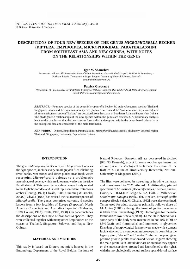
Figs. 17-20: Microphorella malaysiana, new species, 17-20 – female. 17 – abdomen, lateral view, 18 – postabdomen, lateral view, 19 –apical part of ovipositor, dorsal view, 20 – spermatheca, dorsal view. Scale 0.1 mm.

51
THE RAFFLES BULLETIN OF ZOOLOGY 2004
3 with cluster of short setae on each side posteriorly; sternite4 with similar cluster of stronger and longer setae.Postabdomen (Figs. 11, 12) spirally contorted, segments 5-7 confined to left side forming cavity to place hypopygium,somewhat stronger sclerotized; sternite 5 greatly reduced;sternites 5 and 6 with short median posteromarginal process;tergite 5 and segment 7 lacking setation, sternite 6 with 0-1 short lateral setae. Sternite 8 moderately large,subrectangular, lying at hind and exposed ventrolaterad,covered with numerous long setae; tergite 8 atrophied;foramen unformed. Terminalia (Figs. 13-16) lateroflexed tothe right, inverted and with caudal pole directed forward,asymmetrical; hypandrium brown, processes brown to paleyellow, cerci pale. Hypopygium small, somewhat shorterthan apical half of abdomen. Hypandrium very large,occupying most part of hypopygium, produced at apex andwith several accessory processes, bare (except microtrichia).Epandrium greatly reduced and represented only by smallinconspicuous sclerites, separated from hypandrium; rightsurstylus very large, “leaf-like”; left surstylus smaller, withtwo processes more prominent. Cerci weakly sclerotized,moderately large, covered with microtrichia, bearing severalsetae of different length including longest one at tip.Postgonites present, rather large, well sclerotized, ofcomplicated structure. Phallus tubular, more or less smoothlyarcuate, directed forwards, with pointed tip; ejaculatoryapodeme subrectangular.
Female body length 1.5-1.8 mm, wing length 1.3-1.4 mm.Similar to male except the following characters. Palpusbrownish, with ordinary setulae. Legs unmodified, with moredistinct colour pattern; femora largely brown, yellow inapical 1/4; fore tarsus yellowish brown, with tarsomeresbecoming darker from 1st to 5th; mid and hind tarsomere 1largely yellow, yellowish brown at apex, tarsomeres 2-5similar in colour to those on fore tarsus. Abdomen (Fig. 17)more or less gradually tapering, segments 1-6 formingpreabdomen into which posterior segments are retracted andonly partly visible. Postabdomen (Fig. 18, 19) rather slender,brown, mostly covered with scattered setulae; sternite 8articulated with tergite 8 posteriorly, tergite 8 paired, tergite10 and sternite 10 articulated, hemitergites 10 small, hardlydistinguished from cerci, with 3 long bristles each. Cercirather broad, well sclerotized, bearing setae of differentlength (longest one at tip). Spermatheca (Fig. 20) tubular,with receptacle spherical and unpigmented; middle part ofspermathecal duct broadened, finely pigmented and withtracheae-like surface.
Differential diagnosis. – Microphorella malaysiana, newspecies, can be distinguished from all other closely relatedspecies as it is given in the key.
Etymology. – The new species is named after the wholeregion of its origin, Malaysia.
Distribution. – Thailand, Indonesia, Singapore.
Microphorella papuana, new species(Figs. 21-30)
Material examined. – Holotype - male, West Papua or Irian Jaya,Nabire, Kaladiri beach, 970061, 30 Apr.1997, coll. P. Grootaert(in RBINS).
Paratypes – 3 males, 5 females, same data as in holotype (1 maleZRC); 1 male, 2 females, Irian Jaya, Sanoba beach (Nabire), 970021,21 Apr.1994, coll. P. Grootaert & Ph. Hoyois; 3 males, Irian Jaya,Biak, Bosnik beach, 970080, 6 May.1997, coll. P. Grootaert. Alldeposited in RBINS, Brussels, except otherwise indicated.
Diagnosis. – Resembling M. malaysiana, new species, butfore tarsi less thickened and partly yellow in male, scutumwith 4 dorsocentral and 1 postsutural supra-alar bristles; maleabdominal sternite 5 with short and rather slender medianposteromarginal process, sternite 6 with longer, broad (viewedventrally) process; right surstylus broad, crescent-shaped.
Description. – Male (Fig. 21) body length 1.4-1.5 mm, winglength 1.2-1.3 mm. Head and thorax light grey pollinose, withdark-green tinge (in some angle of view). Ocellar tuberclewith 2 long anterior and 2 very short posterior ocellar bristles.Face in middle somewhat narrower than distance betweenposterior ocelli. Scutum with 1 postsutural supra-alar bristle.Dorsocentrals represented by 4 long bristles per row. Legs(Figs. 22-24) with following pattern colour: coxae almostwholly brownish, fore coxa brownish yellow at apex;trochanter of fore leg brownish yellow, those mid and hindlegs brownish; fore femur brownish in basal 2/3, brownishyellow in apical 1/3; mid and hind femora brownish in basal3/4, brownish yellow in apical 1/3; all tibiae wholly yellowish(often with brownish tinge); fore tarsus with tarsomeres 1-2 yellow (sometimes with brownish tinge), tarsomere 3brownish (sometimes indistinctly), tarsomeres 4-5 darkbrown; mid tarsus with tarsomere 1 largely yellowish,yellowish brown apically, tarsomeres 2-5 brown; hind tarsussimilar to mid tarsus but tarsomere 2 paler (in paler specimensmid and hind tarsi with tarsomere 2 yellowish, with slightbrownish tinge); additionally, femora with dark-green tingeon darkened parts (in some angle of view). Wing as in M.malaysiana, new species, but basal section of costa with 1short and 1 long bristles. Abdomen with sternites 3 and 4bearing cluster of long and strong setae on each side (Figs.25, 26); sternite 5 with short and rather slender medianposteromarginal process, sternite 6 with longer, broad (viewedventrally) process; sternites 5-6 and segment 7 lackingsetation. Terminalia (Figs. 27-30) with right surstylus long,broad, crescent-shaped. Otherwise as in M. malaysiana, newspecies.
Female body length 1.4-1.5 mm, wing length 1.2-1.3 mm.Similar to male except the following characters. Palpusbrown, with ordinary setulae. Colour pattern of legs similarto that in male but all femora with broader brown space, tibiaeusually darker and tarsi almost wholly light brown (tarsomere1 paler basally). Postabdomen, including cerci, brown, as inM. malaysiana, new species (spermatheca was not studied).

52
Shamshev & Grootaert: Descriptions of four new species of the genus Microphorella
Figs. 21-26. Microphorella papuana, new species, male. 21 – habitus, right lateral view (scale 1.0 mm), 22 – fore leg, anterior view, 23– mid leg, anterior view, 24 – hind leg, anterior view, 25 – postabdomen (segment 8 and hypopygium not shown), left lateral view, 26 –same, ventral view. Scale 0.1 mm.

53
THE RAFFLES BULLETIN OF ZOOLOGY 2004
Figs. 27-30. Microphorella papuana, new species, male. 27 – terminalia, right lateral view, 28 – same, left lateral view, 29 – same, dorsalview, 30 – same, ventral view. Scale 0.1 mm.
54
Shamshev & Grootaert: Descriptions of four new species of the genus Microphorella
Differential diagnosis. – This species is most closely relatedto M. bira, new species. Main differences between thesespecies are given in the key. The females of these speciesare hardly distinguishable in the colour of legs.
Etymology. – The species is named after the whole region ofits origin, Papua New Guinea.
Distribution. – New Guinea.
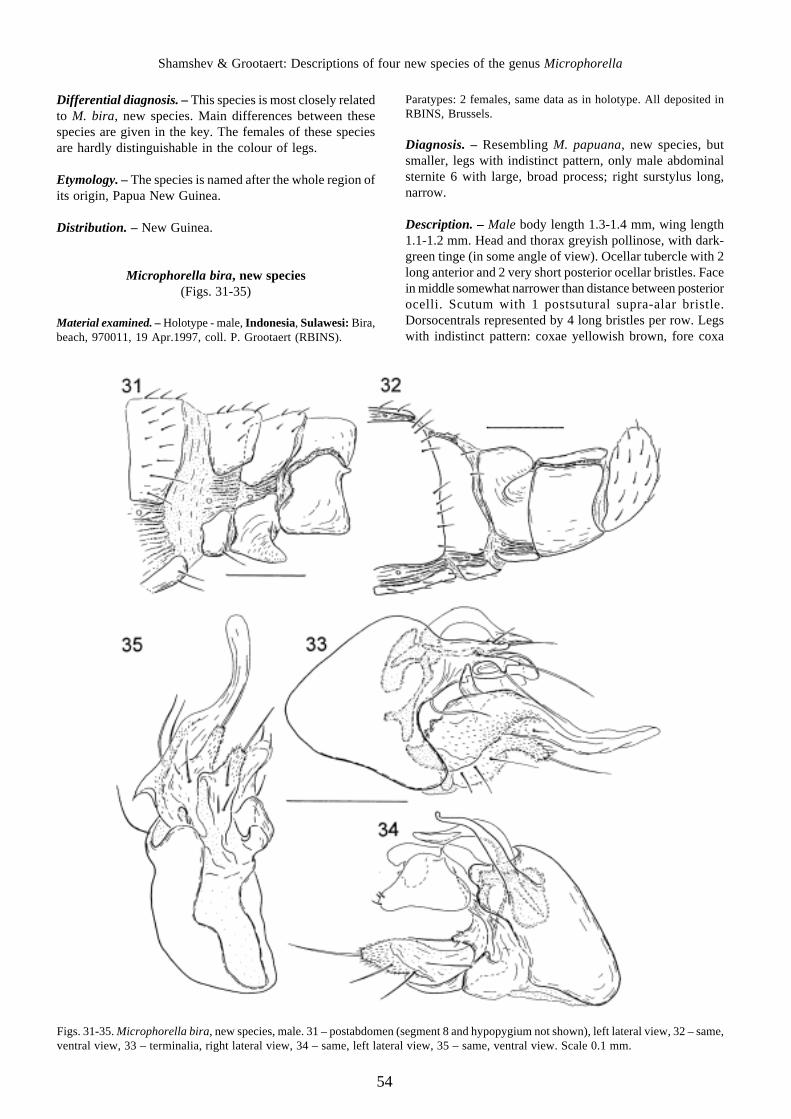
Microphorella bira, new species(Figs. 31-35)
Material examined. – Holotype - male, Indonesia, Sulawesi: Bira,beach, 970011, 19 Apr.1997, coll. P. Grootaert (RBINS).
Paratypes: 2 females, same data as in holotype. All deposited inRBINS, Brussels.
Diagnosis. – Resembling M. papuana, new species, butsmaller, legs with indistinct pattern, only male abdominalsternite 6 with large, broad process; right surstylus long,narrow.
Description. – Male body length 1.3-1.4 mm, wing length1.1-1.2 mm. Head and thorax greyish pollinose, with dark-green tinge (in some angle of view). Ocellar tubercle with 2long anterior and 2 very short posterior ocellar bristles. Facein middle somewhat narrower than distance between posteriorocelli. Scutum with 1 postsutural supra-alar bristle.Dorsocentrals represented by 4 long bristles per row. Legswith indistinct pattern: coxae yellowish brown, fore coxa
Figs. 31-35. Microphorella bira, new species, male. 31 – postabdomen (segment 8 and hypopygium not shown), left lateral view, 32 – same,ventral view, 33 – terminalia, right lateral view, 34 – same, left lateral view, 35 – same, ventral view. Scale 0.1 mm.

55
THE RAFFLES BULLETIN OF ZOOLOGY 2004
yellowish in apical part; trochanters of all legs brownishyellow; femora yellowish, with brownish tinge, moredistinctly brownish dorsally; tibiae yellow; fore tarsus withtarsomeres 1-3 yellow and tarsomeres 4-5 brown, mid andhind tarsi with tarsomeres 1-2 yellow and tarsomeres 3-5becoming gradually darker (tarsomere 5 brown). Tarsi slender(except tarsomere 5). Wing with basal section of costa bearing1 short and 1 long bristles. Abdomen with sternite 4 bearingrow of long, strong setae on each side (Figs. 31, 32); sternite5 lacking process, with several ordinary setae, sternite 6 withlarge broad (viewed ventrally) process; sternite 6 and segment7 lacking setation. Terminalia (Figs. 33-35) with rightsurstylus long, rather narrow. Otherwise as in M. malaysiana,new species.
Female body length 1.3-1.4 mm, wing length 1.2-1.3 mm.Similar to male except the following characters. Palpusbrown, with ordinary setulae. Legs darker, with more distinctpattern; femora largely brownish, paler apically, all tibiae andtarsomeres 1-2 of all tarsi yellowish brown. Postabdomen,including cerci, brown, as in M. malaysiana, new species(spermatheca was not studied).
Differential diagnosis. – The new species can bedistinguished from other species described here as it is givenin the key.
Etymology. – The species is named after the type locality,Bira.
Distribution. – Indonesia, Sulawesi.
Microphorella satunensis, new species(Figs. 36-40)
Material examined. – Holotype - male, Thailand, Pak Bara (Satunprov.), beach, white pan traps, 97135, 28 Oct.1997, coll. P.Grootaert.
Paratypes – 3 males, same data as in holotype. All deposited inRBINS, Brussels, except otherwise indicated.
Diagnosis. – Resembling M. papuana, new species, smaller(1.2-1.4 mm long), scutum with 5 dorsocentrals, abdominalsegments 5 and 6 with equally large processes; right surstylussubtriangular.
Description. – Male body length 1.2-1.4 mm, wing length1.1-1.2 mm. Head and thorax finely greyish pollinose. Ocellartubercle with 2 long anterior and 2 very short posterior ocellarbristles. Face in middle nearly as broad as distance betweenposterior ocelli. Scutum with 2 postsutural supra-alar bristles.Dorsocentrals represented by 5 long bristles per row (2prescutellar pairs somewhat longer). Tarsi with slendertarsomeres (except tarsomere 5). Legs with following pattern:coxae almost wholly brown, fore coxa paler at apex; mid andhind trochanters yellowish brown, fore trochanter somewhatpaler; femora largely brownish; fore femur at extreme baseand in apical 2/5, mid femur in apical 1/3 and hind femur inapical 1/4 yellow; all tibiae pale yellow; all tarsi with
tarsomeres 1-2 yellow, tarsomere 3 brownish yellow andtarsomeres 4-5 brownish. Wing finely infuscate, basal sectionof costa with 1 short and 1 long bristles. Abdomen withsternite 4 bearing clusters of long, strong setae on each side(Figs. 36, 37); sternites 5 and 6 with equally large (viewedventrally) processes, both bearing few setae laterally; segment7 lacking setation. Terminalia (Figs. 38-40) with rightsurstylus rather subtriangular. Otherwise as in M. malaysiana,new species.
Female unknown.
Differential diagnosis. – The new species differs from otherspecies by a set of the characters given in the key.
Etymology. – The species is named after the province of thetype locality, Satun.
Distribution. – Thailand.
DISCUSSION
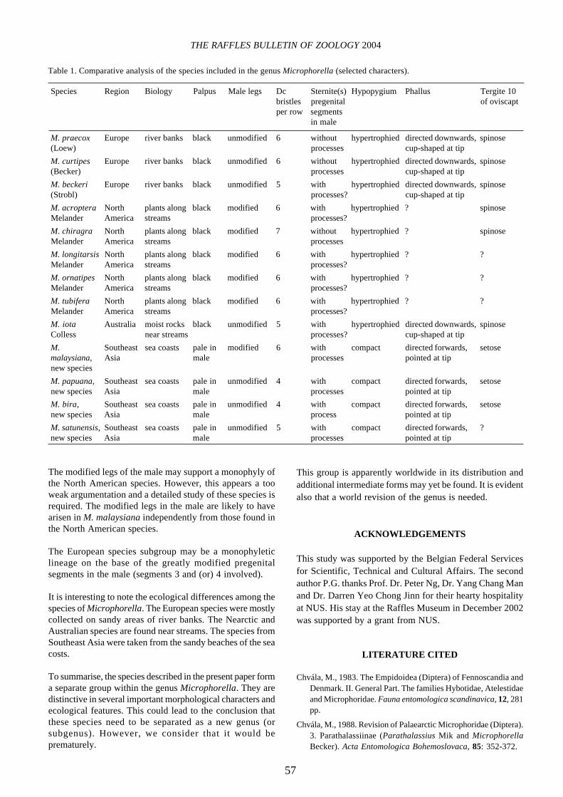
Ulrich (1991) and, quite recently, Cumming & Brooks (2002)discussed the relationships of Microphorella with other generaassigned to the Parathalassiini. However, the relationshipswithin Microphorella have never been examined. Thepreliminary comparative analysis of the species included inthe Microphorella is given in Table 1. We have included onlythose characters, which were noted in the originaldescriptions. Unfortunately, some important morphologicalfeatures (especially of the male and female terminalia) areunknown for a number of species.
Four species known currently from Southeast Asia arehypothesised to form a monophyletic subgroup withinMicrophorella. The support of the subgroup is based on thepresence of a modified palpus and ventral processes onabdominal sternites 5 and 6 in the male and a setose tergite10 in the female. The palpus is sexually uniform in otherMicrophorella species as well as in most Parathalassiini(except for some undescribed taxa). The presence of theprocesses on the abdominal sternites 5 and 6 is a uniquefeature of these species and it is undoubtedly a synapomorphyof this subgroup. Among Parathalassiini, short processes onsternites of pregenital segments in the male are probablypresent in the fossil genus Electrophorella (Cumming &Brooks 2002: 46, Fig. 5). Another character that could supportthis subgroup is a setose tergite 10 of the female. The presenceof the acanthophorous setae on tergite 10 is considered to bean apomorphic feature of Parathalassiini (Sinclair, 1995).However, the female sex is unknown for M. satunensis.
The hypertrophied terminalia of the male and a cup-shapedphallus indicate a closer relationship between European andNorth American species. Although, the last character shouldbe checked in the Nearctic group. The Australian M. iota islikely to belong to this subgroup. This species possesses apeculiar brush of setae on the tip of the phallus that is anautapomorphic state within Microphorella.

56
Shamshev & Grootaert: Descriptions of four new species of the genus Microphorella
Figs. 36-40. Microphorella satunensis, new species, male. 36 – postabdomen (hypopygium not shown), left lateral view, 37 – same, ventralview, 38 – terminalia, right lateral view, 39 – same, left lateral view, 40 – same, ventral view. Scale 0.1 mm.
57
THE RAFFLES BULLETIN OF ZOOLOGY 2004
The modified legs of the male may support a monophyly ofthe North American species. However, this appears a tooweak argumentation and a detailed study of these species isrequired. The modified legs in the male are likely to havearisen in M. malaysiana independently from those found inthe North American species.
The European species subgroup may be a monophyleticlineage on the base of the greatly modified pregenitalsegments in the male (segments 3 and (or) 4 involved).
It is interesting to note the ecological differences among thespecies of Microphorella. The European species were mostlycollected on sandy areas of river banks. The Nearctic andAustralian species are found near streams. The species fromSoutheast Asia were taken from the sandy beaches of the seacosts.
To summarise, the species described in the present paper forma separate group within the genus Microphorella. They aredistinctive in several important morphological characters andecological features. This could lead to the conclusion thatthese species need to be separated as a new genus (orsubgenus). However, we consider that it would beprematurely.
This group is apparently worldwide in its distribution andadditional intermediate forms may yet be found. It is evidentalso that a world revision of the genus is needed.
ACKNOWLEDGEMENTS
This study was supported by the Belgian Federal Servicesfor Scientific, Technical and Cultural Affairs. The secondauthor P.G. thanks Prof. Dr. Peter Ng, Dr. Yang Chang Manand Dr. Darren Yeo Chong Jinn for their hearty hospitalityat NUS. His stay at the Raffles Museum in December 2002was supported by a grant from NUS.
LITERATURE CITED
Chvála, M., 1983. The Empidoidea (Diptera) of Fennoscandia andDenmark. II. General Part. The families Hybotidae, Atelestidaeand Microphoridae. Fauna entomologica scandinavica, 12, 281pp.
Chvála, M., 1988. Revision of Palaearctic Microphoridae (Diptera).3. Parathalassiinae (Parathalassius Mik and MicrophorellaBecker). Acta Entomologica Bohemoslovaca, 85: 352-372.
Table 1. Comparative analysis of the species included in the genus Microphorella (selected characters).
Species Region Biology Palpus Male legs Dc Sternite(s) Hypopygium Phallus Tergite 10bristles pregenital of oviscaptper row segments
in male
M. praecox Europe river banks black unmodified 6 without hypertrophied directed downwards, spinose(Loew) processes cup-shaped at tip
M. curtipes Europe river banks black unmodified 6 without hypertrophied directed downwards, spinose(Becker) processes cup-shaped at tip
M. beckeri Europe river banks black unmodified 5 with hypertrophied directed downwards, spinose(Strobl) processes? cup-shaped at tip
M. acroptera North plants along black modified 6 with hypertrophied ? spinoseMelander America streams processes?
M. chiragra North plants along black modified 7 without hypertrophied ? spinoseMelander America streams processes
M. longitarsis North plants along black modified 6 with hypertrophied ? ?Melander America streams processes?
M. ornatipes North plants along black modified 6 with hypertrophied ? ?Melander America streams processes?
M. tubifera North plants along black modified 6 with hypertrophied ? ?Melander America streams processes?
M. iota Australia moist rocks black unmodified 5 with hypertrophied directed downwards, spinoseColless near streams processes? cup-shaped at tip
M. Southeast sea coasts pale in modified 6 with compact directed forwards, setosemalaysiana, Asia male processes pointed at tipnew species
M. papuana, Southeast sea coasts pale in unmodified 4 with compact directed forwards, setosenew species Asia male processes pointed at tip
M. bira, Southeast sea coasts pale in unmodified 4 with compact directed forwards, setosenew species Asia male process pointed at tip
M. satunensis, Southeast sea coasts pale in unmodified 5 with compact directed forwards, ?new species Asia male processes pointed at tip

58
Shamshev & Grootaert: Descriptions of four new species of the genus Microphorella
Colless, D. H., 1963. An Australian species of Microphorella(Diptera: Empididae), with notes on the phylogeneticsignificance of the genus. Proceedings of the Linnean Societyof New South Wales, 88: 320-323.
Cumming, J. M. & S. E. Brooks, 2002. Electrophorella, a new genusof parathalassiine flies from Baltic amber, with a cladisticanalysis of the Microphorinae + Dolichopodidae lineage(Diptera: Empidoidea). Studia dipterologica, 9: 41-54.
Hennig, W., 1971. Insektenfossilien aus der Unteren Kreide. 3.Empidiformia (“Microphorinae”) aus der Unteren Kreide undaus dem Baltischen Berstein; ein Vertreter der Cyclorrhaphaaus der untere Kreide. Stuttgarter Beitr. z. Naturkunde, 232: 1-28.
McAlpine, J. F., 1981. Morphology and terminology – Adults.[Chapter] 2. In: McAlpine, J. F., B. V. Peterson, G. E. Shewell,H. J. Teskey, J. R. Vockeroth & D. M. Wood (eds.), Manual ofNearctic Diptera, 1: 9-63. Agriculture Canada Monograph, 27.Ottawa.
Melander, A. L., 1928. Diptera, Fam. Empididae. In: Wytsman, P.(ed.), Genera Insectorum, 185, 434 pp., Louis Desmet-Verteneuil, Bruxelles.
Sinclair, B. J., 1995. Generic revision of the Clinocerinae(Empididae), and description and phylogenetic relationships ofthe Trichopezinae, new status (Diptera: Empidoidea). CanadianEntomolologist, 127: 665-752.
Sinclair, B. J., 2000. Morphology and terminology of Diptera maleterminalia. In: Papp, L. & B. Darvas (eds.), Contributions to aManual of Palaearctic Diptera, 1: 53-74, Budapest: ScienceHerald.
Stuckenberg, B. R., 1999. Antennal evolution in the Brachycera(Diptera), with a reassessment of terminology relating to theflagellum. Studia dipterologica, 6: 33-48.
Ulrich, H., 1991. Two new genera of parathalassiine-like flies fromSouth Africa (Diptera, Empidoidea). Bonner ZoologischeBeitraege, 42: 187-216; Bonn.