design and evaluation of soluble ocular insert for...
TRANSCRIPT
Design and Evaluation of Soluble Ocular Insert For Controlled Release of Chloramphenicol
Copyright © 2017 by Kermanshah University of Medical Sciences JRPS, 2017, 6(2), 123-133| 123
Design and Evaluation of Soluble Ocular Insert For Controlled
Release of Chloramphenicol Shahla Mirzaeeia,b*, Moslem Alizadehc
a Pharmaceutical Sciences Research Center, School of Pharmacy, Kermanshah University of Medical Sciences, Kermanshah, Iran bNano Drug Delivery Research Center, School of Pharmacy, Kermanshah University of Medical Sciences, Kermanshah, Iran c Student Research Committee, School of Pharmacy, Kermanshah University of Medical Sciences, Kermanshah, Iran
ARTICLE INFO Article Type: Research Article Article History: Received: 2017-05-07 Revised: 2017-06-20 Accepted: 2017-06-25 ePublished: 2017-07-03 Keywords: Chloramphenicol Controlled Ocular Insert Drug Release Ophthalmic Deliver
A B S T R A C T
Soluble ocular inserts of chloramphenicol were prepared with the aim of achieving once a day administration. Drug reservoir was prepared using hydrophilic polymer and rate-controlling hydrophobic polymer; Eudragit L100, Eudragit S100, Eudragit RL100. All the formulations indicated no interaction between drug and polymer in FTIR studies. The Inserts were evaluated for the several parameters, viscosity, drug–polymer interaction, in vitro drug release, sterility testing. They were also evaluated for % moisture loss, % moisture uptake,thickness, and tensile strength. Ophthalmic inserts provide that prolonged and sustained drug release. Ocular inserts prepared were smooth and passed all the evaluation tests performed. Mechanical properties and in vitro drug release were dependent on film composition. The release profile of all the formulations showed a steady, controlled drug release. Ocular inserts formulated also passed the test for sterility. In vitro studies demonstrated that P5 insert can ensure a sustained drug release on the ocular surface for a prolonged over 20 hours’ time period. Also, reduction in frequency of administration, may improve the patient compliance.
*Corresponding Author: Shahla Mirzaeei, E-mail: [email protected]
Copyright © 2017by Kermanshah University of Medical Sciences
Journal of Reports in Pharmaceutical Sciences 2017, 6(2), 123-133

Mirzaeei and Alizadeh
Copyright © 2017by Kermanshah University of Medical Sciences JRPS, 2017, 6(2), 123-133| 124
Introduction For the treatment of anterior eye segment infections using anti-infective agents, topical ocular application is the most convenient route of administration. However, topical delivery of anti-infective agents is associated with a number of problems and challenges owing to the unique structure of the eye. Topical ocular drug delivery systems can be classified into two forms: conventional and non-conventional. The efficacy of conventional ocular formulations is limited by poor corneal retention and permeation resulting in low ocular bioavailability [1-3]. Ophthalmic solution, suspension, and ointment dosage forms no longer constitute optimal therapy for these indications[2, 4]. Ocular delivery systems like inserts biodegradable polymeric systems, and collagen shields are being developed in order to attain better ocular bioavailability and sustained action of ocular drugs. A number of solid polymeric inserts have been developed as ophthalmic drug delivery systems[5]. Inserts allow for accurate dosing, reduced systemic absorption and in some case, better patient compliance resulting from a reduced frequency of administration. Inserts are affected to lesser extent by nasolacrimal drainage and tear flow than the more conventional dosage forms, and are associated with reliable drug release and longer residence times in conjunctivalcul-de-sac[6-8]. A number of inserts are currently available on the markets or in the latter stages of development. These inserts have been classified as degradable; polyvinyl alcohol, hydroxypropylcelluloseand polyvinyl pyrrolidone or non-degradable; ethylene vinyl acetate copolymers[9]. Ocular inserts of antibiotics were prepared with objectives of reducing the frequency of administration, obtaining controlled release and greater therapeutic efficacy in the treatment of corneal ulcers [10-12]. Inserts for antibiotics, such as natamycin, moxifloxacin hydrochloride, azithromycin were dishinged to improve residence time and corneal absorption [13-15]. Chloramphenicol (ChM) has been an effective agent against external ocular infections by inhibiting prokaryotic protein synthesis. The action of ChM is usually bacteriostatic, but its bactericidal against Haemophilus influenza,
Streptococcus pneumoniae, and Neisseria meningitidis[16, 17]. Eye drops are the most used dosage form by ocular route and chloramphenicol is the main effective drug in the common used eye drops. However the eye drops have several disadvantages, such as a very low bioavailability (1–10%) of the drugs, which must be absorbed at this site and must be inserted several times a day[18]. Also, the effective component, that is chloramphenicol, has very low solubility in water and easily hydrolyzes that cause eye drops turn into unqualified[19]. The aim of the present work was to develop chloramphenicol loaded ocular inserts composed of blends of PVA, PVP and Eudragit® and evaluate their potential for sustained ophthalmic delivery. Therefore this study investigated the drug release pattern of ChM from a hydrophilic, monolithic reservoir system of polyvinyl alcohol, Polyvinyl pyrrolidone cast with rate-controlling hydrophobic polymer as Eudragit®.
Materials and methods
Materials
SinaDarou Laboratories Company, Tehran, Iran provided Chloramphenicol. Polyvinyl pyrrolidone (PVP), poly vinyl alcohol was purchased from Sigma–Aldrich Chemical. Eudragit® polymer, was obtained from Sigma–Aldrich Chemical. All the other reactants were of analytical grade or higher from Merck , Germany.
Methods
Preparation of solutions for film casting
The matrix controlled inserts were prepared by solvent casting technique[20]. Species amounts of Eudragit® polymers and chloramphenicol were dissolved in acetone at room temperature. This organic phase was slowly poured with constant speed into aqueous phase (distilled water) containing 4%(w/v) polyvinyl alcohol (PVA) and 2% (w/v)Polyvinyl pyrrolidone (PVP) or 2% PVA and 1% PVP under moderate magnetic stirring. The final hydrogel were obtained by addition of 4% (w/v) glycerol (G) and 4% (w/v) polyethylene glycol (PG) in solution under magnetic stirring for

Design and Evaluation of Soluble Ocular Insert For Controlled Release of Chloramphenicol
Copyright © 2017 by Kermanshah University of Medical Sciences JRPS, 2017, 6(2), 123-133| 125
1h. Then hydrogels were autoclaved. The hydrogels were casted under aseptic conditions. Some of hydrogel were evaluated for the viscosity study. Different formulations are shown in table1.The matrix solution containing the drug was loaded onto teflon molds and covered with Mylar (Polyester film, DuPont, Hopewell, Va., USA) for film casting preparation and was allowed to dry uniformly under 55ºC temperature for 18 h. All the above experimentation was carried out under laminar airflow to maintain the sterility conditions of ophthalmic product. Six batches of ocular inserts were formulated by the above mentioned method and labeled as P1 to P6 (Table 1). These formulations were sterilized separately by exposing to UV radiation for 90 minutes in a cabinet under aseptic conditions and were finally
packaged in pre-sterilized aluminum foil. The ocular insert of formulation P4 is shown in Fig.1.
Fig. 1. formulations P5 of ocular insert
Table1. Preparative characteristics of the different formulations of ocular inserts.
Formulation PVP %
PVA %
G %
PG %
Chloramphenicol %
EUD L100
%
EUDS100 %
EUD RL100 %
P1 2 4 4 4 1 1 P2 1 2 4 4 1 1 P3 2 4 4 4 1 1 P4 1 2 4 4 1 1 P5 2 4 4 4 1 1 P6 1 2 4 4 1 1
Rheological studies for hydrogel Viscosity determination of the prepared hydrogels were determined using Brookfield’s viscometer. The viscosity of the hydrogel was measured at different rpm (10, 20, 30, 50. 60, 100). The correct viscosity of the hydrogel was noted at particular spindle at which it shows maximum percent torque value (Table 3). The results demonstrate that at low concentration of PVA and PVP were low viscous and at high concentration of PVP and PVA, it changes into a highly viscous preparation.
Evaluation of ophthalmic inserts drug
polymer interaction studies
Drug polymer interaction studies were carried out by infrared spectral analysis. Infrared spectra of chloramphenicol pure drug were scanned by using Shimadzu ir-prestige 21 Fourier transform infrared spectrophotometer. The IR absorbency scans were analyzed between 400 and 4000 cm−1 for changes in the intensity of the sample peaks.
Thickness and Weigh measurement of Insert
Each film was weighed individually, and then the
average weight of films was taken. Thickness of
the inserts (n=3) was measured using Digimatic
Micrometer (Mitutoyo Co., Kanagawa, Japan[21].
Mirzaeei and Alizadeh
Copyright © 2017by Kermanshah University of Medical Sciences JRPS, 2017, 6(2), 123-133| 126
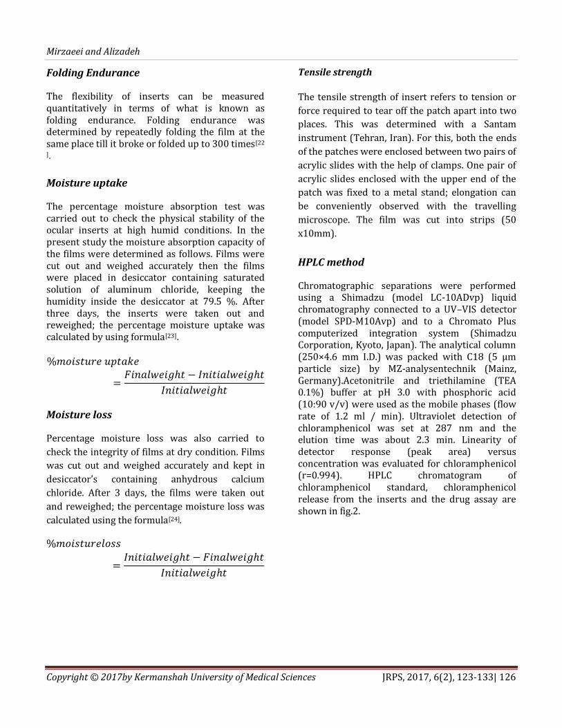
Folding Endurance
The flexibility of inserts can be measured quantitatively in terms of what is known as folding endurance. Folding endurance was determined by repeatedly folding the film at the same place till it broke or folded up to 300 times[22
].
Moisture uptake
The percentage moisture absorption test was carried out to check the physical stability of the ocular inserts at high humid conditions. In the present study the moisture absorption capacity of the films were determined as follows. Films were cut out and weighed accurately then the films were placed in desiccator containing saturated solution of aluminum chloride, keeping the humidity inside the desiccator at 79.5 %. After three days, the inserts were taken out and reweighed; the percentage moisture uptake was calculated by using formula[23].
Moisture loss
Percentage moisture loss was also carried to
check the integrity of films at dry condition. Films
was cut out and weighed accurately and kept in
desiccator’s containing anhydrous calcium
chloride. After 3 days, the films were taken out
and reweighed; the percentage moisture loss was
calculated using the formula[24].
Tensile strength
The tensile strength of insert refers to tension or
force required to tear off the patch apart into two
places. This was determined with a Santam
instrument (Tehran, Iran). For this, both the ends
of the patches were enclosed between two pairs of
acrylic slides with the help of clamps. One pair of
acrylic slides enclosed with the upper end of the
patch was fixed to a metal stand; elongation can
be conveniently observed with the travelling
microscope. The film was cut into strips (50
x10mm).
HPLC method
Chromatographic separations were performed using a Shimadzu (model LC-10ADvp) liquid chromatography connected to a UV–VIS detector (model SPD-M10Avp) and to a Chromato Plus computerized integration system (Shimadzu Corporation, Kyoto, Japan). The analytical column (250×4.6 mm I.D.) was packed with C18 (5 μm particle size) by MZ-analysentechnik (Mainz, Germany).Acetonitrile and triethilamine (TEA 0.1%) buffer at pH 3.0 with phosphoric acid (10:90 v/v) were used as the mobile phases (flow rate of 1.2 ml / min). Ultraviolet detection of chloramphenicol was set at 287 nm and the elution time was about 2.3 min. Linearity of detector response (peak area) versus concentration was evaluated for chloramphenicol (r=0.994). HPLC chromatogram of chloramphenicol standard, chloramphenicol release from the inserts and the drug assay are shown in fig.2.

Design and Evaluation of Soluble Ocular Insert For Controlled Release of Chloramphenicol
Copyright © 2017 by Kermanshah University of Medical Sciences JRPS, 2017, 6(2), 123-133| 127
Fig. 2. HPLC chromatogram of chloramphenicol standard (A), sample of chloramphenicol release from theinserts (B), and the drug assay(C).
Drug release study
To detect the amount of chloramphenicol released from the inserts, an appropriate amount of sample (estimated to containapprox.10 mg of the ocular insert) was introduced into donor compartment containing 1 mL of phosphate buffer at pH 7.4, respectively, separated by a dialysis membrane from the receptor compartment containing 49 ml of the same aqueous buffer. The system was stirred continuously at 100 rpm maintained at 37°C. The receptor compartment was closed to
prevent the evaporation losses from the dissolution medium. A certain amount of sample aliquot was withdrawn at regular time intervals and the same volume was replaced with a fresh dissolution and drug concentration was quantified in the acceptor phase by high performance liquid chromatography (HPLC) following the method described in 2.2.8Drug release data (Figure 3) were fitted to various kinetic equations, first order plots, Higuchi plots and Zero order exponential plots.

Mirzaeei and Alizadeh
Copyright © 2017by Kermanshah University of Medical Sciences JRPS, 2017, 6(2), 123-133| 128
Fig. 3. In vitro release profile of chloramphenicol from inserts. Release assay was performed in PBS solution, pH: 7.4, at
37°C with agitation (n=3).

Design and Evaluation of Soluble Ocular Insert For Controlled Release of Chloramphenicol
Copyright © 2017 by Kermanshah University of Medical Sciences JRPS, 2017, 6(2), 123-133| 129
Sterility testing
The sterility testing of the ophthalmic drug
delivery systems were performed for the aerobic
bacteria, anaerobic bacteria, and fungi by using
alternative thioglycolate medium and soybean
casein digest medium. The positive control
(growth promotion) and negative control
(sterility) test were also carried out.
Statistical analysis
Statistical analysis of release data was carried out
by using a one-way ANOVA for repeated measures
followed by a Student-Newman-Keuls multiple-
comparison test to determine whether type of
rate-controlling membrane affected the release of
ofloxacin from ocular inserts. All results are
reported as means ± SD (n=5); p< 0.05 was
considered to be of statistical significance.
Results and discussion
Evaluation of film casting
In the current study, Chloramphenicol insert was formulated using various polymers such as EUDL100, EUDS100, EUDRL100, mercury surface casting technique using PG and G as plasticizer. Physicochemical data presented in table 2 shows thickness, weight, and Viscosity. The prepared inserts were translucent, colorless, smooth and soft in texture, uniform in appearance and show no visible imperfection (fig. 1). the sizes of Ocular Drug Insert and the amount of drug in each insert, if chloramphenicol amount in each insert reached the MIC level The insert had a thickness varying from 0.0523±0.097 to1.013±0.09mm and weight varying from0.023±0.04to0.61±0.09mg. It was found that the thickness of the inserts was increased by increase in the total polymer concentration. The inserts were found to possess uniform thickness within the batch. Incubation was carried in all cases and growth was checked. The overall results of the sterility test showed that ocular formulation prepared passes the sterility test as there was no evidence of the growth found in the negative control test tubes. Thus all inserts were found sterile in nature.
Table 2. Physicochemical parameters of the ocular inserts of chloramphenicol, (mean ±SD, n=3).
Formulation Weight (g)
Thickness (mm)
Viscosity (cps)
t80%(hour)
P1 0.041±0.01 0.663±0.089 27 8>
P2 0.023±0.04 0.523±0.097 3.12 4 P3 0.033±0.02 0.624±0.021 29 8 P4 0.060±0.01 0.539±0.004 3 8 P5 0.61±0.09 1.013±0.09 26 12 P6 0.50±0.03 0.831±0.040 5.2 8
Tensile strength and Folding Endurance of
Insert
The recorded folding endurance for all batches
was greater than which is considered satisfactory
and reveals good film properties. The folding
endurance was found to be highest for
formulation P5 (370±4) and the lowest for
formulation P6 (170±2). Tensile strength was
within range of0.140–0.874g/mm2(Table 3). The
tensile strength was found to be highest for
formulation P5 (0.874±0.0031g/mm2) and the
lowest for formulation P2 (0.140±0.0033g/mm2).

Mirzaeei and Alizadeh
Copyright © 2017by Kermanshah University of Medical Sciences JRPS, 2017, 6(2), 123-133| 130
Table 3. Mechanical, Moisture loss, MoistureUptake and Folding endurance of ocular inserts(mean ±SD, n=3).
Formulation Tensile
strength(g/mm2)
Elongation at Break
%
Folding endurance
Moisture loss %
Moisture Uptake
%
P1 0.242±0.0021 61.99 300±2 4.3±0.01 3.64±0.03 P2 0.140±0.0033 51.49 273±1 14.7±0.04 3.93±0.01 P3 0.568±0.0012 90.2 310±3 17.5±0.09 3.51±0.02 P4 0.296±0.0023 69.23 246±2 12.5±0.02 4.67±0.06 P5 0.874±0.0031 50.35 370±4 10.6±0.03 3.65±0.03
P6 0.359±0.0047 129.52
170±2 9.4±0.6 3.79±0.03
Moisture uptake and moisture loss
Checking the physical stability of the film at high
humid conditions and integrity of the film at dry
conditions, the films were evaluated for moisture
loss and moisture uptake. The dates from Table 3
indicates that the percentage of moisture loss was
less in the formulation P1 (4.3%) and most in
formulation P3 (17.5%).Amongst all the
formulation the high value of moisture uptake can
be observed in P4 (4.678%).
FTIR analysis
Figure 4 shows the FTIR spectra of
Chloramphenicol, Eudragit, PVA, PVP, and
P1.Chloramphenicol sample showed the main
peaks contributed by the functional groups of
molecule such as carbonyl ––C=O stretching
vibrations (1685.79 cm–1), ––N––H stretching
(3263.56 cm–1), ––O––H stretching vibrations
(3348.42 cm–1),––NO2 as ym. stretching (1519.91
cm–1)(25). Eudragit has characteristics IR
absorption frequency at 3444.87cm-1 (OH
stretch), 2951.09cm-1 (sp3 CH stretch),
1728.22cm-1 (CO stretch)[26]. The FTIR spectra of
PVA showed a broad peak around 3414 cm–1
indicating stretching of hydroxyl groups and peak
at 2924.09 cm–1 due to C-H stretching[27]. The FTIR
spectra of PVP showed peaks in the range 2959–
2879, 1423.47 and 1373.3 cm-1 are ascribed to the
C–H bonding and it is observed that the strong
absorption peaks at around 1288 cm-1, observed
in Figure, are assigned to be due to the C–O
bonding[28].The FTIR spectra of P1showeda peak
around 1728 cm − 1, which is the stretching
vibration of –CO groups in Eudragit and a peak
around1520 cm − 1, which is the ––NO2asym.
Stretching in chloramphenicol and peaks at
2924.09 cm–1 due to C-H stretching in PVA
and1373.3 cm-1 are ascribed to the C–H bonding in
PVP. FTIR spectra were recorded to assess the
interaction between the drug and excipients.
There are some changes in the peaks in the range
of 2800–3500.This indicates that there may be
some physical interactions related to the
formation of intensity hydrogen bonding between
polymers.

Design and Evaluation of Soluble Ocular Insert For Controlled Release of Chloramphenicol
Copyright © 2017 by Kermanshah University of Medical Sciences JRPS, 2017, 6(2), 123-133| 131
Fig. 4. the FTIR spectra of Chloramphenicol, Eudragit RL100, PVA, PVP and P1.
In vitro chloramphenicol release study
All the nine formulations were subjected to invitro drug release studies. The overall cumulative percentage drug release for formulations of ocular inserts (Fig.3.). The released ChM were 80% in almost 8 hours for inserts P1, P3, P4, and P6. In addition, P5 was also released 80% in 12 hours and. The time of 80% release for P2 is the lowest in 4h. The release of drugs from inserts was enhanced by the polymer to drug. As the polymer to the drug increased, the release time increased. The programmed release is due to the formation of hydrogen bonds between the drug and polymers and suspended drug in hydrophobic polymers which have helped in rate control release of drug. The formulations which gave good results with highest percentage were selected for further studies such as in vivo, kinetic treatment and stability studies. Film P5 containing hydrophilic
polymer(PVP/PVA/PG/G) and Eudragit® RL100 showed a release of 99.5 % at the end of 24 hours which indicated that, the polymer combination with same quantities can be used for the formulation of ocular film for therapeutic drug management in the cul-de-sac. OF2 is a combination of hydrophilic polymers. This film has shown good compatible nature in IR studies indicating no drug polymer in-compatibility. PVP also has good adhesive property which is helpful, when the ocular film is inserted in the cul-de-sac. The release data obtained were grouped in there mathematical models of data treatment. Based on the highest regression value (r2), which is nearing to unity, formulations P1, P2, P5 and P6 followed First order kinetics as shown in Table 4. Formulations P3 and P4 were best fitted into Higuchi’s model with ‘r2’ values of 0.96, 0.97, respectively.

Mirzaeei and Alizadeh
Copyright © 2017by Kermanshah University of Medical Sciences JRPS, 2017, 6(2), 123-133| 132
Table 4.The release data of the different formulations of ocular inserts.
FORMULATION FIRST ORDER (R2VALUE)
ZERO ORDER (R2VALUE)
HIGUCHI’S MODEL (R2VALUE)
BEST FIT MODEL
P1 0.9751 0.8589 0.9376 First order P2 0.859 0.4101 0.687 First order P3 NUM 0.829 0.961 Higuchi’s model P4 NUM 0.8929 0.9712 Higuchi’s model P5 0.995 0.756 0.9312 First order P6 0.972 0.8002 0.9312 First order
Conclusion
Chloramphenicol formulation as eye drops suffered the disadvantage of instillation of the dye drops for every 3-4 h and hence maximized patient noncompliance, leading to ineffective therapy. In this study efforts were taken for designing and evaluating chloramphenicol ocular inserts. Ocular inserts of chloramphenicol prepared in PVA/PVP matrix and cast with rate-controlling polymers, Eudragit RL100, Eudragit S100 and Eudragit S100 in combination, were smooth, flexible and transparent. In vitro release studies revealed that the ocular inserts followed First order and Higuchi’s model release kinetics. Studies indicate that the polymer to drug ratio plays a key role in releasing the drug from drug reservoir. Ophthalmic inserts were also evaluated for % moisture loss, % moisture uptake, thickness, and tensile strength, and it was found that prolonged and sustained drug release was observed. Finally, in conclusion to our studies, we suggest that ocular insert formulation are considered as a good choice of ophthalmic drug delivery because allow for accurate dosing, better patient compliance resulting from a reduced frequency of administration. Inserts completely dissolve after 20-24 hours and the residuals exit in ophthalmic channel. The ophthalmic drug delivery systems of this device are, several millimeters in size which are placed in the upper or lower sac of the eye to deliver a complete ophthalmic dosage for a period of 24 hours.
Acknowledgments
This work was financially supported by Research &Technology Center of Kermanshah University of Medical sciencesIran, (Financial Cod 950204). The authors also wish to thank SinaDarouLaboratories Company, Tehran, Iran, for supplying chloramphenicol. Conflict of Interests Authors certify that there is no actual or potential conflict of interest in relation to this article.
References
[1] Duxfield L, Sultana R, Wang R, Englebretsen V, Deo S, Rupenthal ID, et al. Ocular delivery systems for topical application of anti-infective agents.Drug Dev. Ind. Pharm. 2016;42:1-11. [2] Thakur RR, Kashiv M. Modern delivery systems for ocular drug formulations: A comparative overview WRT conventional dosage form. Int J Res Pharm Biomed Sci. 2011; 2: 8-18. [3] Lang JC. Ocular drug delivery conventional ocular formulations.Adv. Drug Del. Rev. 1995;16:39-43. [4] Katz IM. Shaped ophthalmic inserts for treating dry eye syndrome. Google Patents; 1982. [5] Chien YW, Swarbrick J. Novel drug delivery systems. 1992. [6] Sasaki H, Yamamura K, Mukai T, Nishida K, Nakamura J, Nakashima M, et al. Enhancement of ocular drug penetration. Critical Reviews™ in Therapeutic Drug Carrier Systems. 1999;16. [7] Vyas SP, Khar RK. Targeted & controlled drug delivery: Novel carrier systems. CBS publishers & distributors; 2004.
Design and Evaluation of Soluble Ocular Insert For Controlled Release of Chloramphenicol
Copyright © 2017 by Kermanshah University of Medical Sciences JRPS, 2017, 6(2), 123-133| 133
[8] Thakur R, Swami G. Promising implication of ocuserts in ocular disease. JDDTAO. 2012; 2. [9] Patton TF, Robinson JR. Ocular evaluation of polyvinyl alcohol vehicle in rabbits.J.pharm.sci 1975; 64: 1312-1316. [10] Sreenivas S, Hiremath S, Godbole A. Ofloxacin ocular inserts: Design, formulation and evaluation.IJPT. 2006; 5:159-162. [11] Gevariya HB, Patel JK, Girhepunje K, Pal RS. Sustained Ophthalmic Delivery of Levofloxacin from once a day Ocusert. Int J Pharm Sci. 2009;1:24-32. [12] Saisivam S, Manikandar RVM, Nagarajan M. Design and evaluation of ciprofloxacin hydrochloride ocuserts.Indian J. pharm. sci. 1999;61:34-38. [13] Rajasekaran A, Sivakumar V, Karthika K, Preetha JP, Abirami T. Design and evaluation of polymeric controlled release natamycin ocular inserts. Kathmandu University J. Sci, Engineering and Technology. 2010; 6: 108-115. [14] Patel U, Chotai N, Nagda C. Design and evaluation of polymeric ocular drug delivery system for controlled delivery of moxifloxacin hydrochloride: in vitro and in vivo evaluation. Acta Pharm. Sciencia. 2010; 52: 523-535. [15] Gilhotra RM, Nagpal K, Mishra DN. Azithromycin novel drug delivery system for ocular application.Int. J. of pharm.invest. 2011;1:22. [16] Shaw WV. Comparative enzymology of chloramphenicol resistance.Ann. N. Y. Acad. Sci. 1971; 182: 234-242. [17] Rahal JJ, Simberkoff MS. Bactericidal and bacteriostatic action of chloramphenicol against meningeal pathogens.Antimicrob. Agents Chemother. 1979;16:13-8. [18] Järvinen K, Järvinen T, Urtti A. Ocular absorption following topical delivery.Adv. Drug Del. Rev. 1995; 16: 3-19. [19] Prc SPCOT. Pharmacopoeia of the People's Republic of China 2005. BC Decker, Incorporated; 2008. [20] Vijayendra Swamy S, Nanjawade B, Ravichandran M, Manvi F. Development of sustained ocular drug delivery system for betaxolol.Indian J Pharm Educ Res. 2006;40:165. [21] LL CLGG. Design and evaluation of timolol maleate ocuserts. Indian J Pharm Sci. 2005; 67:311-314. [22] Singh S, Jain S, Muthu M, Tiwari S, Tilak R. Preparation and evaluation of buccal bioadhesive films containing clotrimazole. AAPS PharmSciTech. 2008;9:660-667. [23] Kusum Devi V, Saisivam S, Maria G, Deepti P. Design and evaluation of matrix diffusion controlled
transdermal patches of verapamil hydrochloride.Drug Dev. Ind. Pharm. 2003;29:495-503. [24] Deshpande PB, Dandagi P, Udupa N, Gopal SV, Jain SS, Vasanth SG. Controlled release polymeric ocular delivery of acyclovir. Pharm Dev Technol.15:369-378. [25] Mandal B, Halder KK, Dey SK, Bhoumik M, Debnath MC, Ghosh LK. Development and physical characterization of chloramphenicol loaded biodegradable nanoparticles for prolonged release. Die Pharmazie-An Int. J. Pharm. Sci. 2009;64:445-449. [26] Basu SK, Adhiyaman R. Preparation and characterization of nitrendipine-loaded Eudragit RL 100 microspheres prepared by an emulsion-solvent evaporation method.TROP J PHARM RES. 2008;7:1033-1041. Pal K, Banthia AK, Majumdar DK. Preparation and characterization of polyvinyl alcohol-gelatin hydrogel membranes for biomedical applications. AAPS PharmSciTech. 2007;8:E142-E6. [27] Taylor LS, Zografi G. Spectroscopic characterization of interactions between PVP and indomethacin in amorphous molecular dispersions. Pharmaceutical research. 1997;14:1691-1698.