design, synthesis and evaluation of new colourimetric
TRANSCRIPT

Indian Journal of Chemistry
Vol. 60B, May 2021, pp. 755-766
Design, synthesis and evaluation of new colourimetric chemosensors containing
quinazolinones moiety for some cations detection in an aqueous medium and
biological sample
Ayman M Algoharya,b,c, Mohamed M Hassan*d, Sami G Almalkicc & Esam S Al-Malkide
a Department of Chemistry, College of Science Al-zulfi, Majmaah University, P.O. 66 Al-Majmaah, 11952, Saudi Arabia b National Organization for Drug Control and Research (NODCAR), P.O. 29 Giza, Egypt
c Medical Laboratory Department, College of Applied Medical Sciences, Majmaah University, P.O. 66 Majmaah 11952, Saudi Arabia d Chemistry Department, Faculty of Education, Ain Shams University, Roxy, 11711, Cairo, Egypt
e Department of Biology, College of Science Al-zulfi, Majmaah University, P.O. 66 Al-Majmaah 11952, Saudi Arabia
E-mail: [email protected]
Received 20 November 2020; accepted (revised) 4 March 2021
The current project deals with designing and synthesizing of colourimetric chemosensors to detect the cations in the
aqueous medium and biological sample. To achieve this goal a new series of quinazolinone derivatives have been
synthesized via reaction of the novel 6-nitro-2-propyl-4H-benzo[d][1,3]oxazin-4-one 3 with selected nitrogen nucleophiles,
namely, formamide, hydrazine hydrate, hydroxylamine hydrochloride, o-phenylendiamine, o-aminophenol and o-
aminothiophenol, urea and/or thiourea. Structures of the new compounds have been investigated depending on their spectral
data (IR, 1H and 13C NMR and MS) and elemental analyses. Some of the newly synthesized products exhibit significant
response as chemosensors for a few cations detection.
Keywords: Chemosensor, quinazolinone, copper, cadmium, mercury
Currently, synthesis of colourimetric sensors for
detecting of cations and anions is one of the
researcher's targets in the organic synthesis
fieldbecause of the prospect implementation of these
chemosensors as diagnostic tools in medical,
physiological and environmental applications1.
Copper cation is the most abundant ion in the human
body and several proteins utilize Cu2+ as a cofactor
for the transfer of electrons in redox reactions.
Biologically, excess Cu2+ in cells can stimulate the
creation of reactive oxygen species and can harm
lipids, DNA and RNA and cause some of the
dangerous diseases, such as Dementia's disorder,
Prion, Wilson and Menkes disease, which are
directly related to the copper toxicity2. Also, Cd2+ is
extremely poisonous to the living organism and its
compounds pass in the environment from human
activities or geological. Cadmium and its salts are
classified as blacklist compounds because high
exposure level of Cd2+ is associated to raise
cardiovascular risks diseases, cancer, liver diseases
and kidney disorder3. On the other hand, mercury is
considered as the greatest harmful pollutant, which
affects directly on human and environmental health,
so assay of mercury ions in the environment is a
significant target reference4. The most reports of
sensing Hg2+ based on using atomic absorption
spectrometry, Raman spectroscopy, inductively
coupled plasma mass spectrometry, which is
expensive and needs long time pre-treatment5.
Various techniques such as electrochemical, atomic
absorption, inductively coupled plasma atomic
emission and piezoelectric quartz crystals6, have
been used for notifying of metal ions but their
utilization is limited due to its expensive equipment,
laboratories and time consuming procedures. Lin
et al. have applied a ratiometric fluorescent probe for
Cu2+ determination7. Kato et al. used isotope dilution
inductively coupled plasma mass spectrometry for
the detection of silver copper, zinc, nickel, lead and
cadmium in seawater8. Colourimetric techniques are
the most favorable methods to their low cost, lack of
equipment, rapid and we can detect it by naked eye9.
Thus, development of colourimetric chemosensors
depend on changing in absorption accompanied by
sensible colour changes, for heavy metal ions has
produced as an active area of valuable importance.
Kim et al. showed a colourimetric sensor for the

INDIAN J. CHEM, SEC B, MAY 2021
756
determination of Cd, Hg and Pb ions10. Li et al. utilized a turn-on fluorescent sensor for assay of Cd2+, Hg2+ and Zn2+ in water11. Fegade et al, showed selectivity of dualchannel chemosensor for copper in semiaqueous media12. In the last decade, several heterocyclic chemosensors were synthesized for the recognition of different ions13. Synthesized azo dye-based heterocyclic chemosensors designed to produce fluorescent sensors for the detection of zinc without interference with cadmium14. Lu et al.15, designed a sensor containing naphthalimide as the fluorophore, which by undergoing two reverse ICT processes in sensing Zn2+ and Cd2+ distinguished between this ion pair proficiently.
Quinazolinones have attracted notable interest in organic and medicinal chemistry due to their therapeutic potential such as anticancer, antibacterial, antidiabetic, hypnotic, sedative, analgesic, anticonvulsant, antitussive, anti-inflammatory activities16. Few reports were performed for checking the sensor activity of quinazolinone derivatives. Ailin Yuan et al, synthesized novel quinazolinone compound as a fluorescence sensor for the ferric ion17. Quinazolinonebased sensors for amine vapors and Cu2+ ion18 have been developed. As an extension of our awareness in synthesis of quinazolinone heterocyclic, we report that some quinazolin-4(3H)-ones can be used as a simple and efficient ‘turn-on’ fluorescent chemosensor for a highly selective and sensitive detection of some cations, Cu2+, Hg2+ and Cd2+ in aqueous medium and biological sample. Results and Discussion
As part of our concern in the organic synthesis of quinazolinone derivatives19 we produced a new chemosensor carrying quinazolinone moiety. As displayed in (Scheme I), 2-amino-5-nitrobenzoic acid 1 was reacted with butyryl chloride, in dry
pyridine, to yield 2-(butyramido)-5-nitrobenzoic acid 2. Microanalytical and spectral data were used for structure elucidation of the anilide 2. IR spectrum showed absorption bands at ν 1730 and 1640 cm−1 which are characteristic for both C=O carboxylic acid and amide functions stretching vibration, respectively. 1H NMR spectrum of the anilide 2 exposed two deuterium- exchangeable, protons at δ 8.95 and 11.80 related to N–H and CO2H. Mass spectrum showedthe molecular ion peak at m/z 252 which is coincident with the prospective anilide formula.
Using acetic anhydride,intramolecular cyclization for of the anilide 2 was performed leading to our target 6-nitro-2-propyl-4H-benzo[d][1,3]oxazin-4-one 3 as a dynamic benzoxazinone moiety with electronically unsaturated character20. IR spectra of compound 3was very good indication for absence of both carboxylic and amide functions. On the other hand, a characteristic absorption band was observed at ν 1750 cm−1 signifying C=O function of 3,1-benzoxazin-4-ones. Mass spectrum gave a molecular ion peak at m/z 234 which is matching with the predictable molecular formula acquired through loosing of one molecule of water from the anilide 2. These results are deemed confirmatory for the postulation that cyclization process implicated both carboxylic and amidic functions (Scheme I).
Reaction of 6-nitro-2-propyl-4H-benzo[d][1,3] oxazin-4-one 3 with some selected nitrogen nucleophiles, namely, formamide, hydrazine hydrate, hydroxylamine hydrochloride, o-phenylendiamine, o-aminophenol, o-aminothiophenol, urea and/or thiourea furnished a variety of 4(3H)-quinazolinones 4-14 via aminolysis at position 3 (Scheme II). It is expectant that the reactions managed through nucleophilic ring opening ring closure (RORC) process accompanied by ring oxygen replacing with nitrogen. Oxazinone ring splitting takes place by the attack of nitrogen nucleophile followed by cyclization on the highly electrophilic sp2 hybridized carbonyl carbon of the intermediate to give our coveted quinazolinones21.
Scheme I — 6-Nitro-2-propyl-4H-benzo[d][1,3]oxazin-4-one 3
ALGOHARY et al.: NEW COLOURIMETRIC CHEMOSENSORS
757
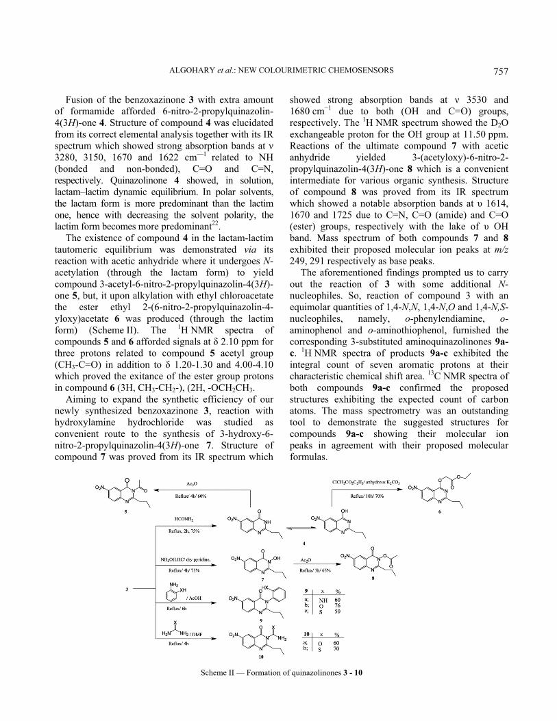
Fusion of the benzoxazinone 3 with extra amount of formamide afforded 6-nitro-2-propylquinazolin-4(3H)-one 4. Structure of compound 4 was elucidated from its correct elemental analysis together with its IR spectrum which showed strong absorption bands at ν 3280, 3150, 1670 and 1622 cm—1 related to NH (bonded and non-bonded), C=O and C=N, respectively. Quinazolinone 4 showed, in solution, lactam–lactim dynamic equilibrium. In polar solvents, the lactam form is more predominant than the lactim one, hence with decreasing the solvent polarity, the lactim form becomes more predominant22.
The existence of compound 4 in the lactam-lactim tautomeric equilibrium was demonstrated via its reaction with acetic anhydride where it undergoes N-acetylation (through the lactam form) to yield compound 3-acetyl-6-nitro-2-propylquinazolin-4(3H)-one 5, but, it upon alkylation with ethyl chloroacetate the ester ethyl 2-(6-nitro-2-propylquinazolin-4-yloxy)acetate 6 was produced (through the lactim form) (Scheme II). The 1H NMR spectra of compounds 5 and 6 afforded signals at δ 2.10 ppm for three protons related to compound 5 acetyl group (CH3-C=O) in addition to δ 1.20-1.30 and 4.00-4.10 which proved the exitance of the ester group protons in compound 6 (3H, CH3-CH2-), (2H, -OCH2CH3.
Aiming to expand the synthetic efficiency of our newly synthesized benzoxazinone 3, reaction with hydroxylamine hydrochloride was studied as convenient route to the synthesis of 3-hydroxy-6-nitro-2-propylquinazolin-4(3H)-one 7. Structure of compound 7 was proved from its IR spectrum which
showed strong absorption bands at ν 3530 and 1680 cm−1 due to both (OH and C=O) groups, respectively. The 1H NMR spectrum showed the D2O exchangeable proton for the OH group at 11.50 ppm. Reactions of the ultimate compound 7 with acetic anhydride yielded 3-(acetyloxy)-6-nitro-2-propylquinazolin-4(3H)-one 8 which is a convenient intermediate for various organic synthesis. Structure of compound 8 was proved from its IR spectrum which showed a notable absorption bands at υ 1614, 1670 and 1725 due to C=N, C=O (amide) and C=O (ester) groups, respectively with the lake of υ OH band. Mass spectrum of both compounds 7 and 8 exhibited their proposed molecular ion peaks at m/z 249, 291 respectively as base peaks.
The aforementioned findings prompted us to carry out the reaction of 3 with some additional N-nucleophiles. So, reaction of compound 3 with an equimolar quantities of 1,4-N,N, 1,4-N,O and 1,4-N,S-nucleophiles, namely, o-phenylendiamine, o-aminophenol and o-aminothiophenol, furnished the corresponding 3-substituted aminoquinazolinones 9a-c. 1H NMR spectra of products 9a-c exhibited the integral count of seven aromatic protons at their characteristic chemical shift area. 13C NMR spectra of both compounds 9a-c confirmed the proposed structures exhibiting the expected count of carbon atoms. The mass spectrometry was an outstanding tool to demonstrate the suggested structures for compounds 9a-c showing their molecular ion peaks in agreement with their proposed molecular formulas.
Scheme II — Formation of quinazolinones 3 - 10

INDIAN J. CHEM, SEC B, MAY 2021
758
In the same sense, equimolar remediation of compound 3 with 1,3-binnucleophiles such as urea and thiourea in boiling DMF furnished smoothly the anticipated quinazolinone derivatives 10a,b. Analytical and spectral data of these products were found in similarity with the proposed formula. IR spectra of both 10a,b compounds showed the C=O stretching vibrations at 1671, 1675 cm−1 and the C=S stretching vibrations at 1220-1050 cm−1 in addition to NH2 stretching vibrations at 3330, 3270 cm−1.The 1H NMR spectra of compounds 10a,b demonstrated the appearance of the emergence of new singlet signals assigned to the characteristic NH2 groups protons at 6.2 and 6.5 ppm for both 10a and 10b, respectively. 1H NMR spectral data displayed a characteristic signal at δ 177.1 and 178.2 ppm attributed to both C=O and C=S carbons, respectively. In addition, mass spectrum represents a good guide to 10a,b structure and showed the molecular ion peak at m/z 276 and 292 which corresponds to the suggested molecular formulas.
Benzoxazinone 3 was reacted with hydrazine hydrate and /or phenyl hydrazine in boiling ethanol, 3-amino-6-nitro-2-propylquinazolin-4(3H)-one 11a and 6-nitro-3-(phenylamino)-2-propylquinazolin-4(3H)-one 11b were produced in a respective yield. 3-Aminoquinazolinone 11a used as many-sided constructing bulk in synthetic heterocyclic chemistry. IR spectrum of the amine 11 exhibited a specific stretching bands at ν 3309 and 3212 cm−1 related to symmetric and asymmetric vibrations of NH2 group. The absorption bands due to C=O group, for ideal quinazolin-4-ones, was observed at ν 1673cm−1.
These outcomes are in accordance with 1H NMR spectral data of compounds 11 which showed a broad singlets chemical shifts at δ 5.50 due to deuterium-exchangeable protons of NH2. In addition, the mass spectra of compound 11a revealed its molecular ion peak at m/z 248 as base peak. All spectral data of 6-nitro-3-(phenylamino)-2-propylquinazolin-4(3H)-one 11b were fully consistent with its suggested formula (Scheme III).
N-(6-nitro-4-oxo-2-propylquinazolin-3(4H)-yl)acetamide 12 was obtained on refluxing of quinazolinone 11a with acetic anhydride. The IR spectrum of compound 12 revealed strong absorption bands assigned to C=N, two C=O group, NH and OH, respectively. 1H NMR spectrum of compound 12 revealed that it exists in solution in a keto-enol tautomerism of an amide functionality, as it showed two singlet signals at δ 6.50 and 8.90 attributable to NH and OH of the keto and the enol forms, respectively.
The carbothioamides 13 was targeted because of its expected biological activity23. Consequently, reacting of the amine 11a with carbon disulfide, in existence of sodium hydroxide as base catalyst and in situ methylation of the non-separable sodium dithiocarbamide salt, with the use of dimethyl sulfate, yielded Methyl(6-nitro-4-oxo-2-propylquinazolin-3(4H)-yl)carbamodithioate 13. Structure of compound 13 was elucidated from its IR spectrum which showed a strong absorption bands at 1450 and 1670 related to υ max C=S and C=O respectively and freed from the NH2 band. 1H NMR spectrum of the product showed a characteristic singlet chemical shift at δ 2.54 due three
Scheme III — Synthesis of quinazolinones 11-14

ALGOHARY et al.: NEW COLOURIMETRIC CHEMOSENSORS
759
protons of SCH3. Mass spectrum exhibited a
molecular ion peak at m/z 338, confirming the
proposed molecular formula.The amine 11awas
condensed with benzaldehyde to give the
corresponding Schiff ’s bases 14 (Scheme III) which
could be utilized as multilateral building bulk in a
variety of heterocyclic synthesis. It will be of interest
in our future project to discuss the behavior of
azomethine 14 which includes an activated
azomethine group (–N=CH)24. Spectral data of the
product pointed to the disappearance of NH2 function,
indicating its embodiment in the condensation
reaction. IR spectra of compounds 14 showed a strong
absorption bands at 1610, 1676; related to υ C=N and
υ C=O, respectively in addition of lacking any
absorption band due to NH2. 1H NMR spectrum of the
trio gave specific chemical shift signals of azomethine
proton (N=C–H) observed at δ 8.80. Mass spectrum
exhibited a molecular ion peak at m/z 336 as base
peak, confirming the proposed structure.
The FT-IR comparison of some of our newly
synthesized chemosensor 7, 10b and 11a,b versus
their related complexs 15, 16, 17 and 18 were
identified as shown in the Table I. The FT-IR
spectrum changes of our produced complexs confirm
that our primary amine, carbonyl groups, thiocarbonyl
and hydroxyl group are sharing in the binding with
our targeted metal cations.
Colourimetric Study
The colourimetric study was applied for 2.5×10⁻⁵ M
of all the synthesized compounds, which were prepared
in DMF- water (1:10). As can be seen from (Table II),
Compounds 3, 9b,c, 10a, 12, 13 and 14 did not give
any changes of colour after adding all metals. The
chemosensor 10b and 7 showed the colour change
from colourless to yellow when adding Cd2+ ion and
the intensity of colour was increased with the addition
of Cd2+ ions. The chemosensors 11a,b exhibited
notable colour change from pale yellow to green when
adding Cu2+ ion which gets more colour intensity on
further addition of Cu2+ cation. The same chemosensor
exhibited a remarkable colour change to rose on
interacting with Hg2+and the intensity of colour also
increased with further addition of Hg2+ion cation. This
important colour change of complex 7, 10b and 11a,b
can be easily applied for detection Cu2+, Cd2+ and Hg2+
ions in an aqueous mediums and biological samples.
UV-vis absorption spectroscopy study
The absorption response of complexes 15, 16, 17
and 18 were examined in the presence of different
solvents, for instance, ethanol, dimethylformamide,
acetonitrile and. The chemosensor 7, 10b, 11a,b
displayed good absorption intensity in the
dimethylformamide solution contrast to other solvents
and so we used dimethylformamide as a solvent for
all UV-Vis spectral studies because it showed good
absorbance shift and absorption intenisty25. Metal ions
were prepared as aqueous solutions of 0.02 M nitrate
salts in distilled water. Chemosensor stock solutions
of 7, 10b, 11a,b (0.02 M) were dissolved in
dimethylformamide. By using a micropipette, we
prepared and diluted various concentrations of metal
Table I — FT-IR variation between chemosensors and formed complexes
Functional group Chemosensor number Stretching vibration chemosensor
(cm–1)
Complex Stretching vibration complex
(cm–1)
(C=O) 7 1680 15 1609
(OH) 3530 3403
(C=O) 10b 1671 16 1620
(NH2) 3330 and 3270 3000 and 3100
(C=O) 11a 1673 17 1615
(NH2) 3309 and 3212 3290 and 3190
(C=O) 11b 1675 18 1630
(NH) 3360 3265
Table II — Colourimetric responses of selected chemosensors toward various cations
Chemosensor Detected cation Colourimetric Responses
7 Cd Colourless to yellow
10b Cd Colourless to yellow
11a Cu Pale yellow to green
Hg Pale yellow to rose
11b Cu Pale yellow to green
Hg Pale yellow to rose
9b,c, 10a, 12, 13, 14 All cations No response

INDIAN J. CHEM, SEC B, MAY 2021
760
ions to 2.5×10⁻⁵ M with the same solution. The diluted chemosensors were added to various concentrations of metal ions. The complex 17-Cu2+ showed the main absorption peak (λ max) at 345 nm. The complex 17-Hg2+ showed λ max at 495 nm. The complex 18-Cu2+ showed λ max at 360 nm. The complex 18-Hg2+ λ max at 505 nm. The complex 15-Cd2+ showed λ max at 362 nm, the complex 16-Cd2+displayed λ max at 425 nm. The calibration curves established standard concentrations of ions and specific absorbance for each one (Figure 1). Suggested Mechanism
According to UV-Vis absorption analysis and the FT-IR spectrum of compounds 7, 10b and 11a,b towards their complexes 15, 16, 17 and 18 respectively, the binding positions were labeled in chemosensor 7, 10b and 11a,b. Some predictable observations can be inferred. The variations in the FT-IR frequencies of 17 and 18 suggest the primary or secondary amine and carbonyl groups are participating in the binding with metal ions and an established binding mechanism is shown in Table III. In the aqueous medium, the nucleophile chemosensors 11a,b attacked cations by lone pairs of electrons from amino and carbonyl groups to form five-membered ring which have high stability and a low amount of ring strained26,27. The change of the FT-IR frequencies of complex 16 indicates that Cd2+ coordinated with oxygen atom of carbonyl group
and C=S group which inform binding mechanism was presented in Table III. Complex 15 have clearly observed shifts of FT-IR spectrum Table I, which indicated the formation of a binding mechanism, is shown in Table III. The nucleophile chemosensor 7 attacked cations by lone pairs of electrons from OH and C=O groups to form a five-member ring26. Application of chemosensor in biological and environmental water samples
The main purpose of the current study was to detect ions traces in a biological sample, so samples were collected from the medical laboratory in Al-zulfi hospital. The relationship between absorption and concentration was obtained by the calibration curve and a linear relationship was established to the determination of the concentration of Cu2+, Cd2+ and Hg2+ ions in the various blood samples. All blood samples were also further assayed by Atomic Absorption Spectroscopy (AAS) to show the chemosensor sensitivity. The synthesized chemosensors 7, 10b and 11a,b were carried as strip paper and added one drop of ion analyte to the chemosensor strip which directly detects the concentration of cation. Experimental Section
Melting points were measured on an Optimelt automated melting point system and are uncorrected.
Figure 1 — Calibration curves of absorbance intensity of different concentrations of ions

ALGOHARY et al.: NEW COLOURIMETRIC CHEMOSENSORS
761
The IR spectra were recorded on a Perkin−Elmer
1800 Series FTIR spectrometer. Samples were
analyzed as thin films on KBr plates. 1H and 13C NMR
spectra were recorded at room temperature in base-
filtered DMSO-d6 on a Varian spectrometer operating
at 400 MHz for proton and 100 MHz for carbon
nuclei. Elemental microanalyses were recorded on a
Perkin−Elmer series II CHNS analyzer 2400. Mass
spectra were obtained using GCMS (QP-1000EX)
Shimadzu gas chromatography instrument mass
spectrometer (70 eV).
2-(Butyramido)-5-nitrobenzoic acid, 2
Butyryl chloride (1.06 g, 10 mmol) was portion-
wise added to a stirred solution of 2-amino-5-
nitrobenzoic acid (1.8 g, 10 mmol), in dry pyridine
(30 mL) during 30 min. The mixture was stirred at RT
for 2 h then poured into ice-cold water (200 mL),
Table III — Proposed binding mechanism of different chemosensors
Chemosensor λ max Complex structure Suggested binding mechanism
18-Cu2+ 345 nm
18-Hg2+ 495 nm
16-Cd 362 nm
17-Cu2+ 360 nm
17-Hg2+ 505 nm
15-Cd2+ 425 nm

INDIAN J. CHEM, SEC B, MAY 2021
762
acidified with hydrochloric acid (2N) up to complete
precipitation. The crude solid product was filtered off,
washed thoroughly with cold water and crystallized
from benzene to give compound 2 (2 g, 80%), yellow
crystals, m.p.130-135°C. IR (KBr): 3300 (O–H, N–
H), 3010 (C–Harom), 2975, 2985 (C–Haliph), 1730
(C=Ocarboxylic), 1640 (C=Oamidic), 1615 cm−1 (bending
N–H); 1H NMR (CDCl3): δ 1.90 (t, 2H, -CH2-CH2-
CH3), 1.50 (m, 2H, -CH2Me) and 1.20 (t, 3H, -CH3),
7.20-8.70 (m, 3H, Harom), 8.95 (bs, 1H exchangeable
with D2O, NH), 11.80 (bs, 1H exchangeable with
D2O, OH); 13C NMR (100 MHZ): δ 143.3 (C-1),
115.15(C-2), 129.7 (C-3), 122.4(C-4), 132.98(C-5),
121.52 (C-6), 168.1 (C-7COOH),
170.2 (C-8NHC=O), 35.1 (C-9), 19.0 (C-10), 13.2 (C-
11); MS: m/z 252(M+,70),253 (M+1,80). Anal. Calcd
for C11H12N2O5 (252): C, 52.38; H, 4.80; N, 11.11; O,
31.72. Found: C, 52.50; H, 4.95; N, 31.94%.
6-Nitro-2-propyl-4H-benzo[d][1,3]oxazin-4-one, 3
The dry solid anthranilide (2.52g, 10 mmol) 2 was
treated with freshly distilled acetic anhydride until
being pasted and then heated over water bath for 2 h
before lifting to cool. The separated out solid was
filtered off and recrystallized from light petroleum
ether 40/60 affording the benzoxazinone 3 (2 g,85%),
pale yellow crystals, m.p.185°C. IR (KBr): 3050 (C-
Haromatic), 2971, 2929,2881 (C-Haliphatic),1710(C=O),
1617(C=N),1159 cm−1 (C-O-C); 1H NMR (400 MHZ,
DMSO-d6): δ 2.10(2H, -CH2-CH2-CH3), 1.60 (m, 2H, -
CH2Me) and 1.10 (t, 3H, -CH3), 7.3-8.2 (m, 3H, Ar-
H);13C NMR (100 MHZ): δ 155.2 (C-1C=N), 159.1 (C-
2C=O), 120 (C-3), 128 (C-4), 127.4 (C-5), 133.5 (C-6),
122 (C-7), 147 (C-8), 25.1 (C-9), 14.6 (C-10), 13.9 (C-
11); MS: m/z 234 (M+,20), 235(M+1,18). Anal. Calcd
for C11H10N2O4 (234): C, 56.41; H, 4.30; N, 11.96.
Found: C, 56.50; H, 4.47; N, 11.99%.
6-Nitro-2-propylquinazolin-4(3H)-one, 4
A solution of benzooxazinone derivative 3 (2.34 g,
10 mmol,) in formamide (15 mL) was refluxed for
2 h, left to cool, then poured into ice. The crude solid
product was collected by filtration, dried and
recrystallised from ethanol to give 4 (1.75g,75%),
yellow crystals, m.p. > 300°C. IR (KBr): 3372, 3280
(NH), 1670(C=O), 1622 cm−1 (C=N); 1H NMR (400
MHZ, DMSO-d6): δ 2.30(2H, -CH2-CH2-CH3), 1.80
(m, 2H, -CH2Me) and 1.22 (t, 3H, -CH3), 7.82-8.90
(m, 3H, Ar-H), 9.40 (s, 1H, NH, D2O exchangeable);
13C NMR (100 MHZ): δ 155.2 (C-1), 164 (C-2), 123
(C-3), 130.5 (C-4), 127.4 (C-5), 133.5 (C-6), 122 (C-
7), 148.8 (C-8), 25.4 (C-9), 14.6 (C-10), 13.7 (C-11);
MS: m/z 233(M+,30), 234(M+1,23). Anal. Calcd for
C11H11N3O3 (233): C, 56.65; H, 4.75; N, 18.02.
Found: C, 56.80; H, 4.90; N, 18.40%.
3-Acetyl-6-nitro-2-propylquinazolin-4(3H)-one, 5
A solution of 4 (2.33 g, 10 mmol) in 20 mL acetic
anhydride was refluxed for 4 h. The reaction mixture
was then allowed to stand at RT for 2 h. The
separated solid product was washed with water
(2×100 mL), dried and crystallized from ethanol to
afford compound 5 (1.65 g, 60%), pale-yellow
crystals, m.p.135-7°C. IR (KBr): 1663 (C=OAcetyl),
1675 (C=OQuinazolinone), 1622 cm−1 (C=N); 1H NMR
(400 MHZ, DMSO-d6): δ 2.40 (s, 3H, CH3-C=O), 2.00
(2H, -CH2-CH2-CH3), 1.85 (m, 2H, -CH2Me), 1.10 (t,
3H, -CH3), 7.55- 7.83 (m, 3H, Ar-H); 13C NMR (100
MHZ): δ 163.1 (C-1), 166.9 (C-2), 129.2 (C-3), 131.8
(C-4), 121.5 (C-5), 136.1 (C-6), 124.0 (C-7), 146.2
(C-8), 23.1 (C-9), 13.4 (C-10), 13.4 (C-11), 175.1 (C-
12C=O acetyl), 20.3 (C-13CH3CO); MS: m/z 275 (M+,10),
276 (M+1,33). Anal. Calcd for C13H13N3O4 (275): C,
56.72; H, 4.76; N, 15.27. Found: C, 53.85; H, 4.90; N,
15.40%.
Ethyl 2-(6-nitro-2-propylquinazolin-4-yloxy)acetate, 6
To a mixture of 4 (2.33 g, 10 mmol) and ethyl
chloroacetate (1.22 g, 10 mol) in dry acetone (20 mL),
anhydrous potassium carbonate (1.4 g, 10 mol) was
added and refluxed on a water-bath for 10 h. The
reaction mixture was washed with water (3×100 mL).
The organic material was extracted with ether
(2×100 mL) and left to be evaporated slowly. The
solid obtained was crystallized from ethanol to furnish
6 (2.23g, 70%), pale-yellow crystals, m.p.165-167°C.
IR (KBr): 2900, 2920, 2880 (C-Haliphatic),1659
(C=Oester), 1615 cm−1 (C=N); 1H NMR (400 MHZ,
DMSO-d6): δ 1.25-1.34 (m, 6H, 2CH3-CH2-), 1.90(2H,
-CH2-CH2-CH3), 1.75 (m, 2H, -CH2Me),4.27 (q, 2H, -
OCH2CH3), 4.78 (s, 2H, O-CH2-C=O), 8.19, 8.38 (m,
3H, Ar-H); 13C NMR (100 MHZ): δ 164 (C-1), 160 (C-
2), 120.9 (C-3), 128.8 (C-4), 127.4 (C-5), 133.5 (C-6),
122.4(C-7), 147.1(C-8), 25.6(C-9), 14.6(C-10), 13.9
(C-11), 65.5(C-12, OCH2), 170 (C-13C=O ester), 28.2(C-
14), 15.5 (C-15); MS: m/z 319 (M+,22), 320 (M+1,38).
Anal. Calcd for C15H17N3O5 (319): C, 56.42; H, 5.37;
N, 13.16. Found: C, 56.80; H, 5.80; N, 1350.80%.
3-Hydroxy-6-nitro-2-propylquinazolin-4(3H)-one, 7
An equimolar mixture of benzoxazinone 3 (2.34 g,
10 mmol) and hydroxylamine hydrochloride (6.9 g,

ALGOHARY et al.: NEW COLOURIMETRIC CHEMOSENSORS
763
10 mmol) in 20 mL of dry pyridine was heated under
reflux for 4 h, left to cool and then poured into cold
water with constant stirring. The solid product that
separated out was filtered off, thoroughly washed
with water, dried and then recrystallized from
benzene to give compound 7 (1.86, 75%), white
crystals, m.p.230-231°C. IR (KBr): 3530 (OH),1680
(C=O), 1615 cm−1 (C=N); 1H NMR (400 MHZ,
DMSO-d6): δ 2.20(2H, -CH2-CH2-CH3), 1.80 (m, 2H,
-CH2Me), 1.15 (t, 3H, -CH3), 11.20 (bs, 1H
exchangeable with D2O, O–H), 7.80, 8.10 (m, 3H, Ar-
H, quinazolone); 13C NMR (100 MHZ): δ 164.2 (C-1),
162.3 (C-2), 131.1 (C-3), 130.7 (C-4), 123.5 (C-5),
139.4 (C-6), 118.7 (C-7), 149.8 (C-8), 23.0 (C-9),
13.4 (C-10), 11.6 (C-11); MS: m/z 249 (M+,100),250
(M+1,12). Anal. Calcd for C11H11N3O4 (249): C, 53.01;
H, 4.45; N, 16.86. Found: C, 53.30; H, 4.80; N, 16.90%.
3-(Acetyloxy)-6-nitro-2-propylquinazolin-4(3H)-
one, 8
1 mol of compound 7 (2.49g, 10 mmol) was heated
under reflux in 30 mL of freshly distilled acetic
anhydride for 3 h. The reaction solution was left to
cool and the solid that deposited was filtered off,
washed several times with light petroleum, dried and
recrystallized from ethanol to afford the desired
product 8 (1.9g, 65%), white crystals, m.p.175-177°C.
IR (KBr): 1669 (C=Oquinazolinone), 1725 (C=Oester), 1614
cm−1 (C=N); 1H NMR (400 MHZ, DMSO-d6): δ 2.02
(2H, -CH2-CH2-CH3), 1.68 (m, 2H, -CH2Me), 1.28 (t,
3H, -CH3), 7.9 (s, 3H, COCH3), 7.80-8.10 (m, 3H, Ar-
H); 13C NMR (100 MHZ): δ 155.2 (C-1), 161 (C-2),
120 (C-3), 128 (C-4), 127.4 (C-5), 133.5 (C-6), 122
(C-7), 147 (C-8), 25.1 (C-9), 14.6 (C-10), 13.9 (C-
11), 169.3 (C-12), 18.5 (C-13); MS: m/z 291
(M+,100), 292 (M+1,12). Anal. Calcd for C13H13N3O5
(291): C, 53.61; H, 4.50; N, 14.43. Found: C, 53.70;
H, 4.80; N, 14.90%.
General procedure for formation of synthesis of
compounds 9a-c
A magnetically stirred solution of benzoxazinone 3
(2.34 g, 10 mmol) in glacial acetic acid (20 mL)
maintained at 100°C was treated with each of o-
phenylendiamine (1.08 g, 10 mmol), o-aminophenol
(1.09 g, 10 mmol) o-aminothiophenol (1.46 mL, 10
mmol). The resulting solution was stirred at 100°C for
additional 4 h before being cooled, poured into water
(30 mL) and extracted with ethyl acetate (2 × 30 mL).
The combined organic phases were washed with brine
(2 × 30 mL) before being dried (Na2SO4), filtered and
concentrated under reduced pressure. The solid
obtained were crystallized from AcOH to give
compounds 9a-c.
3-(2-Aminophenyl)-6-nitro-2-propylquinazolin-
4(3H)-one, 9a: 1.94 g, 60%, pale-yellow crystals,
m.p.164-165°C. IR (KBr): 3360, 3280 (NH2), 3051
(C–Haromatic), 2920 (C–Haliphatic), 1675 (C=Oquinazolinone),
1620 cm−1 (C=N); 1H NMR (400 MHZ, DMSO-d6): δ
2.00 (2H, -CH2-CH2-CH3), 1.70 (m, 2H, -CH2Me),
1.02 (t, 3H, -CH3), 5.2(s, 2H, D2O-exchangeable, NH2),
7.21-7.34 (m, 7H, Ar-H); 13C NMR (100MHz): δ
164.0 (C-1), 164.8 (C-3), 133.0 (C-3), 131.0 (C-4),
124.1 (C-5), 135.1 (C-6), 117.5 (C-7), 148.5 (C-8),
22.1 (C-9), 13.9 (C-10), 11.8 (C-11),, 127.5 (C-12),
136.8 (C-13), 116.9 (C- 14), 125.9 (C-15), 119.0 (C-
16), 120.0 (C-17); MS: m/z 324 (M+,100), 325
(M++1,12). Anal. Calcd for C17H16N4O3 (324): C, 62.95;
H, 4.97; N, 17.27. Found: C, 63.20; H, 5.10; N, 17.40%.
3-(2-Hydroxyphenyl)-6-nitro-2-propylquinazolin-
4(3H)-one, 9b: 2.5 g, 76%, orange crystals, m.p.170-
172°C. IR (KBr): 3360 (OH), 3040 (C–Haromatic), 2929
(C–Haliphatic), 1680 (C=Oquinazolinone), 1625 cm−1 (C=N); 1H NMR (400 MHZ, DMSO-d6): δ 2.20 (2H, -CH2-
CH2-CH3), 1.85 (m, 2H, -CH2Me), 1.12 (t, 3H, -CH3),
7.22-7.85 (m, 7H, Ar-H), 11.81 (s, 1H, OH
exchangeable with D2O); 13C NMR (100MHz): δ
163.0 (C-1), 164.7 (C-3), 132.4 (C-3), 130.4 (C-4),
123.3 (C-5), 137.1 (C-6), 118.1 (C-7), 149.5 (C-8),
23.1 (C-9), 13.4 (C-10), 13.4 (C-11), 127.3 (C-12),
138.4 (C-13), 115.1 (C- 14), 124.6 (C-15), 118.5 (C-
16), 121.1 (C-17); MS: m/z 325 (M+,60), 326 (M+ +
1,22). Anal. Calcd for C17H15N3O4 (325): C, 62.76; H,
4.65; N, 12.92%. Found: C, 62.90; H, 4.80; N, 13.01%.
3-(2-Mercaptophenyl)-6-nitro-2-propylquinazolin-
4(3H)-one, 9c: (1.7 g, 50%), orange crystals, m.p.
210-211°C. IR (KBr): 3400 (SH), 3030 (C–Haromatic),
2910 (C–Haliphatic), 1670 (C=Oquinazolinone), 1618 cm−1
(C=N); 1H NMR (400 MHZ, DMSO-d6): δ 2.10 (2H, -
CH2-CH2-CH3), 1.75 (m, 2H, -CH2Me), 1.10 (t, 3H, -
CH3), 7.52-7.95 (m, 7H, Ar-H), 12.51 (s, 1H, SH
exchangeable with D2O; 13C NMR(100MHz): δ 163.2
(C-1), 164.1 (C-3), 132.0 (C-3), 130.0 (C-4), 123.1
(C-5), 136.1 (C-6), 118.5 (C-7), 149.0 (C-8), 22.9 (C-
9), 13.2 (C-10), 11.4 (C-11),, 127.1 (C-12), 136.4 (C-
13), 116.1 (C- 14), 125.6 (C-15), 119.5 (C-16), 120.1
(C-17); MS: m/z 341 (M+,18),342 (M+ + 1,20). Anal.
Calcd for C17H15N3O3S (341): C, 59.81; H, 4.43; N,
12.31. Found: C, 60.00; H, 4.61; N, 12.44%.

INDIAN J. CHEM, SEC B, MAY 2021
764
General procedure for formation of synthesis of
compounds 10a,b
A mixture of benzoxazinone 3 (2.342 g, 10 mmol)
and the appropriate urea derivatives, urea (0.60g, 10
mmol) and/or thiourea (0.76 g, 10 mmol), in DMF
(20 mL) was refluxed for 4 h. After the completion of
reaction, the reaction mixture was poured into ice water
to give a precipitate that was filtered off and
recrystallized from ethanol to give 10a,b.
6-Nitro-4-oxo-2-propylquinazoline-3(4H)-
carboxamide 10a: 1.66 g, 60%, pale-yellow crystals,
m.p.180-182°C. IR (KBr): 3300-3240 (NH2), 3050
(C–Haromatic), 2950 (C–Haliphatic), 1675
(C=Oquinazolinone), 1640 (C=O), 1618 cm−1 (C=N); 1H NMR (400 MHZ, DMSO-d6): δ 1.90 (2H, -CH2-
CH2-CH3), 1.72 (m, 2H, -CH2Me), 1.15 (t, 3H, -CH3),
4.5 (s, 2H, D2O-exchangeable, NH2), 7.50-7.70 (m, 3H,
Ar-H); 13C NMR (100MHz): δ 164 (C-1), 160 (C-2),
120.9 (C-3), 128.8 (C-4), 127.4 (C-5), 133.5 (C-6),
122.4(C-7), 147.1(C-8), 25.6(C-9), 14.6(C-10), 13.9
(C-11), 168(C-12C=ONH2); MS: m/z 276 (M+,100), 277
(M+ + 1,12). Anal. Calcd for C12H12N4O4 (276): C,
52.17; H, 4.38; N, 20.28. Found: C, 52.20; H, 4.60; N,
20.65%.
6-Nitro-4-oxo-2-propylquinazoline-3(4H)-
carbothioamide10b: 2g, 70%, white crystals,
m.p.200-201°C. IR (KBr): 3320, 3270 (NH2), 3000
(C–Haromatic), 2900 (C–Haliphatic), 1671 (C=Oquinazolinone),
1621 (C=N), 1240 cm−1 (C=S); 1H NMR (400 MHZ,
DMSO-d6): δ 2.10 (2H, -CH2-CH2-CH3), 1.82 (m, 2H,
-CH2Me), 1.10 (t, 3H, -CH3), 5.4 (s, 2H, D2O-
exchangeable, NH2), 7.90-8.50 (m, 7H, Ar-H);
13C NMR (100MHz): δ 162 (C-1), 161 (C-2), 121.9
(C-3), 127.5 (C-4), 126.2 (C-5), 131.5 (C-6), 120.4(C-
7), 146.2(C-8), 24.6(C-9), 13.6(C-10), 11.9 (C-11),
172(C-12C=S); MS: m/z 292 (M+,32),293 (M+ + 1,18).
Anal. Calcd for C12H12N4O3S (292): C, 49.31; H,
4.14; N, 19.17. Found: C, 49.70; H, 14.30; N,
19.40%.
General procedure for formation of synthesis of
compounds 11a,b
An equimolar mixture of benzoxazinone 3 (2.34 g,
10 mmol) and hydrazine hydrate (0.75 g, 10 mmol)
and/or phenylhydrazine (1.08 g, 10 mmol), in ethanol
was heated at refluxing temperature for 4 h. The solid
that settled down on cooling was filtered off and
crystallized from light petroleum 60/80 to give 11a,b.
3-Amino-6-nitro-2-propylquinazolin-4(3H)-one,
11a: 2.23g, 90%, pale-yellow crystals, m.p.133-134°C.
IR (KBr): 3309, 3212 (NH2), 3010 (C–Haromatic), 2930
(C–Haliphatic), 1673 (C=Oquinazolinone), 1620 cm−1 (C=N); 1H NMR (400 MHZ, DMSO-d6): δ 2.20 (2H, -CH2-
CH2-CH3), 1.80 (m, 2H, -CH2Me), 1.15 (t, 3H, -CH3),
5.50 (s, 2H, D2O-exchangeable, NH2), 7.20-7.80 (m,
3H, Ar-H); 13C NMR (100MHz): δ 163.5 (C-1), 167.1
(C-2), 129.2 (C-3), 131.7 (C-4), 121.5 (C-5), 136.7 (C-
6), 124.3 (C-7), 146.4 (C-8), 23.0 (C-9), 13.4 (C-10),
12.6 (C-11); MS: m/z 248 (M+,100), 249 (M+ + 1,12).
Anal. Calcd for C11H12N4O3 (248): C, 53.22; H, 4.87;
N, 22.57. Found: C, 53.55; H, 4.95; N, 22.90%.
6-Nitro-3-(phenylamino)-2-propylquinazolin-4(3H)-
one, 11b: 2.75g, 85%, pale-yellow crystals, m.p.192-
193°C. IR (KBr): 3385 (NH), 3000 (C–Haromatic), 2910
(C–Haliphatic), 1675 (C=Oquinazolinone), 1622 cm−1 (C=N); 1H NMR (400 MHZ, DMSO-d6): δ 2.10 (2H, -CH2-
CH2-CH3), 1.70 (m, 2H, -CH2Me), 1.10 (t, 3H, -CH3),
8.90 (bs, 1H exchangeable with D2O, N–H), 7.50-8.80
(m, 8H, Ar-H); 13C NMR (100MHz): δ 163.1 (C-1),
166.9 (C-2), 129.2 (C-3), 131.8 (C-4), 121.5 (C-5),
136.1 (C-6), 124.0 (C-7), 146.2 (C-8), 23.1 (C-9), 13.4
(C-10), 12.3 (C-11), 136.0 (C-12), 130.1 (C- 13), 128.4
(C-14), 132.1 (C-15), 128.4 (C-16), 130.1 (C-17); MS:
m/z 324 (M+,100), 325 (M+ + 1,12). Anal. Calcd for
C17H16N4O3 (324): C, 62.95; H, 4.97; N, 17.27. Found:
C, 62.02; H, 5.10; N, 17.90%.
N-(6-Nitro-4-oxo-2-propylquinazolin-3(4H)-yl)
acetamide, 12
A solution of 11 (2.48 g, 10 mmol) and acetic
anhydride (20 mmol) in glacial acetic acid (30 mL) was
heated at refluxing temperature for 4 h. When the
reaction mixture was left to cool and diluted with cold
water, the solid which separated out was filtered off
and recrystallized from ethanol to afford 12 (2 g, 70%),
yellow crystals, m.p.170-172°C. IR (KBr): 3487 (OH),
3280 (NH), 3080 (C–Harom), 2910 (C–Haliph), 1673
(C=Oquinazolinone), 1635 (C=Oamidic) 1610 cm−1 (C=N); 1H NMR (400 MHZ, DMSO-d6): δ 2.30 (2H, -CH2-
CH2-CH3), 2.00 (2, 3H, CH3-C=O), 1.70 (m, 2H, -
CH2Me), 1.30 (t, 3H, -CH3), 8.00-7.85 (m, 3H, Ar-H),
6.50 (bs, 1H exchangeable with D2O, NH), 8.90 (bs,
1H exchangeable with D2O, OH); 13C NMR (100
MHz): δ 155.2 (C-1), 161 (C-2), 120 (C-3), 128 (C-4),
127.4 (C-5), 133.5 (C-6), 122 (C-7), 147 (C-8), 25.1
(C-9), 14.6 (C-10), 13.9 (C-11), 173.0 (C-12 NHCO),
12.0 (C-13COCH3); MS: m/z 290 (M+,100), 291 (M+ +

ALGOHARY et al.: NEW COLOURIMETRIC CHEMOSENSORS
765
1, 12). Anal. Calcd for C13H14N4O4 (290): C, 53.79; H,
4.86; N, 19.30. Found: C, 53.90; H, 4.95; N, 19.90%.
Methyl (6-nitro-4-oxo-2-propylquinazolin-3(4H)-
yl)carbamodithioate, 13
An aqueous solution of sodium hydroxide (12 mL,
24 mmol, 2 N) was drop-wise added to a stirred
mixture of the amine 11 (2.48 g, 10 mmol) and carbon
disulfide (1.5 mL, 24 mmol), in DMSO (20 mL), in
an ice bath, over a period of 30 min. Then the reaction
mixture was stirred for additional 3 h. Afterwards,
dimethyl sulphate (1.9 mL, 20 mmol) was gradually
added with continuous stirring in 2 h. Then the
reaction mixture was diluted with ice-cold water
(40 mL) to give solid deposits which were filtered,
washed with water, dried and crystallized from
acetonitrile to yield compound 13 (2 g, 60%), yellow
crystals, m.p.145-146°C. IR (KBr): 3280, 3150 (N–
H), 3060 (C– Haromatic), 2980, 2900, 2870 (C–Haliphatic),
1670 (C=O), 1610 (C=N, N–H), 1450 cm−1 (C=S); 1H NMR (400 MHZ, DMSO-d6): δ 2.50 (s, 3H, SCH3),
2.10 (2H, -CH2-CH2-CH3), 1.65 (m, 2H, -CH2Me),
1.15 (t, 3H, -CH3), 7.20-7.80 (m, 3H, Ar-H), 9.95 (bs,
1H exchangeable with D2O, N–H); 13C NMR (100
MHz): δ 155.2 (C-1), 161 (C-2), 120 (C-3), 128 (C-4),
127.4 (C-5), 133.5 (C-6), 122 (C-7), 147 (C-8), 25.1
(C-9), 14.6 (C-10), 13.9 (C-11), 179.3 (C-12CS), 12.2
(C-13 SCH3); MS: m/z 338 (M+,100), 339 (M+ + 1,
12). Anal. Calcd for C13H14N4O3S2 (338): C, 46.14; H,
4.17; N, 16.56. Found: C, 46.30; H, 4.25; N,
16.90%.
3-(Benzylideneamino)-6-nitro-2-propylquinazolin-
4(3H)-one, 14
A mixture of 11 (2.48 g, 10mol) and benzaldehyde
(0.01 mol) in 60 mL of ethanol, containing a few
drops of piperidine as a catalyst, was refluxed for 4h.
The solid separated out upon cooling was filtered off
and recrystallized from benzene to produce compound
14 (2.7g, 80%), white crystals, m.p.180-181°C. IR
(KBr): 3020 (C– Haromatic),2900(C–Haliphatic), 161676
(C=O), 1610 cm−1 (C=N); 1H NMR (400 MHZ,
DMSO-d6): δ 2.20 (2H, -CH2-CH2-CH3), 1.85 (m, 2H,
-CH2Me), 1.10 (t, 3H, -CH3), 7.40-8.20 (m, 8H, Ar-
H), 8.80 (s, 1H, N=CH); 13C NMR (100 MHz): δ
163.1 (C-1), 166.9 (C-2), 129.2 (C-3), 131.8 (C-4),
121.5 (C-5), 136.1 (C-6), 124.0 (C-7), 146.2 (C-8),
23.1 (C-9), 13.4 (C-10), 13.4 (C-11), 164.5(C-12C=N),
136.0 (C-13), 130.1 (C- 14), 128.4 (C-15), 132.1 (C-
16), 128.4 (C-17), 130.1 (C-18); MS: m/z 336
(M+,100), 337 (M+ +1,18). Anal. Calcd for
C18H16N4O3 (336): C, 64.28; H, 4.79; N, 16.66.
Found: C, 64.30; H, 4.90; N, 16.90%.
Conclusion
The current study established new chemosensors
quinazolinones 9a and 11 for the quantitative
determination of Cu2+, Cd2+ at exact wavelengths in
an aqueous medium and in blood samples. Similarly,
chemosensors 10b and 7 have been synthesized for
determination of Hg2+ ion in an aqueous medium and
the environmental samples like water samples. Hence,
we designed a paper strip carrying chemosensor, so
the determination of Cu2+, Cd2+ and Hg2+ will become
easy and inexpensive.
Acknowledgments
The authors are grateful to Deanship of Scientific
Research at Majmaah University for financial
assistance under project number (15-1439).
References 1 (a) Xu Z, Chen X, Kim H N & Yoon J, Chem Soc Rev, 39
(2010) 127; (b) Yoon J, Kim S K, Singh N J & Kim K S,
Chem Soc Rev, 35 (2006) 355; (c) Kundu A, Hariharan P S
& Prabakaran K, Sen Actuators, 206 (2015) 524; (d) Lee H
N, Xu Z, Kim S K, Swamy K M K, Kim Y, Kim S J & Yoon
J, J Am Chem Soc, 129 (2007) 3828; (e) Chen C F & Chen Q
Y, New J Chem, 30 (2006) 143; (f) Ghosh S, Choudhury A
R, Guru Row T N & Maitra U, Org Lett, 7 (2005) 1441.
2 (a) Barnham K J & Masters C L, Nat Rev Drug Discov, 3
(2004) 205; (b) Brown D R, Brain Res Bull, 55 (2001) 165;
(c) Waggoner D J, Bartnikas T B & Gitlin J D, Neurobiol
Dis, 6 (1999) 221.
3 Lauwerys R R, Bernard A M, Roels H A & Buchet J P, Clin
Chem, 40 (1994) 1391.
4 Rahim M, Ullah I, Khan A & Haris M R H M, J Chem Soc
Pak, vol. 38 (2016) 177.
5 Ivan S, Irina K, Trajce S & Juli J, Microchemical Journal, 89
(2008) 42.
6 (a) Butler O T, Cook J M, Harrington C F, Hill S J,
Rieuwerts J & Miles D L, J Anal Atom Spectrom, 21
(2006) 217; (b) Li Y, Chen C, Li B, Sun J, Wang J,
Gao Y, Zhao Y & Chai Z, J Anal Atom Spectrom, 21
(2006) 94; (c) Leermakers M, Baeyens W,
Quevauviller P & Horvat M, Trends Anal Chem,
24 (2005) 383.
7 Lin W, Yuan L, Tan W, Feng J & Long L, Chemistry,
15 (2009) 1030.
8 Kato T, Nakamura S & Morita M, Anal Sci, 6 (1990) 623
9 (a) Cho E J, Ryu B J, Lee Y J & Nam K C, Org Lett,
7 (2005) 2607; (b) Duke R M, Veale E B, Pfeffer F M,
Kruger P E & Gunnlaugsson T, Chem Soc Rev, 39 (2010)
3936; (c) Kaur P & Sareen D, Dyes Pigm, 88 (2011) 296.
10 Kim H N, Ren W X, Kim J S & Yoon J, Chem Soc Rev, 41
(2012) 3210.
INDIAN J. CHEM, SEC B, MAY 2021
766
11 Li M, Lu H Y, Liu R L, Chen J D & Chen C F, J Org Chem,
77 (2012) 3670.
12 Fegade U, Saini A, Sahoo S K, Singh N, Bendre R & Kuwar
A, RSC Adv, 4 (2014) 39639.
13 (a) Razi S S, Ali R, Srivastava P, Shahid M & Misra A,
RSC Adv, 4 (2014) 16999; (b) Mahapatra A K, Manna S K &
Sahoo P, Talanta, 85 (2011) 2673; (c) Velmathi S,
Reena V, Suganya S & Anandan S, J Fluoresc, 22
(2012) 155; Isaad J & El-Achari A, Tetrahedron, 67
(2011) 4939.
14 Bai X, Ren J, Zhou J & Song Z, Heterocycl Commun, 24
(2018) 135.
15 Lu C, Xu Z, Cui J, Zhang R & Qian X, J Org Chem, 72
(2007) 3554.
16 (a) Azab M E, Kassab E A, El-Hashash M A & Ali R
S, Phosphorus Sulfur Silicon Relat Elem, 184 (2009)
610; (b) Giri R S, Thaker H M, Giordano T,
Williams J & Rogers D, Eur J Med Chem, 44 (2009)
2184; (c) El-Hashash M A, Guirguis D B & El-
Badry Y A, Der Pharma Chem, 3 (2011) 147; (d) El-
Hashash M A, Darwish K M, Rizk S A & El-
Bassiouny F A, Pharmaceutical, 4 (2011) 1032;
(e) Jessy E M, Sambanthan A T, Alex J, Sridevi
C H & Srinivasan K K, Indian J Pharm Sci,
69 (2007) 476; (f) Jatav V, Mishra P & Kashaw S,
Eur J Med Chem, 43 (2008) 1945; (g) Kadi A A,
Azab A S, Alafeefy A M & Abdel S G, J Pharm Sci,
34 (2006) 147; (h) Alagarsamy V,
Thangathiruppathy A, Mandal S C & Rajasekaran S,
Indian J Pharm Sci, 68 (2006) 108; (i) Cao S L,
Feng Y P, Jiang Y Y, Liu S Y & Ding G Y, Bioorg
Med Chem Lett, 15 (2005) 1915; (j) Xia Y, Yang Z
Y, Hour M J, Kuo S C & Xia P, Bioorg Med Chem
Lett, 11 (2001) 1193.
17 (a) Yuan A, Zheng C, Zhang Z, Lu-Yang L C & Wang H, J
Fluoresc, 24 (2014) 557; (b) Gao M, Li S-W, Lin Y-H, Geng
Y, Ling X & Wang X, ACS Sens, 1 (2016) 179.
18 Borase P N, Thale P B & Shankarling G S, Dyes Pigments,
134 (2016) 276.
19 (a) Algohary A M, Hassan M & Abass M, Der Pharmacol
Chem, 3 (2011) 1; (b) Eissa A M F, El-Metwally A M & El-
Hashash M A, J Korean Chem Soc, 3 (2008) 328; (c) El-
Hashash M A, Azab M E, Morsy J M & Mahmoud N, J
Heterocyclic Chem, 92 (2016) 316.
20 El-Hashash M A, Azab M E & Morsy J M, J Heterocyclic
Chem, 53 (2016) 95.
21 Ghorab M M & Hassan A Y, Phosphorus Sulfur Silicon
Relat Elem, 141 (1998) 251.
22 Joule J A & Smith G F, Heterocyclic Chemistry (Van
Nostrand Reinhold, London), 61 (1972).
23 (a) Pilátová M, Sarisský M, Kutschy P, Mirossay A,
Mezencev R, Curillová Z, Suchý M, Monde K, Mirossay L
& Mojzis J, Leuk Res, 29 (2005) 415; (b) Csomós P, Zupkó I,
Réthy B, Fodor L, Falkay G & Bernáth G, Bioorg Med Chem
Lett, 16 (2006) 6273; (c) Hou X, Ge Z, Wang T, Guo W, Cui
J, Cheng T, Lai C & Li R, Bioorg Med Chem Lett, 16 (2006)
4214; (d) Zahran M A-H, Salem T A-R, Samaka R M, Agwa
H S & Awad A R, Bioorg Med Chem, 16 (2008) 9708; (e)
Gaspari P, Banerjee T, Malachowski W P, Muller A J,
Prendergast G C, DuHadaway J, Bennett S & Donovan A M,
J Med Chem, 49 (2006) 684.
24 (a) Kumar A & Rajput C S, Eur J Med Chem, 44 (2009) 83;
(b) Alafeefy A M, El-Azab A S, Mohamed M A, Bakhat M
A & Abdel-Hamid S G, J Saudi Chem Soc, 15 (2011) 319.
25 Venkatadri T, Darshak R & Trivedi A, Analytica Chimica
Acta, 972 (2017) 81.
26 Wiberg K B, Lampman G M, Ciula R P, Connor D S,
Schertler P & Lavanish J, Tetrahedron, 21 (1965) 2749.