designing a new concept of lc-dcp broad type bone
TRANSCRIPT
DESIGNING A NEW CONCEPT OF LC-DCP BROAD TYPE BONE PLATE
MUHAMMAD KAMAL ASYRAF BIN PUB1N
A report submitted in partial fulfillment of the
requirements for the award of the degree of
Bachelor of Mechanical Engineering
Faculty of Mechanical Engineering
Universiti Malaysia Pahang
NOVEMBER 2007
ABSTRACT
Bone plate design has evolved dramatically in recent years. The main
research of the study is to improve the previous design of the Dynamic Compression
Plate (DCP) broad type bone plates that was used in bone fracture as internal
fixation. The DCP bone plate has been superseded by new plating system that claim
a reduced interface contact between the plate and the underlying bone. It is believed
that contact reduction can reduce the risk of Ischaemia - deficiency of blood in a
part, usually due to functional constriction or actual obstruction of blood vessel that
could lead to other side effect on the patient. To develop the new proposed design,
CAD (Computer Aided Design) software is used to design and simulate the model.
The interface characteristics of the actual Dynamic Compression Plate (DCP) and the
new Low-Contact Dynamic Compression Plate (LC-DCP) plating systems have been
analyzed using a Finite Element Analysis (FEA) software. The final result of model
simulation is displayed in form of stress von Mises, and heat transfer distribution.
The maximum value of von-Mises stress for the new LC-DCP is 768195.2 N/rn 2 and
heat transfer distribution temperature is 57.15 °C. The LC-DCP system was
consistently lower, in terms of interface contact, than the DCP in each of the CAD
model tested.
111
ABSTRAK
Rekaan plat tulang telah berubah secara rnenyeluruh sejajar dengan
perkembangan teknologi kejuruteraan dan perubatan sejak beberapa tahun
kebelakangan mi. Tujuan utarna kajian mi dijalailkan adalah untuk memperbaiki
rekaan Plat Tulang Mampatan Dinamik jenis lebar yang berfungsi untuk
memperbaiki kecederaan tulang secara dalaman. Plat Tulang Mampatan Dinamik
telah diubahsuai dengan system plat barn yang mernpunyai permukaan yang
dikurangkan di antara plat serta tulang yang terletak di bawahnya. Adalah dipercayai
bahawa dengan pengurangan luas permukaan bersentuhan tersebut, maka ia dii angka
akan dapat rnengurangkan masalah 'Ischemia', iaitu masalah kerosakan salur darah
(pernbuluh darah) yang terletak di bawah set plat dengan tulang tersebut. Kerosakan
pernbuluh darah di bawah plat tulang rnenyebabkan sel-sel tulang pada bahagian
tersebut tidak mendapat bekalan darah dan seterusnya menjurus kepada masalah
pereputan tulang (Osteporosis) setempat di bahagian yang terlibat. Perisian CAD 3D
digunakan untuk membina dan menggambarkan model plat tulang tersebut. Dalam
kajian mi, ciri-ciri permukaan Plat Tulang Mampatan Dinarnik serta Plat Tulang
Mampatan Dinarnik Dengan Pengurangan Permukaan telah di analisis menggunakan
perisian Finite Element Analysis (FEA). Keputusan analisis adalah dalam bentuk
daya von-Mises serta penyebaran haba. Nilai maksimum daya von-Mises yang
dicatatkan pada plat tulang Mampatan Dinamik Dengan Pengurangan Permukaan
adalah 768195.2 N/rn2 rnanakala penyeberan haba telah rnernberikan nilai iaitu pada
suhu 57.15 °C. Plat Tulang Mampatan Dinarnik Dengan Pengurangan Pennukaan
mernpunyai pengurangan dari segi luas permukaan bersentuhan berbanding dengan
Plat Tulang Mampatan Dinamik pada setiap model CAD 3D yang di kaji.
lv
TABLE OF CONTENTS
CHAPTER TITLE PAGE
DECLARATION i•
ACKNOWLEDGEMENT ii
ABSTRACT. iii
ABSTRAK iv
TABLE OF CONTENT v
LIST OF TABLES viii
LIST OF FIGURES ix
LIST OF APPENDICES xi
1 INTRODUCTION
1.1 Project title 1
1.2 Objectives 1
1.3 Project Scopes 1
1.4 Problem Statements 2
1.5 Project Background 2
Ii
vi
2 LITERATURE REVIEW
2.1 Introduction 4
2.2 Human Bone System 4
2.3 Bone Characteristic 6
2.4 Mechanical Properties of Bone 7
2.5 Bone Fracture 9
2.6 Fracture Fixation 10
2.7 Bone Plate (The Dynamic Compression Plate-DCP Plate) 12
2.8 LC-DCP Plate 13
2.9 Common Metals Used In Bone Plate Applications 14
2.10 Stainless Steel 14
2.11 A Finite Element Analysis Study (FEA) 17
2.12 ALGOR 19
2.13 Typical Steps in FEA using Algor 19
3 METHODOLOGY
3.1 Introduction 21
3.2 Methodology of Flow Chart 22
3.3 Literature Review 24
3.4 Designing The Bone Plate CAD Model 24
3.5 Analysis and Simulation with FEA software 26
3.6 Method of analysis 28
• 3.7 Stress Analysis Method 26
3.8 Heat Transfer Analysis Method 31
3.9 Report Writing 33
vi'
4 RESULT & DISCUSSION
4.1 Introduction 34
4.2 Result For Stress Analysis 34
4.3 Result Discussion For Stress Analysis 36
4.4 Result For Heat transfer Analysis 37
4.5 Result Discussion For Heat Transfer Analysis 39
5 CONCLUSIONS & RECOMMENDATION
5.1 Conclusions 40
5.2 Recommendation 41
REFERENCES
42
APPENDICES A - D 44-64
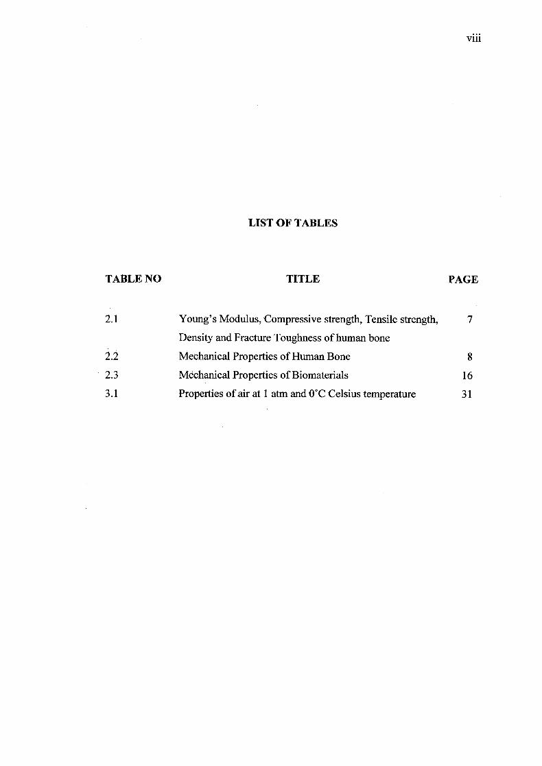
LIST OF TABLES
TABLE NO TITLE PAGE
2.1 Young's Modulus, Compressive strength, Tensile strength, 7
Density and Fracture Toughness of human bone
2.2 Mechanical Properties of Human Bone
2.3 Mechanical Properties of Biomaterials 16
3.1 Properties of air at I atm and 0°C Celsius temperature 31
viii
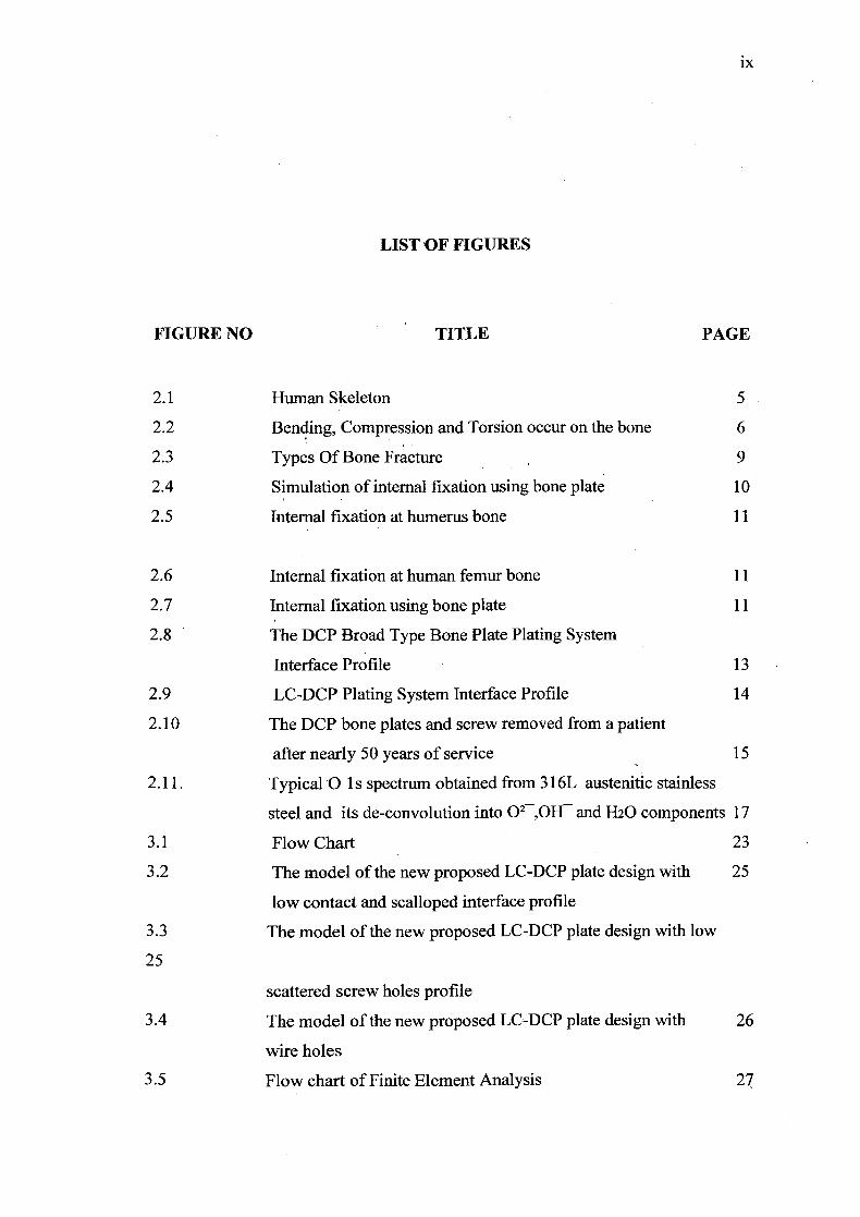
LIST OF FIGURES
FIGURE NO TITLE PAGE
2.1 Human Skeleton 5
2.2 Bending, Compression and Torsion occur on the bone 6
2.3 Types Of Bone Fracture 9
2.4 Simulation of internal fixation using bone plate 10
2.5 Internal fixation at humerus bone 11
2.6 Internal fixation at human femur bone 11
2.7 Internal fixation using bone plate 11
2.8 The DCP Broad Type Bone Plate Plating System
Interface Profile 13
2.9 LC-DCP Plating System Interface Profile 14
2.10 The DCP bone plates and screw removed from a patient
after nearly 50 years of service 15
2.11. Typical 'O is spectrum obtained from 316L austenitic stainless
steel and its de-convolution into 0 2 ,0H and H20 components 17
3.1 Flow ChartV
23
3.2 The model of the new proposed LC-DCP plate design with 25
low contact and scalloped interface profile
3.3 The model of the new proposed LC-DCP plate design with low
25
scattered screw holes profile
3.4 The model of the new proposed LC-DCP plate design with 26
wire holes
3.5 Flow chart of Finite Element Analysis 27
ix
x
3.6 A single part of DCP plate in FEA environment under three point
bending simulation. 29
3.7 A single part of LC-DCP plate in FEA environment under three 29
point bending simulation
3.8 An assembly part of DCP plate in FEA environment under heat
transfer simulation. 32
3.9 An assembly part of LC-DCP plate in FEA environment under
heat transfer simulation 32
4.1 The simulation result of the stress distribution acted on the
DCP bone plate 35
4.2 The simulation result of the stress distribution acted on
the LC-DCP bone plate 35
4.3 The simulation result of the heat transfer distribution in DCP
bone plate 38
4.4 The simulation result of the heat transfer distribution in
4.5 LCDCP bone plate 39
LIST OF APPENDICES
xi
APPENDIX TITLE
Al DCP Stress Analysis Data
A2 LC-DCP Stress Analysis Data
A3 DCP Heat Transfer Analysis Data
A4 LC-DCP Heat Transfer Analysis Data
B Properties table of Stainless Steel 316L
C Formulas
D Properties table of air at 1 atm pressure
PAGE
45
49
53
58
63
64
65
CHAPTER 1
INTRODUCTION
1.1 Project title
Designing a new concept of LC-DCP broad type bone plate.
1.2 Objectives
The objective of this project is to design a new concept of broad type LC-DCP bone
plate and analyze the bone plate stress and heat transfer analysis
1.3 Project scopes
Scopes of project:
1. Conducting literature review on LC-DCP bone plate.
2. Conceptual design of slender, easy wired and low contact LC-DCP plate using
SolidWorks CAD software.
3. Conducting heat transfer and stress analysis on the DCP plate using 3-point
bending technique with Algor FEA simulation.
2
1.4 Problem Statement
1. Patient will suffer from pain after the plate had been assembled especially
when they was in an air conditioned room. The uncomforting feeling happens
because of the high surfaces contact between the plates and the bone itself.
2. Traditional design of DCP broad bone plate does not have any wire hole to tie
the plate onto the bone prior drilling. This phenomenon causes difficulties to
the surgeon to place the plate and drill screw holes with a bit accurate.
1.5 Project Background
Bone is the main constituent of the skeletal system and it differs from the
connective tissue primarily due to its rigidity and strength. A strong and stiff bone
enables the skeleton to maintain body shape, to protect the internal organs, and to
transmit force of the muscular contraction from one part of the body to another
during movement. Fatigue and impact loads are the most common reasons for bone
fracture. Fractured bones need to be fixed surgically for it's healing and proper
functioning as early as possible. Fracture fixation by bone plate provides rigid
immobilization at the fracture site and reduces the fracture gap thus allowing primary
bone healing by new formation. The role of bone plate and screws is to hold the
fragments of the bone in position till the bone heals. In this study, the main research
is to improve the previous design of DCP broad type bone plates that was used in
bone fracture fixation.
3
Today's DCP broad type bone plate looks like a plain holed square 4mm
thick stainless steel plat. When the plate had been assembled, the patient will suffer
from the pain especially when they were in an air conditioned room. The plate's
metal will contract and shrink when cold. The uncomforting feeling happens because
of the high surfaces contact between the plates and the bone itself. When the plates
metal shrink, the deformation in compression resulting a change in stress shielding
on the bone surfaces. Another complications associated is effects on vascularity of
the cortex beneath the plate, blocking the normal blood flow. (Ischemia - deficiency
of blood in a part, usually due to functional constriction or actual obstruction of
blood vessel).
During surgery, the surgeon place the plate and drill screw holes on the
fragment part where the bone fracture. The traditional design of DCP broad type
bone plate does not have any wire hole to tie the plate onto the bone prior drilling.
This phenomenon causes difficulties to the surgeon to place the plate and drill screw
holes with a bit accurate.
Therefore in this project, a new concept and design of LC-DCP broad type
bone plate will be introduced to give benefit on orthopedics field.

CHAPTER 2
LITERATURE REVIEW
2.1 Introduction
In this chapter, focus will be to find out the related information from internet,
books and journals. These information can use as a guide and procedure to conduct
the project. Areas of studies are human bone system, the bone fracture, the bone
fracture fixation, the DCP broad type bone plate, the new concept of Low Contact -
DCP broad type bone and others. The understanding of the literature review will
helps to achieve the project objective.
2.2 Human Bone System
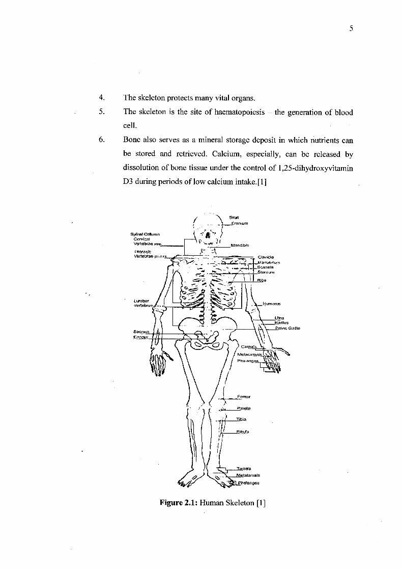
The human skeleton contains 206 bones. Each of them has a different size
and length. Figure 2.1 show that the human skeleton. The human skeleton has six
main functions:
1. The skeleton provides the framework to support our body allow us to
maintain the body shape.
2. The bones of the skeleton provide the attachment surface for muscle,
tendons and ligaments.
3. Our movement is dependent on the skeletal muscle. Without the
skeleton to give the leverage, movement would be greatly restricted.
5
4. The skeleton protects many vital organs.
5. The skeleton is the site of haematopoiesis - the generation of blood
cell.
6. Bone also serves as a mineral storage deposit in which nutrients can
be stored and retrieved. Calcium, especially, can be released by
dissolution of bone tissue under the control of 1 ,25-dihydroxyvitamin
D3 during periods of low calcium intake.[ 1]
/ \ 5kuH I 'I LfirIm
Slimn Ci'n'Ial 1 1
•. Mnd-b thuJr
1eIaett.r,fl-_
I i\ -';-- 7_ 1/ L H
L&flI
Figure 2.1: Human Skeleton [1]
2.3 Bone Characteristic
Bones is the one of the hardest structures of the human body. Bones come in
a variety of shapes and have a complex internal and external structure, allowing them
to be lightweight yet strong and hard, while fulfilling their many other functions.
Bone have they special characteristic.
Bone is anisotropic. Its modulus is dependent upon the direction of
loading.Mechanical properties dependent upon direction of loading. Bone is weakest
in shear, then tension, then compression.
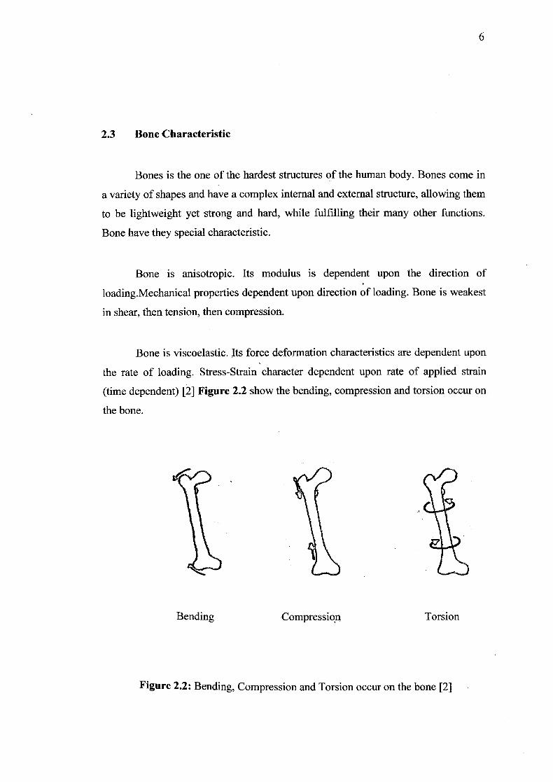
Bone is viscoelastic. Its force deformation characteristics are dependent upon
the rate of loading. Stress-Strain character dependent upon rate of applied strain
(time dependent) [2] Figure 2.2 show the bending, compression and torsion occur on
the bone.
Bending Compression Torsion
Figure 2.2: Bending, Compression and Torsion occur on the bone [2]
2.4 Mechanical Properties of Bone
Compact bone has a porosity of 5-30% and cancellous bone of approximately
30-90%, which is the proportion of the volume occupied by nonmineralized tissue
[3].
A key requirement in bone is compressive strength, and the most important
factor in compressive strength is the degree of mineralization. Loss of mineralization
results in increased risk of fracture [4].
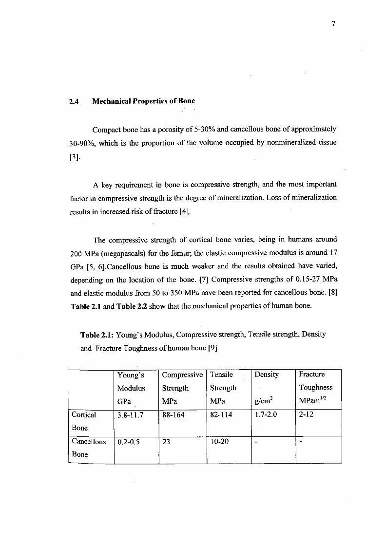
The compressive strength of cortical bone varies, being in humans around
200 MPa (megapascals) for the femur; the elastic compressive modulus is around 17
GPa [5, 6].Cancellous bone is much weaker and the results obtained have varied,
depending on the location of the bone. [7] Compressive strengths of 0.15-27 MPa
and elastic modulus from 50 to 350 MPa have been reported for cancellous bone. [8]
Table 2.1 and Table 2.2 show that the mechanical properties of human bone.
Table 2.1: Young's Modulus, Compressive strength, Tensile strength, Density
and Fracture Toughness of human bone [9]
Young's Compressive Tensile Density Fracture
Modulus Strength Strength Toughness
OPa MPa MPa g/crn3 MPam1"2
Cortical 3.8-11.7 88-164 82-114 1.7-2.0 2-12
Bone
Cancellous 0.2-0.5 23 10-20 - -
Bone
7
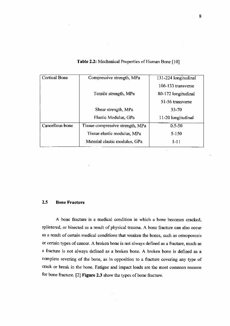
Table 2.2: Mechanical Properties of Human Bone [10]
Cortical Bone Compressive strength, MPa 131-224 longitudinal
106-133 transverse
Tensile strength, MPa 80-172 longitudinal
51-56 transverse
Shear strength, MPa 53-70
Elastic Modulus, GPa 11-20 longitudinal
Cancellous bone Tissue compressive strength, MPa 0.5-50
Tissue elastic modulus, MPa 5-150
Material elastic modulus, GPa 1-11
2.5 Bone Fracture
A bone fracture is a medical condition in which a bone becomes cracked,
splintered, or bisected as a result of physical trauma. A bone fracture can also occur
as a result of certain medical conditions that weaken the bones, such as osteoporosis
or certain types of cancer. A broken bone is not always defined as a fracture, much as
a fracture is not always defined as a broken bone. A broken bone is defined as a
complete severing of the bone, as in opposition to a fracture covering any type of
crack or break in the bone. Fatigue and impact loads are the most common reasons
for bone fracture. [2] Figure 2.3 show the types of bone fracture.
8
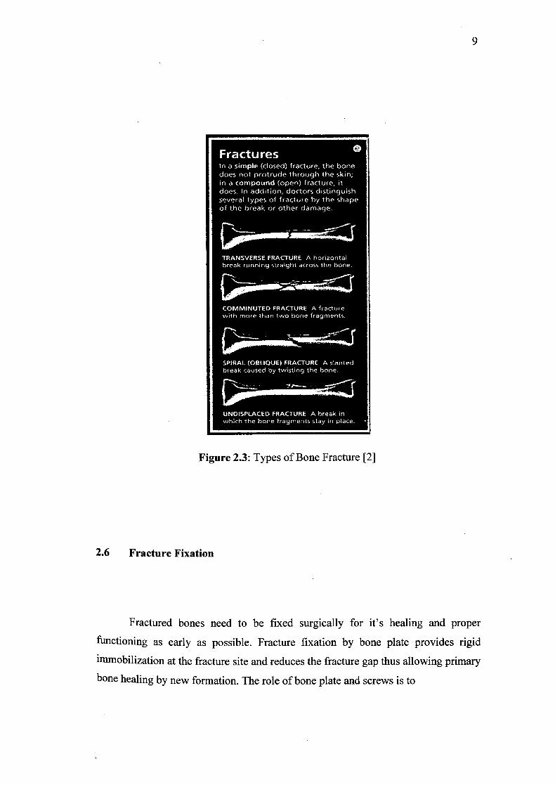
Figure 2.3: Types of Bone Fracture [2]
2.6 Fracture Fixation
Fractured bones need to be fixed surgically for it's healing and proper
functioning as early as possible. Fracture fixation by bone plate provides rigid
immobilization at the fracture site and reduces the fracture gap thus allowing primary
bone healing by new formation. The role of bone plate and screws is to
10
hold the fragments of the bone in position till the bone heals. DCP broad bone plate
is used when the actual bone is fracture or broken. The plate fixation is according
to the place. DCP broad bone plate is submerge at broad bone like at humerus, tibia
and femur bone. The function of the plate is as an internal splint an compress the
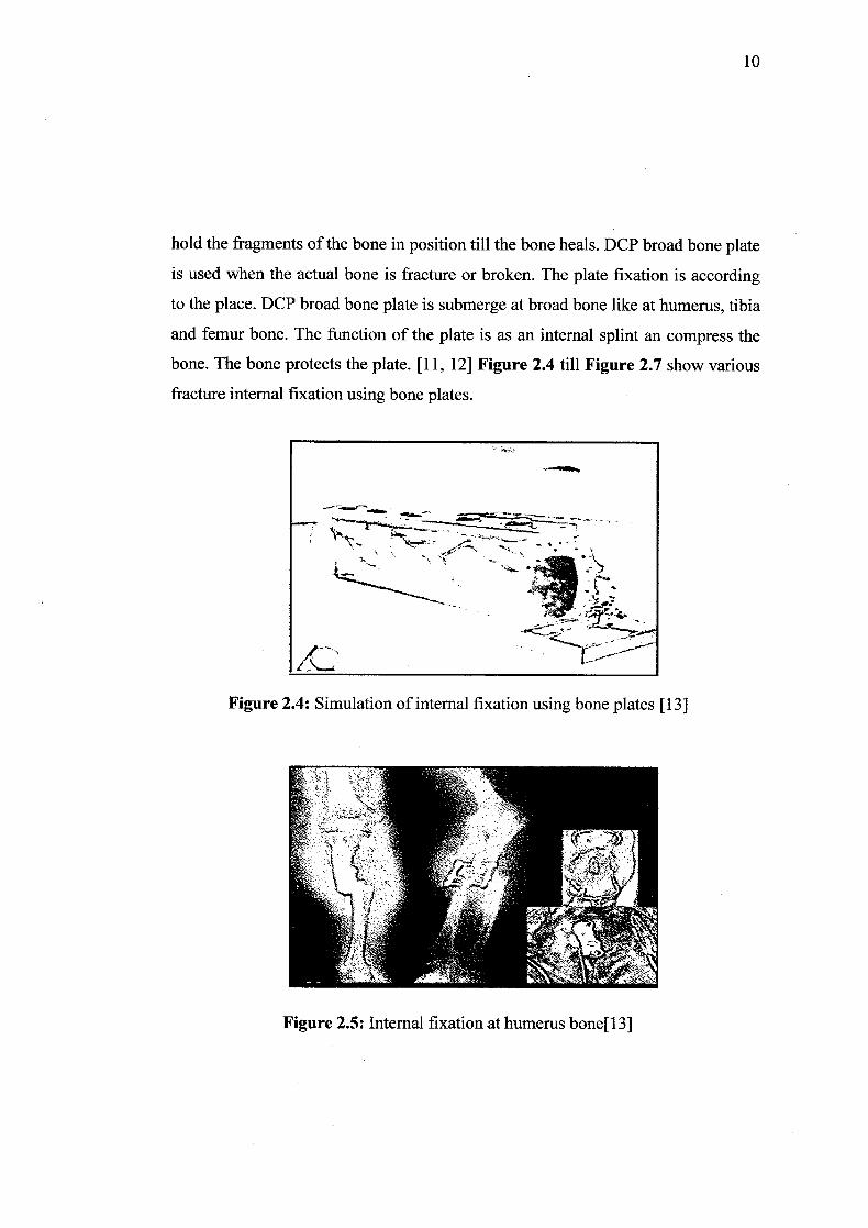
bone. The bone protects the plate. [11, 12] Figure 2.4 till Figure 2.7 show various
fracture internal fixation using bone plates.
- -
-.-
tC - b--. ---
Figure 2.4: Simulation of internal fixation using bone plates [13]
Figure 2.5: Internal fixation at humerus bone[13]
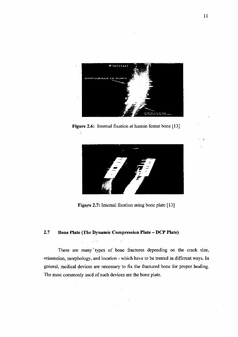
Figure 2.6: Internal fixation at human femur bone [13]
Figure 2.7: Internal fixation using bone plate [13]
2.7 Bone Plate (The Dynamic Compression Plate - DCP Plate)
There are many types of bone fractures depending on the crack size,
orientation, morphology, and location - which have to be treated in different ways. In
general, medical devices are necessary to fix the fractured bone for proper healing.
The most commonly used of such devices are the bone plate.
11
12
Bone plates, also known as osteosynthesis plates are conventionally made of
stainless steel, Cr-Co, and Ti alloys. The rigid fixation is designed to provide high
axial pressures (also known as Dynamic Compression) in the fragments of the bone,
in order to facilitate primary bone healing without the formation of external callus. A
secondary operation is generally required to remove the plate once the bone healing
is completed - which may take one to two years. However, the high rigidity of the
metal plate fixation can result in bone atrophy. The bone underneath the plate adapts
to the low stress and becomes less dense and weak. Due to bone atrophy, there is a
possibility of bone refracture after the plate is removed. This is recognized as the
"stress shielding" effect. It may be noted that the modulus of stainless steel (210-230
GPa) is much higher than the 10-18 GPa modulus of the bone. In the plate and the
fractured bone system, the amount of stress carried by each of them is directly related
to its stiffness. 191
Thus bone is insufficiently loaded compared to the implanted plate, resulting
in "stress-shielding" or stress protection. Many investigators have shown that the
degree of stress protection is proportional to the degree of stiffness mismatch. This
suggests that 'less rigid fixation plates' diminish the stress-shielding problem, and
that it is desirable to use plates whose stiffness is close to that of the bone. However,
low stiffness should not be accompanied with low fatigue strength, since the
plate/bone system will have to sustain severe cyclic loading while the bone is healing.
Polymer composite materials offer the desired high strength and bone-like elastic
properties, and hence have been proposed for bone plate applications. They may be
grouped into non-resorbable, partially resorbable, and fully resorbable bone plates.
[14] Figure 28 show the DCP Broad Type Bone Plating System Interface profile.
14
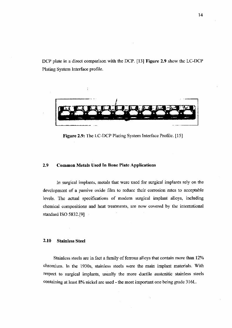
DCP plate in a direct comparison with the DCP. [13] Figure 2.9 show the LC-DCP
Plating System Interface profile.
Figure 2.9: The LC-DCP Plating System Interface Profile. [15]
2.9 Common Metals Used In Bone Plate Applications
In surgical implants, metals that were used for surgical implants rely on the
development of a passive oxide film to reduce their corrosion rates to acceptable
levels. The actual specifications of modern surgical implant alloys, including
chemical compositions and heat treatments, are now covered by the international
standard ISO 5832.[9]
2.10 Stainless Steel
Stainless steels are in fact a family of ferrous alloys that contain more than 12%
chromium. In the 1930s, stainless steels were the main implant materials. With
respect to surgical implants, usually the more ductile austenitic stainless steels
containing at least 8% nickel are used - the most important one being grade 316L.
15
316L stainless steel has a nominal composition of Mr, 8Ni, 2Mo, balanced Fe, and
an extremely low carbon content to prevent chromium depletion, hence the suffix 1!.
Occasionally nitrogen is added at about one-quarter percent level to improve the
corrosion resistance of the alloy. The relative corrosion resistance of stainless steels
can be estimated from their pitting resistance number (PREN):
PREN = %Cr + 3.3x%Mo +16x%N
However, 316L stainless steel can corrode within the body - especially in
regions where there is insufficient oxygen to maintain the passive film or where
crevices are formed (e.g., under the heads of screws). In addition, stainless steel
femoral components can fracture. Therefore stainless steel is more suitable for
temporary implant devices. Nevertheless, there are cases where 316L fracture plates
have.been removed from patients after more than 20 years of service, yet show no
evidences of corrosion. One final word of caution on stainless steels is that different
grades should not be mixed as this can result in galvanic corrosion. Since it is not
possible to visually distinguish one grade of stainless steel from another, careful
quality control must be exercised. Thebecause one out of a group of was fabricated
using the lower 304L grade. [9] Figure 2.10 show The DCP bone plates and screw
removed from a patient after nearly 50 years of service.