detecting signatures of selection from dna sequences …classic.datamonkey.org/help/tutorial.pdf ·...
TRANSCRIPT

Detecting signatures of selection from
DNA sequences using Datamonkey
Art F.Y. Poon, Simon D.W. Frost and Sergei L. Kosakovsky Pond*
Antiviral Research Center, Department of Pathology, University of California San Di-
ego, La Jolla, California, USA
*Corresponding Author
Sergei L Kosakovsky Pond, PhD
Assistant Project Scientist
Department of Pathology,
Antiviral Research Center
150 W. Washington St., #100
San Diego, CA 92103-2005
Phone: 619-543-8899
E-mail: [email protected]
Keywords: Positive selection, adaptive evolution, dN and dS estimation, HyPhy,
phylogenetic analysis, maximum likelihood inference, parallel algorithms, web serv-
ice.
2
Abstract
Natural selection is a fundamental process affecting all evolving populations. In the
simplest case, positive selection increases the frequency of alleles that confer a fit-
ness advantage relative to the rest of the population, or increases its genetic diver-
sity, and negative selection removes those alleles that are deleterious. Codon-based
models of molecular evolution are able to infer signatures of selection from align-
ments of homologous sequences by estimating the relative rates of synonymous
(dS) and non-synonymous substitutions (dN). Datamonkey
(http://www.datamonkey.org) provides a user-friendly web interface to a wide collec-
tion of state-of-the-art statistical techniques for estimating dS and dN and identifying
codons and lineages under selection, even in the presence of recombinant se-
quences.

3
1. Introduction
Natural selection plays a pivotal role in shaping the genetic variation of populations
and driving the differentiation of biological taxa. The study of causes and mecha-
nisms of molecular adaptation is one of the fundamental goals of evolutionary biol-
ogy, with numerous important applications.
Most current techniques for the inference of selection from protein-coding sequences
are based on the following observation: a nonsynonymous (or replacement) substi-
tution in a protein-coding sequence changes the primary sequence of the encoded
protein and is more likely to influence the fitness of an organism than a random syn-
onymous substitution that leaves the amino acid sequence unchanged. If nonsyn-
onymous mutations at a particular codon site in the sequence have a negligible ef-
fect on the function or expression of the protein (and hence on its fitness), then the
rate of nonsynonymous substitutions (dN) should be comparable to the rate of syn-
onymous substitutions (dS), and the site evolves neutrally. An excess of nonsyn-
onymous substitutions (dN>dS) can be interpreted as positive selection – suggestive
that replacement substitutions increase fitness. A paucity of replacement changes
(dN<dS) indicates that negative selection is working to remove such substitutions
from the gene pool.
The ratio
€
ω=dN/dS (sometimes also denoted KA/ KS) has seen wide adoption as a
measure of selective pressure (1, 2). Datamonkey (http://www.datamonkey.org) (3)
is one of the many available software tools for estimating
€
ω (Datamonkey actually
reports dN – dS, see Note 1) using a variety of evolutionary models, with several
unique advantages. Datamonkey has an intuitive and streamlined interface that pro-

4
vides easy access to complex, state-of-the-art evolutionary models. Complex models
are quickly fitted using a remote computer cluster; an analysis that would otherwise
take hours to run on a conventional desktop computer will finish in minutes on a
cluster. The collection of available methods is constantly updated as novel tech-
niques are published, obviating the need for a practicing scientist to keep apace of
new methodological advances and to install and learn how to use a plethora of soft-
ware packages. Finally, Datamonkey can analyze selection in the presence of re-
combination – something few other publicly-accessible programs can currently do.
Datamonkey uses the HyPhy package (4) as its computational engine. All of the se-
lection analyses implemented in Datamonkey, as well as a number of other analy-
ses, can also be carried out directly in HyPhy. For an in-depth discussion of the
methods and a tutorial on how sequences can be analyzed for selection in HyPhy,
we direct the interested reader to ref. (5).
Currently, Datamonkey may be used to address the following questions:
• Which codon sites in the alignment are subject to positive or negative selec-
tion? SLAC/FEL/REL methods (6) can estimate dN and dS at each codon
site. More specialized hypotheses can also be tested. For instance, if the
alignment contains sequences from multiple individuals (e.g. viruses) which
sites are positively selected at the level of a population? (the IFEL method
(7)).
• At what point in the evolutionary history of sequences did selection occur?
The GA Branch method (8) can be used to assign values of dN/dS to every
branch (lineage) in the phylogenetic tree.

5
• Does a sequence alignment contain recombinant sequences (GARD (9))?
Many traditional selection techniques can be misled by recombination (10),
but recombination can be corrected for by identifying non-recombinant frag-
ments in the alignment and reconstructing a phylogenetic tree for each frag-
ment (9).
• Is there evidence of positive selection operating within recombining fragments
of the alignment? The PARRIS test (11) is used to determine whether a pro-
portion of sites have dN>dS in the context of recombination.
2. Program Usage
To perform a selection analysis, Datamonkey requires an uploaded alignment
of at least three homologous coding nucleotide sequences (see Note 2 for browser
compatibility). Codon-based methods for estimating dN and dS can be applied to any
sequence alignment, but there are several considerations to keep in mind. Ideally,
the alignment should represent a single gene or a part thereof (e.g. a subunit), sam-
pled over multiple taxa (e.g. mammalian interferon genes) or a diverse population
sample (e.g. Influenza A viruses infecting different individuals;see Note 3). The
number of sequences in the alignment is important: too few sequences will contain
too little information for meaningful inference, while too many may take too long to
run. At the time of this writing, Datamonkey permits up to 150 sequences for SLAC
analyses, 100 for FEL/IFEL analyses, 40 for REL and PARRIS and 25 for GA-
Branch. These numbers are determined by current hardware availability and will be
6
increased in the future (see section 2.1). As a rule of thumb, at least 10 sequences
are needed to detect selection at a single site (SLAC/FEL/IFEL/REL) with any de-
gree of reliability, while as few as 4 may be sufficient for alignment-wide inference
(PARRIS/GA-Branch). The median number of sequences in an alignment submitted
to Datamonkey is 19. In addition, comparative methods may be ill suited to study
certain kinds of selection (see Note 4).
It is a good practice to visually inspect your data to make sure that the sequences
are aligned correctly. Of course, one can never be sure that an alignment is objec-
tively “correct”, but gross misalignments (e.g. sequences that are out of frame) are
easy to spot with software that provides a graphical visualization of the alignment,
such as HyPhy(4), Se-Al (http://tree.bio.ed.ac.uk/software/seal/), or BioEdit
(http://www.mbio.ncsu.edu/BioEdit/bioedit.html). You should verify that the alignment
is in frame, i.e. that it does not contain stop codons, including premature stop
codons, indicative of a frame shift, e.g. due to misalignment, or a non-functional
coding sequence, and the terminal stop codon. Your alignment should exclude any
non-coding region of the nucleotide sequence, such as introns or promoter regions,
for which existing models of codon substitution would not apply. When coding nu-
cleotide sequences are aligned directly, frameshifting (i.e. not in multiples of 3) gaps
may be inserted, since the alignment program often does not take the coding nature
of the sequence into account. Therefore it is generally a good idea to align translated
protein sequences and then map them back onto constituent nucleotides. Datamon-
key will perform a number of checks when it receives coding sequences and report
all problems it encounters (see Section 2.1.).

7
Since Datamonkey uses the HyPhy package as its processing engine, it will accept
files containing alignments in FASTA, PHYLIP, MEGA, and NEXUS formats.
If the alignment contains identical sequences, Datamonkey will discard all but one of
the duplicate sequences before proceeding. This is done to speed up the analyses,
because identical sequences do not contribute any information to the likelihood in-
ference procedure (except via base frequencies), but the computational complexity
of phylogenetic analyses grows with the number of sequences.
Finally, Datamonkey may rename some of the sequences to conform to HyPhy
naming conventions for technical reasons (all sequence names must be valid identi-
fiers, e.g. they cannot contain spaces). This is done automatically and has no effect
on the subsequent analyses.
2.1 Common issues when preparing the data for Datamonkey.
2.1.1. Non-text files. Datamonkey expects sequence alignments to be uploaded as
text files. Any other format (Word, RTF, PDF) will not be recognized and must be
converted into plain text prior to submission.
2.1.2. Nonstandard characters in the alignment. For instance, BioEdit may use
the tilde (‘~’) character to denote a gap. The dot (‘.’) character is sometimes used
as ‘match the first sequence’ character and sometimes as the gap character.
Datamonkey will accept IUPAC nucleotide characters (ACGT/U and ambiguity
characters) and ‘?’, ‘X’, ‘N’ or ‘-’ for gap or missing data (Datamonkey is not case
sensitive). All other characters in sequence data will be skipped and could result
in frame shifts, which will be reported upon upload.

8
2.1.3. Uploading an amino-acid alignment. Datamonkey employs codon models
which require the knowledge of silent substitutions, lost upon translation to
amino-acids.
2.1.4. Termination codons. Datamonkey will reject any alignments that contains
stop codons, even if the stop codon is at the end of the sequence (i.e. is a proper
termination codon). Please strip all stop codons out of the alignment prior to up-
loading it (the HyPhy standard analysis Data File
Tools:CleanStopCodons.bf can do this by replacing all stop codons with in-
dels).
2.1.5. Alignments that are too gappy. If an alignment contains more than 50% of
indels, it may not be properly processed (e.g. it could be read as a protein align-
ment, depending on the alignment format).
2.1.6. Alignments that are too large. If your alignment exceeds the size currently
allowed by Datamonkey, consider running your analysis locally in HyPhy. A de-
tailed discussion of how HyPhy can be used for that purpose can be found in ref.
(5)
2.1.7. Incorrect genetic code. If the genetic code is misspecified (e.g. the mito-
chondrial code is applied to nuclear sequences), valid alignments may fail to up-
load and if they do, then the results may be compromised (because codons are
mistranslated). Make sure the correct genetic code is selected on the data upload
page.
9
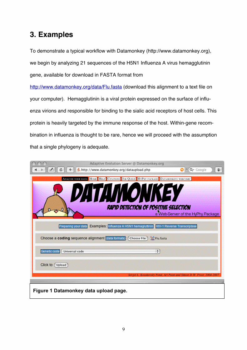
3. Examples
To demonstrate a typical workflow with Datamonkey (http://www.datamonkey.org),
we begin by analyzing 21 sequences of the H5N1 Influenza A virus hemagglutinin
gene, available for download in FASTA format from
http://www.datamonkey.org/data/Flu.fasta (download this alignment to a text file on
your computer). Hemagglutinin is a viral protein expressed on the surface of influ-
enza virions and responsible for binding to the sialic acid receptors of host cells. This
protein is heavily targeted by the immune response of the host. Within-gene recom-
bination in influenza is thought to be rare, hence we will proceed with the assumption
that a single phylogeny is adequate.
Figure 1 Datamonkey data upload page.

10
3.1. Upload the alignment.
3.1.1 Select the alignment file. The front page of http://www.datamonkey.org
includes a link to the data upload page (Fig. 1), either in the tool bar at the top of the
page, or via a large graphical button at the bottom of the page. Click on the
“Browse” button, located next to the field labeled “Choose a sequence
file:” to use your browser's interactive window to locate the file on your com-
puter.
3.1.2. Choose the genetic code. Datamonkey can interpret codons using
one of the twelve standard genetic codes, but it will employ the “Universal” genetic
code by default, which is appropriate for Influenza A.
3.1.3. Examine the uploaded file. Upon a successful upload, Datamonkey
reports some basic statistics on the alignment, including the number of sequences,
columns (codon sites) and partitions (for alignments with recombinant sequences,
there will be multiple partitions — one for each non-recombinant fragment), base fre-
quencies and an amino-acid translation of the alignment — the PDF version is handy
for an at-a-glance sequence consensus and minor variants report (Fig. 2). You can
also verify that the alignment was uploaded correctly by selecting a sequence (using
the drop-down menu labeled “BLAST your sequences?”) to BLAST against the NCBI
non-redundant nucleotide sequence database.

11
3.1.4. Phylogeny. Datamonkey will automatically detect whether a tree topology (or
multiple topologies for recombinant data) is present in the file, e.g. the TREES block
in a NEXUS-formatted file. If each tree tip can be matched up with a sequence in the
alignment, Datamonkey will make this tree available for analysis. If no tree(s) were
found in the file (as is the case for Flu.fasta), then Datamonkey can estimate a
neighbor joining (NJ, (12)) phylogeny from the alignment, using Tamura-Nei (13) nu-
cleotide distances. There is empirical evidence that selection analyses are robust to
some error in the phylogeny, hence a “quick and dirty” method like NJ should be suf-
Figure 2 Data upload summary page.

12
ficient in most cases. Click on the “Proceed” button to move on to the analysis
setup page.
3.2. Analysis setup page. The analysis setup page (Fig. 3) is used to configure all
selection analyses available via Datamonkey.
3.2.1. Job Status. The first thing to note is the Job ID bar. Each successfully up-
loaded file will be assigned a random identifier, which can be used to track all the
analyses performed on the alignment. Clicking on the [get info] link brings up
the status page for this alignment. Nearly every Datamonkey page will have a link to
Figure 3 Analysis setup page

13
the status page, and we will later discuss how the status page can be used in more
detail.
3.2.1. Method. This drop-down menu lists all analyses that can be run on the up-
loaded alignment. Some of the options may not be present for large and/or recombi-
nant alignments. We will perform SLAC, FEL and REL analyses (for detecting sites
under selection) on Flu.fasta,
3.2.2. Nucleotide substitution bias model. Each of the methods implemented by
Datamonkey makes use of a nucleotide substitution model to estimate the branch
lengths and nucleotide substitution biases (such as transition/transversion biases) of
the tree from your alignment. Datamonkey can make use of one of the 203 time-
reversible nucleotide substitution models. The most general supported time-
reversible model (denoted as REV) is comprised of eight free parameters (3 nucleo-
tide frequencies + 5 substitution rates). Four of the most frequently used models
(F81, HKY85, TrN93 and REV) are predefined as “named” options.
A parameter-rich model could conceivably overfit a small alignment, while a model
that is too simple may lead to biased inference; for this reason, Datamonkey pro-
vides an automated tool (link at the bottom of the analysis setup page, see Fig. 3)
that will select the best-fitting nucleotide model (see Note 5) from all 203 reversible
models (14) . Run the model selection procedure on Flu.fasta and verify that the
HKY85 (15) model is the best fitting model for influenza hemagglutinin. After the
model selection analysis is finished, you can return to the analysis setup page from
the model results page by clicking on the [get info] link in the Job ID bar, and
then on the link offering to set-up the SLAC analysis.

14
3.2.3. Analysis Options. There are up to three analysis options (the first two only
apply to SLAC, see Note 6). Each option has a link to the relevant help page, ex-
plaining what each settings means. Significance level determines how con-
servative each method should be, but this option only affects how the results are
presented and can be adjusted after the analysis has finished. We begin by submit-
ting a SLAC analysis with the HKY85 model and default analysis options.
3.3. The job queue page. After you submit the analysis by clicking on ‘Run’ button,
Datamonkey will display a page with all the jobs currently queued for execution. The
newly submitted page analyses are inserted at the end of the queue, and must wait
for the jobs in front of it to finish. You may bookmark the queue page in your web
browser and return to it later to check on the progress update. Once Datamonkey
begins processing your analysis, it will display intermediate progress reports for the
task and present a result page when it becomes available.
3.4. SLAC results page.
All Datamonkey analysis result pages consist of the following three parts (Fig. 4).
3.4.1 The job ID bar including the universal [get info] link to bring up the central
job summary and page.
3.4.2. Data and analysis summary. For SLAC, Datamonkey reports descriptive
statistics of the alignment (partitions, codons and estimated evolutionary tree lengths
– note that very long trees can be indicative of misaligned sequences and are
flagged as such), the inferred nucleotide substitution biases, log likelihood scores for
the fitted models and the estimate of the alignment-wide dN/dS.

15
Figure 4 SLAC analysis results page
3.4.3. Links to more detailed results. A detailed or graphical output of various,
analysis specific quantities, can be accessed via these links. For SLAC you can, for
instance, plot dN-dS across sites, or map the number of inferred synonymous and
non-synonymous substitutions to each branch of the tree (Fig. 4).
3.4.4. (SLAC/FEL/IFEL/REL) Summary of selected sites. Given a specific signifi-
cance level, all those codons, which the method detected to be under positive or
negative selection are reported. This section can be regenerated on the fly for a dif-
ferent significance level (Retabulate).

16
3.5. Interpreting SLAC results.
The summary output of selected sites lists every site where dN/dS ≠ 1 with statistical
significance (p-value) no greater than the value supplied by the user. The p-value
bounds the rate of false positives, e.g. p=0.05 means that up to 5% of neutrally
evolving sites may be incorrectly classified as selected (i.e. false positives). The p-
value should be taken as a guideline, because the statistical properties of the test
may vary from alignment to alignment. For instance, SLAC tends to be a very con-
servative test (6), hence the actual rate of false positives can be much lower than the
significance level (hence the default of 0.1, see Note 7). For each codon site,
Datamonkey will report the estimated the unadjusted dN–dS (see Note 1), dN–dS
scaled by the total length of the tree (to facilitate direct comparison between different
data sets), p-value for the test dN≠dS at that codon, and links to investigate the in-
ferred mutations at that site.
You may notice that at p=0.1, SLAC reports no positively selected sites. Increase p
to 0.2 and Retabulate the results to find that 4 codons (154,156,157,172) have p-
values for positive selection in the 0.1-0.2 range (borderline selection). For codon
156, Normalized dN-dS = 14.93. If you now click on the [Counts] link in the Ad-
ditional Info column, Datamonkey will display the list of inferred substitutions at
that codon, showing at least 6 non-synonymous and 0 synonymous substitutions,
mapped to 5 branches of the tree (Fig. 5). The p-value of 0.12 corresponds to the
binomial probability that all six substitutions at that site will be non-synonymous by
chance (see Note 8). SLAC estimates a number of quantities, which enable it to
compute this probability (accessible via the detailed HTML report from the SLAC re-

17
sult page). For site 156, its inferred codon composition and relative branch lengths
predict that a random substitution is synonymous with probability 0.30, assuming
neutral evolution, such that the probability of observing 0/6 synonymous substitutions
equals (1–0.3)6 ≈ 0.12, yielding the p-value.
Figure 5 FEL results page for the influenza analysis and substitution maps forcodon 156
3.6. FEL.
Next, return to the analysis setup page (Fig. 2), via the Job ID bar, and run the FEL
analysis with default options (note that if you have run a substitution model selection,

18
Datamonkey will automatically select the appropriate model on the analysis setup
page). The FEL result page (Fig. 5) is similar to the SLAC result page.
Like SLAC, FEL evaluates dS and dN at each site, but instead of basing its inference
on the expected and inferred numbers of synonymous and non-synonymous substi-
tutions, FEL directly estimates dN and dS based on a codon-substitution model, and
derives the p-value for the test dN≠dS using a likelihood ratio test (6). FEL tends to
be quite a bit more powerful than SLAC, e.g. note that all 4 borderline positively se-
lected SLAC sites, have p-value in 0-0.1 for FEL), but it is an order of magnitude
more computationally expensive.

19
Figure 6 REL results page for the influenza analysis.
3.7. REL
Run the REL analysis with default options (Fig. 6). REL is similar to the popular like-
lihood methods implemented in the PAML package (16), with several important addi-
tions (e.g. synonymous rate variation, see (6)). Instead of directly estimating dS and
dN at each site, REL estimates the parameters for discretized distributions of dS and

20
dN (with 3 rate categories for a total of 9 possible rate combinations) from the entire
alignment, and then infers which of these each site is most likely to have. To decide
if a codon has dN>dS, the REL method computes a Bayes factor (BF) defined as the
ratio of posterior odds of having dN>dS to the prior odds. A large Bayes factor sug-
gests that the data lend strong support to the hypothesis that a site is positively se-
lected (as a rule of thumb, 1/BF is similar to the p-value). REL tends to be the most
powerful of the three tests because it uses the entire alignment to make inferences
about rates at each site, but also generally has the highest rate of false positives,
because the distribution of rates to be fitted must be defined a priori, and it may not
adequately model the unobserved distribution of rates.
For our example, 18 sites are called positively selected (dN>dS). However, REL
does not take the magnitude of dN-dS into account, and nearly neutral or invariable
sites (such as those from rate classes 5 or 7, Fig. 6) may count towards the posi-
tively selected category.
3.8. Integrative selection analysis.
Practical statistical inference is never 100% accurate, and there will be some false
positives and negatives. As our example illustrates, different approaches to inferring
the same effect can lead to different inferences. Fortunately, for larger datasets (e.g.
> 100 sequences), all three methods tend to agree (6), with the SLAC being the
most conservative (i.e. it may miss some selected sites, but will not identify many
neutral sites as selected), followed by FEL (more detected sites), and then REL
(most detected sites, but highest errors). For smaller datasets, like the example in-
fluenza alignment, it is a good idea to run all three methods, and compare their re-

21
sults side by side. When the methods corroborate each other’s findings, we may be
more confident in their inference.
Figure 7 Integrative selection page for the influenza analysis.

22
Once SLAC, FEL and REL have been run, the Integrative Selection Analy-
sis option is enabled on the job status page. The integrative selection page (Fig. 7)
tabulates all codons, which are detected by at least one of the methods, based on
the specified significance levels. For the influenza analysis, codons 154, 156, 157,
172 and 191 are supported by FEL and REL, and all have large dN-dS values for
SLAC with borderline (0.1-0.25) p-values. Hence, they are likely to be under diversi-
fying selection. On the other extreme, codon 69 is only identified by REL, while FEL
assigned dN<dS (negative selection, but not significant), and SLAC assigns a very
large p-value for positive selection; this discordance does not inspire confidence!
It also helps to find confirmatory evidence for why detected sites may be under posi-
tive selection (e.g. based on the structure of the protein or in vitro experiments). For
example, codons 154,156 and 157 reside in the previously characterized antigenic
domain of hemagluttinin (17), while codon 172 is involved in the formation of a new
glycosylation site - an immune evasion mechanism (18).
3.9. Lineage-specific selection.
If sequences in the alignment come from different selective environments, dN/dS
may vary from branch to branch in the phylogenetic tree. Consider 9 HIV-1 envelope
sequences isolated from two patients (19) where one (the source, 5 sequences),
transmitted the virus to the other (the recipient, 4 sequences), available for download
in NEXUS format from http://www.datamonkey.org/data/env.nex (download this
alignment to a text file on your computer). The evolution of HIV envelope protein is
strongly influenced by selective pressures exerted by the humoral immune response
mounted by the host, and in our example is subject to three potentially different se-

23
lective environments: the source, the recipient, and the transmission period repre-
sented by the branch separating the sequences from each host. We run the GA
Branch analysis (see Note 9) to demonstrate how Datamonkey can be used to seg-
regate all branches into a smaller number of different selective regimes. First, upload
the alignment, perform model selection (HKY85 should be selected) and start the GA
Branch analysis from the model setup page (Fig. 3).
Figure 8 Lineage-specific selection in a two-patient HIV-1 envelope sample.
The resulting page (Fig. 8) shows that the data support 3 rate classes, with a large
number of models, which can credibly describe the data (over 2500 in the 95% con-
fidence set). The summary table for model-averaged dN/dS shows statistical de-

24
scription for estimated dN/dS at each branch. For instance, Node1, which is the in-
ternal branch (see tree plot in Fig. 8) representing the transmission event, has
dN>dS with high degree (over 0.999 probability support) of confidence, as also evi-
denced by the histogram of dN/dS tabulated from all credible models (weighted ap-
propriately, see (8)). The tree plot is a useful visual representation of the results:
each tree branch is labeled with percent support for positive selection along that
branch (4 branches have over 95% support), while the coloring reflects the selection
pattern according to the best fitting model.
As expected, there is evidence of complex and variable selective pressures on the
envelope of HIV-1 in this sample, with strong support of positive selection along
some (but not all) branches representing the evolution in each patient, as well as
along the connecting (transmission) branch (see Note 4).
3.10. Selection in the presence of recombination.
The presence of recombinants among aligned sequences frequently makes it impos-
sible to represent the evolutionary history of the sample as a single phylogenetic
tree. Most traditional selection detection techniques (19-21) assume a single
phylogeny, and can be misled if recombinants are present, e.g. (9, 10). Datamonkey
can avoid this shortcoming by first screening the alignment to locate all non-
recombinant fragments (partitions) using the GARD method (9), and then allowing
each partition to have its own phylogenetic tree. We demonstrate this approach on
an alignment of 16 HIV-1 polymerase genes sampled from the Democratic Republic
of Congo – an area where multiple divergence variants of HIV are in common circu-

25
lation (22). When a single host is infected with multiple HIV-1 viruses, this creates
the potential for the generation and transmission of recombinant HIV variants, be-
cause of very high recombination rates in multiply infected cells (23).
Figure 9 GARD recombination screen on HIV-1 pol alignment.
Download the file from http://www.datamonkey.org/data/pol.nex. When screened
with GARD (http://www.datamonkey.org/GARD), this alignment shows evidence of 3
recombination breakpoints (Fig. 9). As one of outputs of a GARD analysis (which we
encourage the interested users to run), there is NEXUS format file, which includes
the information about all non-recombinant partitions (ASSUMPTIONS block), and the
trees inferred for each partition.

26
This information will be recognized by Datamonkey and incorporated into the analy-
ses (for instance the data upload page will show that 4 partitions were read, and
what they were). The rest of the analyses can be carried out exactly as we did previ-
ously with a non-recombinant example alignment.
As an exercise in the effect of recombination, compare the multiple-segment analysis
with the results on the same alignment assuming a single phylogeny
(http://www.datamonkey.org/data/pol-1.nex). In many cases the difference is not
dramatic, but noticeable nonetheless (Fig. 10). For example, both analyses agree of
codon 476, the evidence for selection at codon 273 is stronger when multiple trees
are allowed, codon 371 is no longer detected as positively selected when multiple
trees are considered.

27
Figure 10 Effect of including multiple trees on the detection of sites under se-lection (HIV-1 pol alignment).

28
4. Notes.
1. The difference is used in place of a more common ratio dN/dS, because dS could
be 0 for some sites, rendering the ratio infinite.
2. Datamonkey.org has been developed and tested using Mozilla based browsers
(Firefox, Camino) and the Safari browser. While every attempt has been made to
write standard compliant HTML and JavaScript code, certain compatibility issues
may exist (e.g. when using Internet Explorer). Certain features require that
JavaScript be enabled.
3. Because comparative methods estimate relative rates of synonymous and non-
synonymous substitution, substantial sequence diversity is needed for reliable
inference. For example when Suzuki and Nei (24) applied a REL-type method to
a very low divergence (1 or 2 substitutions per sequence along a star phylogeny)
sample of the Human T-lymphotropic virus (HTLV), they found that the method
performed poorly. Yang and colleagues (25, 26) have suggested that the total
length of the phylogenetic tree should be at least one expected substitution per
codon site, but this is merely a guideline, not a requirement. However, sequences
that are too divergent could lead to saturation, i.e. the inability to reliably infer
branch lengths and substitution parameters.
4. For example, comparative methods should not be applied to the detection of se-
lective sweeps (rapid replacement of one allele with a more fit one, resulting in a
homogeneous population), unless sequences sampled prior to and following the

29
selective sweep are included in the sample. We refer an interested reader to (5)
for further insight.
5. The model test procedure is based on repeated likelihood ratio tests between
nested model, and AIC comparisons for non-nested models. Akaike’s Information
Criterion - a goodness-of-fit criterion that rewards the model for higher log-
likelihood score (logL) but penalizes it for each additional parameter (p) as fol-
lows: AIC = -2logL+2p. The model with the lowest AIC explains the data best.
See (27) for an excellent treatment on model selection in the phylogenetic con-
text.
6. If JavaScript is enabled in your browser, selecting an analysis will automatically
hide all the inapplicable options.
7. Readers interested in technical details should see (28) for discussion and a prac-
tical example of determining an appropriate significance level in the context of
detecting sites under selection.
8. The binomial distribution provides a tractable approximation to the expected pro-
portions of synonymous and non-synonymous substitutions at a site assuming no
selection (see http://www.math.cornell.edu/~durrett/sg/sgnote0123.pdf for techni-
cal details). Our simulation studies (unpublished) suggest that the binomial ap-
proximation is adequate for a wide range of scenarios.
9. The GA branch analysis (8) considers a large number of potential models, where
each of the branches is allocated to one of several selective regimes, evaluates
the fitness of each model by its small sample AIC score and uses a genetic algo-

30
rithms to evolve the population of potential models towards the best fitting one.
The unique benefit of this approach is that the information from all models can be
combined (weighted by the credibility of each model) to avoid incorrect statistical
inference due to model mis-specification. The main advantage of the GA ap-
proach, as opposed to the alternative branch-site family (21) of tests is that the
GA procedure will automatically mine the data in search of selection patterns,
whereas in branch-site tests, one has to select “interesting” branches a priori,
potentially oversimplifying or misleading the inference procedure (5).

31
References
1. Nielsen, R., and Yang, Z. (1998) Genetics 148, 929-36.2. Yang, Z. H., Nielsen, R., Goldman, N., and Pedersen, A. M. K. (2000) Genet-
ics 155, 431-49.3. Kosakovsky Pond, S. L., and Frost, S. D. W. (2005) Bioinformatics 21, 2531-
33.4. Kosakovsky Pond, S. L., Frost, S. D. W., and Muse, S. V. (2005) Bioinformat-
ics 21, 676-9.5. Kosakovsky Pond, S. L., Poon, A. F. Y., and Frost, S. D. W. (2007) in "Phylo-
genetic Handbook" (Lemey, P., and Pybus, O., Eds.), pp. (in press; preprintavailable at http://www.hyphy.org/pubs/hyphybook2007.pdf), Cambridge Uni-versity Press.
6. Kosakovsky Pond, S. L., and Frost, S. D. W. (2005) Mol Biol Evol 22, 1208-22.
7. Kosakovsky Pond, S. L., Frost, S. D. W., Grossman, Z., Gravenor, M. B.,Richman, D. D., and Brown, A. J. L. (2006) PLoS Computational Biology 2,e62.
8. Kosakovsky Pond, S. L., and Frost, S. D. W. (2005) Mol Biol Evol 22, 478-85.9. Kosakovsky Pond, S. L., Posada, D., Gravenor, M. B., Woelk, C. H., and
Frost, S. D. W. (2006) Mol Biol Evol 23, 1891-901.10. Shriner, D., Nickle, D. C., Jensen, M. A., and Mullins, J. I. (2003) Genet Res
81, 115-21.11. Scheffler, K., Martin, D. P., and Seoighe, C. (2006) Bioinformatics 22, 2493-9.12. Saitou, N., and Nei, M. (1987) Mol Biol Evol 4, 406-25.13. Tamura, K., and Nei, M. (1993) Mol Biol Evol 10, 512--26.14. Kosakovsky Pond, S. L., and Frost, S. D. W. (2005) Mol Biol Evol 22, 223-34.15. Hasegawa, M., Kishino, H., and Yano, T. A. (1985) J Mol Evol 22, 160-74.16. Yang, Z. H. (1997) Computer Applications In The Biosciences 13, 555-56.17. Caton, A. J., Brownlee, G. G., Yewdell, J. W., and Gerhard, W. (1982) Cell 31,
417-27.18. Perdue, M. L., and Suarez, D. L. (2000) Vet Microbiol 74, 77-86.19. Nielsen, R., and Yang, Z. H. (1998) Genetics 148, 929-36.20. Suzuki, Y., and Gojobori, T. (1999) Mol Biol Evol 16, 1315-28.21. Yang, Z., and Nielsen, R. (2002) Mol Biol Evol 19, 908-17.22. Vergne, L., Peeters, M., Mpoudi-Ngole, E., Bourgeois, A., Liegeois, F., Toure-
Kane, C., Mboup, S., Mulanga-Kabeya, C., Saman, E., Jourdan, J., Reynes,J., and Delaporte, E. (2000) J Clin Microbiol 38, 3919-25.
23. Posada, D. (2002) Mol. Biol. Evol. 19, 708-17.24. Suzuki, Y., and Nei, M. (2004) Molecular Biology And Evolution 21, 914-21.25. Anisimova, M., Bielawski, J. P., and Yang, Z. H. (2002) Molecular Biology And
Evolution 19, 950-58.26. Anisimova, M., Bielawski, J. P., and Yang, Z. H. (2001) Molecular Biology And
Evolution 18, 1585-92.27. Posada, D., and Buckley, T. R. (2004) Syst Biol 53, 793-808.28. Sorhannus, U., and Kosakovsky Pond, S. L. (2006) J Mol Evol 63, 231-9.