detection and drug resistance profile of escherichiacoli ... · detection and drug resistance...
TRANSCRIPT

Submitted 20 February 2017Accepted 17 May 2017Published 13 June 2017
Corresponding authorNitaya Indrawattana,[email protected]
Academic editorPaul Tulkens
Additional Information andDeclarations can be found onpage 12
DOI 10.7717/peerj.3431
Copyright2017 Hinthong et al.
Distributed underCreative Commons CC-BY 4.0
OPEN ACCESS
Detection and drug resistance profile ofEscherichia coli from subclinical mastitiscows and water supply in dairy farms inSaraburi Province, ThailandWoranich Hinthong1, Natapol Pumipuntu2,3, Sirijan Santajit2, SuphangKulpeanprasit2, Shutipen Buranasinsup4, Nitat Sookrung5, Wanpen Chaicumpa6,Pisinee Aiumurai5 and Nitaya Indrawattana2
1 Faculty of Medicine and Allied Health, HRH Princess Chulabhorn College of Medical Science, Bangkok,Thailand
2Department of Microbiology and Immunology, Faculty of Tropical Medicine, Mahidol University, Bangkok,Thailand
3 Faculty of Veterinary Sciences, Mahasarakham University, Mahasarakham, Thailand4Department of Pre-clinic and Applied Animal Science, Faculty of Veterinary Medicine, Mahidol University,Nakornpathom, Thailand
5Department of Research and Development, Faculty of Medicine Siriraj Hospital, Mahidol University,Bangkok, Thailand
6Center of Excellence on Therapeutic Proteins and Antibody Engineering, Department of Parasitology, Facultyof Medicine Siriraj Hospital, Mahidol University, Bangkok, Thailand
ABSTRACTSubclinical mastitis is a persistent problem in dairy farms worldwide. EnvironmentalEscherichia coli is the bacterium predominantly responsible for this condition. InThailand, subclinical mastitis in dairy cows is usually treated with various antibiotics,which could lead to antibiotic resistance in bacteria. E. coli is also a reservoir ofmany antibiotic resistance genes, which can be conveyed to other bacteria. In thisstudy, the presence of E. coli in milk and water samples was reported, among whichenteropathogenic E. coli was predominant, followed by enteroaggregative E. coli andenterohemorrhagic E. coli, which was found only in milk samples. Twenty-one patternsof antibiotic resistance were identified in this study. Ampicillin- and carbenicillin-resistant E. coliwas the most common among the bacterial isolates from water samples.Meanwhile, resistance to ampicillin, carbenicillin, and sulfamethoxazole-trimethoprimwas the pattern found most commonly in the E. coli from milk samples. Notably,only the E. coli from water samples possessed ESBL phenotype and carried antibioticresistance genes, blaTEM and blaCMY-2. This indicates that pathogenic E. coli in dairyfarms is also exposed to antibiotics and could potentially transfer these genes to otherpathogenic bacteria under certain conditions.
Subjects Microbiology, Infectious DiseasesKeywords Subclinical bovine mastitis, Escherichia coli, Antibiotic resistance, Extend spectrumbeta-lactamase
How to cite this article Hinthong et al. (2017), Detection and drug resistance profile of Escherichia coli from subclinical mastitis cows andwater supply in dairy farms in Saraburi Province, Thailand. PeerJ 5:e3431; DOI 10.7717/peerj.3431

INTRODUCTIONIn dairy farms, mastitis is a persistent problem resulting in economic losses and prematureculling of cows. Staphylococcus aureus (S. aureus) is considered a major causative pathogenwhich is a threat to farmers, although easily identifiable, whereas other gram negativebacteria is overlooked or not considered to be a cause for concern by farmers. Subclinicalmastitis, which is defined as a somatic cell count (SCC) of >200,000 cells/mL in milk,is usually caused by gram negative bacteria, such as Escherichia coli (E. coli), Klebsiellapneumoniae, and Serratia marcescens (Schukken et al., 2012; Azevedo et al., 2016). Thesebacteria are commonly found in environmental settings, such as bedding, clothes,farmers’ hands, and water used on farms (Perkins et al., 2009; Iraguha, Hamudikuwanda& Mushonga, 2015; Azevedo et al., 2016). Among gram negative bacteria, E. coli is themost notable cause of mastitis. E. coli was found to usually infected mammary gland ofcows parturition and early lactation period which could lead to local and acute mastitis(Burvenich et al., 2003). In a study in Portugal, E. coli was found to be the second mostcommon bacteria after non-coagulative staphylococci found in bulk tank milk (Azevedo etal., 2016). In Uruguay, E. coli was second only to S. aureus in bovine subclinical mastitiscases (Gianneechini et al., 2002), whereas in China, it was one of the leading types ofcoliform bacteria found in milk from cows with subclinical mastitis (Memon et al., 2013;Wang et al., 2015).
The treatment of bovine subclinical mastitis usually depends on the severity of thesymptoms. In Thailand, the disease is usually treated with antibiotics or the infected cowsare culled. Antibiotics are also used for prevention in some farms. However, this can lead tobacteria developing resistance to them. For example, increased resistance to antibiotics inS. aureus in the form of oxacillin- or gentamicin-resistant strains was reported in Thailanddue to their excessive use (Suriyasathaporn, 2011; Suriyasathaporn et al., 2012). Despite thisbackground, there is little information on antibiotic resistance and drug resistance genes inother bacteria related to bovine mastitis in Thailand. E. coli can be antibiotic-resistant as itis also exposed to antibiotics from wastewater from farms. Furthermore, E. coli that carriesresistance genes can transfer those genes to other pathogenic bacteria (Hu et al., 2016).The discovery of antibiotic resistance in E. coli isolates from farms could possibly show thetrend or specific characteristic of antibiotic resistance and facilitate better prevention orthe more effective treatment for mastitis on dairy farm. This study was thus conducted todetect E. coli from water sources and milk from cows with subclinical mastitis, and theirantibiotic resistance patterns.
MATERIALS AND METHODSSample collectionAll procedures performed in this study are in accordance with the ethical standards of theFaculty of Tropical Medicine–Animal Care and Use Committee (FTM-ACUC), MahidolUniversity, Thailand (protocol no. 002-2016). Water and milk samples were collected from17 dairy farms in Saraburi Province, Thailand, where agriculture and livestock are themain source of income of the people. A total of 35 water samples were collected in 500-ml
Hinthong et al. (2017), PeerJ, DOI 10.7717/peerj.3431 2/16
sterile bottles from drinking water for cows in a milking area and also from washing water.Thirty-eight milk samples were collected in sterile falcon tubes from cows with subclinicalmastitis, which had an SCC of >200,000 cells/ml inmilk, after the teats had been disinfectedwith 70% ethanol and 4–5 streams of milk had been removed. Both water andmilk sampleswere stored at 4 ◦C and transported to the laboratory within 24 h for the experiment.
Bacterial isolationEach water and milk sample was centrifuged at 6,000 rpm for 10 min, and the precipitantwas subjected to 10-fold dilution and spread on MacConkey agar (Becton, Dickinson, andCompany). Suspected E. coli lactose-fermenting colonies (pink colonies) were subjectedto gram staining and standard biochemical tests, including triple sugar iron agar, lysinedecarboxylase, ornithine decarboxylase/deaminase, motility, and indole production tests.
Antibiotic susceptibility testsAll E. coli isolates were subjected to antibiotic susceptibility tests following the Clinicaland Laboratory Standards Institute (CLSI) guidelines (Clinical and Laboratory StandardsInstitute, 2016). The antimicrobial disks used in the experiment included 10 µg ampicillin(≤13 mm), 100 µg piperacillin (≤17 mm), 10 µg carbenicillin (≤17 mm), 20 µgamoxicillin-clavulanic acid (≤13 mm), 30 µg cefepime (≤14 mm), 30 µg cefotaxime(≤22mm), 30µg ceftriaxone (≤19mm), 30µg ceftazidime (≤17mm), 75µg cefoperazone(≤15 mm), 30 µg cefuroxime (≤14 mm), 10 µg imipenem (≤19 mm), 10 µg meropenem(≤19 mm), 10 µg gentamicin (≤12 mm), 30 µg amikacin (≤14 mm), 15 µg tigecycline(≤14 mm), 5 µg ciprofloxacin (≤15 mm), 10 µg norfloxacin (≤12 mm), and 23.75 µgtrimethoprim–sulfamethoxazole (≤10 mm) (Oxoid). E. coli strain ATCC 25922 was usedas a control in this experiment.
Extended spectrum β-lactamase (ESBL) production was tested by double disk synergy(DDS) method modified from Clinical and Laboratory Standards Institute (2012). The testuses 30µg antibiotic disks of cefepime, cefotaxime ceftriaxone, ceftazidime, and cefuroxime.The antibiotic disks were placed on the E. coli spreaded MHA culture plate, 30 mm (centerto center) from the amoxicillin-clavulanic acid (30 µg) disk. Plates were incubated at 37 ◦Covernight and observed for the presence of an extended spectrum beta-lactamase (ESBL)phenotype by an extension of the edge of inhibition zone of antibiotic disks toward theamoxicillin-clavulanic acid.
Gene detection by polymerase chain reactionAll E. coli isolates from both water and milk samples were determined using specific geneand plasmid, and the isolates that showed resistance to antibiotics were selected andsubjected to PCR to investigate their drug resistance genes. The bacteria were cultured in1.5 ml of tryptic soy broth (Oxoid) and incubated overnight; they were then harvestedand centrifuged for 10 min at 6,000 rpm. The pellet was resuspended with 800 µl of steriledistilled water, boiled for 10 min, centrifuged at 6,000 rpm for 10 min, and then thesupernatant was collected for use as a DNA template in PCR. PCR primers, conditions,and positive control strains for the detection of target gene and drug resistance genes arepresented in Tables 1 and 2. All PCR reactions with a total volume of 25 µl were performed
Hinthong et al. (2017), PeerJ, DOI 10.7717/peerj.3431 3/16

Table 1 Primers and PCR conditions used for virulence gene detection.
Target genes Positivecontrol
Sequences (5′–3′) Annealingtemperature(◦C)
Productsize (bp)
References
Heat-labile toxin (lt ) ETEC tctctatgcatacggagccatactgattgccgcaatt
55 322 Deng et al. (1996)
Hest-stable toxin (st ) ETEC tgctaaaccagtagagtcttcaaaagcaggcttacaacacaattcacagcag
55 138 Mercado et al. (2011)
Shiga-like enterotoxins 1 (evt ) EHEC caacactggatgatctcagccccctcaactgctaata
55 349 Khan et al. (2002)
Shiga-like enterotoxins 2 (evs) EHEC atcagtcgtcactcactggtctgctgtcacagtgacaaa
55 110 Khan et al. (2002)
Transcriptional activatorof the aggregativeadherence fimbriae (aggR)
EAEC 17-2 ctaattgtacaatcgatgtaatgaagtaattcttgaat
55 308 Nataro et al. (1994)
pCVD432 plasmid EAEC 17-2 ctggcgaaagactgtatcatcaatgtatagaaatccgctgtt
55 630 Aranda, Fagundes-Neto &Scaletsky (2004)
Intimin (eaeA) Plasmid-eaeA aaacaggtgaaactgttgcctctcgcctgatagtgtttggta
55 350 Yu & Kaper (1992)
Bundle-forming pilus (bfpA) – aatggtgcttgcgcttgctgcgccgctttatccaacctggta
57 326 Zhang et al. (2016)
Notes.ETEC, enterotoxigenic E. coli; EHEC, enterohemorrhagic E. coli; EAEC, enteroaggregative E. coli.
in 1× Taq buffer, 1 mM MgCl2, 0.2 mM dNTP, 1 µM of each of the forward and reverseprimers, and 2 units of Taq DNA polymerase (Thermo Scientific). The PCR amplicon wassubjected to 1.5% agarose gel electrophoresis in TAE buffer. For gene amplification withno reference control, the PCR product from positive samples was subjected to nucleotidesequencing and sequence analysis for gene confirmation.
SerotypingE. coli isolates with virulence genes were serotyped using Serosystem (Serosystem, Clinag,Thailand) to identify O and H antigens present on the surface of the pathogenic E. coliisolates with slide agglutination test. The experiment was performed following themanufacturer’s protocol. EAEC, EHEC, EPEC, and ETEC strains were used as positivecontrol in the experiment.
RESULTSE. coli isolation and antibiotic resistance patternsA total of 185 E. coli isolates were collected from water (116 isolates) and milk (69 isolates)samples and subjected to antibiotic susceptibility tests. Among these isolates, a total of 77(51 isolates from water and 26 isolates from milk samples) showed resistance to at leastone of the antibiotics use in the experiment. Penicillin-resistant E. coli (71/77, 92.2%) wasfound to be the largest group in this study followed by folate pathway inhibitor-resistantE. coli (20/77, 26%). E. coli resistant to cephems (14/77, 18.2%), aminoglycosides (14/77,18.2%), β-lactamase inhibitor combination (4/77, 5.2%), fluoroquinolone (12/77, 14.3%),and carbapenem (1/77, 1.3%) were also found. Among antibiotic resistant E. coli, 84.31%
Hinthong et al. (2017), PeerJ, DOI 10.7717/peerj.3431 4/16
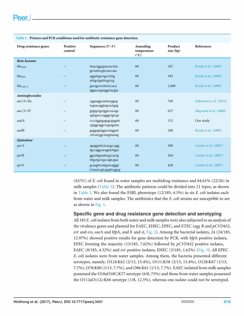
Table 2 Primers and PCR conditions used for antibiotic resistance gene detection.
Drug resistance genes Positivecontrol
Sequences (5′–3′) Annealingtemperature(◦C)
Productsize (bp)
References
Beta-lactamsblaTEM – ttaactggcgaactacttac
gtctatttcgttcatccata60 247 Kozak et al. (2009)
blaSHV – aggattgactgccttttgatttgctgatttcgctcg
60 393 Kozak et al. (2009)
blaCMY-2 – gacagcctctttctccacatggacacgaaggctacgta
60 1,000 Kozak et al. (2009)
Aminoglycosidesaac(3)-IIa – cggaaggcaataacggag
tcgaacaggtagcactgag60 740 Soleimani et al. (2014)
aac(3)-IV – gtgtgctgctggtccacagcagttgacccagggctgtcgc
60 627 Maynard et al. (2004)
aadA – cccctggagagagcgagattcgtggctggctcgaagatac
60 152 Our study
aadB – gaggagttggactatggattcttcatcggcatagtaaaag
60 208 Kozak et al. (2009)
QuinoloneqnrA – agaggatttctcacgccagg
tgccaggcacagatcttgac60 580 Cattoir et al. (2007)
qnrB – ggcattgaaattcgccactgtttgctgctcgccagtcgaa
60 264 Cattoir et al. (2007)
qnrS – gcaagttcattgaacagggttctaaaccgtcgagttcggcg
60 428 Cattoir et al. (2007)
(43/51) of E. coli found in water samples are multidrug resistance and 84.61% (22/26) inmilk samples (Table 3). The antibiotic patterns could be divided into 21 types, as shownin Table 3. We also found the ESBL phenotype (12/185, 6.5%) in six E. coli isolates eachfrom water and milk samples. The antibiotics that the E. coli strains are susceptible to areas shown in Fig. 1.
Specific gene and drug resistance gene detection and serotypingAll 185 E. coli isolates frombothwater andmilk samples were also subjected to an analysis ofthe virulence genes and plasmid for EAEC, EHEC, EPEC, and ETEC (agg R and pCVD432,evt and evs, eaeA and bfpA, and lt and st, Fig. 2). Among the bacterial isolates, 24 (24/185,12.97%) showed positive results for gene detection by PCR, with bfpA positive isolates,EPEC forming the majority (13/185, 7.02%) followed by pCVD432 positive isolates,EAEC (8/185, 4.32%) and evt positive isolates, EHEC (3/185, 1.62%) (Fig. 3). All EPECE. coli isolates were from water samples. Among them, the bacteria presented differentserotypes, namely, O124:K62 (2/13, 15.4%), O111:K58 (2/13, 15.4%), O128:K67 (1/13,7.7%), O78:K80 (1/13, 7.7%), and O86:K61 (1/13, 7.7%). EAEC isolated frommilk samplespossessed the O18aO18C:K77 serotype (6/8, 75%) and those from water samples possessedthe O112aO112c:K66 serotype (1/8, 12.5%), whereas one isolate could not be serotyped.
Hinthong et al. (2017), PeerJ, DOI 10.7717/peerj.3431 5/16
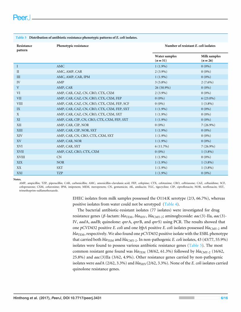
Table 3 Distribution of antibiotic resistance phenotypic patterns of E. coli isolates.
Resistancepattern
Phenotypic resistance Number of resistant E. coli isolates
Water samples(n= 51)
Milk samples(n= 26)
I AMC 1 (1.9%) 0 (0%)II AMC, AMP, CAR 2 (3.9%) 0 (0%)III AMC, AMP, CAR, IPM 1 (1.9%) 0 (0%)IV AMP 3 (5.8%) 2 (7.6%)V AMP, CAR 26 (50.9%) 0 (0%)VI AMP, CAR, CAZ, CN, CRO, CTX, CXM 2 (3.9%) 0 (0%)VII AMP, CAR, CAZ, CN, CRO, CTX, CXM, FEP 0 (0%) 6 (23.0%)VIII AMP, CAR, CAZ, CN, CRO, CTX, CXM, FEP, SCF 0 (0%) 1 (3.8%)IX AMP, CAR, CAZ, CN, CRO, CTX, CXM, FEP, SXT 1 (1.9%) 0 (0%)X AMP, CAR, CAZ, CN, CRO, CTX, CXM, SXT 1 (1.9%) 0 (0%)XI AMP, CAR, CIP, CN, CRO, CTX, CXM, FEP, SXT 1 (1.9%) 0 (0%)XII AMP, CAR, CIP, NOR 0 (0%) 7 (26.9%)XIII AMP, CAR, CIP, NOR, SXT 1 (1.9%) 0 (0%)XIV AMP, CAR, CN, CRO, CTX, CXM, SXT 1 (1.9%) 0 (0%)XV AMP, CAR, NOR 1 (1.9%) 0 (0%)XVI AMP, CAR, SXT 6 (11.7%) 7 (26.9%)XVII AMP, CAZ, CRO, CTX, CXM 0 (0%) 1 (3.8%)XVIII CN 1 (1.9%) 0 (0%)XIX NOR 1 (1.9%) 1 (3.8%)XX SXT 1 (1.9%) 1 (3.8%)XXI TZP 1 (1.9%) 0 (0%)
Notes.AMP, ampicillin; TZP, piperacillin; CAR, carbenicillin; AMC, amoxicillin–clavulanic acid; FEP, cefepime; CTX, cefotaxime; CRO, ceftriaxone; CAZ, ceftazidime; SCF,cefoperazone; CXM, cefuroxime; IPM, imipenem; MEM, meropenem; CN, gentamicin; AK, amikacin; TGC, tigecycline; CIP, ciprofloxacin; NOR, norfloxacin; SXT,trimethoprim–sulfamethoxazole.
EHEC isolates from milk samples possessed the O114:K serotype (2/3, 66.7%), whereaspositive isolates from water could not be serotyped (Table 4).
The bacterial antibiotic-resistant isolates (77 isolates) were investigated for drugresistance genes (β-lactam: blaTEM, blaSHV, blaCMY-2; aminoglycoside: aac(3)-IIa, aac(3)-IV, aadA, aadB; quinolone: qnrA, qnrB, and qnrS) using PCR. The results showed thatone pCVD432 positive E. coli and one bfpA positive E. coli isolates possessed blaCMY-2 andblaTEM, respectively. We also found one pCVD432 positive isolate with the ESBL phenotypethat carried both blaTEM and blaCMY-2. In non-pathogenic E. coli isolates, 43 (43/77, 55.9%)isolates were found to possess various antibiotic resistance genes (Table 5). The mostcommon resistant gene found was blaTEM (38/62, 61.3%) followed by blaCMY-2 (16/62,25.8%) and aac(3)IIa (3/62, 4.9%). Other resistance genes carried by non-pathogenicisolates were aadA (2/62, 3.3%) and blaSHV(2/62, 3.3%). None of the E. coli isolates carriedquinolone resistance genes.
Hinthong et al. (2017), PeerJ, DOI 10.7717/peerj.3431 6/16

Figure 1 Antibiotic susceptibility test results. (A) E. coli isolates from water samples. (B) E. coli isolatesfrom milk samples.
Figure 2 Agarose gel electrophoresis of 1% agarose of the amplification products of virulence genesand plasmid for ETEC (lt, st ), EHEC (evt, evs), EAEC (pCVD432, aggR), and EPEC (eaeA, bfpA). M,DNAMarker.
Figure 3 Prevalence of pathogenic E. coli detected fromwater andmilk samples.
Hinthong et al. (2017), PeerJ, DOI 10.7717/peerj.3431 7/16

Table 4 Target genes, serotyping, antibiotic resistance pattern, resistant gene profile, and ESBL phenotype of pathogenic E. coli isolates.
Isolate number Origin Target gene Serotype Antibioticresistancepattern
Resistance geneprofile
ESBLphenotype
M-W910-1 LF1 Water bfpA a *− −
M-W910-1 LF3 Water bfpA a * − −
M-W1010-1 LF1 Water bfpA a V blaTEM −
M-W1110-1 LF3 Water bfpA a XX − −
M-W1110-1 LF5 Water bfpA O124:K62 * − −
M-W1110-1 LF6 Water bfpA O111:K58 * − −
M-W12UD LFB6 Water bfpA O111:K58 I − −
M-W13UD LFB6 Water bfpA O128:K67 * − −
M-W13UD LFB7 Water bfpA O124:K62 * − −
M-W13UD LFB10 Water bfpA O78:K80 * − −
M-W15UD LFB4 Water bfpA a V − −
M-W22UD LF9 Water pCVD432 a VI blaCMY-2, aac(3)IIa −
M-W23UD LF2 Water pCVD432 O112aO112c:K66 VI blaTEM, blaCMY-2, aac(3)IIa, aadA +
M-W32UD LF1 Water bfpA a V − −
M-W33UD LF1 Water bfpA O86:K61 * − −
M-M10UD LFB4 Milk evt O114:K * − −
M-M10UD LFB5 Milk evt O114:K * − −
M-M35UD LFB2 Milk pCVD432 O18aO18c:K77 VII − +
M-M35UD LFB3 Milk pCVD432 O18aO18c:K77 VII − +
M-M35UD LFB4 Milk pCVD432 O18aO18c:K77 VII − +
M-M35UD LFB5 Milk pCVD432 O18aO18c:K77 VIII − +
M-M35UD LFB6 Milk pCVD432 O18aO18c:K77 VII − +
M-M35UD LFB7 Milk pCVD432 O18aO18c:K77 VII − +
M-M37UD LFB4 Milk evt a XVI − −
Notes.aNot typable.*Susceptible.+Positive.−Negative.
DISCUSSIONE. coli is known to be the most common gram negative bacteria that potentially causessubclinical mastitis and exhibits antibiotic resistance. However, pathogenic E. coli in theenvironment has often been overlooked. Many studies have reported the presence of E. coliamong subclinical mastitis cases in dairy farms in many regions of the world, particularlyin developing countries, such as Uruguay, Turkey, Brazil, Ethiopia, Mexico, and China(Gianneechini et al., 2002; Guler & Gunduz, 2007; Fernandes et al., 2011; Haftu et al., 2012;Abera et al., 2012; Olivares-Perez et al., 2015; Wang et al., 2015). This study demonstratedthe existence of pathogenic E. coli in environmental sources and also in milk from cowswith subclinical mastitis by detecting specific genes associated with the pathogenic types ofthis species. bfpA-positive E. coli was found to be the most common strain of pathogenicE. coli residing in water sources. pCVD432-positive isolate was found in both water and
Hinthong et al. (2017), PeerJ, DOI 10.7717/peerj.3431 8/16

Table 5 Target genes, serotyping, antibiotic resistance pattern, resistant gene profile, and ESBL phenotype of non-pathogenic E. coli isolates.
Isolate number Origin Target gene Serotype Antibioticresistance pattern
Resistance geneprofile
ESBL phenotype
M-W610-1 LF1 Water − b V blaTEM −
M-W910-1 LF2 Water − b IV blaCMY-2−
M-W1110-1 LF9 Water − b XVI blaTEM −
M-W12UD LFB6 Water − b I blaCMY-2−
M-W16UD LF1 Water b V blaTEM, blaSHY, blaCMY-2−
M-W16UD LF2 Water − b V blaTEM, blaCMY-2−
M-W1910-1 LF6 Water − b V blaTEM −
M-W20UD LF6 Water − b V blaTEM, blaCMY-2−
M-W20UD LF9 Water − b V blaTEM, blaCMY-2−
M-W22UD LF3 Water − b XIV blaTEM, blaCMY-2+
M-W22UD LF7 Water − b X blaTEM, blaCMY-2, aac(3)IIa +
M-W24UD LF1 Water − b V blaTEM −
M-W24UD LF3 Water − b XI blaTEM, blaCMY-2, aac(3)IIa, aadA +
M-W24UD LF4 Water − b V blaTEM −
M-W24UD LF5 Water − b XVI blaTEM −
M-W24UD LF7 Water − b * blaTEM, blaCMY-2−
M-W26UD LF8 Water − b IV blaTEM −
M-W27UD LF4 Water − b XVI blaTEM −
M-W28UD LF1 Water − b XV blaTEM −
M-W28UD LF5 Water − b V blaSHV, blaCMY-2−
M-W28UD LF7 Water − b V blaTEM, blaCMY-2−
M-W29UD LF1 Water − b IX blaTEM, blaCMY-2, aac(3)IIa, aadA +
M-W29UD LF10 Water − b XVI blaTEM −
M-W31UD LF6 Water − b V blaTEM −
M-W33UD LF2 Water − b V blaTEM −
M-W33UD LF6 Water − b V blaTEM −
M-W33UD LF9 Water − b V blaTEM −
M-W33UD LF10 Water − b V blaTEM −
M-W34UD LF3 Water − b V blaTEM −
M-W34UD LF7 Water − b XIII blaTEM −
M-M1610-1 LFB2 Milk − b IV blaTEM −
M-M37UD LFB1 Milk − b XVI blaTEM −
M-M37UD LFB3 Milk − b XVI blaTEM −
M-M37UD LFB5 Milk − b XVI blaCMY-2−
M-M37UD LFB6 Milk − b XVI blaTEM, blaCMY-2−
M-M37UD LFB8 Milk − b XVI blaCMY-2−
M-M38UD LFB1 Milk − b XII blaTEM −
M-M38UD LFB2 Milk − b XII blaTEM −
M-M38UD LFB3 Milk − b XII blaTEM −
M-M38UD LFB4 Milk − b XII blaTEM −
(continued on next page)
Hinthong et al. (2017), PeerJ, DOI 10.7717/peerj.3431 9/16

Table 5 (continued)
Isolate number Origin Target gene Serotype Antibioticresistance pattern
Resistance geneprofile
ESBL phenotype
M-M38UD LFS2 Milk − b XII blaTEM −
M-M38UD LFS3 Milk − b XII blaTEM −
M-M38UD LFS4 Milk − b XII blaTEM −
Notes.bNot serotype.*Susceptible.+Positive.−Negative.
milk samples. evt-positive E. coli was the least common and was only identified in milksamples; it was not present in any of the water samples. In this study, EPEC possessedonly bfpA, which encodes bundle-forming pili that are a specific characteristic of EPEC(Cleary et al., 2004). The presence of EPEC in water sources in dairy farms could lead tointramammary infection of cows. A study by Dopfer, Nederbragt & Almeida (2001) alsoreported the isolation of bfpA-positive EPEC from persistent cases of bovine mastitis(Dopfer, Nederbragt & Almeida, 2001). Although none of the E. coli isolates was positive foreaeA in this study, there are reports of the presence of eaeA-positive EPEC among E. colifound in cows with mastitis in Brazil and Turkey (Correa & Marin, 2002; Guler & Gunduz,2007). However, in Iran, eaeA-positive E. coli was not found in clinical mastitis cases(Ghanbarpour & Oswald, 2010). This indicates that bfpA- and eaeA-positive EPEC may bedistributed unevenly across the globe. EAEC (pCVD432-positive isolates) was found inboth water and milk samples. However, the serotypes of those isolates differed. This mayindicated different sources of EAEC in water and infected cows. The results also designatedthat EAEC may be an epidemic strain in dairy farms in Saraburi Province, and EAEC andEPEC could be causative agents of mastitis considering their potential infection throughwater in farms. EHEC was the least common group found only in milk samples in thisstudy and positive only for evt (shiga-toxin 1-encoding gene). These results raise concernsregarding the bacterial distribution to nearby areas via the contaminated water whichworkers should be aware of. Studies by Lira, Macedo & Marin (2004) and Momtaz (2010)also reported shiga-toxin 1-producing E. coli from cases of subclinical mastitis in cows inBrazil and Iran. Momtaz et al. (2012) later reported that shiga-toxin 1-producing E. coliwas the most common type of E. coli in milk samples from cows with subclinical mastitis inIran (Momtaz et al., 2012). These results also correlated with many studies on clinical casesof bovine mastitis. For example,Momtaz et al. (2012) reported the presence of EHEC withshiga-toxin 1-encoding gene as themost common virulence gene inmilk samples from caseswith subclinical and clinical mastitis (Momtaz et al., 2012), which also correlated with thestudy by Suojala et al. (2011), in which shiga-toxin 1-encoding gene was among the mostcommon virulence genes found in clinical cases of bovine mastitis (Suojala et al., 2011).
Among the 21 antibiotic resistance patterns, the most common pattern found in E. colifrom water sources was pattern V (ampicillin and carbenicillin resistance), followedby pattern XVI (ampicillin, carbenicillin, gentamicin, ceftriaxone, cefotaxime, andtrimethoprim/sulfamethoxazole resistance). Among the antibiotic patterns in the E. coli
Hinthong et al. (2017), PeerJ, DOI 10.7717/peerj.3431 10/16

from milk, pattern XII (ampicillin, carbenicillin, ciprofloxacin, and norfloxacin resistance)and XVI were the most common. This may indicate that E. coli in milk could potentiallyderive from water or other environmental sources. A study by Sayah et al. (2005) alsoreported the difference in antibiotic resistance patterns between E. coli isolated from farmwater and fecal samples (Sayah et al., 2005). Our results call for a more cautious approachwith antibiotics usage in dairy farms in the Saraburi province area, since the antibioticsthat the E. coli isolates were susceptible to are from the high generation cephalosporin andβ-lactam classes which are normally used for the treatment of drug-resistance bacteria.
In another study, Geser, Stephan & Hachler (2012) reported on ESBL-positive E. coli inmilk from cows with mastitis Geser, Stephan & Hachler (2012), and ESBL-producing E.coli was shown to be able to spread from infected animals to the environment, such as airand slurry, as reported in a pig farm in Germany (Von Salviati et al., 2015). In this study,EAEC was the only pathogenic strain that possessed the ESBL phenotype. Notably, only theESBL-producing EAEC isolates from water samples contained antibiotic resistance genes(blaTEM and blaCMY-2). The results also correlate with the study by Franz et al. (2015), whoreported that EAEC found in surface water and wastewater dominates over other strainsof pathogenic E. coli in terms of possessing the ESBL phenotype (Franz et al., 2015). Inthis study, we found that non-pathogenic E. coli isolates carried ESBL-associated genes(blaTEM, blaSHV, and blaCMY-2. However, only four isolates (M-W22UD LF3, M-W22UDLF7, M-W24UD LF3, and M-W29UD LF1) presented the ESBL phenotype and all ofthese carried blaTEM and blaCMY-2. These results imply that the presence of drug-resistantstrains of non-pathogenic E. coli isolates from the environment is possible. This can pose athreat to mastitis management programs for farm since one study also reported that non-pathogenic E. coli can serve as a reservoir of antibiotic resistance genes and could possiblytransfer the genes to other pathogenic E. coli if conditions are suitable (Hu et al., 2016).
CONCLUSIONThis study provides evidence that E. coli isolates from cows with subclinical mastitis andfrom water at dairy farms in Saraburi Province of Thailand consisted of pathogenic E. colistrains that are resistant to many groups of antibiotics, including the fluoroquinolonegroup, which should raise concerns regarding the improper use of antibiotics in thisarea. However, the information on which antibiotics are being used on the farms is verylimited. Identification of the ESBL phenotype and β-lactamase genes was also a concern asthese can be transferred to other E. coli strains, including pathogenic strains, and bacterialspecies. This could lead to more serious problems associated with antibiotic resistancein the future. It should be recommended that farms prevent mastitis by promote cleanenvironments for cows such as frequently changing bedding at the stalls and milkingareas by cleaning the areas thoroughly. The use of dry and clean cloths to clean theteats before milking and effective teat dips should reduce mastitis on farms. The useof antibiotics, mastitis control programs, and milking hygiene should be consideredand supervised by veterinarians to improve mastitis status and treatment in this area.
Hinthong et al. (2017), PeerJ, DOI 10.7717/peerj.3431 11/16

ADDITIONAL INFORMATION AND DECLARATIONS
FundingThe work was supported by a NSTDA Chair professor grant (P-1450624) funded by theCrown Property Bureau of Thailand, Thailand Research Fund (RSA5980048) and RoyalGolden Jubilee (RGJ) grant (PHD58K0073). The funders had no role in study design, datacollection and analysis, decision to publish, or preparation of the manuscript.
Grant DisclosuresThe following grant information was disclosed by the authors:Crown Property Bureau of Thailand, NSTDA: P-1450624.Thailand Research Fund: RSA5980048.Royal Golden Jubilee (RGJ): PHD58K0073.
Competing InterestsThe authors declare there are no competing interests.
Author Contributions• Woranich Hinthong conceived and designed the experiments, performed theexperiments, analyzed the data, wrote the paper, prepared figures and/or tables, revieweddrafts of the paper.• Natapol Pumipuntu, Sirijan Santajit, Suphang Kulpeanprasit and Pisinee Aiumuraiperformed the experiments.• Shutipen Buranasinsup analyzed the data, contributed reagents/materials/analysis tools,prepared figures and/or tables.• Nitat Sookrung analyzed the data, contributed reagents/materials/analysis tools, revieweddrafts of the paper.• Wanpen Chaicumpa analyzed the data, contributed reagents/materials/analysis tools.• Nitaya Indrawattana conceived and designed the experiments, analyzed the data,contributed reagents/materials/analysis tools, wrote the paper, prepared figures and/ortables, reviewed drafts of the paper.
Animal EthicsThe following information was supplied relating to ethical approvals (i.e., approving bodyand any reference numbers):
The ethical standards of the Faculty of Tropical Medicine–Animal Care and UseCommittee (FTM-ACUC), Mahidol University, Thailand.
Data AvailabilityThe following information was supplied regarding data availability:
The raw data has been supplied as a Supplementary File.
Supplemental InformationSupplemental information for this article can be found online at http://dx.doi.org/10.7717/peerj.3431#supplemental-information.
Hinthong et al. (2017), PeerJ, DOI 10.7717/peerj.3431 12/16

REFERENCESAberaM, Habte T, Aragaw K, Asmare K, Sheferaw D. 2012.Major causes of mastitis
and associated risk factors in smallholder dairy farms in and around Hawassa,Southern Ethiopia. Tropical Animal Health and Production 44:1175–1179DOI 10.1007/s11250-011-0055-3.
Aranda KRS, Fagundes-Neto U, Scaletsky ICA. 2004. Evaluation of multiplex PCRs fordiagnosis of infection with diarrheagenic Escherichia coli and Shigella spp. Journal ofClinical Microbiology 42:5849–5853.
Azevedo C, Pacheco D, Soares L, Romao R, MoitosoM,Maldonado J, Guix R, SimoesJ. 2016. Prevalence of contagious and environmental mastitis-causing bacteria inbulk tank milk and its relationships with milking practices of dairy cattle herds inSao Miguel island (Azores). Tropical Animal Health and Production 48:451–459DOI 10.1007/s11250-015-0973-6.
Burvenich C, Merris VV, Mehrzad J, Diez-Fraile A, Duchateau L. 2003. Severity ofE. colimastitis is determined by cow factors. Veterinary Research 34:521–564DOI 10.1051/vetres:2003023.
Cattoir V, Poirel L, Rotimi V, Soussy C, Nordmann P. 2007.Multiplex PCR fordetection of plasmid-mediated quinolone resistance qnr genes in ESBL-producingenterobacterial isolates. Journal of Antimicrobial Chemotherapy 60:394–397DOI 10.1093/jac/dkm204.
Cleary J, Lai LC, Shaw RK, Straatman-Iwanowska A, DonnegbergMS, Frankel G,Knutton S. 2004. Enteropathogenic Escherichia coli (EPEC) adhesion to intestinalepithelial cells: role of bundle-forming pili (BFP), EspA filaments and intimin.Microbiology 150:527–538 DOI 10.1099/mic.0.26740-0.
Clinical and Laboratory Standards Institute. 2012. Performance standards for antimi-crobial susceptibility testing; Twenty second informational supplement. In: CLSIdocument M100-S22. Wayne: Clinical and Laboratory Standards Institute.
Clinical and Laboratory Standards Institute. 2016. Performance standards for an-timicrobial susceptibility testing. In: CLSI document M100S. 26th edition. Wayne:Clinical and Laboratory Standards Institute.
Correa MGP, Marin JM. 2002. O-serogroups, eae gene and EAF plasmid in Escherichiacoli isolates from cases of bovine mastitis in Brazil. Veterinary Microbiology85:125–132 DOI 10.1016/S0378-1135(01)00413-8.
DengMY, Cliver DO, Day SP, Fratamico PM. 1996. Enterotoxigenic Escherichia colidetected in foods by PCR and an enzyme-linked oligonucleotide probe. InternationalJournal of Food Microbiology 30:217–329 DOI 10.1016/0168-1605(96)00942-7.
Dopfer D, Nederbragt H, Almeida RA. 2001. Studies about the mechanism of internal-ization by mammary epithelial cells of Escherichia coli isolated from persistent bovinemastitis. Veterinary Microbiology 80:285–296 DOI 10.1016/S0378-1135(01)00307-8.
Fernandes JBC, Zanardo LG, Galvao NN, Carvalho IA, Nero LA, Moreira MAS. 2011.Escherichia coli from clinical mastitis: serotypes and virulence factors. Journal ofVeterinary Diagnostic Investigation 23:1146–1152 DOI 10.1177/1040638711425581.
Hinthong et al. (2017), PeerJ, DOI 10.7717/peerj.3431 13/16

Franz E, Veenman C, Van Hoek AHAM, De Roda Husman A, Blaak H. 2015.Pathogenic Escherichia coli producing Extended-Spectrum β-Lactamases isolatedfrom surface water and wastewater. Scientific Reports 5:14372 DOI 10.1038/srep14372.
Geser N, Stephan R, Hachler H. 2012. Occurrence and characteristics of extended-spectrum β-lactamase (ESBL) producing Enterobacteriaceae in food pro-ducing animals, minced meat and raw milk. BMC Veterinary Research 8:21DOI 10.1186/1746-6148-8-21.
Ghanbarpour R, Oswald E. 2010. Phylogenetic distribution of virulence genes inEscherichia coli isolated from bovine mastitis in Iran. Research in Veterinary Science88:6–10 DOI 10.1016/j.rvsc.2009.06.003.
Gianneechini R, Concha C, Rivero R, Delucci I, Moreno Lopez J. 2002. Occurrenceof clinical and sub-clinical mastitis in dairy herds in the West Littoral Region inUruguay. Acta Veterinaria Scandinavica 43:221–230 DOI 10.1186/1751-0147-43-221.
Guler L, Gunduz K. 2007. Virulence properties of Escherichia coli isolated from clinicalbovine mastitis. Turkish Journal of Veterinary and Animal Science 31:361–365.
Haftu R, Taddele H, Gugsa G, Kalayou S. 2012. Prevalence, bacterial causes, andantimicrobial susceptibility profile of mastitis isolates from cows in large-scale dairyfarms of Northern Ehtiopia. Tropical Animal Health and Production 44:1765–1771DOI 10.1007/s11250-012-0135-z.
Hu Y, Yang X, Li J, Lv N, Liu F,Wu J, Lin IYC,WuN,Weimer BC, Gao GF, Liu Y, ZhuB. 2016. The transfer network of bacterial mobile resistome connecting animaland human microbiome. Applied and Environmental Microbiology 82:6672–6681DOI 10.1128/AEM.01802-16.
Iraguha B, Hamudikuwanda H, Mushonga B. 2015. Bovine mastitis prevalence andassociated risk factors in dairy cows in Nyagatare District, Rwanda. Journal of theSouth African Veterinary Association 86(1) DOI 10.4102/jsava.v86i1.1228.
Khan A, Yamasaki S, Sata T, Ramamurthy T, Pal A, Datta S, Chowdhury NR, Das SC,Sikdar A, Tsukamoto T, Bhattacharya SK, Takeda Y, Nair GB. 2002. Prevalenceand genetic profiling of virulence determinants of non-O157 shiga toxin-producingEscherichia coli isolated from cattle, beef, and humans, Calcutta, India. EmergingInfectious Diseases 8:54–62 DOI 10.3201/eid0801.010104.
Kozak GK, Boerlin P, Janecko N, Reid-Smith RJ, Jardine C. 2009. Antimicrobialresistance in Escherichia coli isolates from swine and wild animal mammals in theproximity of swine farms and in natural environments in Ontario, Canada. Appliedand Environmental Microbiology 75:559–566 DOI 10.1128/AEM.01821-08.
LiraWM,Macedo C, Marin JM. 2004. The incidence of shiga toxin-producing Es-cherichia coli in cattle with mastitis in Brazil. Journal of Applied Microbiology97:861–866 DOI 10.1111/j.1365-2672.2004.02384.x.
Maynard C, Bekal S, Sanschagrin F, Levesque RC, Brousseau R, Masson L, Lariviere S,Harel J. 2004.Heterogeneity among virulence and antimicrobial resistance gene pro-files of extraintestinal Escherichia coli isolates of animal and human origin. Journal ofClinical Microbiology 42:5444–5452 DOI 10.1128/JCM.42.12.5444-5452.2004.
Hinthong et al. (2017), PeerJ, DOI 10.7717/peerj.3431 14/16

Memon J, Kashif J, YaqoobM, LipingW, Yang Y, Hongjie F. 2013.Molecular charac-terization and antimicrobial sensitivity of pathogens from sub-clinical and clinicalmastitis in eastern China. Pakistan Veterinary Journal 33:170–174.
Mercado EH, Ochoa TJ, Eckar L, Cabello M, Durand D, Barletta F, MolinaM, GilAI, Huicho L, Lanata CF, Cleary TG. 2011. Fecal leukocytes in children infectedwith diarrheagenic Escherichia coli. Journal of Clinical Microbiology 49:1376–1381DOI 10.1128/JCM.02199-10.
Momtaz H. 2010. Investigation of virulence factors in Escherichia coli isolated fromclinical and subclinical bovine mastitis. Bulgarian Journal of Veterinary Medicine13:122–126.
Momtaz H, Dehkordi FS, Taktaz T, Rezvani A, Yarali S. 2012. Shiga toxin-producingEscherichia coli isolated from bovine mastitic milk: serogroups, virulence factors,and antibiotic resistance properties. The Scientific World Journal 2012:618709DOI 10.1100/2012/618709.
Nataro JP, Yikang D, Yingkang D,Walker K. 1994. AggR, a transcriptional activatorof aggregative adherence fimbria I expression in enteroaggregative Escherichia coli.Journal of Bacteriology 176:4691–4699 DOI 10.1128/jb.176.15.4691-4699.1994.
Olivares-Perez J, Kholif AE, Rojas-Hernandez S, ElghandourMMMY, Salem AZMS,Bastida AZ, Velazquez-Reynoso D, Cipriano-Salazar M, Camacho-Diaz LM,Alonso-FresanMU, DiLorenzo N. 2015. Prevalence of bovine subclinical mastitis,its etiology and diagnosis of antibiotic resistance of dairy farms in four municipalitiesof a tropical region of Mexico. Tropical Animal Health and Production 47:1497–1504DOI 10.1007/s11250-015-0890-8.
Perkins NR, Kelton DF, Hand KJ, MacNaughton G, Berke O, Leslie KE. 2009. Ananalysis of the relationship between bulk tank milk quality and wash water qualityon dairy farms in Ontario, Canada. Journal of Dairy Science 92:2009–2030.
Sayah RS, Kaneene JB, Johnson Y, Miller R. 2005. Patterns of antimicrobial resistanceobserved in Escherichia coli isolates obtained from domestic- and wild-animal fecalsamples, human septage, and surface water. Applied and Environmental Microbiology71:1394–1404 DOI 10.1128/AEM.71.3.1394-1404.2005.
Schukken Y, Chuff M, Moroni P, Gurjar A, Santisteban C,Welcome F, Zadoks R.2012. The ‘‘other’’ gram-negative bacteria in mastitis: Klebseilla, Serratia, and more.Veterinary Clinics of North America: Food Animal Practice 28:239–256.
Soleimani N, Aganj M, Ali L, Shokoohizadeh L, Sakinc T. 2014. Frequency distributionof genes encoding aminoglycoside modifying enzymes in uropathogenic E. coli iso-lates from Iranian hospital. BMC Research Notes 7:842 DOI 10.1186/1756-0500-7-842.
Suojala L, Pohjanvirta T, Simojoki H, Myllyniemi AL, Pitkala A, Pelkonen S, PyoralaS. 2011. Phylogeny, virulence factors and antimicrobial susceptibility of Escherichiacoli isolated in clinical bovine mastitis. Veterinary Microbiology 147:383–388DOI 10.1016/j.vetmic.2010.07.011.
SuriyasathapornW. 2011. Epidemiology of subclinical mastitis and their antibacterialsusceptibility in smallholder dairy farm, Chiang Mai province, Thailand. Journal ofAnimal and Veterinary Advances 10:316–321 DOI 10.3923/javaa.2011.316.321.
Hinthong et al. (2017), PeerJ, DOI 10.7717/peerj.3431 15/16

SuriyasathapornW, Chupia V, Sing-Lah T,Wongsawan K, Mektrirat R, Chaisri W.2012. Increases of antibiotic resistance in excessive use of antibiotics in smallholderdairy farms in northern Thailand. Asian-Australasian Journal of Animal Sciences25:1322–1328 DOI 10.5713/ajas.2012.12023.
Von Salviati C, Laube H, Guerra B, Roesler U, Friese A. 2015. Emission of ESBL/AmpCproducing Escherichia coli from pig fattening farms to surrounding areas. VeterinaryMicrobiology 30:77–84 DOI 10.1016/j.vetmic.2014.10.010.
Wang L, Yang F,Wei X, Luo Y, Zhou X, GuaW, Niu J, Guo Z. 2015. Investiga-tion of bovine mastitis pathogen in two northwestern provinces of Chinafrom 2012–2014. Journal of Animal and Veterinary Advances 14:237–243DOI 10.3923/javaa.2015.237.243.
Yu J, Kaper JB. 1992. Cloning and characterization of the eae gene of enterohemorrhagicEscherichia coli O157:H7.Molecular Microbiology 6:411–417.
Zhang S,WuQ, Zhang J, Zhu X. 2016. Occurrence and characterization of en-teropathogenic Escherichia coli (EPEC) in retail ready-to-eat foods in China.Foodborne Pathogen and Diseases 13:49–55 DOI 10.1089/fpd.2015.2020.
Hinthong et al. (2017), PeerJ, DOI 10.7717/peerj.3431 16/16