detection and molecular characterization of … and molecular... · professor dr. kasing apun for...
TRANSCRIPT

DETECTION AND MOLECULAR CHARACTERIZATION OF SHIGA-LIKE TOXIN PRODUCING Escherichia coli AND Escherichia coli
0157: H7 ISOLATED FROM RAW BEEF MARKETED IN EAST MALAYSIA
Chang Pheh Ping
Master of Science (Molecular Microbiology)
2003

Pusat Khidmat Maklumat Akademik UNtVERSCI'1 MALAYSIA SARAWAK
P. KMIDMAT MAKLUMAT AKADEMIK MAR
III1I ýiiiIiT 11ýý I11ý IIII 1000246210 DETECTION AND MOLECULAR
CHARACTERIZATION OF SHIGA-LIKE TOXIN PRODUCING Escherichia coli AND Escherichia coli
0157: H7 ISOLATED FROM RAW BEEF MARKETED IN EAST MALAYSIA
Chang Pheh Ping
A thesis submitted
in fulfillment of the requirements for the degree of
Master of Science
Faculty of Resource Science and Technology
UNIVERSITI MALAYSIA SARAWAK
2003

ACKNOWLEDGEMENTS
I wish to express my deepest gratitude and appreciation to my supervisor, Associate
Professor Dr. Kasing Apun for her support, guidance, advice and encouragement
throughout the Master project and thesis preparation. My sincere thanks to Professor
Dr. Mohd. Azib Salleh for his support and some advice during the initial part of the
work. My gratitude also goes to the Ministry of Science, Technology and
Environment (MOSTE) for providing the National Science Fellowship (NSF) to
enable me to further my studies at University Malaysia Sarawak. I wish to thank the
University Malaysia Sarawak for providing the funding for this project with the grant
No. 243/2001 (2).
I would like to extend my sincere thanks to Associate Professor Dr. Son Radu,
University Putra Malaysia for providing the reference culture E. coli 0157: H7 strain
EDL933 and Dr. Abdul Karim Russ Hassan, Faculty of Medicine and Health Science,
University Malaysia Sarawak for the clinical isolate of E. coli 0157: H7 strain SGH1.
My deepest appreciation to the lecturers of Faculty of Resource Science and
Technology, especially Dr. Edmund Sim for his invaluable suggestions and opinions
on the polymerase chain reaction (PCR) and sequencing part. My deepest gratitude
goes to Miss Ooi Wai Ling, University Putra Malaysia, for her invaluable opinions
and advice on some aspects of the experimental procedures. I am pleased to thank
Miss Ivyna Bong, Limjatai, Aziz, Haji Karni Taha, Puan Dayang, Puan Siti Fatimah,
Puan Roki, and Cik Jennifer for their friendship and excellence technical assistance.
ii

My thanks also to Goh Soon Hian for some guidance on the use of the software
RAPDistance and to the postgraduate office staffs for their excellent assistance.
Finally, I would like to thank my parents, family members, my friends and
those who have given their moral support as well as encouragement so that I can
complete my thesis.
Ill
ABSTRACT
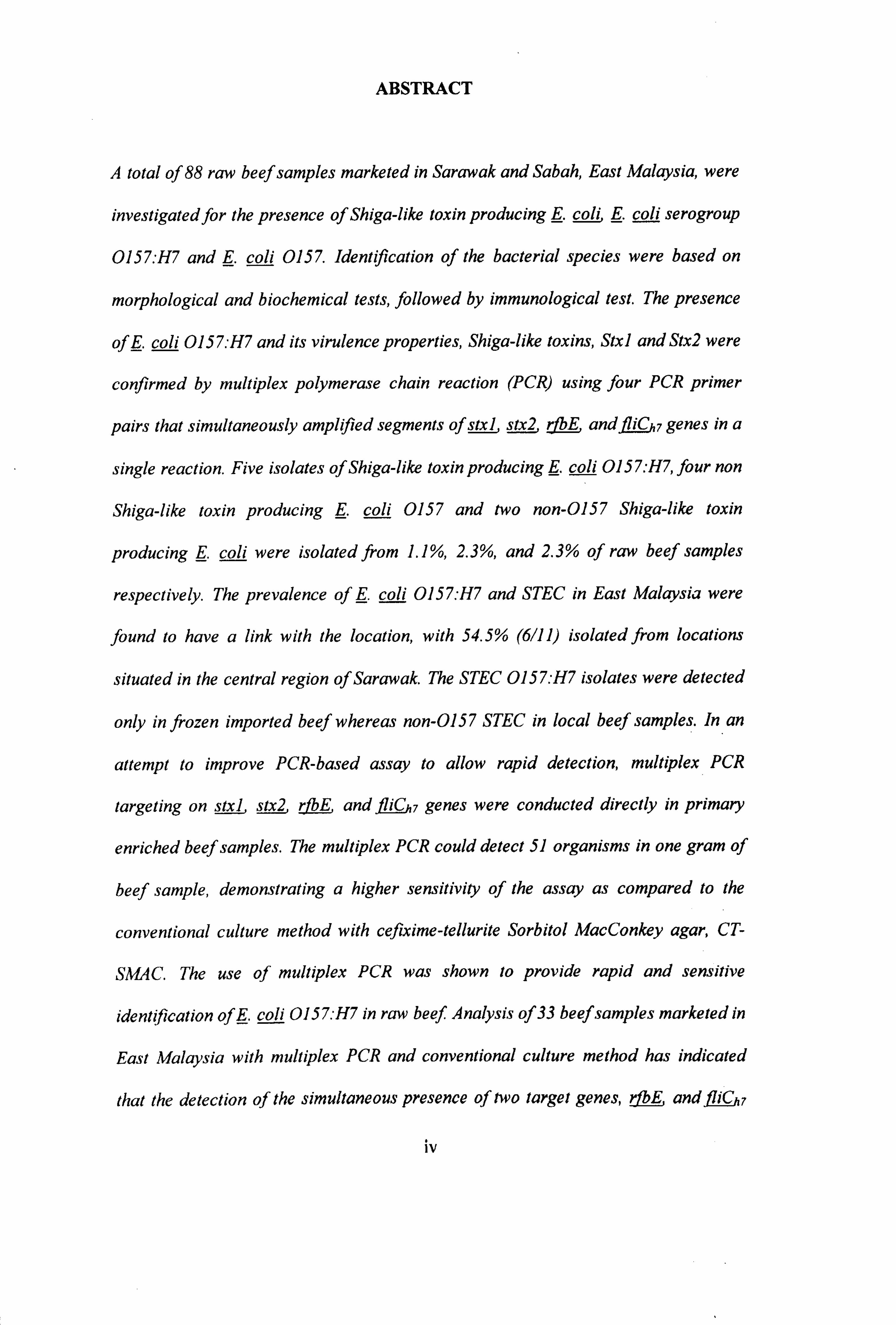
A total of 88 raw beef samples marketed in Sarawak and Sabah, East Malaysia, were
investigated for the presence of Shiga-like toxin producing coli, . coli serogroup
0157: H7 and E. coli 0157. Identification of the bacterial species were based on
morphological and biochemical tests, followed by immunological test. The presence
off. coli 0157. -H7 and its virulence properties, Shiga-like toxins, Stxl and Stx2 were
confirmed by multiplex polymerase chain reaction (PCR) using four PCR primer
pairs that simultaneously amplified segments of sal, stx2, rE and iC 7 genes in a
single reaction. Five isolates of Shiga-like toxin producing E. coli 0157: H7, four non
Shiga-like toxin producing E. coli 0157 and two non-0157 Shiga-like toxin
producing E. coli were isolated from 1.1%, 2.3%, and 2.3% of raw beef samples
respectively. The prevalence of E. coli 0157: H7 and STEC in East Malaysia were
found to have a link with the location, with 54.5% (6/11) isolated from locations
situated in the central region of Sarawak. The STEC 0157: H7 isolates were detected
only in frozen imported beef whereas non-0157 STEC in local beef samples. In an
attempt to improve PCR-based assay to allow rapid detection, multiplex PCR
fl- iC K genes were conducted directly in primary targeting on stxl, stx2, rE and
enriched beef samples. The multiplex PCR could detect 51 organisms in one gram of
beef sample, demonstrating a higher sensitivity of the assay as compared to the
conventional culture method with cefixime-tellurite Sorbitol MacConkey agar, CT-
SMAC. The use of multiplex PCR was shown to provide rapid and sensitive
identification of E. coli 0157. H7 in raw beef. Analysis of 33 beef samples marketed in
East Malaysia with multiplex PCR and conventional culture method has indicated
that the detection of the simultaneous presence of two target genes, rE and iC 7
iv

genes was sufficient for a 0157: H7 positive diagnosis in a beef sample. The inclusion
of primer sets for stxl and stx2 genes in the same reaction provided additional
information on the toxin profiles of a sample and were useful for detecting STEC
0157: H7 and other STEC. Pulsed-field gel electrophoresis (PFGE) with restriction
enzyme, Xbal was used to assess the genetic relatedness of the isolated E. coli
0157: H7, E. coli 0157, non-0157 STEC and other E. coli strains. The PFGE profiles
indicated that five E. coli 0157: H7 and three E. coli 0157 isolates presumably
represented a single strain of E. toll 0157: H7 and E. coli 0157 respectively.
Identical PFGE profiles found in the same E. coli serotype of the same beef sample
indicated that each individual beef marketed in East Malaysia harboured only one
type of STEC 0157: H7 or E. coli 0157. A large variety of PFGE patterns (45
patterns) were found among E. coli isolates demonstrating a high E. coli diversity in
the beef marketed in East Malaysia. From the dendrogram generated, there was no
close relation observed between STEC 0157: H7 and non-0157 STEC to the non-
pathogenic E. coli strains. In this study, PFGE typing method was shown to possess
high discriminatory power and proved its usefulness in differentiating among E. coli
0157: H7 and STEC. The PFGE profiles obtained from the E. coli beef isolates could
provide a database that would aid in the epidemiological investigation of the study
area in future.
V
ABSTRAK
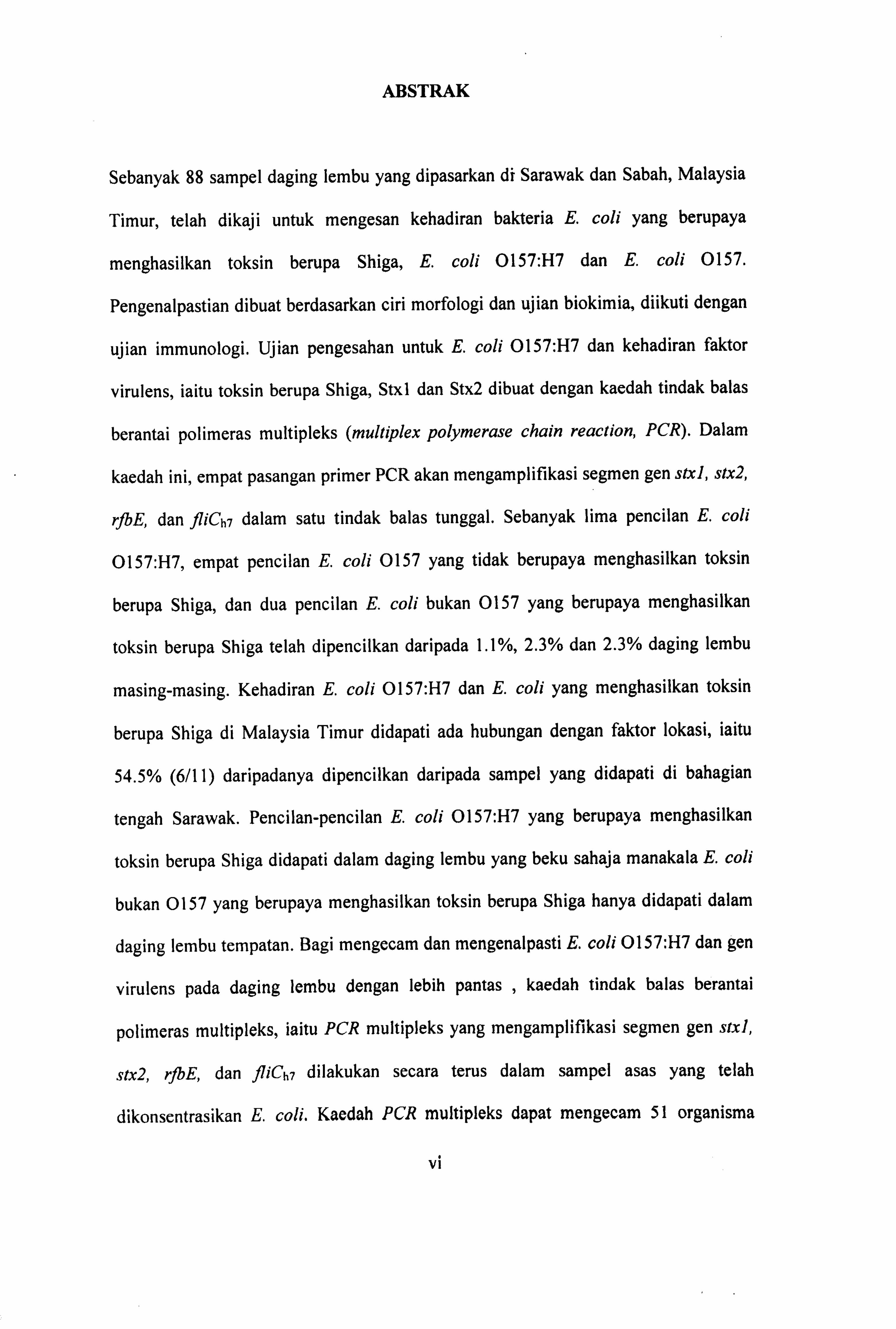
Sebanyak 88 sampel daging lembu yang dipasarkan di Sarawak dan Sabah, Malaysia
Timur, telah dikaji untuk mengesan kehadiran bakteria E. coli yang berupaya
menghasilkan toksin berupa Shiga, E. coli 0157: H7 dan E. coli 0157.
Pengenalpastian dibuat berdasarkan ciri morfologi dan ujian biokimia, diikuti dengan
ujian immunologi. Ujian pengesahan untuk E. coli 0157: H7 dan kehadiran faktor
virulens, iaitu toksin berupa Shiga, Stxl dan Stx2 dibuat dengan kaedah tindak balas
berantai polimeras multipleks (multiplex polymerase chain reaction, PCR). Dalam
kaedah ini, empat pasangan primer PCR akan mengamplifikasi segmen gen stxl, stx2,
rfbE, dan fliCh7 dalam satu tindak balas tunggal. Sebanyak lima pencilan E. coli
0157: H7, empat pencilan E. coli 0157 yang tidak berupaya menghasilkan toksin
berupa Shiga, dan dua pencilan E. coli bukan 0157 yang berupaya menghasilkan
toksin berupa Shiga telah dipencilkan daripada 1.1%, 2.3% dan 2.3% daging lembu
masing-masing. Kehadiran E. coli 0157: H7 dan E. coli yang menghasilkan toksin
berupa Shiga di Malaysia Timur didapati ada hubungan dengan faktor lokasi, iaitu
54.5% (6/11) daripadanya dipencilkan daripada sampel yang didapati di bahagian
tengah Sarawak. Pencilan-pencilan E. coli 0157: H7 yang berupaya menghasilkan
toksin berupa Shiga didapati dalam daging lembu yang beku sahaja manakala E. coli
bukan 0157 yang berupaya menghasilkan toksin berupa Shiga hanya didapati dalam
daging lembu tempatan. Bagi mengecam dan mengenalpasti E. coli 0157: H7 dan gen
virulens pada daging lembu dengan lebih pantas , kaedah tindak balas berantai
polimeras multipleks, iaitu PCR multipleks yang mengamplifikasi segmen gen stxl,
stx2, rfbE, dan fliCh7 dilakukan secara terus dalam sampel asas yang telah
dikonsentrasikan E. coli. Kaedah PCR multipleks dapat mengecam 51 organisma
V1

dalam satu gram daging lembu dan kebolehan ini juga menunjukkan bahawa kaedah
PCR multipleks mempunyai sensitiviti yang lebih tinggi berbanding dengan kaedah
kultur tradisional yang menggunakan agar cefixime-tellurite Sorbitol MacConkey, CT-
SMAC. Penggunaan kaedah PCR multipleks tertunjuk membolehkan pengenalpastian
E. coli 0157: H7 dalam daging lembu dilakukan dengan cepat dan sensitif. Analisis
menggunakan kaedah PCR multipleks dan kaedah kultur tradisional yang
menglibatkan 33 sampel daging lembu yang dipasarkan di Malaysia Timur ini telah
menunjukkan bahawa sistem PCR dengan kombinasi dua gen, rfbE, danfliCh7 sudah
memadai dalam pengecaman E. coli 0157: H7 dalam sampel. Penambahan gen
stxldan stx2 ke dalam sistem PCR ini bertujuan memberi informasi tambahan tentang
profil toksin sesuatu sampel dan adalah berguna untuk mengecam E. coli 0157: H7
dan E. coli yang berupaya menghasilkan toksin berupa Shiga. Dalam usaha untuk
mengenalpasti perhubungan genetik di antara pencilan, kaedah elektroforesis "gel
pulse field" (Pulsed-field gel electrophoresis, PFGE) yang menglibatkan enzim
pemotong, Xbal telah digunakan untuk mendapatkan kesamaan dan perbezaan. bagi E.
coli 0157: H7, E. coli 0157, E. coli bukan 0157 yang berupaya menghasilkan toksin
berupa Shiga dan E. coli lain yang telah dipencilkan. Profil PFGE yang diperolehi
menunjukkan bahawa lima pencilan E. coli 0157: H7 dan tiga pencilan E. coli 0157
masing-masing mewakili stren E. coli 0157: H7 dan E. coli 0157 yang tunggal. Profil
PFGE yang serupa hanya didapati pada serotaip E. coli yang sama yang dipencilkan
daripada sampel daging lembu yang sama. Ini menunjukkan bahawa daging lembu
yang dipasarkan di Malaysia Timur secara individunya merupakan habitat bagi satu
jenis E. coli 0157: H7 atau E. coil 0157. Pelbagai jenis profil PFGE (45 jenis) telah
diperolehi daripada pencilan-pencilan E. coli dan ini menunjukkan bahawa daging
lembu yang dipasarkan di Malaysia Timur mempunyai diversiti E. coli yang tinggi.
vii

Daripada dendrogram yang dihasilkan, tiada perhubungan rapat didapati di antara E.
coli 0157: H7 yang berupaya menghasilkan toksin berupa Shiga dan E. coli bukan
0157 yang menghasilkan toksin berupa Shiga dengan stren E. coli yang bukan
patogenik. Kaedah PFGE telah tertunjuk mempunyai kuasa diskriminasi yang tinggi
dan berguna dalam pembezaan di antara E. coli 0157: H7 dan E. coli yang
menghasilkan toksin berupa Shiga. Butiran data daripada profil PFGE yang diperolehi
daripada E. coli yang dipencil daripada daging lembu turut akan membantu dalam
kajian epidemiologi berkenaan kelak.

Pusat Khidmat Maklumat Akademik UNIVERSITI MALAYSIA SARAWAK
TABLE OF CONTENTS
Acknowledgements
Abstract
Abstrak
Table of Contents
List of Tables
List of Figures
List of Abbreviations
1. General Introduction
2. Literature Review
2.1 Escherichia coli
2.1.1 Characteristic
2.1.2 Taxonomy
2.1.3 Habitat
2.1.4 Growth
2.2 Shiga-like toxin producing Escherichia coli (STEC)
2.3 Escherichia coli 0157 and 0157: H7
2.3.1 Biochemical characteristic
2.3.2 The disease
2.3.3 Prevalence and incidence
2.3.4 Mode of transmission
2.3.5 Pathogenicity
2.3.5.1 Shiga-like toxins (Stx)
2.3.5.2 Attaching and effacing adherence
2.3.5.3 Pathogenesis
11
iv
V1
ix
xiv
xv
xvi
I
8
8
9
11
12
13
13
14
14
16
22
24
25
27
28
ix
2.4 Isolation of Escherichia coli 0157 and Escherichia coli 0157: H7 29
2.4.1 Enrichment media 30
2.4.2 Plating media 31
2.4.3 Incubation condition 34
2.5 Identification of Escherichia coli 0157 and Escherichia coli 0157: H7 34
2.6 Detection of Escherichia coli 0157 and Escherichia coli O157: H7 35
2.6.1 Immunological detection systems 35
2.6.2 Polymerase chain reaction (PCR)
2.6.2.1 Multiplex PCR
2.6.2.2 Direct multiplex PCR
2.7 Nucleic acid sequencing
2.8 Molecular typing: Pulsed-field gel electrophoresis
3. The occurrence of Shiga-like toxin producing Escherichia coli, Escherichia coli 0157: H7, and Escherichia coli 0157 in raw beef marketed in East Malaysia
38
39
43
43
46
3.1 Introduction 49
3.2 Materials and methods 51
3.2.1 Meat samples 51
3.2.2 Isolation of Shiga-like toxin producing E. coli (STEC) and E. 52 coli 0157: H7
3.2.3 DNA isolation 55
3.2.4 Primers
3.2.5 Multiplex polymerase chain reaction (PCR)
3.2.6 PCR optimization
3.2.7 Gel electrophoresis
3.2.8 Automated DNA sequencing
56
57
57
58
58
X
3.2.9 Data analysis 60
3.3 Results 60
3.3.1 Comparison of the methods used in the isolation of Shiga-like 60 toxin producing E. coli and E. coli 0 157: H7
3.3.2 The prevalence of E. coli based on location 61
3.3.3 Prevalence of Shiga-like toxin producing E. coli 0 157: H7, E. 67
coli 0157 and Shiga-like toxin E. coli (STEC)
3.3.4 Presence of E. coli 0157: H7,0157 and STEC based on the 69
country source of beef samples
3.3.5 Presence of E. coli 0 157: H7,0157 and STEC based on the 69
storage condition
3.3.6 Presence of E. coli 0157: H7,0157 and STEC based on the 70 different beef part
3.3.7 The specificity of the multiplex PCR assay 70
3.3.8 DNA sequencing 74
3.4 Discussion 80
3.5 Conclusion 91
4. Rapid detection and identification of Escherichia coli 0157: H7 and the virulence genes in raw beef marketed in East Malaysia by direct
multiplex PCR
4.1 Introduction 92
4.2 Materials and methods 94
4.2.1 Meat samples 94
4.2.2 Bacterial strain and culture condition 94
4.2.3 Seeding experiment 95
4.2.4 Culture preparation for detection of E. coli 0157: H7 in raw 96 beef samples
4.2.5 Conventional culture method used for comparison 96 4.2.6 Preparation of DNA template for multiplex PCR assay 97
4,23 Primers 99
X1

4.2.8 Multiplex PCR
4.2.9 Automated DNA sequencing
4.3 Results
99
100
102
4.3.1 The sensitivity of the direct multiplex PCR 102
4.3.2 Detection of virulence genes and identification of E. coli 104 0 157: H7 in raw beef
4.3.3 The effectiveness of boiling cell extraction method in the 106
preparation of template DNA
4.3.4 The specificity of direct multiplex PCR and the generated 110
products
4.4 Discussion 114
4.5 Conclusion 121
5. Molecular characterization of Shiga-like toxin producing Escherichla
coli and serogroup 0157: H7, Escherichia coli 0157, and other Escherichia coli strains isolated from raw beef by pulsed-field gel electrophoresis
5.1 Introduction
5.2 Materials and methods
5.2.1 Bacterial strains
5.2.2 Bacterial culture conditions
5.2.3 DNA preparation
122
124
124
124
125
5.2.4 Restriction endonuclease digestion 126
5.2.5 Running the pulsed-field gel electrophoresis (PFGE) 127
5.2.6 Data analysis 128
5.3 Result 130
5.3.1 Optimization of running condition and gel resolution 130
5.3.2 PFGE profiles 133
xu
5.4 Discussion
5.5 Conclusion
6. General Conclusion
7. References
8. Appendix
143
147
149
153
177
PUBLICATION/ CONFERENCE PROCEEDINGS
Apun, K., Chang, P. P., and Sim, E. U. H. (2003) Survey of raw beef marketed in East Malaysia for Escherichia coli 0157 and Escherichia coli 0157: H7. Journal of Bioscience (accepted for publication).
Apun, K. and Chang, P. P. (2003) Application of pulsed-field gel electrophoresis to characterize pathogenic and non-pathogenic Escherichia coli isolates obtained from beef marketed in East Malaysia. Presented at 3rd Federation of Asia Pacific Microbiology Societies Conference, Kuala Lumpur, October 2003.
Chang, P. P., Apun, K., and Sim, E. U. H. (2002) A multiplex polymerase chain reaction assay for identification of Escherichia coli 0157: H7. Proceedings NSF Seminar 2002, Kuala Lumpur, December 2002, pg. 46-49.
Chang, P. P. (2002) The occurrence of Escherichia coli 0157 in raw beef from local
and imported sources marketed in Sarawak. Proceedings of the Postgraduate Colloquium in Science and Technology 2002, Kuching, February 2002, pg. 9- 12.
xiii
LIST OF TABLES
TABLE 2.1 Clinical features of E. coli 0157: H7 infection 16
TABLE 3.1 Oligonucleotide primers used in this study 56
TABLE 3.2 Bacterial count (aerobic plate counts, total coliform counts and 62
presumptive E. coli counts) in fresh, frozen, and thawed samples purchased according to two bacterial isolation methods used
TABLE 3.3 Biochemical tests for the identification of E. coli 64
TABLE 3.4 E. coli 0157: H7, E. coli 0157 and non-0157 STEC isolated 68 from 88 raw beef samples marketed in East Malaysia
TABLE 3.5 The nucleotide sequence homology and the total nucleotides 76
sequenced (in percentage) of the PCR products obtained with primer pairs SLTIF-SLTIR, SLTIIF-SLTIIR, RfbF-RfbR, and FLICh7F-FLICh7R from the E. coli strains
TABLE 4.1 The evaluation of the sensitivity of multiplex PCR and culture 103
method with seeding experiments
TABLE 4.2 Detection of stxl, stx2, rfbE and/ orliCh, genes with multiplex 107 PCR in 33 raw beef samples collected in Sarawak and Sabah
TABLE 4.3 The detection of E. coli 0 157: H7 directly from enrichment 107
cultures with multiplex PCR assay and conventional culture method, and the identification of the specific genes according to location of beef purchased
TABLE 4.4 The detection of E. coli O157: H7 directly from enrichment 109
cultures with multiplex PCR assay and conventional culture method, and the identification of the specific genes according to the country source of beef samples
TABLE 4.5 The detection of E. coli O157: H7 directly from enrichment 109
cultures with multiplex PCR assay and conventional culture method, and the identification of the specific genes according to the types of retailers
TABLE 4.6 The nucleotide sequence homology and the total nucleotides 112
sequenced (in percentage) of the PCR products obtained with primer pairs SLTIF-SLTIR, SLTIIF-SLTIIR, RfbF-RfbR, and FLICh7F-FLICh7R
TABLE 5.1 The source and characteristics of E. coli isolates 135
TABLE 5.2 Distance matrix of 51 E. coli isolates, including 49 isolates from 139 beef samples marketed in East Malaysia
xiv

LIST OF FIGURES
FIG 2.1 The illustration of the polymerase chain reaction (PCR) process 41
FIG 2.2 The illustration of automated DNA sequencing using the 45 dideoxynucleotide triphosphate chain termination method
FIG 3.1 The biochemical tests for the identification of E. coli 64
FIG 3.2 The phenotypic characteristics on different selective agar of E. coli 65
FIG 3.3 The geographic distribution of typical and atypical E. coli isolates 66
which were categorized according to the ability to ferment sorbitol (SF) and exhibit ß-glucuronidase (GUD) activity
FIG 3.4 Amplicons obtained by multiplex PCR of the respective 12 E. coli 71
strains carrying virulence gene(s) were analyzed by agarose (1.2%)
gel electrophoresis
FIG 3.5 The amplified products of the independent rfbE- andliCh7-specific 73 PCR assay and duplex PCR of both for KKW1,3,5 and KCD8 isolates
FIG 3.6 Multiple sequence alignment of fliCh7 gene of beef isolates BTC 11,77 BTC20, and KKA7 with respect to reference strain EDL933
FIG 3.7 Multiple sequence alignment of stx2 gene of beef isolates BTC 11, BTC20, and KKA7 with respect to reference strain EDL933
78
FIG 3.8 PHYLIP rooted tree-a Phenogram of the stx2 genes as determined by DRAWGRAM
79
FIG 4.1 A representative gel for seven combinations of genetic profiles 108
obtained from 33 beef samples
FIG 4.2 An example of a sequencing result analyzed with the blastn 113
program
FIG 5.1 PFGE of chromosomal DNA digested with XbaI of E. coli isolates 131
with 1 program block and initial parameters
FIG 5.2 PFGE of chromosomal DNA digested with Xbal of E. coli isolates 132
with two program blocks: Comparison of the gel resolution before
and after optimization
FIG 5.3 PFGE of chromosomal DNA digested with XbaI of 51 E. coli 134 isolates with two program blocks
FIG 5.4 Dendrogram of E. coli 0157: H7, E. coli 0157, STEC, and E. coli 138
strains isolated in beef marketed in East Malaysia
xv
LIST OF ABBREVIATIONS
A BLAST BCIG bp C cm CDC cfu CTAB CT-SMAC DNA EHEC dNTPs ELISA EMB ERIC-PCR
g G GUD HC HUS IMS IMViC kb LB LPS mEC+n MgC12 M Mda Mb mg min ml mm µm µg µl mol sec
NaCI Na-EDTA NCBI PATS PCI PCR PFGE
adenine Basic Local Alignment Search Tool chromogen 5-bromo-4-chloro-3-indolyl-b-D-glucuronide base pairs cytosine centimeter Centers for Disease Control and Prevention colony forming unit cetyltrimethylammonium bromide cefixime-tellurite Sorbitol MacConkey agar deoxyribonucleic acid Enterohemorrhagic Escherichia coli deoxynucleotide triphosphates enzyme-linked immunosorbent assay Eosin Methylene Blue agar Enterobacterial repetitive intergenic consensus sequence-based polymerase chain reaction gram guanine 13-glucuronidase activity Hemorrhagic colitis Hemolytic-uremic syndrome Immunomagnetic separation Indole-Methyl red-Voges-Proskauer- Citrate tests kilobase pairs Luria Bertani lipopolyssaccharide modified Escherichia coli medium with novobiocin magnesium chloride Molar or molarity (moles of solute per liter of solution) megadalton megabase pairs milligram minute(s) milliliter milliMolar micrometer microgram microliter mole second(s) sodium chloride sodium ethylenediamine tetra-acetic acid National Center for Biotechnology Information Polymorphic amplified typing sequences phenol/chloroform/isoamyl alcohol Polymerase Chain Reaction Pulsed-Field Gel Electrophoresis
xvi
psi pound(s) per square inch (Ib/in2) RAPD Random Amplification of Polymorphic DNA rpm revolution per minute SDS sodium dodecyl sulfate SF sorbitol-fermenting/fermenter SNF non-sorbitol-fermenting/fermenter SMAC Sorbitol MacConkey agar SMAC-BCIG Sorbitol MacConkey agar supplemented with chromogen 5-bromo-4-
chloro-3-indolyl-b-D-glucuronide sp. species STEC Shiga-like toxin producing Escherichia coli Stx Shiga-like toxin T thymidine Taq Thermus aquaticus DNA polymerase TAE Tris-acetate electrophoresis buffer TBE Tris-Borate EDTA electrophoresis buffer TE Tris-EDTA buffer Tris Tris (hydroxymethyl) methylamine TTP Thrombotic thrombocytopenic purpura USA United States of America USDA United States Department of Agriculture USFDA United States Food and Drug Administration VT Verotoxin V volts % percent > more than < less and the same as °C degree Celsius
xvii
CHAPTER 1
GENERAL INTRODUCTION
Escherichia coli is a common commensal of the human gastrointestinal tract.
However, under certain conditions strains of E. coli can cause disease. Shiga-like
toxin-producing E. coli (STEC) has been implicated as the causative agent in several
human diseases (Nataro and Kaper 1998, Paton and Paton 1998a), with E. coli
0157: H7 to be the most well known among them. These diseases range from mild
diarrhea to severe and life threatening conditions, such as hemolytic-uremic syndrome
(HUS) and thrombotic thrombocytopenic purpura (TTP). Cattle are generally
considered to be the major reservoir of both E. coli 0157 and non-0157 STEC
(Bettelheim 2000). The organisms can be transmitted efficiently via contaminated,
undercooked food of animal origin, cross-contaminated food (Griffin 1995; Smith
1997), water (Olsen et al. 2002) and from human to human (Griffin 1995). The first
recorded outbreak associated with HUS in 1982 was attributed to E. coli O157: H7
(Riley et al. 1983), a serotype that still accounts for the greatest proportion of STEC
disease in North America, Europe and Japan. However, non-0157 STEC strains may
account for 20 to 70 percent of STEC infections overall in different countries (World
Health Organization 1999).
E. coli 0157: H7 and non-0157 STEC produce toxins, verotoxin 1 or Shiga-like
toxin (Stxl) and verotoxin 2 or Stx2. According to O'Brien and Holmes (1987), the
Stxs are cytotoxic for some cell lines, enterotoxic and paralytic-lethal when injected
I
intravenously in mice and rabbits. In addition, these Shiga-like toxins are reported to
be responsible in part for hemorrhagic colitis (HC) and hemolytic-uremic syndrome,
HUS (Griffin 1995).
STEC and E. coli 0157: H7 possess very low infectious dose, as few as 10
viable bacteria can initiate the pathogenesis of disease (Willshaw et al. 1994). Despite
the very low infectious dose, as well as the high pathogenicity of E. coli 0 15 7: H7 and
STEC, there is no current available specific treatment for the disease HUS. Hence,
there is an urgent need for effective preventive measures based on a detailed
understanding of the epidemiology of STEC infections. Such measures will also
depend on the availability of rapid, sensitive, simple and reproducible procedures for
the detection of these pathogens and for the characterization among the pathogens and
those of non-pathogenic E. coli strains. Consequently, attempts to develop suitable
and sensitive methods are initiated and the development has progressed rapidly for
better detection. Initial and traditional methods for the detection and identification of
STEC 0157 and other serogroups in samples involve enrichment cultures, selection of
bacterial colonies, biochemical analysis of the isolates and determination of the main
virulence markers, such as Shiga-like toxins (Tarr 1995, Clifton-Hadley 2000). This
approach is laborious, time-consuming and insufficiently sensitive in identifying
STEC organisms. To improve the sensitivity and reduce the total time involved in
detection, commercial rapid ELISA procedures have been introduced (Padhye and
Doyle 1991). With the advent of molecular biology techniques, genetically based
assay has been developed for the detection improvement of pathogenic E. coli and E.
2
coli 0157: H7. The available methods include DNA hybridization (Feng 1993) and
PCR-based assay (Hu et al. 1999).
A number of PCR-based assays have been developed for the detection of E. coli
0157 and other serogroups (Meng et al. 1997, Kumar et al. 2001, Toma et al. 2003).
Although significant developments have been made in the molecular diagnosis of
STEC bacteria, there is still a need to improve PCR-based assays so that specific and
direct identification of the important virulence factors and genotypic identification
factors can be made in a single reaction. Multiplex PCR is a PCR approach that uses
two or more primer sets to simultaneously amplify multiple target sequences in a
single reaction. Hence, the use of multiplex PCR has been widely studied with a
varying combination of target gene sequences in order to improve the sensitivity of
the method (Meng et al. 1997, Nagano et al. 1998). Most of the tests are based on the
identification of stx (Shiga-like toxin) and eaeA (intimin) gene sequences (Meng et al.
1997, Fagan et al. 1999). Currently, the use of multiplex PCR to amplify the presence
of the target genes in the primary sample, rather than in the broth culture of pure
colony has introduced a direct approach for detection and identification STEC (Osek
2002, Hu et al. 1999). This direct multiplex PCR approach not only reduces the time
consumed, it also eliminates the necessity for strain isolation, thereby negating the
potential biases during selection of bacterial colonies, in particular the non-0157
STEC.
The detection and identification of pathogenic E. coli and E. coli 0157: H7 are
very much of importance. Subserovar characterization of these pathogenic E. coli and
3
E. coli 0157: H7 is no less essential in order to investigate the genetic relationship
among pathogenic E. coli and E. coli 0157: H7, as well as to differentiate them from
non-pathogenic E. coli. Initial typing methods routinely used during outbreak
investigations include phage typing (Khakhria et al. 1990) and Shiga-like toxin typing
(Paros et al. 1993). In addition to that, plasmid profiling (Meng et al. 1995),
ribotyping (Apun et al. 1995), enterobacterial repetitive intergenic consensus (ERIC)
sequence-based PCR (ERIC-PCR) (Giammanco et al. 2002), randomly amplified
polymorphic DNA (RAPD) (Hopkins and Hilton 2000), pulsed-field gel
electrophoresis (Gautom 1997) and, recently, in a novel approach for differentiating
E. coli 0157: H7, polymorphic amplified typing sequences (PATS) (Kudva et al.
2002) have been used to characterize STEC E. coli and E. coli 0157: H7. Plasmid
profiling is a relatively simple and economical technique for subtyping, yet they are
not universally applicable to all bacteria as many bacterial species harbour plasmids
infrequently. Phage typing, Shiga-like toxin typing, and ribotyping have been reported
to be relatively lacking of discriminatory power (Saari et al. 2001, Martin et al. 1996).
However, they can exhibit superior discriminatory performance when combined with
other typing technique. Preston et al. (2000) reported that combination of phage typing
and PFGE fingerprinting could provide optimal discrimination for E. coli 0157
subtyping. Likewise, combined use of PFGE and ribotyping in the characterization of
E. coli 0157 from animal origin food, retail meats and cases of human disease has
demonstrated superior discriminatory performance (Avery et al. 2002). The PCR-
based assay, ERIC-PCR has been successfully used in differentiating laboratory
strains of E. coli by Versalovic et al. (1991) and suggested its potential for subtyping
gram-negative enteric bacteria. In contrast, Giammanco et al. (2002) reported that
4
Pusat Kliidmat Maklumat Akademik UMVEFLSITI MALAYSIA SARAWAK
ERIC-PCR was unable to distinguish epidemiologically unrelated strains of E. coli
0157: H7.
Another subtyping method, PFGE, was widely used for bacterial
characterization. PFGE has been described as the "gold standard" of genetic
fingerprinting methods for E. coli 0157 with high discriminatory performance and
reproducible result (Preston et al. 2000). An alternative to PFGE for E. coli 0157
subtyping is RAPD analysis and it was shown to be more discriminatory than phage
typing (Grif et al. 1998), ribotying and serotyping (Kärkkitinen et al. 1996).
Nevertheless, unlike PFGE, it often presents intra- and inter-laboratory reproducibility
problems in particular when subtyping data are used to track strains over long periods
and in different laboratories. Polymorphic amplified typing sequences (PATS)
analysis is a very newly devised simple PCR-based strain typing technique for E. coli
0157. In a study of Kudve et al. (2002), PATS was able to type all the E. coli 0157
tested while PFGE was unable to type a few of the isolates. However, PFGE had
higher discriminatory power compared to PATS.
In recent years, PFGE has increasingly been used for the molecular subtyping of
a wide range of bacterial and fungal pathogens. For E. coli 0157: H7, the usefulness of
PFGE fingerprinting during outbreak investigations has been demonstrated by Barrett
et al. (1994) and Preston et al. (2000). In 1993, the Centers for Disease Control. and
Prevention (CDC) has applied PFGE to characterize clinical and food isolates of E.
coli 0157: H7 during a large outbreak of foodborne illness caused by E. coli 0157: H7
that occurred in the western United States of America. In addition, CDC has
5

standardized PFGE methods in order to allow isolates to be compared from different
parts of the country, enabling recognition of nationwide outbreaks attributable to a
common source of infection, particularly those in which cases are geographically
separated.
E. coli 0157: H7 is a newly emerged foodborne pathogen and has been
associated with human disease such as hemorrhagic colitis and hemolytic-uremic
syndrome (HUS). This important emerging pathogen, E. coli 0157, has been reported
in West Malaysia (Son et al. 1998a). However, there is no published report on the
occurrence of this pathogen in East Malaysia, either in food or in clinical samples. As
cattle is the main reservoir of E. coli 0157 and non-0157 STEC, it is necessary to
initiate a study on the occurrence of the E. coli 0157: H7 and STEC in beef marketed
in Sarawak and Sabah. The data generated would assist the relevant authorities
(Health and Agriculture sectors) in planning proper procedure for detection of these
bacteria in beef. Most laboratories in East Malaysia detect the pathogens with
conventional culture method, involving enrichment cultures, selection of bacterial
colonies, biochemical analysis of the isolates and determination of the main virulence
markers. This is laborious and time consuming. Therefore, a rapid and reproducible
method for the detection and identification of E. coli 0157: H7 is needed for the
diagnosis and detection of this pathogen in the laboratories in East Malaysia. The
characterization of E. coli 0157: H7 and STEC detected are as vital. The data on the
genetic relatedness or divergence among the E. coli 0157: H7 and STEC can be used
in studying possible clonal relationship among strains. Hence, under the condition
whereby any outbreak occurs, investigation and determination of foodborne illness
6