detection, isolation, and characterization of avian
TRANSCRIPT
Detection, isolation, and characterization of avian influenza viruses from waterfowl
in wildlife refuges in the southeastern United States (2006-2011)
by
Teresa Villan Dormitorio
A dissertation submitted to the Graduate Faculty of
Auburn University
in partial fulfillment of the
requirements for the Degree of
Doctor of Philosophy
Auburn, Alabama
December 12, 2015
Keywords: avian influenza virus, wild waterfowl, real-time reverse transcriptase PCR
(RRT-PCR), antigen capture-ELISA, phylogeny, composting
Copyright 2015 by Teresa Villan Dormitorio
Approved by
Joseph J. Giambrone, Chair, Professor of Poultry Science
Gary R. Hepp, Professor Emeritus of Forestry and Wildlife Sciences
Kenneth S. Macklin, Professor and Extension Specialist of Poultry Science
Hongzhuan Wu, Associate Professor at Alabama State University
ii
ABSTRACT
Avian influenza (AI) is an infectious disease of birds caused by type A influenza
viruses. These AI viruses (AIV) commonly infect poultry and wild birds; however, some
AIV strains have infected and caused mortality in a variety of mammals including
humans. Direct transmission of AIV from poultry to man have been demonstrated, and
human influenza pandemic viruses have been reported to contain two or more novel
genes that were very similar to those found in wild birds.
Wild aquatic birds represent major natural reservoirs of influenza A viruses and have
been implicated as a continuous source of virus for domestic birds and other animal
species including humans. All of the known hemagglutinin (H1-H16) and neuraminidase
(N1-N9) influenza subtypes have been detected in wild waterfowl. While the virus does
not usually cause clinical disease in these birds, severe illness may occur when the virus
crosses the species border to poultry.
In this study, cloacal swabs were collected from hunter-killed or nesting waterfowl
from wildlife refuges in Alabama, Georgia, and Florida. Out of 1260 swab samples
inoculated into embryonated eggs, 64 allantoic fluids (AF) agglutinated red blood cells
and 29 were RRT-PCR positive for the matrix gene of AIV. Nineteen AIV and 3 avian
paramyxovirus subtypes were identified. No H5 or H7 isolates were found. RRT-PCR
was found to be more sensitive and specific than AC-ELISA, since it detected AIV from
AF with a hemagglutination titer as low as 4. Phylogenetic analysis of the H gene
sequence of an Alabama H10N7 isolate showed close similarity (98%) to more recent
iii
isolates, but only 90% related to an H10N7 isolated 38 years ago. Sequencing analysis of
the H gene of four H1N1 isolates revealed that they were 92-97% similar to previously
published H1N1 isolates including one, which came from swine. Experimental studies
showed that an H1N1 isolate in embryonated eggs was eliminated within 24 hours of
litter composting when the temperature reached 660C. Continuous surveillance and
characterization of AIVs in wild birds will help in the understanding of the origin,
evolution, transmission and control of present and future influenza outbreaks.
4
ACKNOWLEDGMENTS
The author expresses sincere appreciation and gratefulness to Dr. Joseph J.
Giambrone, her mentor all throughout her career and professional life since coming to the
United States in the late 1990, for his never-ending support, guidance, patience, trust, and
friendship. To Dr. Gary Hepp she wants to acknowledge with profound gratitude his help
in getting her research project started; it was he who collected the first samples from
resident wood ducks of Lake Eufaula, and provided names of persons to contact
regarding sampling of waterfowl from the different refuges in Alabama, Georgia, and
Florida. The author is very thankful to Dr. Ken Macklin for his valuable comments,
questions, and corrections to the dissertation. She would also like to thank Dr.
Hongzhuan Wu, for his bright suggestions, explanations, and advice regarding molecular
techniques and virology. Special thanks go to Dr. Bernhard Kaltenboeck who willingly
accepted my request for him to be my University Reader.
The author is forever grateful to Dr. Kejun Guo, who played a big part during the
conduct of this research, thus he was co-author of two publications that came out of this
work. He went with me or Dr. Giambrone in almost all the field sampling trips. He was
my biggest help in the laboratory: inoculating eggs; virus detection tests; molecular
cloning; statistical and phylogenetic analyses; and anything computer-related. The author
gives special thanks to Dr. Shan-chia Ou, Leah Donahue, Caroline Benjamin, and
Puttitorn Saengrungruang for their help in the field or in the lab.
Not the least, the author wants to express her deepest gratitude to her beloved family
and friends, near and far, who in their own countless ways, have given their

5
unconditional love, support, encouragement, respect, faith, and trust. This work is
dedicated to her late father, Agustin D. Dormitorio who was her most ardent supporter
and avid fan. Above all, the author gives thanks to God for all the blessings that made
things possible.
6
TABLE OF CONTENTS
ABSTRACT .................................................................................................................................. ii
ACKNOWLEDGMENTS ............................................................................................................ iv
LIST OF TABLES ..................................................................................................................... viii
LIST OF FIGURES ..................................................................................................................... ix
LIST OF ABBREVIATIONS ....................................................................................................... x
CHAPTER I: GENERAL INTRODUCTION .............................................................................. 1
CHAPTER II: LITERATURE REVIEW ..................................................................................... 9
2.1 The history of influenza A viruses ............................................................................ 9
2.2 The biology of avian influenza virus ...................................................................... 12
2.3 The natural hosts of avian influenza virus .............................................................. 14
2.4 Avian influenza virus testing and diagnostics ......................................................... 15
2.5 Avian influenza spread and transmission ................................................................ 17
2.5 Avian influenza prevention and control .................................................................. 18
2.6 References ............................................................................................................... 20
CHAPTER III: EVALUATION OF FIELD AND LABORATORY PROTOCOLS USED
TO DETECT AVIAN INFLUENZA VIRUSES IN WILD AQUATIC BIRDS ........................ 25
3.1 Abstract ................................................................................................................... 25
3.2 Introduction ............................................................................................................. 26
3.3 Materials and Methods ............................................................................................ 29
3.4 Results and Discussion ............................................................................................ 33

7
3.5 Acknowledgements ................................................................................................. 38
3.6 References ............................................................................................................... 39
CHAPTER IV: DETECTION AND CHARACTERIZATION OF AVIAN INFLUENZA
AND OTHER AVIAN PARAMYXOVIRUSES FROM WILD WATERFOWL IN
PARTS OF THE SOUTHEASTERN UNITED STATES .......................................................... 48
4.1 Abstract ................................................................................................................... 48
4.2 Introduction ............................................................................................................. 49
4.3 Materials and Methods ............................................................................................ 50
4.4 Results and Discussion ............................................................................................ 52
4.5 Acknowledgements ................................................................................................. 54
4.6 References ............................................................................................................... 55
CHAPTER V: LITTER COMPOSTING PROCEDURES FOR INACTIVATION OF
AVIAN INFLUENZA VIRUS IN EGGS ................................................................................... 62
5.1 Abstract ................................................................................................................... 62
5.2 Introduction ............................................................................................................. 63
5.3 Materials and Methods ............................................................................................ 64
5.4 Results and Discussion ............................................................................................ 66
5.6 References ............................................................................................................... 68
DISSERTATION CONCLUSIONS ........................................................................................... 73
APPENDIX ................................................................................................................................. 77
8
LIST OF TABLES
Table 3.1 Detection and isolation of avian influenza virus (AIV) and avian
paramyxovirus (APMV) from cloacal swabs of hunter-killed wild birds ............... 44
Table 3.2 Effect of egg passage (P1 vs. P2) on hemagglutination (HA) titer, antigen
capture-ELISA (AC-ELISA), and real-time reverse transcription-PCR (RRT-
PCR; matrix gene) detection assays on allantoic fluid of swab inoculated
embryonated eggs .................................................................................................... 46
Table 4.1 Hemagglutination (HA) titer, Directigen, real-time reverse transcription-PCR
(RRT-PCR; matrix gene) assays, hemagglutination inhibition (HI) testing,
and subtyping of viruses isolated from 2006 to 2007 .............................................. 58
Table 4.2 Hemagglutination (HA) titer, Directigen, real-time reverse transcription-PCR
(RRT-PCR; matrix gene) assays, hemagglutination inhibition (HI) testing,
and subtyping of viruses isolated from 2007 to 2008 .............................................. 59
9
LIST OF FIGURES
Figure 3.1 Flowchart of experimental procedures used to detect avian influenza viruses
(AIV) in wild aquatic birds…………………………………….………………… 47
Figure 4.1 Phylogenetic analysis of the hemagglutinin (H) gene of A/Northern
Shoveler/AL/26/2006 in comparison with other published H10N7 isolates ......... 60
Figure 4.2 Sequence comparison of the complete hemagglutinin (H) gene of 4 Alabama
isolates with those of previously published avian influenza virus isolates ............ 61
Figure 5.1 Compost layout of Trial 2 ....................................................................................... 70
Figure 5.2 The rate (%) of survival of AIV in composted or non-composted eggs ................. 71
Figure 5.3 Temperature (0C) readings from probes placed at different sites of the compost pile
and composting room for Trial 1 (T1) and Trial 2 (T2) ..…….…………………..72

10
LIST OF ABBREVIATIONS
AIV Avian influenza virus
APMV Avian paramyxovirus
CRBC Chicken red blood cells
NDV Newcastle Disease Virus
NVSL National Veterinary Services Laboratories
AC-ELISA Antigen Capture-Enzyme Linked Immunosorbent Assay
RRT-PCR Real-time Reverse Transcription-Polymerase Chain Reaction
HPAI High Pathogenic Avian Influenza
LPAI Low Pathogenic Avian Influenza
USDA United States Department of Agriculture
USAID United States Agency for International Development
1
CHAPTER I: GENERAL INTRODUCTION
Avian influenza viruses (AIVs) can affect domestic poultry, as well as exotic pet
birds, and wild migratory birds. AIVs have also been isolated, although less frequently,
from mammalian species including rats, mice, weasels, ferrets, pigs, cats, tigers, dogs,
horses, as well as humans.
Wild aquatic birds are the natural reservoir of AIVs. Ecological studies have
established them as a primary source of virus for domestic poultry as well as other animal
species including humans (Slemons et al., 1974; Webster et al., 1992). From this
reservoir, viruses are sometimes transmitted to other host species and can continue to
spread and sporadically cause mortality. Avian influenza viruses (AIVs) from wild birds
usually cause no disease; however, some subtypes (H5 and H7) may undergo mutation
and become highly pathogenic, especially in more susceptible domestic species (Capua
and Alexander, 2009). All of the known 16 hemagglutinin (H) and 9 neuraminidase (N)
AIV subtypes, and most H-N combinations have been detected in wild birds,
predominantly ducks, geese, and shorebirds (Fouchier et al., 2005). AIVs are known to
replicate in the intestinal tracts of ducks and contaminate water habitat through shedding
of virus in feces. This process plays an important role in spreading AIVs. Occasionally,
AIVs are transmitted directly to man and one recent example is the H5N1 virus isolated
in 1997 from patients in Hong Kong. The same virus caused an outbreak on Hong Kong’s
chicken farms and resulted in a high mortality of birds. In an attempt to eradicate the
disease, all of Hong Kong’s poultry (approx.1.5 million birds) were slaughtered, which

2
may well have prevented adaptation of the avian H5N1 virus to man and, cause a
subsequent human pandemic.
Because most wild bird species considered to be AIV reservoir are migratory, they
are suspected of having a role in the spread of AIV. With the recognition of the human
health implications due to AI, especially as a result of the spread of Asian H5N1 virus, AI
infections have attracted the attention of veterinary and human scientists. Moreover,
influenza A viruses of the H1N1 subtype have caused substantial morbidity and mortality
in humans such as the pandemics of 1918 and 2009. Thus, there has been increased virus
surveillance and establishment of AIV databases around the world. There is a global
effort to collect and characterize AIV to identify genetic mutations and antigenic
properties, which are useful in developing vaccines and controlling influenza.
Influenza viruses are inherently unstable. As these RNA viruses lack a genetic proof-
reading mechanism, small errors that occur when the virus replicates, go undetected and
uncorrected. Specific mutations and evolution in influenza viruses cannot be predicted,
making it difficult if not impossible to know if or when a virus might acquire the
properties needed to spread easily and infect humans. This difficulty is increased by the
present lack of understanding concerning specific mutations, which may lead to increased
transmissibility of the virus among humans. If and when, such a mutation occurs, there
will be little warning when AI becomes a major threat to animals and humanity.
Therefore, it is essential to prepare before a disaster occurs, as the time scale for the start
of a pandemic disease cannot be predicted.

3
Since 1977, when the first human infections with the H5N1 (also known as bird flu)
were documented, the virus has undergone changes. These changes have affected patterns
of virus transmission and spread among domestic and wild birds. Studies have shown that
H5N1 viruses from current outbreaks, when compared with viruses from 1997 and 2003,
have become more lethal in experimentally infected chickens and mice, and are also
hardier, surviving several days longer in the environment (WHO, 2006; Stallknecht et al.,
2010). Other studies have shown that the virus is not yet fully adapted to poultry and
continues to evolve (Webster and Govorkova, 2006; WHO, 2006).
The H5N1 outbreak has since spread to 16 countries in Asia, Africa, the Pacific Rim,
Europe, Canada, and the Middle East and had resulted in the death and slaughtering of
billions of poultry. People from these areas have also become ill with these avian viruses,
in which more than half of those infected died. As of July 17, 2015, the cumulative
number of confirmed human cases of H5N1infection reported to WHO is 844 with 449
deaths (53%) (WHO, 2015). The majority of these infections and deaths have occurred
in rural areas of developing countries. It could well be argued that, given a lack of
resources for diagnosis and reporting, an accurate figure would be somewhat higher.
Although WHO reports only laboratory confirmed cases, it is evident from these numbers
that H5N1 is lethal to humans, but difficult to spread. Residing deep in airways, the virus
is not easily expelled by coughing and sneezing, the usual route of spread. Two research
groups reported results that help explain the difficulty of virus spread in humans. The
group of Yoshihiro Kawaoka of University of Wisconsin, Madison, tested various tissues
of the human respiratory tract for receptors to which the virus can bind. Human flu
preferentially bind to α2,6 galactose receptors that populate the human respiratory tract

4
from nose to lungs (Shinya et al., 2006). Avian viruses prefer α 2,3 galactose receptors
that are common in birds but thought to be absent in humans. Using marker molecules
that bind to one receptor or the other, the team found that humans also have α 2,3
galactose receptors, but only in and around the alveoli, structures deep in the lungs.
Another group from Rotterdam, Netherlands, led by Thijs Kuiken, used more direct
techniques to show that H5N1 readily binds to alveoli but not to tissues higher up in
respiratory tract (Kuiken, 2006). This pattern is consistent with autopsies that have
shown heavy damage to lungs but little involvement of upper respiratory tract. Findings
explain, at least in part, the localization and severity of H5N1 viral pneumonia in
humans.
Although more than half of H5N1 infected human beings have died, so far, human
infection is still considered rare. Close contact with infected poultry and other domestic
birds remains the most important source of human infections and to date, no human cases
have been linked to exposure to wild birds. The virus does not spread easily from birds to
humans or readily from person to person. Unfortunately, there are some indications that
such a pattern of sustained human-to-human transmission may be eminent. Recent
findings suggest that the 1918 “Spanish flu” pandemic may have resulted from a similar
interspecies transmission event in which a purely avian virus adapted directly to human-
to-human transmission without prior reassortment (Taubenberger, J., 2005).
Few scientists are prepared to say that H5N1 will not cause a human pandemic,
because ingredients for it to happen are evident. We are seeing in real time an
unprecedented evolution of a virus that has a pandemic potential. It is the first time in
history that such an epizootic occurred. Nevertheless, it is heartening to note that

5
scientists, government agencies and regulators, biologics and poultry industries all over
the world get together to attempt to deal with the problem. There is currently a “World
Health Organization Global Influenza Program Surveillance Network” with nearly 100
members and collaborating laboratories, comprised of AI experts worldwide. Daily
information and updates on research findings, reviews and commentaries indicate a
round-the-clock effort of everyone concerned. In the USA, millions of dollars have been
released for AI pandemic preparation. United States Agency for International
Development (USAID) and other private agencies have recruited AI experts to be
included in the database of volunteers, who may be called to help other countries if
needed. The USDA and other departments and agencies of the Federal Government have
taken steps to safeguard the country and its livestock populations from HPAI.
The spread of H5N1 across the globe highlights our vulnerability for the emergence
of novel subtypes of influenza virus. Yet despite our fears of pandemic human disease,
H5N1 is primarily a disease of birds. The establishment of regional sublineages suggests
that H5N1 virus is perpetuated in poultry largely through a lapse in biosecurity rather
than by continued reintroduction of viruses by migrating birds. A study has shown that
H5N1 virus has persisted in its birthplace, southern China, for almost 10 years and has
been repeatedly introduced into neighboring (e.g., Vietnam) and distant (e.g., Indonesia)
regions, establishing “colonies” of H5N1 virus throughout Asia that exacerbate the
pandemic threat. The best approach to avert the threat is to control H5N1 virus infection
at its source, domestic poultry. Sustained and aggressive efforts to control H5N1
circulation in commercial and backyard poultry are mandatory to avoid catastrophic
public health consequences.

6
New outbreaks of AI reported to the World Organization for Animal Health (OIE)
have decreased, as might be expected from the seasonal pattern of the disease, but new
cases of H5, H7, and H9 were reported during August 2015 in Africa, Asia, the Americas
and Europe. Since December 2014, the United States Department of Agriculture
(USDA) has confirmed 223 cases of highly pathogenic avian influenza (HPAI) in the
Pacific, Central, and Mississippi flyways (USDA-APHIS, 2015a). The HPAI virus
causing the US outbreaks is believed to have originated from a wild bird that was
infected with an HPAI H5N8 virus from Asia and a low pathogenic North American
strain of avian influenza at the same time. The viruses then recombined creating a new
mixed origin viruses containing the Asian-origin H5 part of the virus, which is highly
pathogenic to poultry (USDA, 2015). USDA has identified two mixed-origin viruses in
the Pacific flyway: the H5N2 virus and new H5N1 virus. The new H5N1 virus is not the
same virus as the H5N1 virus found in Asia that has caused human illness and spread
globally (USDA-APHIS, 2015b).
The current widespread disease in the USA has been found in wild birds, as well as
in a backyard and commercial poultry flocks, mostly in the Midwest. Since April, 2015,
the outbreak has not moved south, or spread to the Atlantic flyway, however, experts
predict the disease maybe in all four flyways this fall (USDA-APHIS, 2015a). As of
June 17, 2015, 223 cases infected chickens, turkeys or mixed poultry from 10
Midwestern states have been reported and confirmed with a total of 48,091,293 affected
birds (USDA-APHIS, 2015c). No new outbreaks have been reported since June. So far,
the cost of depopulation, clean up, and indemnification by USDA to farmers, and loss in
profits is about $3.3 billion (USDA-APHIS, 2015c). As a result of culling and mortality

7
due to infection, 10% of egg layers and 3.5% of turkeys in the Midwest were lost. In
addition, sixteen percent of US poultry exports were embargoed by other countries
(USDA-APHIS, 2015d).
The objectives of this research work were to detect, isolate, and characterize AIVs
from wild waterfowl in wildlife refuges of the southeastern USA (Alabama, Georgia, and
Florida). Virus isolation in embryonating eggs, AC-ELISA, and real-time PCR were used
then compared to determine the most rapid, sensitive and cost-effective method for
detection of AIV in wild birds. Subtypes of the AIV isolates were determined by the
National Veterinary Services Laboratories (Ames, Iowa). Gene segments of some H1N1
isolates were cloned or amplified and DNA sequences determined. Phylogenetic analyses
were conducted to determine genetic differences or similarity with other published
H1N1s, including those that caused the 2009 pandemic. In addition, experimental studies
were conducted to determine virus survival during composting.
AIVs undergo rapid and continuous mutation events as a natural consequence of
adaptation in poultry. The rate is slower in the natural hosts, namely wild waterfowl, but
even in these hosts evolution generates novel strains. The information generated here is
useful for scientists and will contribute to the understanding of the influenza virus.
Knowledge gained from these findings will be used for developing a more proactive and
effective intervention program for AI. These data will be integrated into existing poultry
health practices as well as state management systems, and will help poultry producers
prevent AIV’s entrance into their flocks. In addition, this project provided baseline data
in support of the USA Homeland Security efforts to assist in distinguishing between

8
naturally emerging and purposely introduced type A influenza viruses in poultry in the
southeastern USA.

9
CHAPTER II: REVIEW OF LITERATURE
2.1 The history of influenza A viruses
Avian influenza (AI) was first described when the deadly form of the disease caused
massive outbreaks in poultry in northern Italy in 1878 (Lupiani and Reddy, 2009). It was
originally termed “fowl plague”, and was confused with the acute septicemic form of
fowl cholera. In 1901, the causative agent of this disease was shown to be ultra-filterable
(i.e. virus) and, it was demonstrated in 1955 that there was a close relationship between
this agent and mammalian influenza viruses (Alexander and Brown, 2000).
Infections in susceptible avian species can be divided into two forms based on the
severity of the clinical disease produced. The very virulent viruses (formerly termed fowl
plague) now called highly pathogenic avian influenza (HPAI) may cause poultry
mortality as high as 100% (Alexander and Brown, 2000). These viruses have been
restricted to subtypes H5 and H7, although not all viruses of these subtypes cause HPAI.
For over 100 years, HPAI rarely occurred, which was often self-limiting or rapidly
controlled by slaughter (Alexander and Brown, 2000). However, on some occasions,
infection has become widespread in poultry and had severe implications for animal health
and economy of the country. For example, in the Pennsylvania, USA epidemic (1983-84),
more than 17,000,000 birds were slaughtered or died with an estimated cost to the US
government of $60,000,000, excluding cost borne by the farmers (Lasley, 1986).
The first HPAI outbreak affecting poultry in the USA occurred in 1924-25. In 1975,
outbreaks in chicken flocks caused by low pathogenic (LP) H4N8 were reported in

10
Alabama, with 69% mortality, including 31% mortality in a single day in one flock
(Johnson et al., 1977). In 1995, 178 turkey farms in Minnesota were infected with H9N2,
resulting in the worst economic loss due to influenza recorded in a single year
(approximately $6,000,000) (Halvorson et al., 1998).
Outbreaks of HPAI in the last 50 years have been relatively uncommon around the
world and have had limited spread within a country or region with one major exception,
Asian lineage H5N1 that was first identified in 1996 (Suarez, 2010). This lineage of virus
has spread to over 60 countries and has become endemic in poultry in at least four
countries. The highly pathogenic strain of the H5N1 was first isolated and characterized
in a domestic goose in the southern Guangdong province of China in 1996 (Whitworth et
al., 2007). In the following year, the first H5N1 outbreak occurred in domestic poultry in
Hong Kong, resulting in the culling of over 1.5 million chickens in an effort to contain
and eliminate the disease. This outbreak also led to the infection of 18 people (with 6
fatalities) in what would be the first documented human death from the H5N1 virus. In
the majority of cases, people obtained HPAI Asian H5N1 virus infection after direct or
close contact with infected or dead poultry. Currently, HP Asian H5N1 virus does not
transmit efficiently from person to person. Some cases of limited, non-sustained human-
to-human transmission have likely occurred.
The ongoing outbreak of H5N1 among birds with occasional transmission to human
beings is of major concern because of intriguing parallels between the H5N1 virus and
the 1918 H1N1 influenza strain. This most famous and lethal pandemic named Spanish
flu lasted from 1918 to 1919, and spread across Europe, Asia, and North America
11
(Taubenberger and Morens, 2006). This pandemic has been described as “the greatest
medical holocaust in history”, and it is generally assumed that 50 million people died.
The unusually severe disease killed from 20 to 2% of those infected, who were mostly
young adults (20-40 yr old), and it is estimated that 2.5% to 5% of the world’s population
at that time was killed. The complete coding of the sequences of all 8 viral RNA
segments of the 1918 H1N1 virus was enabled with the recovery of the genomic RNA
from archived formalin-fixed autopsy material and from frozen tissue from an influenza
victim who was buried in permafrost in 1918 (Taubenberger, 2003). The 1918 virus
according to this investigation was not a reassortant virus like those of the 1957 and 1968
pandemics, but more likely an entirely avian-like virus that adapted to humans. Research
has shown that just 10 amino acid changes in the polymerase proteins differentiate the
1918 virus sequences from that of avian viruses, and that a number of the same changes
have been found in recently circulating , highly pathogenic H5N1 viruses (Taubenberger
et al., 2005). Later pandemic such as the 1957 Asian flu (H2N2 strain), the 1968 Hong
Kong flu (H3N2 strain), and 2009 Swine flu (H1N1 strain), were not as devastating
although it killed millions of people. In later pandemics antiviral drugs and antibiotics to
control secondary infections were available and so may have helped reduce mortality
compared to the 1918 Spanish flu.
The 2009 H1N1 pandemic is now known to involve a re-assorted virus produced
from 2 kinds of porcine influenza, one of which is itself already a “triple-re-assortant”
strain containing segments originating in human seasonal H3N2, AIV, and porcine
influenza virus. As of August 1, 2010, more than 214 countries and overseas territories or
communities worldwide have reported laboratory confirmed cases of pandemic influenza

12
H1N1 2009, including over 18,449 deaths (WHO, 2010). On August 10, 2010, the World
Health Organization (WHO) International Health Regulations (IHR) Emergency
Committee declared an end to the 2009 H1N1 pandemic globally. The U.S. Public Health
Emergency for 2009 H1N1 Influenza expired on June 23, 2010.
The most important thing to remember when talking about pandemic influenza is that
its severe form has little in common with seasonal influenza.
2.2 The biology of avian influenza virus
Influenza viruses are RNA viruses that make up 3 of the 5 genera of the family
Orthomyxoviridae. The viral particles are about 80-120 nm in diameter and can be
spherical or pleomorphic. There are 3 types of influenza viruses based on the molecular
nature of its internal proteins, namely A, B, and C. Influenza A viruses are the most
prevalent pathogens for both humans and animals. Only viruses of the influenza A genus
are known to infect birds, and have been shown to cause devastating infections in poultry,
which spread throughout the world. Influenza B viruses almost exclusively infect
humans, causing significant flu-like illnesses, and is less common than influenza A
viruses. B subtypes H3 and H2 are in human vaccines. Influenza C viruses infect humans,
dogs and pigs and sometimes causing mild respiratory illness and are not thought to cause
epidemics.
The influenza A viral genome consists of 8 segments of single negative strand RNA.
Each RNA segment contains either 1 or 2 genes which code for a gene product (protein).
Segments that codes for single gene products are: PB1, PB2, and PA proteins that
constitute the viral polymerase; the hemagglutinin (HA) and neuraminidase (NA)
13
glycoproteins; and the nucleoprotein (NP) (Steinhauer, 1999). The two glycoproteins
determine the subtypes of influenza A, and are associated with virulence, thus these
proteins are targets for antiviral drugs. The HA protein is the most abundant and is
responsible for binding of virus particles to sialic acid-containing cell surface receptors
and, for mediating fusion of the virus to host cell membranes after endocytosis
(Steinhauer, 1999). NA is involved in the release of new virions from infected host cells,
by cleaving sugars that bind mature viral particles. Antibody responses to these 2 proteins
form the basis for the division of influenza A viruses into their antigenic subtypes. To
date, 16 HA (H1-H16) and 9 NA (N1-N9) subtypes have been detected in birds and
mammals worldwide.
New epidemic influenza A strains arise every 1 to 2 years by the introduction of
selected point mutations within 2 surface glycoproteins: haemagglutinin (HA) and
neuraminidase (NA). These permanent and usually small changes in the antigenicity of
influenza A viruses are termed “antigenic drift” and are the basis for the occurrence of
influenza epidemics. In addition, there is now evidence that multiple lineages of the same
virus subtype can co-circulate, persist, and reassort in epidemiologically significant ways
(Holmes et al., 2005)
In contrast to epidemics, pandemics are rare events that occur every 10 to 50 years.
The 1918 pandemic was caused by a H1N1 virus of apparently avian origin ((Reid et al.,
1999), whereas the subsequent pandemic strains – H2N2 in 1957 and H3N2 in 1968 –
were reassortant viruses containing genes from avian viruses: 3 in 1957 (HA, NA, and the
RNA polymerase PB1) and 2 (HA and PB1) in 1968 (Kawaoka et al., 1989). These major
changes in the antigenicity of an influenza virus are called “antigenic shift”.
14
2.3 Natural hosts of avian influenza
Only viruses of the influenza A genus have been isolated from birds and termed
avian influenza viruses (AIVs), but all 16 hemagglutinin (H1-H16) and all 9
neuraminidase (N1-N9) subtypes, and most of the possible H-N combinations have been
detected or isolated from wild birds, particularly ducks, geese, and shorebirds (Fouchier
et al., 2005). Since wild birds are considered the natural reservoir of influenza A viruses,
they are thought to be the source of these viruses for all other animals. The highly
pathogenic avian influenza (HPAI) viruses, which are restricted to the H5 and H7, are
rarely isolated from wild birds. On the other hand, the low pathogenic (LPAI) viruses,
which causes mild respiratory disease have extremely high isolation rates of about 11%
for ducks and geese and around 2% for all other species (Alexander, 2007). LPAI viruses
have been isolated from at least 109 wild bird species with 1.6 to 10% prevalence in
ducks (Olsen et al., 2006).
AIVs from wild birds usually cause no disease; however, some subtypes (H5 and
H7) may undergo mutation and become highly pathogenic, especially in more susceptible
domestic species (Capua and Alexander, 2008). Influenza viruses that wild birds carry
have been implicated in major human pandemics, including the most devastating
“Spanish Flu” of 1918, which killed an estimated 20-50 million people around the world.
HPAI, subtype H5N1 has been occurring in domestic poultry in Southeast Asia since
2003, but it was only in 2005 when deaths of migratory birds due to H5N1 were reported.
In April to June 2005, a large-scale outbreak of H5N1 infection where more than 6000
migratory birds were reported at the Qinghai Lake Nature Reserve in Qinghai Province,
15
China (Gall-Reculé et al., 2008), which is a protected nature reserve with no poultry
farms in the vicinity. This was the first observation of sustained transmission within
migratory fowls. In early August, 2005, Mongolia reported the death of some 90
migratory birds at two lakes in the northern part of the country (Brown et al., 2008).
AIV was reported as an etiological agent of an epizootic outbreak among Common
Terns in South Africa in 1961 (Becker, 1967). The strain-specific antigen of this virus
was found to be closely related to the Chicken/Scotland/1959, which caused an outbreak
in chickens in Scotland. This relationship supported the hypothesis that migrating sea-
birds such as Common Tern may transmit AIVs to domestic poultry.
HPAI H5N8 viruses emerged in China in 2010 and continued to spread to poultry in
South Korea and Japan in early 2014, and to Germany, the Netherlands, UK, Italy and
North America by late 2014. This unprecedented spread coincided with direction and
timing of bird migration from Russia; however, the role of migrating birds in the global
dispersal of H5N8 virus is unclear. By 2015, HPAI H5N8 virus has been detected in 16
migratory bird species of the orders Anseriformes, Charadriiformes and Gruiformes. In
Germany, H5N8 was detected in common teal, mallar, and great black-backed gull. In the
Netherlands, H5N8 was detected in two Eurasian wigeons (Verhagen et al., 2015).
2.4 Avian influenza virus testing and diagnostics
Detection of AIV virus infection may be accomplished by isolating the virus in
embryonating eggs or cell culture or by detecting viral protein, viral RNA, or antibodies
(Spackman Suarez, D. L. and Senne, D.A., 2008). The use of embryonated chicken eggs
for the isolation of AIVs remains the gold standard for detection of the virus. It has been
16
the most sensitive and widely used approach. Most importantly, isolation of the virus is
necessary for viral characterization. Although virus isolation (VI) has many advantages,
it does present challenges for testing large numbers of clinical samples. One of the main
problems is that specific pathogen free (SPF) embryonated eggs are perishable,
expensive, and supply may be limited. Virus isolation also requires laboratory space with
high levels of biosecurity, and takes days or weeks for results. Working with live
influenza viruses requires additional precautions to prevent the virus from spreading
outside the laboratory or cross contamination inside the laboratory. Furthermore, it
should be noted that AIVs pose a risk of human infection.
In recent years, detection of influenza A virus nucleic acids has been used for rapid
detection and diagnosis of influenza in domestic poultry and surveillance in wild birds
(Spackman et al., 2002; Fouchier et al., 2005; Spackman, E., Suarez, D. L., and Senne,
D.A., 2008). Real-time RT-PCR (RRT-PCR) detects viral RNA during both infectious
and non-infectious phases, and has been found to be as sensitive as VI. A one-step RRT-
PCR was developed for typing and subtyping AIVs using specific primers (matrix gene,
H5 or H7 subtypes) and fluorescent Taqman probes (Spackman et al., 2002). Lastly, it
has proven to be the best combination of sensitivity, ease of use, timeliness, and cost.
Antigen detection using commercially available antigen capture-ELISA (AC-ELISA)
kits have been used successfully, although of lower sensitivity or specificity than VI and
RRT-PCR. A test kit licensed for human use (Directigen Flu A test) has been used as a
diagnostic test in poultry for several years (Spackman, E., Suarez, D. L., and Senne,
D.A., 2008). The only immunoassay kit licensed for use in poultry in the US is the
FluDetect test (Synbiotics Inc., San Diego, CA). These AC-ELISA kits use a monoclonal

17
antibody directed against the highly conserved AI nucleoprotein to bind viral antigen on a
membrane or filter strip. Results are shown as a pattern or band on the membrane disk or
paper strip following a chromatographic immunochemical reaction.
2.5 Avian influenza spread and transmission
Poor biosecurity and the migratory flight paths of wild birds are the two key points of
how avian influenza spreads and how it should be controlled. It should also be
remembered that poverty, poor human and veterinary infrastructure - as well as ignorance
- also play a part.
AIVs can be transmitted among birds through direct contact with secretions from
infected birds, especially feces or through contaminated feed, water, equipment, and
human clothing and shoes. They are readily transmitted from farm to farm by the
movement of domestic live birds, people, and contaminated vehicles, equipment, feed,
and cages. Airborne transmission has not been noted as a means of AIV spread, although
it was considered to have been possible in an HPAI outbreak in Frazer Valley, British
Columbia in 2004 (Halvorson, 2008). However, attempts to detect airborne live virus
outside poultry houses in that outbreak were not successful (Halvorson, 2008).
Swine has been considered to serve as an intermediate host in the transmission of
AIVs to humans, but cases of direct transmission from domestic poultry to humans have
been reported (Kaye and Pringle, 2005). The influenza viruses are easily spread by
fomites and survive and spread in water. Illegal trade in wild birds without quarantine
procedures are also a H5N1 transmission vehicle.

18
Certain species of wild waterfowl and shorebirds (orders Anseriformes and
Chadriiformes) can carry AIVs without exhibiting any clinical signs. High titers of virus
occur in late-summer and thus readily detected, when birds leave their northern breeding
areas than in birds migrating north from overwintering areas in the spring (Runstadler et
al., 2007). A duck can excrete 108
embryo infectious doses per milliliter (EID50/ml), and
shed virus for 30 days (Webster et al., 1978). Thus, virus contamination of the water
habitat, transmission via oral-fecal route, and movement of waterfowl and shorebirds
provide the mechanisms for AIV survival and spread in nature (Halvorson, 2008). Cold
environments will allow AIVs to survive for weeks at 40C or for months in a frozen state,
but at 210C, the virus is usually inactivated in 7 days (Swayne and Halvorson, 2008).
Thus, not only are wild waterfowl a main source virus, but also its environment can be an
important means of its survival and spread.
Several factors can contribute to the spread of all AIVs including: movement of
people and goods, marketing practices (live bird markets), farming practices and the
presence of the viruses in migratory wild birds, insects, rodents, etc. (OIE.int)
2.6 Avian influenza prevention and control
Due to the natural existence and maintenance of LPAI viruses in a variety of wild
aquatic birds, causing mostly asymptomatic infections, control of AI within the natural
reservoir is of minimal consequence. However, prevention of wild bird-origin AI viruses
into domestic poultry is of utmost importance for AI control strategies.
The most important component of all AI control plans is biosecurity, which is the
prevention of exposure to disease agents such as AIVs, through management practices.

19
Preventing the introduction of AI by eliminating all contact between commercial poultry
and wild birds, swine farms, and live bird markets (LBMs) is a common, routine and
successful practice. The majority of AIVs that are enzootic in wild aquatic birds are
unable to replicate efficiently in chickens. However, multiple subtypes of AIVs have been
isolated from healthy and diseased chickens; and occasionally, these viruses have
acquired high virulence (Webster et al., 1992). It is not known whether AIVs isolated
from chickens were transmitted directly from aquatic birds, or if the viruses acquired
expanded host-range capabilities by replication in other avian species, e.g. quail, prior to
infection of the chicken. When an AI outbreak occurs in an area with a high population
density, the application of rigorous biosecurity measures might not be possible, and the
disease may spread very rapidly. In this case, especially when culling capacities might
not be adequate, ring vaccination around infected farms can be taken to attempt to reduce
disease spread. The expected results on the dynamics of infection are: reducing the
susceptibility of infection (i.e. a higher dose of virus is necessary for establishing
productive infection); and reducing the amount of virus shed into the environment (Capua
and Marangon, 2005). A higher infective dose necessary to establish infection and fewer
virus contaminating the environment as a result of vaccination, represents a valuable
support to the eradication of AIV infection. Therefore, limited vaccination represents a
tool to support eradication, and will be successful tool only if coupled with restriction and
increased biosecurity. However, use of vaccines risks embargo of export markets.
AI vaccines have been used for decades to protect against low and high pathogenic
strains of AIVs. Recent successful application of vaccines to control HPAI incorporated
additional well-defined control measures that included early reporting, culling,
20
enforcement of strict biosecurity, closure of live bird markets, and compensation to
farmers. Vaccination reduces virus shedding and, when properly applied, can be of
significant benefit in controlling the rapid spread of the disease and reducing risk of
human infection. According to the OIE, poultry receiving killed vaccines are not
excluded from the export trade, although specific technical guidelines must be followed
to ensure that the vaccine is being applied properly and monitored effectively (Lubroth et
al., 2008). However, countries are not mandated to adhere to OIE regulations.
The use of vaccines has been limited by the impossibility of differentiating
vaccinated/infected from vaccinated/non-infected animals (DIVA). The major concern is
that through trade or movement of apparently uninfected animals or products, the disease
could spread further or might be exported to other countries (Capua and Marangon,
2005). According to the United Egg Producers, USA egg farms are repopulating, but it
could take a year or more before the US egg industry reaches the production levels it
attained before the recent H5N2 2015 avian influenza outbreak in the Midwestern USA.
2.7 References
Alexander, D. J. 2007. An overview of the epidemiology of avian influenza. Vaccine
25:5637–5644.
Alexander, D. J., and I. H. Brown. 2000. Recent zoonoses caused by influenza A viruses.
Rev. Sci. Tech. 19:197–225.
Becker, W. B. 1967. Experimental Infection of Common Terns with Tern Virus:
Influenza Virus A/Tern/South Africa/1961. J. Hyg. (Lond). 65:61–65.
Brown, J. D., D. E. Stallknecht, and D. E. Swayne. 2008. Experimental Infection of
Swans and Geese with Highly Pathogenic Avian Influenza Virus (H5N1) of Asian

21
Lineage. Emerg. Infect. Dis. 14:136–142.
Capua, I., and D. J. Alexander. 2008. Ecology, Epidemiology and Human Health
Implications of Avian Influenza Viruses: Why do We Need to Share Genetic Data?
Zoonoses Public Health 55:2–15.
Capua, I., and D. Alexander. 2009. Ecology, Epidemiology and Human Health
Implications of Avian Influenza Virus Infections.Pages 1–18 in Avian Influenza and
Newcastle Disease SE - 1. Capua, I., Alexander, D., eds. Springer Milan.
Capua, I., and S. Marangon. 2005. Currently available tool and strategies for emergency
vaccination in case of avian influenza.Pages 59–74 in Avian Influenza Prevention
and Control. Schrijver, R.S., Koch, G., eds. Springer, Dordrecht, The Netherlands.
Fouchier, R. A. M., V. Munster, A. Wallensten, T. M. Bestebroer, S. Herfst, D. Smith, G.
F. Rimmelzwaan, B. Olsen, and A. D. M. E. Osterhaus. 2005. Characterization of a
Novel Influenza A Virus Hemagglutinin Subtype (H16) Obtained from Black-
Headed Gulls. J. Virol. 79:2814–2822.
Gall-Reculé, G. Le, F.-X. Briand, A. Schmitz, O. Guionie, P. Massin, and V. Jestin.
2008. Double introduction of highly pathogenic H5N1 avian influenza virus into
France in early 2006. Avian Pathol. 37:15–23.
Halvorson, D. A. 2008. Control of low pathogenicity avian influenza.Pages 513–536 in
Avian Influenza. Swayne, D.E., ed. Blackwell Publishing Ltd, Ames, IA.
Halvorson, D. A., D. D. Frame, K. A. J. Friendshuh, and D. P. Shaw. 1998. Outbreaks of
Low Pathogenicity Avian Influenza in U. S. A. Avian Diseases , Vol . 47 , Special
Issue , Fourth International Symposium on Avian influenza.Pages 36–46 in Proc.
4th International Symposium on avian influenza, 28-31 May 1997, Athens Georgia

22
USA. American Association of Avian Pathologists.
Holmes, E. C., E. Ghedin, N. Miller, J. Taylor, Y. Bao, K. St George, B. T. Grenfell, S.
L. Salzberg, C. M. Fraser, D. J. Lipman, and J. K. Taubenberger. 2005. Whole-
Genome Analysis of Human Influenza A Virus Reveals Multiple Persistent Lineages
and Reassortment among Recent H3N2 Viruses (S Levin, Ed.). PLoS Biol. 3:e300
Johnson, D. C., G. M. Burton, and J. I. Moulthrop. 1977. Epidemiologic Studies of the
1975 Avian Influenza Outbreak in Chickens in Alabama. Avian Dis. 21:167–177
Available at http://www.jstor.org/stable/1589337.
Kawaoka, Y., S. Krauss, and R. G. Webster. 1989. Avian-to-human transmission of the
PB1 gene of influenza A viruses in the 1957 and 1968 pandemics. J. Virol.
63:4603–4608.
Kaye, D., and C. R. Pringle. 2005. Avian Influenza Viruses and their Implication for
Human Health. Clin. Infect. Dis. 40 :108–112.
Kuiken, T. 2006. Host Species Barriers to Influenza Virus Infections. Science (80).
312:394–397.
Lasley, F. A. 1986. Economics of Avian Influenza : Control vs Noncontrol Source: Avian
Diseases, Vol 47, Special Issue.Pages 390–399 in Second international Symposium
on Avian Influenza. American Association of Avian Pathologists.
Lubroth, J., M. Subhash, and A. B. Thiermann. 2008. Global strategy for highly
pathogenic avian influenza: progressive control and eradication, and postoutbreak
recovery.Pages 561–585 in Avian Influenza. Swayne, D.E., ed. Blackwell
Publishing Ltd, Ames, IA.
Lupiani, B., and S. M. Reddy. 2009. The history of avian influenza. Comp. Immunol.
23
Microbiol. Infect. Dis. 32:311–323.
Olsen, B., V. J. Munster, A. Wallensten, J. Waldenström, A. D. M. E. Osterhaus, R. a M.
Fouchier, and B. Olsen Munster, V.J., Wallensten, A., Waldenstrom, J., Osterhaus,
A.D.M.E and Fouchier, R.A.M. 2006. Global patterns of influenza a virus in wild
birds. Science 312:384–388.
Reid, a H., T. G. Fanning, J. V Hultin, and J. K. Taubenberger. 1999. Origin and
evolution of the 1918 “Spanish” influenza virus hemagglutinin gene. Proc. Natl.
Acad. Sci. U. S. A. 96:1651–1656.
Runstadler, J. A., G. M. Happ, R. D. Slemons, Z.-M. M. Sheng, N. Gundlach, M. Petrula,
D. Senne, J. Nolting, D. L. Evers, A. Modrell, H. Huson, S. Hills, T. Rothe, T.
Marr, and J. K. Taubenberger 2007. Using RRT-PCR analysis and virus isolation to
determine the prevalence of avian influenza virus infections in ducks at Minto Flats
State Game Refuge, Alaska, during August 2005. Arch. Virol. 152:1901–1910.
Shinya, K., M. Ebina, S. Yamada, M. Ono, N. Kasai, and Y. Kawaoka. 2006. Avian flu:
influenza virus receptors in the human airway. Nature 440:435–436.
Slemons, R. D., D. C. Johnson, J. S. Osborn, and F. Hayes. 1974. Type-A Influenza
Viruses Isolated from Wild Free-Flying Ducks in California. Avian Dis. 18:119-124.
Spackman Suarez, D. L. and Senne, D.A., E. 2008. Avian influenza diagnostics and
surveillance methods.Pages 299–308 in Avian Influenza. Swayne, D.E., ed. 1st ed.
Wiley-Blackwell Publishing , Ames, IA.
Spackman, E., D. A. Senne, T. J. Myers, L. L. Bulaga, L. P. Garber, M. L. Perdue, K.
Lohman, L. T. Daum, and D. L. Suarez. 2002. Development of a Real-Time
Reverse Transcriptase PCR Assay for Type A Influenza Virus and the Avian H5 and
24
H7 Hemagglutinin Subtypes. J. Clin. Microbiol. 40:3256–3260.
Stallknecht, D. E., V. H. Goekjian, B. R. Wilcox, R. L. Poulson, and J. D. Brown. 2010.
Avian Influenza Virus in Aquatic Habitats: What Do We Need to Learn? Avian Dis.
54:461–465.
Steinhauer, D. A. 1999. MINIREVIEW Role of Hemagglutinin Cleavage for the
Pathogenicity of Influenza Virus. 20:1–20.
Suarez, D. L. 2010. Avian influenza: our current understanding. Anim. Heal. Res. Rev.
11:19–33.
Swayne, D. E., and D. A. Halvorson. 2008. Avian influenza.Pages 135–160 in Diseases
of Poultry. Saif, Y.M., ed. 11th ed. Iowa State University Press, Ames, IA.
Taubenberger, J. K. 2003. Fixed and frozen flu: the 1918 influenza and lessons for the
future. Avian Dis. 47:789–791.
Taubenberger, J. K., and D. M. Morens. 2006. 1918 Influenza: The mother of all
pandemics. Emerg. Infect. Dis. 12:15–22.
Taubenberger, J. K., A. H. Reid, R. M. Lourens, R. Wang, G. Jin, and T. G. Fanning.
2005. Characterization of the 1918 influenza virus polymerase genes. Nature
437:889–893.
USDA. 2015. USDA Questions and Answers: Biology of Avian Influenza and Recent
Outbreaks.:5–8.
USDA-APHIS. 2015a. Fall 2015 HPAI Preparedness and Response Plan.
USDA-APHIS. 2015b. Epidemiologic and Other Analyses of HPAI-Affected Poultry
Flocks: June 15, 2015 Report.
USDA-APHIS. 2015c. Epidemiologic and Other Analyses of HPAI - Affected Poultry
25
Flocks : June 15 , 2015 Report.
USDA-APHIS. 2015d. Epidemiologic and Other Analyses of HPAI-Affected Poultry
Flocks: June 15, 2015 Report. Available at http://www.aphis.usda.gov/animal
health/animal_dis_spec/poultry/downloads/Epidemiologic-Analysis-June-15-
2015.pdf.
Verhagen, J. H., H. P. van der Jeugd, B. A. Nolet, R. Slaterus, T. Kuiken, and R. A.
Fouchier. 2015. Spread of H5N8 to Europe: role of wild birds in epidemiology
(abstract).
Webster, R., W. Bean, O. Gorman, T. Chambers, and Y. Kawaoka. 1992. Evolution and
ecology of influenza A viruses. Microbiol. Rev. 56:152–179.
Webster, R. G., and E. a Govorkova. 2006. H5N1 Influenza — Continuing Evolution and
Spread — NEJM. N. Engl. J. Med. 355:2174–2177.
Webster, R. G., M. Yakhno, V. S. Hinshaw, W. J. Bean, and K. G. Murti. 1978. Intestinal
influenza: replication and characterization of influenza viruses. Virology 84:268–
278.
Whitworth, D., S. Newman, T. Mundku, P. Harris, W. Birds, and A. Influenza. 2007.
Wild birds and avian influenza.
WHO. 2006. WHO | Avian influenza: significance of mutations in the H5N1 virus.
World Health Organization.
WHO. 2010. Pandemic (H1N1) 2009 - update 112. Available at
http://www.who.int/csr/don/2010_08_06/en/ (verified 20 August 2015).
WHO. 2015. Cumulative number of confirmed human cases for avian influenza A
(H5N1) reported to WHO, 2003-2015. Available at

26
http://www.who.int/influenza/human_animal_interface/EN_GIP_201503031
cumulativeNumberH5N1cases.pdf (verified 23 August 2015).
27
CHAPTER III: EVALUATION OF FIELD AND LABORATORY PROTOCOLS
USED TO DETECT AVIAN INFLUENZA VIRUSES IN WILD AQUATIC BIRDS
T. V. Dormitorio,* J. J. Giambrone,* K. Guo,† and G. R. Hepp‡
*Department of Poultry Science, 260 Lem Morrison Drive, Auburn University, Auburn,
AL 36849; †Department of Microbiology, University of Colorado Denver School of
Medicine, Aurora 80014; and ‡School of Forestry and Wildlife Sciences, Auburn
University, Auburn, AL 36849
3.1 ABSTRACT
Careful selection and observance of standard field and laboratory protocols are
critical for successful detection and characterization of avian influenza viruses (AIV)
from wild birds. Cloacal swabs were collected from hunter-killed or nesting waterfowl
and shorebirds from wildlife refuges in Alabama, Georgia, and Florida during 2006 to
2008. Swab samples were inoculated into embryonated eggs followed by
hemagglutination (HA) test to determine presence of hemagglutinating agents. Antigen
capture-ELISA (AC-ELISA) and real-time reverse transcription PCR (RRT-PCR) were
used to detect AIV from both allantoic fluids (AF) and swab specimens of HA-positive
samples. Hemagglutination inhibition test was used to detect Newcastle disease virus, a
paramyxovirus that causes respiratory disease and can hemagglutinate red blood cells. It
is also common in wild birds. The HA-positive AF were sent to the National Veterinary
Services Laboratory for subtyping of the isolates. Out of 825 samples tested, 19 AIV and

28
3 avian paramyxovirus subtypes were identified by the National Veterinary Services
Laboratory. Without egg passage, AC-ELISA did not detect virus, whereas matrix gene
of 13 AIV were detected using RRT-PCR. When testing was done on AF, 14 were
positive for influenza A by AC-ELISA and 20 by RRT-PCR. AC-ELISA did not detect
influenza A when the HA titer was lower than 125, whereas RRT-PCR detected AIV
from AF with HA titer as low as 4. The highest isolation rate was from Florida, where out
of 109 samples analyzed, 14 AIV were detected by RRT-PCR from AF. Real-time
reverse transcription-PCR was more sensitive, specific, and cost-effective than AC-
ELISA. However, to avoid false negative results, testing should be performed on AF and
not directly from cloacal swabs. Our procedures to detect AIV directly from cloacal
swabs from wild birds need further optimization for improved sensitivity.
Key words: avian influenza virus, wild aquatic bird, real-time reverse transcription
polymerase chain reaction, antigen capture-enzyme-linked immunosorbent antibody
assay
3.2 INTRODUCTION
Wild aquatic birds are a natural reservoir of avian influenza viruses (AIV).
Ecological studies have established them as a primary source of virus for domestic
poultry as well as other animal species including humans (Slemons, et al., 1974; Webster,
et al., 1992). AIVs from wild birds usually cause no disease; however, some subtypes
(H5 and H7) may undergo mutation and become highly pathogenic especially in more
susceptible domestic species (Capua and Alexander, 2008). All 16 hemagglutinin (HA)
and 9 neuraminidase AIV subtypes, and most HA-neuraminidase combinations have been
detected in wild birds, predominantly ducks, geese, and shorebirds (Fouchier et al.,

29
2005). Low pathogenic AIV have been isolated from at least 109 wild bird species with
1.6 to 10% prevalence in ducks (Olsen et al., 2006). Avian influenza viruses have been
found to replicate in the intestinal tract of ducks and thus, viral contamination of the
water habitat through shedding of virus in feces may play an important role in spreading
type A influenza viruses (Slemons and Easterday, 1978).
Because most wild bird species considered to be AIV reservoirs are migratory, they
are suspected of having a role in the spread of AIV. Surveillance studies on prevalence of
AIV in wild birds have been conducted in North America (Slemons, et al., 1974;
Stallknecht, et al., 1991; Hanson, et al., 2003; 2005; Krauss, et al., 2004; Dugan, et al.,
2008; Ferro, et al., 2008; Stallknecht and Brown, 2008). With the recognition of the
human health implications of AI, especially as a result of the spread of Asian H5N1
virus, AI infections have attracted the attention of veterinary and human scientists. Thus,
there has been increased virus surveillance and establishment of AIV databases, not only
in the United States, but around the world. There is a global effort to collect and
characterize AIV to identify genetic mutations and antigenic properties (Capua and
Alexander, 2008).
To establish a collaborative database to explain the natural history and ecology of
AIV in wild birds, it is important to observe standard field and laboratory protocols
(Runstadler et al., 2007). Maintenance of cold temperatures during sampling and
transport, use of specific-pathogen-free or commercial eggs for virus isolation, and RNA
extraction procedures are steps that require standardization. After virus detection, further
viral characterization requires in-depth and standardized techniques.
30
Virus isolation in embryonating eggs is the most sensitive and widely used approach
to demonstrate presence of AIV in clinical samples (Spackman et al., 2008). Moreover,
viral isolates are available for further genetic and antigenic characterization. However,
embryonated eggs are costly and perishable and supplies are limited. Since the viruses are
amplified during virus isolation, there is also a concern of cross-contamination among
samples and exposure of laboratory personnel to infectious virus (Spackman et al., 2008).
Antigen capture-ELISA (AC-ELISA) using a commercially available kit licensed for
human diagnostic use (Directigen Flu A test; Becton-Dickinson, 2004), has been used in
wild bird surveillance and poultry for several years ( Slemons and Brugh, 1997; Davison
et al., 1998; Woolcock and Cardona, 2005; Runstadler et al., 2007). This test is easy to
use, doesn’t require expensive equipment, and provides results in 15 min (Spackman et
al., 2008).
Avian influenza virus detection using real-time reverse transcription-PCR (RRT-
PCR) has been used worldwide (Spackman, et al., 2002; Cattoli et al., 2004; Karlsson, et
al., 2007; Runstadler et al., 2007). This technique is sensitive, specific, rapid, and has
high-throughput (Suarez et al, 2006). Real-time reverse transcription-PCR can identify
H5 and H7, which have the potential to become highly pathogenic, using specific primers
and probes (Spackman, et al., 2002).
In this study, we isolated and characterized AIV and avian paramyxoviruses
(APMV) from wild aquatic birds in Alabama, Georgia and Florida. We examined AC-
ELISA and RRT-PCR for use in detection of AIV directly from swabbed material and
allantoic fluids (AF) of hemagglutination-positive (HA+) samples. We showed that
31
successful AIV detection and isolation from wild aquatic birds was dependent on
sampling and laboratory protocols.
3.3 MATERIALS AND METHODS
Swab Collection
Most sampling was conducted during the hunting season, which is in late September
to early February 2005 to 2008. Sampling of non-migratory, nesting wood ducks (Aix
sponsa) occured in the summer during the breeding and egg-laying season. Samples were
from hunter-killed and nesting waterfowl from wildlife refuges in Alabama (geographic
information system coordinates near 31° 58’ N, 85° 06’ W; 34° 34’ N, 86° 06’ W),
Georgia (31° 59’ N, 85° 03 W), and Florida (30° 25’ N, 82° 47’ W). Data such as
species, sex, age, habitat, and time of isolation were recorded. There were about 13
species of birds sampled, but mainly wood ducks (Aix sponsa), mallards (Anas
platyrhynchos), ring-necked ducks (Aythya collaris), northern shovelers (Anas clypeata),
hooded mergansers (Lophodytes cucullatus), American green-winged teals (Anas crecca
carolinensis) and blue-winged teals (Anas discors; BWTE). Most sampling was
conducted by Auburn employees, but some samples were collected by federal or state, or
both, government wildlife employees, who placed samples in polystyrene foam coolers
with cold packs and sent to Auburn via overnight mail.
Experimental Design
A cloacal swab was collected from each bird by swirling a sterile Dacron swab with
polyester shaft (14-959-90; Fisher Scientific, Hampton, NH) around the interior mucosa
of the cloaca. Swabs were placed in a tube containing 2 ml virus-transport medium
(brain-heart infusion broth supplemented with 10,000 IU/ml of penicillin G and 10 g/ml

32
of streptomycin sulfate), kept cold in the field with wet ice or frozen in dry ice and
transferred to a -70°C freezer within 5 h. Frozen tubes containing cloacal swabs in
transport medium were thawed and centrifuged at 2000 x g for 10 min. The cotton-tipped
swab was discarded and the supernatant was aliquoted into three vials for use in egg
embryo inoculation (aliquot 1), AC-ELISA (aliquot 2) and RRT-PCR (aliquot 3) as
shown in Figure 1. Egg embryo inoculation was accomplished first, and after HA+
samples were identified from AF, the corresponding aliquots were then tested by AC-
ELISA and real-time reverse transcription-PCR. Thus, AC-ELISA and real-time reverse
transcription-PCR were used to detect AIV only from HA+ samples, both from swab
material and AF. HA+ AF were also tested for presence of Newcastle disease virus
(NDV) by hemagglutination inhibition (HI) test. Allantoic fluids that were found positive
for the matrix gene of AIV were further tested if they were of the H5 and H7 subtypes by
RRT-PCR, and then sent to the National Veterinary Services Laboratories (NVSL),
Ames, Iowa for virus identification.
Virus Isolation in Embryonated Eggs
Aliquot 1 of each of the 825 samples was inoculated into 10-day-old embryonating
specific-pathogen-free or commercial chicken eggs following standard egg inoculation
procedures via allantoic route (Senne, 1998). Samples were thawed quickly in a 37°C
water bath, vortexed for 1 min, and then centrifuged at 2000 x g for 10 min. Four eggs
were inoculated with 150µl (per egg) of each swab sample. Eggs were incubated at 37°C
and 62% RH. After 48 hours of incubation, eggs were chilled overnight and AF was
harvested (Hitchner, 1980).
33
Hemagglutination and HI Tests
Hemagglutination (HA) titers in AF were determined with chicken erythrocytes by
standard procedures (Alexander, 1997). The HA+ were identified by adding one drop
(duplicate) of AF from each egg into a labeled 96-well plate. Fifty microliters of 0.5%
chicken red blood cells (CRBC) was added to each well and after 45 min of incubation at
room temperature, results were recorded as positive (HA+) when CRBC were
hemagglutinated (formed in a latticework, giving a solid pink appearance to the entire
well). Wells that had pelleted CRBC, which ran in a tear-drop shape upon tilting the
plate 45°, were negative. Allantoic fluid from HA+ eggs was harvested and the HA titers
determined. HA+ AF were tested for NDV by HI (Thayer & Beard, 1998) using NDV
antisera (Charles River SPAFAS Inc., North Franklin, CT).
Antigen Capture Enzyme Linked Immunosorbent Assay
Type A influenza viruses were identified using a commercially available AC-ELISA
kit (Directigen Flu A assay, Becton Dickinson). Instructions of the manufacturer were
followed to perform the test. Attempts to detect AIV were done directly on original swab
specimens and AF that were HA+.
Real-time Reverse Transcription Polymerase Chain Reaction
The HA+ AF were tested for AIV (matrix, H5, and H7 genes) by RRT-PCR using
specific primers and probes developed by (Spackman et al., 2002). Same reagents and
procedures were used to detect AIV directly from swab specimens, except that TRIzol
reagent (Invitrogen Corp., Carlsbad, CA) was used to extract RNA instead of RNeasy kit
(Qiagen Inc., Valencia CA) used for AF.
34
RNA extraction. Ribonucleic acid from AF was extracted with the RNeasy kit
following the instructions of the manufacturer. Briefly, 500 µL of AF was mixed with
500 µL of kit-supplied RLT buffer (Qiagen Inc., Valencia, CA) and the entire sample was
applied to the RNeasy spin column. The column was washed with buffers and then RNA
was eluted in 50 µL of nuclease-free water and 5 µL per RRT-PCR reaction was used for
template. Extraction of RNA directly from swabs was accomplished by adding 750 µL
of Trizol reagent to 250 µL of the thawed aliquot of each HA+ sample. After a series of
chloroform, isopropanol and ethanol additions followed by incubations and
centrifugations, the pelleted RNA was hydrated in 50 µL nuclease-free water. Five
microliters per RRT-PCR reaction was used for template.
Primers and probes. Published primers and probes targeting the matrix, H5 and H7
genes of type A influenza virus were used, namely: forward primer M+25, reverse primer
M-124, probe M+64; forward primer H5+1456, reverse primer H5-1685, probe
H5+1637; and forward primer H7+1244, reverse primer H7-1342, probe H7+1281
(Spackman et al., 2002)
PCR. The One-step RT-PCR kit (Qiagen Inc.) was used to make reaction mixtures
containing: 4 µL of 5x buffer, 3.75 mM of MgCl2 (Promega, Madison, WI) 325 µM of
deoxynucleoside triphosphate (each), 10 pmol of each forward or reverse primer, 6.5 U of
RNase inhibitor (Promega), 0.8 µL of enzyme mix, and 0.15 µM of probe. The PCR
reactions were performed in a Lightcycler (Roche Applied Science, Indianapolis, IN)
with a 20 µL volume, containing 5 µL viral RNA sample and 15 µL of reaction mixture,
using the following protocol: 30 min at 50°C, 15 min at 95°C; followed by 45 cycles at

35
94°C for 0 s and at 60°C for 20 s (matrix); 40 cycles at 94°C for 0 s, at 57°C for 20 s, and
72°C for 5 s (H5); and 40 cycles at 94°C for 0 s and at 58°C for 20 s (H7).
The RRT-PCR results are reported as positive or negative (amplification or no
amplification) for the target gene, with a corresponding crossing point (Cp) value, which
represented the point at which amplification of DNA is detected above background
fluorescence (Lightcycler 2.0 Instrument Operator’s Manual). A sample with a lower
initial concentration of target DNA required more cycles to reach the Cp. Samples with a
higher concentration required fewer cycles.
AIV Subtyping
The HA+ AF were submitted to NVSL for subtyping of the isolates. When there was
difficulty subtyping, (e.g., reaction to more than 1 HE subtype), a limiting dilution study
was conducted and the highest dilution exhibiting HA was resubmitted to NVSL.
Second Passage in Embryonating Eggs
Five RRT-PCR-positive and 10 negative samples (first passage) from the first 2 yr of
sampling (713 samples) were used to determine the effect of egg passage on AIV
detection. These samples were repassed on 10-d-old embryonating eggs following
procedures as in the initial pass. The HA titers, AC-ELISA and RRT-PCR results of egg
passage1 and passage2 were compared.
3.4 RESULTS AND DISCUSSION
Out of 825 swab samples inoculated into embryonated eggs, 21 were HA+ (Table 1).
Real-time reverse transcription-PCR and AC-ELISA were used to detect or identify AIV
from AF and original swab material of these HA+. Subtypes of the virus isolates were
determined by NVSL. Newcastle disease virus and other APMV were identified by HI
36
test and NVSL. When testing was done on AF, RRT-PCR revealed 20 positive results for
the matrix gene of AIV, with Cp ranging from 12 to 40. None of the matrix positives
were H5 or H7. ELISA tests of the same HA+ allantoic fluids gave only 14 positives.
Furthermore, AC-ELISA could not detect AIV when the HA titer was lower than 128. In
contrast, RRT-PCR detected AIV from AF having an HA titer as low as 4, although the
Cp was high (40), indicating that the concentration of the virus was low.
During the first year of sampling (2006), only one virus (H10N7) was isolated. At
this time, procedures and techniques were still being developed. In the second year, more
viruses were isolated, although there was difficulty in determining its subtypes. The
NVSL reported that all submitted samples except AL381 were AIV by PCR and positive
for N1 by subtyping, but the HA could not be subtyped. Limiting dilution studies were
conducted on five samples (AL121, AL167, AL253, AL299 and AL253). It took one
experimental run to determine the highest dilution that exhibited HA for the 4 samples,
but took several for AL381. On first egg passage for AL381: HA titer was high (2048);
AIV matrix gene positive by RRT-PCR (Cp=40); and NDV+ by HI (Table 1). On further
passages, there was difficulty determining the end point of agglutination. Two samples
(AL381a and AL381b) of the fourth passage (109
dilutions), were re-submitted to NVSL.
The virus H1N1 was identified from AL381a, and APMV-1 from AL381b. It was found
that AL381 was the only virus-positive sample from those collected and sent via
overnight mail by wildlife employees. This sample came from an American widgeon,
which was found dead in a wildlife refuge in North Alabama. These results showed that
detection and subtyping of AIVs may be difficult when there is a mixed infection with
APMV.

37
Table 1 showed that 15 samples (out of 109 swabs) were positive for AIV or APMV
or both, from one sampling day (9/22/07). These samples were included in the third
sampling year (2008) batch, where we were more trained and careful with sampling and
laboratory techniques. The hosts of the virus isolates were all BWTE from Lake City,
Florida. Based on testing of the 15 HA+ samples, AIV detection rate from AF by PCR
was 93% (14/15) and 73% (12/15) by AC-ELISA. The RRT-PCR detection directly from
swabs was also high (80%), whereas AC-ELISA has not yet been done on these swabs. In
addition, H3N8, H4N6, H4N8, APMV-1 and APMV-4 were identified by NVSL from
AF of these samples. These higher isolation rates may be attributed to the use of dry ice
to freeze the samples in the field, which was not done in the previous two yr. Other
factors that may have contributed to the higher isolation rate are: 1) time or location of
sampling; 2) bird species or age; and 3) improved sampling and laboratory techniques. It
is interesting to note that 97% of the samples collected in September were BWTE,
whereas on another sampling time (1/21/06) conducted on this site, more green-winged
teals were sampled versus BWTE (40:1) and there were other species such as ruddy duck
(Oxyura jamaicensis), northern shoveler, etc.
One HA+ sample (FL20) had an HA titer of 256, but was negative for all other tests
except for HI. This sample was not submitted to NVSL because following the
experimental flowchart in Figure 1; only samples that were found positive by RRT-PCR
were considered for subtyping. Since this sample was positive for NDV with a titer of
480, this virus could be the hemagglutinating agent and not AIV.
None of the AIV isolates were detected by AC-ELISA when testing was done
directly on cloacal swabs. In contrast, 13 were detected by RRT-PCR from the swabs,

38
although the Cp were high (>22). It was noted that the cloacal specimens were
inadequately absorbed in the membrane of the AC-ELISA device. The manual of the
manufacturer suggested dilution of the specimen with saline, but this was not done.
Possible reasons for failure of RRT-PCR to detect AIV directly from swabs (RRT-PCR-
1) when it could from AF (RRT-PCR-2) may be inhibitory substances in the cloacal
swabs, low or undetectable virus concentration, or loss of virus during RNA extraction.
Limiting dilutions need to be done on AF (FL52 and FL108) that were RRT-PCR
positive, but no AIV was identified by NVSL. Through the use of subtyping and HI tests,
APMV-1 and APMV-4 were identified or detected in these samples. It was noted that Cp
of these AF were higher compared with others within this sampling group, plus they were
AC-ELISA-negative. Co-infection with APMV may have masked AIV detection by
RRT-PCR or AC-ELISA, or APMV might have produced a higher titer than AIV in
chicken embryo culture; thus, they were the ones picked up and identified by NVSL.
During the first two yr of sampling, out of 713 swabs, 16 were HA+. Six (GA26,
AL121, AL167, AL253, AL299, AL381) of the AF were positive for the matrix gene of
AIV by RRT-PCR, with Cp ranging from 19 to 40 (Table 1). Avian influenza was
detected directly from the swab by RRT-PCR from only 1 sample (AL167). Because the
Cp were high and there was difficulty with subtyping, an experiment was conducted to
determine the effect of egg passage on AIV detection from AF. Following the flowchart
in Figure 1, HA+ that were negative by RRT-PCR were included in the samples tested in
the experiment. Results for 3 samples (GA26, AL121 and AL299) are reported in Table
2. There were problems with data obtained from samples AL253 and AL381, and thus
were not included in the table. Samples that were negative by RRT-PCR test on the first

39
passage remained negative after repassage and therefore were likewise not included.
Results presented on Table 2 showed that amplification of the virus as a result of passage
occurred and resulted in increased HA titer. As a consequence of an increase in HA titer,
AF (GA26 and AL299) that were negative by AC-ELISA on the first passage, tested
positive on the second. The effect of egg passage on AIV detection by RRT-PCR was
lowered Cp, indicating that there was an increase in the detectable virus. Therefore, a
second passage into embryonated eggs is helpful in confirming presence of AIV in
samples that are AC-ELISA-negative and RRT-PCR-positive but with a high Cp.
However, for detection purposes, it is not productive to do a second passage on samples
that are negative by RRT-PCR on initial passage.
It was found that RRT-PCR was more sensitive than AC-ELISA in detecting AIV
from swabs and AF. However, AC-ELISA assay using the Directigen kit has advantages.
This kit is commercially available, needs no additional reagents or expensive equipment,
and provides results in 15 minutes. It is simple to use and unlike RRT-PCR, testing can
be performed without intensive training and molecular expertise. Moreover, this kit was
developed for the rapid diagnosis of type A influenza infections in humans using
nasopharyngeal specimens and therefore may not be best for use in AIV detection in
healthy wild birds, especially when used on cloacal specimens. Sensitivity of detection
from cloacal swabs may be enhanced by pre-treatment of samples such as filtering to
remove particles which clog the membrane on the ColorPAC device (Becton-Dickinson,
Sparks, NJ). Other licensed AC-ELISA or antigen detection kits for AIV are
commercially available and maybe sensitive or convenient, or both, for use with AIV
detection in wild birds.
40
Results showed that more work is needed to optimize our RRT-PCR procedures for
use in detecting AIV directly from cloacal swab material without passage in embryonated
eggs. Identification of PCR inhibitors and more efficient RNA extraction procedures,
such as use of robotics are some of the factors that need to be explored.
We detected and characterized AIV and APMV from wild aquatic birds of several
wildlife refuges in the southeastern states. It was found that RRT-PCR was more
sensitive, cost-effective, and specific than AC-ELISA for the detection of AIV in wild
aquatic birds. It could detect AIV from AF with HA titer as low as 4. Samples that are
positive for AIV, with a high Cp (<32) and were mixed infections with APMV, required
further limiting dilution to separate the viruses. Our procedures to detect AIV directly
from cloacal swabs need to be further optimized for improved sensitivity.
3.5 ACKNOWLEDGEMENTS
This research was supported by the USDA-National Research Initiative-Coordinated
Agricultural Project on “Prevention and Control of Avian Influenza in the US” and a
USDA grant on “Detection and Subtyping of Influenza Viruses in Birds”. We appreciate
the cooperation of the waterfowl hunters and Federal and State wildlife employees of
Alabama, Georgia, and Florida, who allowed us to take cloacal swab samples from wild
waterfowl. We are grateful to the following for their assistance during swab collection or
swab collection and overnight shipping, or both: Francisco Castiov (Florida Fish and
Wildlife Conservation Commission, Tallahassee), Troy Littrell (Eufaula National
Wildlife Refuge, Eufaula, AL), Milton Hubbard (Eufaula Wildlife Refuge), Judd
Easterwood (Alabama Department of Conservation and Natural Resources,
Montgomery), Chuck Sharp (Alabama Department of Conservation and Natural

41
Resources), and Shan-chia Ou (Department of Poultry Science, Auburn University), and
to Dennis Senne of the NVSL for subtyping HA+ samples.
3.6 REFERENCES
Alexander, D. J. 1997. Newcastle disease and other Paraymxoviridae infections. Pages
542-570 in Diseases of Poultry, 10th
ed. B. W. Calneck, H. J. Barnes, C. W Beard, W.
M. Reid, and H. Yoder, eds. Iowa State University Press, Ames, IA.
Becton Dickinson. 2004 BD Directigen Flu A. Manufacturer’s Instruction Manual. Becto
Dickinson, Sparks, MD.
Capua, I. and D. J. Alexander. 2008. Ecology, Epidemiology and human health
implications of avian influenza viruses: Why do we need to share genetic data?
Zoonoses Public Health 55:2-15.
Cattoli, G., Drago, A., Maniero, S., Toffan, A., Bertoli E., Fassina, S., Terregino, C.,
Robbi, C., Vicenzoni, G. and Capua, I. 2004. Comparison of three rapid detection
systems for type A influenza virus on tracheal swabs of experimentally and naturally
infected birds. Avian Pathol. 33:432-437.
Davison, S., Ziegler, A. F., and Eckroade, R. J. 1998. Comparison of an antigen-capture
enzyme immunoassay with virus isolation for avian influenza from field samples.
Avian Dis. 42:791-795.
Dugan, V. G., R. Chen, D. J. Spiro, N. Sengamalay, J. Zaborsky, E. Ghedin, J. Nolting,
D. E. Swayne, J. A. Runstadler, G. M. Happ, D. A. Senne, R. Wang, R. D. Slemons, E.
C. Holmes, and J. K. Taubenberger. 2008. The evolutionary genetics and emergence
of avian influenza viruses in wild birds. PLoS Pathog 4:e1000076.
42
Ferro, P. J., J. El-Attrache, X. Fang, S. N. Rollo, A. Jester, T. Merendino, M. J. Peterson,
and B. Lupiani. 2008. Avian Influenza Surveillance in Hunter-harvested Waterfowl
from the Gulf Coast of Texas (November 2005-January 2006). J Wildl Dis 44:434-
439.
Fouchier, R.A.M., Munster, V., Wallensten, A., Bestebroer, T.M., Herfst, S., Smith, D.,
Rimmelzwaan, G.F., Olsen, B., and Osterhaus, A.D.M.E. 2005. Characterization of a
novel influenza A virus hemagglutinin subtype (H16) obtained from black-headed
gulls. J. Virol. 79:2814-2822.
Hanson, B. A., D. E. Stallknecht, D. E. Swayne, L. A. Lewis, and D. A. Senne. 2003.
Avian Influenza Viruses in Minnesota Ducks during 1998-2000. Avian Diseases
47:867-871.
Hanson, B. A., D. E. Swayne, D. A. Senne, D. S. Lobpries, J. Hurst, and D. E.
Stallknecht. 2005. Avian Influenza Viruses and Paramyxoviruses in Wintering and
Resident Ducks in Texas. J Wildl Dis 41:624-628.
Hitchner, S. B. 1980. Virus propagation in embryonating eggs. Pages 120-121 in
Isolation and indentification of avian pathogens. S.B. Hitchner, C. H. Domermuth, H.
G. Purchase, and J.E. Williams, eds. The American Association of Avian
Pathologists, College Station, TX.
Karlsson, M., A. Wallensten, A. Lundkvist, O. B., and M. Brytting. 2007. A real-time
PCR assay for the monitoring of influenza a virus in wild birds. J. of Virological
Methods 144:27-31.

43
Krauss, S., D. Walker. S. Paul-Pryor, L. Niles, L. Chenghong, V. S. Hinshaw, and R. G.
Webster. 2004. Influenza A viruses in migrating wild aquatic birds in North America.
Vector-borne and Zoonotic Diseases 4:177-189.
Olsen, B., Munster, V.J., Wallensten, A., Waldenstrom, J., Osterhaus, A.D.M.E and
Fouchier, R.A.M. 2006. Global patterns of influenza A virus in wild birds. Sci.
312:384-388.
Qiagen Inc. 2002 RNeasy® Minihandbook. Qiagen Inc., Valencia, CA.
Runstadler, J. A. H., G.M., Slemons, R.D., Sheng, Z.-M., Gundlach, N., Petrula M.,
Senne, D., Nolting, J. Evers, D.L., Modrell, A. Huson, H., Hills, S. Rothe, T. Marr, T.,
and Taubenberger, J.K. 2007. Using RRT-PCR analysis and virus isolation to
determine the prevalence of avian influenza virus infections in ducks at Minto Flats
State Game Refuge, Alaska, during August 2005. . Arch Virol 152:1901-1910.
Senne, D.A. 1998. Virus propagation in embryonating eggs. Pages 235-240 in A
Laboratory Manual for the Isolation and Identification of Avian Pathogens. D.E.
Swayne, J.R. Glisson, M.W. Jackwood, J. E. Pearson and W.M. Reed, eds. American
Association of Avian Pathologists, Kenneth Square, PA.
Slemons, R. D., and M. Brugh. 1997. Rapid antigen detection as an aid in early diagnosis
and control of avian influenza. Proc. 4th International Symposium on Avian
Influenza., Athens, GA.
Slemons, R. D., and B. C. Easterday. 1978. Virus replication in the digestive tract of
ducks exposed by aerosol to type-A influenza. Avian Dis. 22:367-377.

44
Slemons, R. D., D. C. Johnson, J. S. Osborn, and F. Hayes. 1974. Type A influenza
viruses isolated from wild free-flying ducks in California. Avian Dis. 18:119-124.
Spackman, E, Senne, D.A., Myers, T.J., Bulaga, L.L., Garber, L.P., Perdue, M.L.,
Lohman, K., Daum, L.T., and Suarez, D.L. 2002. Development of a real-time reverse
transcriptase PCR assay for Type A influenza virus and the avian H5 and H7
hemagglutinin subtypes. J. Clin. Microbiol. 40:3256-3260.
Spackman, E., Suarez, D. L. and Senne, D.A. 2008. Avian influenza diagnostics and
surveillance methods. Pages 299-308 in Avian Influenza. D. E. Swayne ed. Wiley-
Blackwell Publishing Ames, IA.
Stallknecht, D., D. Senne, P. Zwank, S. Shane, and M. Kearney. 1991. Avian
paramyxoviruses from migrating and resident ducks in coastal Louisiana. J Wildl Dis
27:123-128.
Stallknecht, D. E., and J.D. Brown. 2008. Ecology of avian influenza in wild birds. Pages
43-58 in Avian Influenza. D. E. Swayne ed. Wiley-Blackwell Publishing Ames, IA.
Suarez, D. L., A. Das, and E. Ellis. 2006. Review of rapid molecular diagnostic tools for
avian influenza virus. Avian Dis. 51:201-208.
Thayer, S.G. and Beard, C. W. 1998. Serologic Procedures. Pages 255-266 in A
Laboratory Manual for the Isolation and Identification of Avian Pathogens. D.E.
Swayne, J.R. Glisson, M.W. Jackwood, J. E. Pearson and W.M. Reed, eds. American
Association of Avian Pathologists, Kenneth Square, PA.
Webster, R. G., W. J. Bean, O. T. Gorman, T. M. Chambers, and Y. Kawaoka. 1992.
Evolution and ecology of influenza A viuses. Microbiol. Rev. 56:152-179.
45
Woolcock, P. R., and C. J. Cardona. 2005 Commercial immunoassay kit for the detection
of influenza virus type A: Evaluation of their use in poultry. Avian Dis. 49:477-481.

46
Table 3.1. Detection and isolation of avian influenza virus (AIV) and avian paramyxovirus (APMV) from cloacal swabs of hunter-
killed wild birds1
Sample ID2
Sampling Date Host3
HA titer
AC-ELISA
RRT-PCR-14
(Cp5)
RRT-PCR-26
(Cp) HI
(NDV titer)
Subtype(s)
GA26 1/7/06 NSHO 64 negative negative 36 negative H10N7 AL121 1/6/07 RNDU 256 positive negative 24 negative H1N1, H1N4
AL167 1/6/07 BWTE 256 positive 34.2 19 negative H1N1
AL253 1/20/07 MALL 512 positive negative 20 negative H1N1
AL299 1/27/07 AMWI 4 negative negative 40 negative H1N1 AL381 1/21/07 AMWI 2048 negative negative 40 + (640) H1N1, APMV-1
FL20 9/22/07 BWTE 256 negative negative negative +(480) ND7
FL21 9/22/07 BWTE 512 positive 27.4 12.4 negative H3N8
FL28 9/22/07 BWTE 1024 positive 38.4 28.0 negative H3N8
FL36 9/22/07 BWTE 1566 positive 33.6 22.4 negative H3N8
FL43 9/22/07 BWTE 2048 positive 33.8 26.7 negative H4N6
FL52 9/22/07 BWTE 384 negative 40.0 32.2 +(1280) APMV-1
FL55 9/22/07 BWTE 1024 positive 22.9 13.8 negative H3N8
FL64 9/22/07 BWTE 1536 positive negative 16.8 negative H4N6
FL77 9/22/07 BWTE 1536 positive negative 14.2 negative H3N8
FL93 9/22/07 BWTE 128 positive 36.3 19.6 negative H3N8
FL101 9/22/07 BWTE 128 positive 28.9 19.3 negative H4N8
FL102 9/22/07 BWTE 1024 positive 34.2 18.7 negative H3N8
FL105 9/22/07 BWTE 128 positive 36.6 15.6 negative H3N8
FL107 9/22/07 BWTE 128 negative 40.0 15.9 negative H4N8
FL108 9/22/07 BWTE 64 negative 38.4 32.3 +(10) APMV-4
47
1Out of 825 virus isolation samples, 21 were hemagglutination-positive (HA+) in allantoic fluid and further characterization of these
21 samples is shown. Hemagglutination (HA) titer, antigen capture-ELISA (AC-ELISA), real-time reverse transcription-PCR (RRT-
PCR) of the AIV matrix gene, HA Inhibition (HI) Newcastle disease virus (NDV) titer, and subtyping test results. All tests except
RRT-PCR-1 were conducted on allantoic fluids collected from swab inoculated embryonating eggs (passage1). RRT-PCR-1 tests were
done directly on swabs. The AC-ELISA test results done directly on swabs; of these, 21 HA+ samples were all negative and were not
included in the table.
2ID = sample indentification.
3NSHO = northern shoveler; RNDU = Ring-necked duck; MALL = mallard; BWTE = blue-winged teal; AMWI = American wigeon.
4RRT-PCR-1 = AIV detection directly from swab by RRT-PCR.
5Cp = crossing point.
6RRT-PCR-2 = AIV detection from allantoic fluids of swab-inoculated embryonating eggs by RRT-PCR.
7ND = not determined
Table 3.2. Effect of egg passage (P1 vs. P2) on hemagglutination (HA) titer, antigen
capture-ELISA, and real-time reverse transcription-PCR (RRT-PCR; matrix gene)
detection assays on allantoic fluid of swab inoculated embryonated eggs1.
Sample ID
1 HA titer P1/P2
3 AC-ELISA P1/P2 RRT-PCR (Cp
4) P1/P2
GA26 64/256 -/+ 36/24
AL121 256/1024 +/+ 24/17
AL299 4/640 -/+ 40/17
1Five RRT-PCR positive and 10 negative samples on first passage were included in the
experiment. Results for 3 samples are included in the table.
2ID = sample indentification.
3P1/P2 = passage 1/passage 2
4Cp = crossing point
48

49
49
Figure 3.1. Flowchart of experimental procedures used to detect avian influenza viruses in
wild aquatic birds. After hemagglutination positive (HA+) samples were identified from
allantoic fluids of inoculated eggs (Aliquot 1), the corresponding aliquots (Aliquot 2 and
Aliquot 3) were then tested by AC-ELISA and RRT-PCR.
50
50
CHAPTER IV: DETECTION AND CHARACTERIZATION OF AVIAN
INFLUENZA VIRUSES AND OTHER AVIAN PARAMYXOVIRUSES FROM WILD
WATERFOWL IN PARTS OF THE SOUTHEASTERN UNITED STATES
T. V. Dormitorio,* J. J. Giambrone,*2 K. Guo,* and G. R. Hepp†
*Department of Poultry Science, 260 Lem Morrison Drive, and †School of Forestry and Wildlife Sciences, Alabama Agriculture Experiment Station, Auburn University, Auburn 36849-5416.
4.1 ABSTRACT
Cloacal swabs were taken from migratory hunter-killed, non-migratory, nesting
waterfowl and migratory shorebirds from wildlife refuges in Alabama, Georgia, and Florida
during 2006 to 2008. Samples were processed in embryonated eggs followed by
hemagglutination (HA), Directigen, and real-time reverse transcription-PCR tests. Sequence
analysis of the hemagglutinin (H) gene of the H10N7 Alabama isolate revealed that it was
closely related (98%) to recent isolates from Delaware and Canada, but only 90% related to
an H10N7 isolated 30 yr ago. Four isolates had 94 to 97% similarity to published H1N1
isolates including one from swine. No H5 or H7 isolates were found. One sample was highly
pathogenic in embryos, produced a high HA titer, and was positive for both avian influenza
(AIV) and Newcastle disease virus (NDV) or avian paramyxovirus-1 (AMPV-1). In recent
(2008) sampling, more (14%) AIV, AMPV, or both were isolated than in 2006 to 2007 (1%
isolation rate). The higher isolation rate during 2008 may be attributed to optimized sample
collection, storage in dry ice, new egg incubator, healthier eggs, time or habitat for isolation,
species sampled, migratory status of birds, and more experience with detection procedures.
An additional egg passage resulted in increased viral titer; however, no HA negative samples

51
51
became HA positive. The chance of transmission of APMV or low-nonpathogenic AIV from
wild waterfowl to commercial poultry is possible. However, the chance of transmission of
H5 or H7 AIV isolates from waterfowl to commercial farms in Alabama, Georgia, or Florida
is unlikely. Therefore, continual testing of these birds is justified to insure that H5 or H7 AIV
are not transmitted to commercial poultry.
Key words: avian influenza virus, paramyxovirus, wild waterfowl, real time-reverse
transcription polymerase chain reaction, phylogenetic analysis
4.2 INTRODUCTION
Wild waterfowl are a natural reservoir of avian influenza A viruses and have been
implicated as a source of virus for domestic birds as well as other animal species including
humans. In addition, avian paramyxovirus (APMV) is routinely found in wild birds. All of
the 16 hemagglutinin (H) and nine neuraminidase (N) influenza virus (AIV) subtypes, and
most H-neuraminidase combinations have been detected in bird reservoirs, predominantly
ducks, geese and shorebirds (Fouchier, et al, 2005). The AIV usually replicate in the
intestinal tract in these birds, cause no disease, and are spread by fecal contamination of the
water habitat. Some APMV-1 (Newcastle disease virus; NDV) are pathogenic in chickens.
Avian influenza virus was reported as an aetiological agent of an epizootic among
common terns in South Africa in 1961 (Becker, 1966). The strain-specific antigen of this
virus was closely related to the Chicken/Scotland/1959, which caused an outbreak in
chickens in Scotland (Becker, 1967). This supported the hypothesis that migrating sea-birds
such as the Common Tern may transmit AIV to domestic poultry. Another case of lethal AIV
infection in wild aquatic birds occurred in late 2002 when H5N1 caused deaths of many
resident avian species, including waterfowl and greater flamingo in 2 Hong Kong parks

52
52
(Sturm-Ramirez, et al., 2004). The most noteworthy AIV outbreak happened in April 2005
when more than 6,000 migratory wild birds died of H5N1 infection at Qinghai Lake Nature
Reserve (protected nature reserve with no poultry farms in the vicinity) in China (Wang, et
al., 2008). This is the first observation of sustained transmission within migratory fowls. In
addition, low pathogenic AIV have been isolated from 109 wild bird species with 1.6 to10%
prevalence in ducks (Olsen, et al. 2006).
We reported the isolation and characterization of AIV and APMV from wild waterfowl
from parts of the Southeastern United States. This is important because Alabama, Georgia,
and Florida produce 20% of commercial poultry in the United States. Poultry in these areas
are highly concentrated and are reared close to wildlife refuges. In addition, the isolation of
any H5 or H7 isolate in these flocks will result in the temporary loss of importation into
many foreign countries. These states export approximately 25% of their production to other
countries.
4.3 MATERIALS AND METHODS
Swab Collection
Samples were from hunter-killed and trapped-nested waterfowl from wildlife refuges in
Alabama, Georgia and Florida. A cloacal swab was collected from each bird and placed in a
vial containing 2 ml virus-transport medium (Brain-heart infusion broth supplemented with
10000IU/ml of penicillin G and 10 g/ml of streptomycin sulfate; Sigma-Aldrich Inc., St.
Louis, MO). Bird data such as species, sex, age, habitat, and time of isolation were recorded.
Most sampling was done by Auburn employees. These samples were placed in vials and
stored in wet ice or dry ice, and transferred to a -70°C freezer within 5 h. Some samples were

53
53
collected by federal or state government wildlife employees and these were placed in
Styrofoam coolers with cold packs and sent to Auburn via overnight mail.
The majority of sampling was during the hunting season, which is between late
September to early February of 2006, 2007, and 2008. Sampling of nonmigratory, nesting
wood ducks occurred in the Summer during the breeding and egg laying season.
Virus Isolation
Standard virus isolation procedures were accomplished using 10-day-old embryonating
specifi-pathogen-free or commercial chicken eggs (Slemons et al., 1973). Four eggs
inoculated with 150µL (per egg) of original specimen were propagated for each sample.
After 48 hr of incubation, the eggs were chilled overnight and allantoic fluids (AF) harvested
(Hitchner, 1980).
Hemagglutination and Hemagglutination Inhibition (HI) tests
The hemagglutination (HA) titers in AF were determined with chicken erythrocytes by
standard procedures (Alexander, 1979). The HA-positive AF were tested for NDV by
hemagglutination-inhibition (HI) test using NDV antisera.
Directigen Flu A Test
Type A influenza viruses were identified using a commercially available antigen
capture-ELISA (Directigen Flu A assay, Becton Dickinson Microbiology Systems,
Cockeysville, MD). Due to the high cost of the kit, only HA-positive samples were tested and
only for the 2006-2007 years.
Real-Time Reverse Transcription-PCR and AIV Subtyping
The HA-positive AF were tested for AIV (matrix, H5 and H7 genes) by real-time reverse
transcription-PCR (RRT-PCR) using specific primers and procedures (with modifications)
54
54
developed by Spackman, E., et al (2002). Subtyping of AIV and APMV was done by the
National Veterinary Services Laboratories (NVSL), Ames, Iowa.
Reverse Transcription-PCR
Viruses were characterized by nucleotide sequence analysis of the AIV H gene. A 2-step
reverse transcription-PCR amplified the H segment of each AIV isolate (Hoffman, et al.,
2001).
Sequencing
The PCR products were run on a gel, and H fragments (~1800bp) were excised and
purified using the Wizard PCR Preps DNA purification system (Promega Co., Madison, WI).
Purified PCR products served as templates for automated sequencing by ABI PRISM 3100
Genetic Analyzer (Applied Biosystems, Foster City, CA) at Auburn University Genetic
Analysis Lab.
Phylogenetic Analyses
Sequences generated by the ABI Genetic Analyzer were entered into the Vector NTI
software (Invitrogen, Carlsbad, CA) for phylogenetic analyses. Percent identity and sequence
alignment were compared with published isolates. Their H genes were sequenced, and
regions of similarity with other AIV isolates were performed using the BLAST tool of
GenBank. A phylogenetic tree was generated with Mega2 program by neighbor-joining
method with 1,000 bootstrap replicates. A dendrogram was drawn using PHYLO_WIN
(Galtier et al., 1996).
4.4 RESULTS AND DISCUSSION
Out of 704 swabs collected during September 2006 to February 2007, 16 samples
agglutinated red blood cells. From these 16 HA-positive samples, 7 AIV and 1 APMV-1
55
55
were identified (Table 1). No samples from nonmigratory, nesting ducks, migratory
shorebirds, or samples shipped overnight were positive for AIV or APMV. No AIV was
detectable directly from cloacal swabs by antigen capture-ELISA, but one AIV was detected
by RRT-PCR. Allantoic fluid from HA negative for the first egg passage were re-passed in
eggs for additional HA testing. Although an additional egg passage increased HA titer and
lower crossing point, no HA-negative sample became positive.
Initially, there was difficulty in determining the subtypes of the viruses found in 5
samples. Additional passage in embryos and limiting dilution experiments were conducted.
After resubmitting samples to NVSL, H1N1 were identified. Sample 381 had both H1N1 and
NDV, confirming gene sequencing analysis, RRT-PCR, and HI-positive results for both AIV
(H1N1) and NDV.
Figure 1 showed the H sequence of isolate 26 [A/Northern shoveler/AL/26/2006
(H10N7)] compared with other published H10N7 isolates. The Alabama isolate was closely
related (98%) to recent isolates from Delaware and Canada, but only 90% related to an
H10N7 isolated 30 yr ago. The Alabama isolates may have evolved from older isolates,
producing changes as they circulated in ducks over the years.
Figure 2 showed that the 4 isolates from Alabama (121, 167, 253, and 299) had 94-97%
similarity to published H1N1 isolates including one from swine [A/swine/Saskatchewan/
18789/02]. These findings were confirmed by the NVSL, which reported H1N1 subtypes
from these samples, as shown in Table 1.
Table 2 showed results from swabs collected during the early teal hunting season
(September 2007) in a former Florida Potash mine (Potash Corp. Wildlife Refuge, White
Springs, Florida). Out of 109 swabs tested so far, 15 were HA positive, all were either AIV

56
56
or AMPV, and were isolated from all teals. We are currently analyzing the remaining 700
samples from the recent (September 2007 to February 2008) hunting season in Alabama,
Georgia, and Florida, for HA positive viruses. We are also looking at separately analyzing
the AIV isolates from green-winged-teals and blue-winged-teals because they most likely
originated from different breeding areas: green-winged-teals (eastern Canada) and blue-
winged-teals (prairie Canada).
The higher isolation rate in 2007-2008 (14 vs 1%) may be due to improved sampling,
transportation in dry ice, use of new egg incubator, month of the year, species of birds,
habitat, migratory status, use of more viable commercial eggs compared with specific-
pathogen-free, and more experience with the procedures.
Three of the 15 isolates were NDV (APMV-1) or APMV-4, 8 were H3N8, 2 were H4N6,
and 2 were H4N8. Because NDV and other paramyxoviruses were isolated, there is a
possibility of transmission of these viruses to commercial poultry. As of yet, no attempt has
been made to examine their pathogenicity in chickens. The APMV-4 are not pathogenic for
chickens.
No H5 or H7 isolates were detected. Therefore, the chance of transmission of these
isolates from waterfowl to commercial farms in Alabama, Georgia, or Florida is unlikely.
However, continual testing of these birds is justified to ensure they they do not transmit H5
or H7 to commercial poultry.
4.5 ACKNOWLEDGEMENTS
This research was supported by the USDA-NRI-CAP on “Prevention and Control of
Avian Influenza in the US” and USDA-OID grant on “Detection and Subtyping of Influenza
Viruses in Birds. We appreciate the cooperation of the waterfowl hunters and Federal and

57
57
State wildlife employees of Alabama, Georgia, and Florida, who allowed us to take cloacal
swab samples from wild waterfowl. We are grateful to the following for their assistance
during swab collection or swab collection and overnight shipping: Francisc Castiov, Troy
Littrell, Milton Hubbard, Judd Easterwood, Chuck Sharp, and Shan-chia, Ou; and to Dennis
Senne of the NVSL for subtyping HA-positive samples.
4.6 REFERENCES
Alexander, D. J. 1997. Newcastle disease and other Paraymxoviridae infections. Pages 542-
570 in Diseases of Poultry, 10th
ed. B. W. Calneck, H. J. Barnes, C. W Beard, W. M.
Reid, and H. Yoder, eds. Iowa State University Press, Ames, IA.
Becker, W.B. 1966. The isolation and classification of tern virus: Influenza Virus
A/Tern/South Africa/1961. J. Hyg-Cambridge. 64:309-320.
Becker, W. B., 1967. Experimental infection of common terns with tern virus: Influenza
Virus A/Tern/South Africa/1961. J. Hyg-Cambridge. 65:61-65.
Fouchier, R.A.M., Munster, V., Wallensten, A., Bestebroer, T.M., Herfst, S., Smith, D.,
Rimmelzwaan, G.F., Olsen, B., and Osterhaus, A.D.M.E. 2005. Characterization of a
novel influenza A virus hemagglutinin subtype (H16) obtained from Black-Headed gulls.
J. Virol. 79:2814-2822.
Galtier, N., Gouy, M. and Gautier, C. 1996. SEAVIEW AND PHYLO_WIN:two graphic
tools for sequence alignment and molecular phylogeny. Comput. Appl. Biosci. 12:543-
548.
58
58
Hitchner, S. B. 1980. Virus propagation in embryonating eggs. Pages 120-121 in Isolation
and indentification of avian pathogens. S.B. Hitchner, C. H. Domermuth, H. G. Purchase,
and J.E. Williams, eds. The American Association of Avian Pathologists, College Station,
TX.
Hoffman, E., Stech, J., Guan, Y. Webster, R.G. and Perez, D.R. 2001. Universal primer set
for the full-length amplification of all influenza viruses. Arch. Virol. 146:2275-2289.
Olsen, B., Munster, V.J., Wallensten, A., Waldenstrom, J., Osterhaus, A.D.M.E and
Fouchier, R.A.M. 2006. Global patterns of influenza A virus in wild birds. Science,
312:384-388.
Slemons, R. D., R. S. Cooper, and J. S. Orsborn. 1973. Isolation of type A influenza viruses
from imported exotic birds. Avian Dis. 34:746-751.
Spackman, E, Senne, D.A., Myers, T.J., Bulaga, L.L., Garber, L.P., Perdue, M.L., Lohman,
K., Daum, L.T., and Suarez, D.L. 2002. Development of a real-time reverse transcriptase
PCR assay for Type A influenza virus and the avian H5 and H7 hemagglutinin subtypes.
J. Clin. Microbiol. 40:3256-3260.
Sturm-Ramirez, K. M., Ellis, T., Bousfield, B., Bissett, L., Dyrting, K., Rehg, J. E., Poon,
L., Guan, Y., Peiris, M., Webster, R. G. 2004. Reemerging H5N1 Influenza Viruses in
Hong Kong in 2002 Are Highly Pathogenic to Ducks. J. Virol. 78:4892-4901.
Wang, G., Zhan, D., Li, L., Lei, F., Liu, B., Liu, D., Xiao, H., Feng, Y., Li, J., Yang, B.,
Yin, Z., Song, X., Zhu, X., Cong, Y., Pu, J., Wang, J., Liu, J., Gao, G. F., Zhu, Q., 2008.
H5N1 avian influenza re-emergence of Lake Qinghai: phylogenetic and antigenic
59
59
analyses of the newly isolated viruses and roles of migratory birds in virus circulation. J.
Gen. Virol. 89:697-702.

Table 4.1. Hemagglutination (HA) titer, Directigen, real-time reverse transcription-PCR
(RRT-PCR; matrix gene) assays, hemagglutination inhibition (HI) testing, and subtyping
of viruses from 2006 to 2007.
Sample ID1 HA titer Directigen RRT-PCR (Cp
2) HI (NDV
3) Subtype
26 64 negative 36 negative H10N7
121 256 positive 24 negative H1N1, H1N4
167 256 positive 19 negative H1N1
253 512 positive 20 negative H1N1
299 4 negative 40 negative H1N1
381 2048 negative 40 positive H1N1, AMPV14
1ID = identification number.
2Cp = crossing point.
3NDV = Newcastle Disease virus.
4APMV = avian paramyxovirus.

61
61
Table 4.2. Hemagglutination (HA) titer, real-time reverse transcription-PCR (RRT-PCR;
matrix gene)assay, hemagglutination inhibition (HI) testing, and subtyping of viruses
from 2007 to 2008.
Sample ID1 HA titer RRT-PCR (Cp
2) HI (NDV
3) Subtype
20 256 Negative + (480) ND4
21 512 12.4 Negative H3N8
28 1024 28.0 Negative H3N8
36 1566 22.4 Negative H3N8
43 2048 26.7 Negative H4N6
52 384 32.2 + (1280) APMV15
55 1024 13.8 Negative H3N8
64 1536 16.8 Negative H4N6
77 1536 14.2 Negative H3N8
93 128 19.6 Negative H3N8
101 128 19.3 Negative H4N8
102 1024 18.7 Negative H3N8
105 64 15.6 Negative H3N8
107 128 15.9 Negative H4N8
108 64 32.3 + (10) APMV4
1ID = identification number.
2Cp = crossing point.
3NDV = Newcastle Disease virus.
4ND = not determined.
5APMV = avian paramyxovirus.

62
62
Figure 4.1. Phylogenetic analysis of the hemagglutinin (H) gene of of A/Northern
Shoveler/AL/26/2006 in comparison with other published H10N7 isolates. Numbers in
parentheses are percent identities with of A/Northern Shoveler/AL/26/2006.

63
63
Figure 4.2. Sequence comparison of the complete hemagglutinin (H) gene of 4 Alabama
isolates with those of previously published avian influenza virus isolates.
64
64
CHAPTER V: LITTER COMPOSTING PROCEDURES FOR INACTIVATION
OF AVIAN INFLUENZA VIRUS IN EGGS
5.1 ABSTRACT
This study examined optimal litter composting conditions for the rapid inactivation
of avian influenza virus (AIV) in eggs. AIV infected and non-infected eggs were placed
into boxes that were then placed either in the middle of a litter compost pile or outside the
compost pile. Hourly temperatures were monitored for 72 hours using data loggers. The
compost pile consisted of used broiler litter containing pine wood shavings, feathers,
insects, feed and manure. It was 1.2 m high by 3 m wide and water was added to obtain
approximately 40% moisture. Twenty-four hours after compost set-up, seven boxes
containing AIV infected and non-infected embryonated chicken eggs (ECEs) were placed
in the positions described above and were removed after 24, 30, 36, 48, 54, 60, and 72
hours. At those designated times the allantoic fluid (AF) from each egg was inoculated
into ECEs for determination of AIV survival by the hemagglutination (HA) test. Trial 1
showed that there was 62.5% AIV survival at 48 hours of composting, however, the virus
was eliminated after 54 hours. In contrast, no viable virus was re-isolated from the eggs
after 24 hours of composting in the 2nd
trial when the internal compost temperature
reached 570C. The rate of temperature increase and peak, varied upon the area within the
compost pile where the eggs were located. This could account for differences in virus
survival between trials. Results showed that optimum composting conditions and
procedures are required to eliminate AIV in eggs within 24 hours. Litter should be
65
65
adequately moistened and thoroughly mixed, so that all areas of the compost pile will
attain maximum temperature. It is also necessary to bury the infected eggs in the middle
of the compost pile since temperature on the top only reached 340C. At 30
0C room
temperature, AIV survived for 3 days. Using these results, a more effective composting
strategy to rapidly inactivate AIV infected eggs can be implemented.
5.2 INTRODUCTION
AI outbreaks continue to occur resulting in economic loss world-wide from
morbidity, mortality, reduced growth rate, lowered egg production/shell quality, culling
of infected flocks, shutting down of live bird markets, and vaccination. In addition, AIV
infections have resulted in trade sanctions against countries and consumers have often
stopped eating poultry products from fear of becoming infected with AIV (Swayne, D. E.
et al, 2013).
To control AI, most countries implement “stamping out” policies established by the
OIE, by which all poultry that are potentially exposed to the virus are killed and disposed
of in an efficient and humane manner (OIE, 2006). Inactivation of AIV in organic
material including litter, carcasses, and eggs and the removal of this matter must be done
as soon as possible to prevent the spread of the virus.
Methods for pathogen inactivation in litter, carcasses, eggs, and other organic matter
include: windrow composting, incineration, burial, rendering, landfill disposal, and
alkaline hydrolysis. In-house windrow composting is the fastest and most
environmentally acceptable pathogen inactivation method and is a commonly used
management practice to prevent the spread of infectious diseases in the USA (Bautista,
D. A. 2008; Wilkinson, K. G. 2007). It has been shown to reduce or eliminate bacterial,

66
66
viral, and protozoan loads between grow-outs: due to thermal inactivation, exposure to
high ammonia levels, and to antimicrobial action of heat-tolerant bacteria (Macklin, K. S.
et al, 2006). Coliforms are inactivated by composting litter at 54°C-64°C in 3.5-6.5 days
(Bautista, D. A., 2008). Adenovirus and AIV were inactivated in carcasses after
composting for 10-12 days (Senne, D. A. et al, 1994). AIV was inactivated in chicken
manure, litter, and feed at 3 days when the temperature in the compost pile reached 47°C
and in carcasses, liver, breast meat, and broken and whole eggs after composting by 7
days, when the temperature reached 54°C (Guan, J. C. et al, 2008). To be a viable method
for poultry carcass, eggs, and litter disposal, the compost process must inactivate
pathogenic microorganisms completely before removal from the premise. Composting of
contaminated litter and carcasses destroys pathogens in poultry houses, thus reducing the
potential of disease spread. This present study determined optimum procedures needed to
inactivate AIV in eggs during litter composting.
5.3 MATERIALS AND METHODS
An AIV-H1N1 isolate (A/Blue-winged teal/AL/167/2007) (Dormitorio, T. V. et al,
2009) was propagated in egg embryo culture and then used to infect ten-day-old ECE for
composting. Each egg was inoculated with 0.15 ml of virus suspension via the allantoic
sac, and then incubated for 48 hrs to allow viral propagation before use for the
composting trials. In trial 2, the initial virus titer in each egg for composting was 2096
HA units. After each composting trial, the AFs from the eggs were harvested and then re-
inoculated into ECE for determination of AIV survival using the HA test. A preliminary
run was performed to establish composting mechanics and setup, after which two
experimental trials were conducted.

67
67
For all the trials, used chicken litter consisting of pine wood shavings, feed, feather,
insects, and chicken manure was utilized to create a compost pile. Water was added to the
chicken litter to achieve litter moisture of approximately 40%, which had been
determined to be optimal for composting (Macklin, K. S. et al, 2006). The compost pile,
1.2 meters high and 3 meters wide was placed on the floor of a concreate pathogen
isolation building (Figure 5.1). Twenty four hours after compost was initiated, the upper
half of the pile was removed, then 7 plastic boxes (23 cm x 36 cm) containing 4 AIV-
infected and 4 non-infected (inoculated with phosphate buffered saline) eggs, were placed
on this newly exposed area and immediately covered with composting litter. The boxes
were lined in a row at the center portion of the pile. Each box was labeled (left to right):
24, 30, 36, 48, 54, 60, and 72, which corresponded to the number of hours it was to be
composted (Figure 5.1). Each box was withdrawn using attached strings at the designated
hour. Another set of seven similar boxes containing infected and non-infected eggs were
placed on a table near the compost pile, to determine survival of AIV outside the compost
(non-composted eggs). Non-composted boxes were collected at the same time
corresponding composted eggs were taken.
Five temperature data logging probes were utilized to monitor hourly temperatures.
These probes were placed in the following sites: 1) each of three boxes (24, 48, and
72hrs) that were buried in the compost pile, 2) on top of the compost pile, and 3) room
where the study was conducted in. After each trial, the AFs from composted and non-
composted eggs were inoculated unto 10-day old ECEs, and then incubated at 37.50C,
60% humidity for 48 hours to allow surviving virus to multiply. AFs from these eggs
were tested by HA to determine AIV survival (Swayne, D. E. et al, 2008). Rate of

68
68
survival was measured by the number of wells on a plate per egg sample, that were
positive for HA activity.
5.4 RESULTS AND DISCUSSION
Preliminary litter composting data (data not presented) showed that 6% of
embryonated eggs inoculated with AIV and composted for 72 hours still contained viable
virus. Efficiency of this composting trial was not known since temperatures were not
recorded. Therefore, an improved compost setup was made by thoroughly and evenly
mixing used litter with water to obtain optimum moisture content of approximately 40%.
From this setup, the compost pile was created and its temperature monitored. The
compost pile was “incubated” for 24 hours to allow generation of optimum temperatures
for use in subsequent trials.
Results of HA testing in trial 1 (T1) indicated that AIV was 62.5% viable after 48
hours, but was totally inactivated after 54 hours (Figure 5.2). Temperature readings from
probes placed in the middle-center of the compost pile at this time and beyond were
57.5°C and above. The speed at which temperatures rose and when the temperature
reached their peaks was dependent upon the site of the compost pile where a box
containing the eggs was buried. For example, as illustrated in Figure 5.3, after 10 hours
of composting, the probe placed in the left-center (T1) registered a temperature of
31.5°C, whereas the one in the right-center (T1) was 40°C. Although it peaked at 45.5°C,
heat generation on the far-left side of the pile where Box-24 was placed was slow to rise
compared to those in the middle and far right. Figure 5.2 showed that with T1, there was
a 100% virus survival of the eggs placed on 24, 30 and 36 hr boxes. It is possible that the
69
69
areas of the compost pile, where these eggs were buried, did not attain temperatures high
enough to inactivate the virus quickly.
In contrast to Trial 1, AIV in eggs in trial 2 (T2) was fully eliminated within 24 hours
of study initiation (Figure 5.2). Temperatures in all boxes with probes were higher and
reached peak temperature (66°C) faster compared to those in Trial 1 (Figure 5.3).
Temperature in the far-left center box of Trial 2 rose from 25°C to 43°C in one hour and
reached a high of 66°C (was only 440C for Trial 1) after 24 hours. Maximum
temperatures in the probes placed on top of compost pile and outside the pile (compost
room) were 34°C and 29°C, respectively (Figure 5.3). As expected, AIV-infected eggs
placed outside the compost (non-composted) still contained viable virus after three days
in both trials. Therefore, it is imperative that AIV infected eggs be buried in the center of
the compost pile where the temperature increase is quicker and at maximum, as long as
other required components of composting is optimal.
Data from a prior study showed that the virus was destroyed in AIV infected whole
and broken eggs, breast meat, and liver when placed in the top or middle of a compost
pile for 7 days when the temperature reached 560C (Guan, J. C. et al, 2008). Other work
showed that the virus can survive in whole non-composted eggs for 7 days (Bendfelt, E.
S. et al, 2009); or at least 21 days if the temperature is not above 300C (Guan, J. C. et al,
2008). Other factors that influence AIV survival for a longer time (7 days) in whole eggs
compared to our study (1 day) could be due to differences in the subtype and titer of the
initial virus, composting conditions including type of litter, % moisture of the litter,
organic matter in the litter, and position of the eggs in the pile. The proper amount of air,
water, nutrients, and carbon must be balanced to allow the composting process to initiate

70
70
and continue at a rate sufficient to produce adequate heat to reduce the level of pathogens
in the organic matter (Macklin, K. S. et al, 2006).
Results of the present experiment indicated that optimum composting conditions and
procedures are required to inactivate AIV in eggs within 24 hours. Litter should be
thoroughly mixed and have enough moisture, so that all areas of the compost pile will
achieve maximum heat. AIV was inactivated in eggs after 24 hours of composting when
the temperature reached 660C. Therefore, composting period for AIV-infected eggs could
be reduced, assuming the proper composting procedures are initiated and internal
composting temperature of the pile where the eggs are buried is at least 570C. Regardless
of the time allowed for the samples to be composted, samples should be collected and
tested to verify that the virus has been inactivated.
5.5 REFERENCES
Bautista, D. A. Pathogen inactivation by in-house composting of poultry litter using an
aerator/windrowing equipment. 57th
WPDC/XXXIII ANECA Proceedings. Page
197. 2008.
Bendfeldt, E. S, R.W. Peer, and G. W. Flory. In house composting as a rapid response to
avian influenza. Biocycle, 47, 38-40. 2009.
Dormitorio, T. V., J. J. Giambrone, K. Guo, and G. R. Hepp. Detection and
characterization of avian influenza and other avian paramyxoviruses from wild
waterfowl in parts of the southeastern United States. Poult. Sci. 88, 851-855. 2009.
Guan, J., C. Chan, B, C. Grenier, D. C. Wilkie, B. W. Brooks, and J. L. Spencer. Survival
of avian influenza and Newcastle disease viruses in composted and at ambient

71
71
temperatures based on virus isolation and real time reverse transcription PCR.
Avian Dis. 53:26-33. 2008.
Office International des Epizooties (OIE). International Animal Health Code. OIE, Paris,
France. Available from: http://www.upc-online.org/winter0607/foam.html .
Accessed: 18 February 2015. 2006.
Macklin, K.S., J.B. Hess, S.F. Bilgili and R.A. Norton. The use of in-house composting
to reduce bacterial numbers between growouts. Journal of Applied Poultry
Research, 15: 531-537. 2006
Senne, D.A., B. Panigraphy, and R. L. Morgan. Effect of composting on survival of
exotic avian viruses: highly pathogenic avian influenza (HPAI) virus and
adenovirus of egg-drop syndrome-76. Avian Dis. 38:733-737. 1994.
Swayne, D.E., D. A. Senne, and D. L. Suarez. Avian Influenza. In: Isolation and
identification of avian pathogens. Dufour-Zavala, DE. ed. American Association of
Avian Pathologist. Jacksonville, FL, 128-134. 2008.
Swayne, D. E., D. L. Suarez, and D. L. Sims. Influenza. In: Diseases of Poultry
D. E. Swayne, J. R. Glisson, L. R. McDougal, L. K. Nolan, D. L. Suarez, and V.
Nair. eds. Wiley Blackwell, Ames Iowa. pp.181-219. 2013.
Wilkinson, K.G. The biosecurity of on-farm mortality composting. J. Appl. Microbiol.
102:609-618. 2007.

72
72
Figure 5.1. Compost layout of Trial 2.
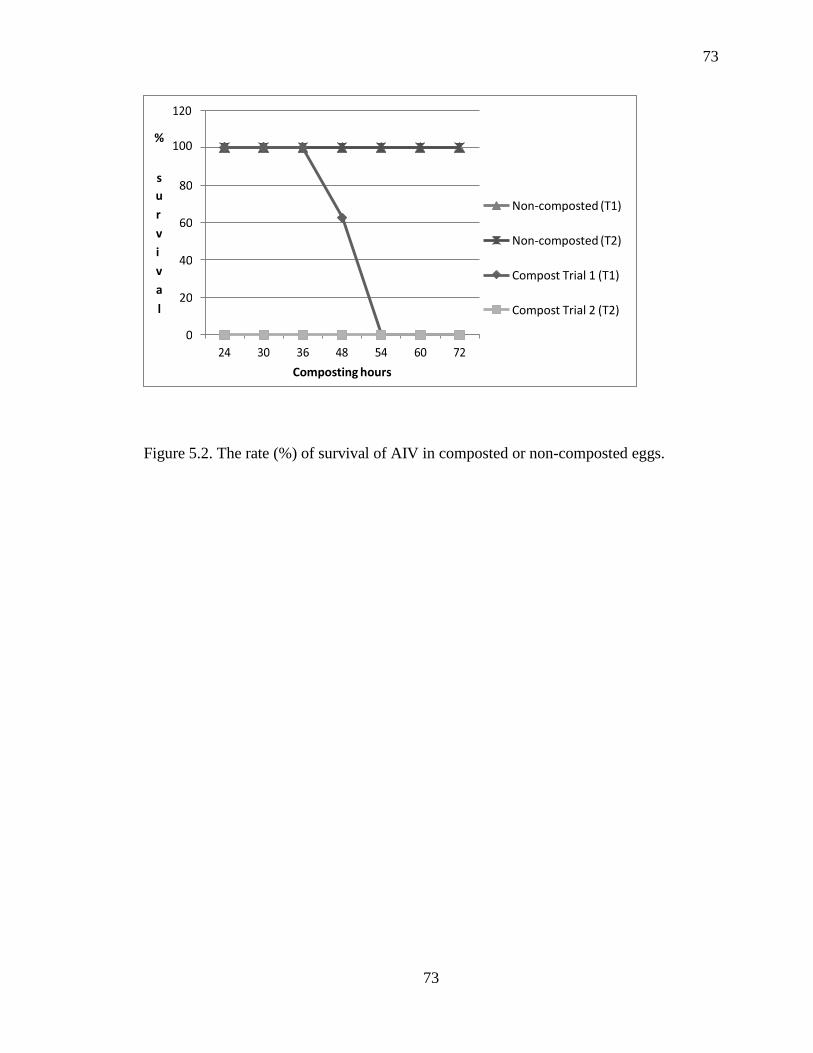
73
73
Figure 5.2. The rate (%) of survival of AIV in composted or non-composted eggs.
% 100
s
u
r
v
i
v
a
l
80
Non-composted (T1)
60
Non-composted (T2)
40 Compost Trial 1 (T1)
20 Compost Trial 2 (T2)
0
Composting hours

74
74
Figure 5.3. Temperature (°C) readings from probes placed at different sites of the
compost pile and composting room for Trial 1(T1) and Trial 2 (T2).
e
n
t
i
g
r
a
d
e
left-center T2
middle-center T2
right-center T2
right-center T1
middle-center T1
left-center T1
top T2
compost room T2 1 4 7 10 13 16 19 22 25 28 31 34 37 40 43 46 49 52 55 58 61 64
Composting hours
75
75
DISSERTATION CONCLUSIONS
Based on available epidemiological data, migrating wild aquatic birds are the
primary reservoir of influenza A viruses, and could be the initial source of virus, which
can be passed on through contact with resident waterfowl or domestic poultry. In these
birds, influenza viruses usually replicate in the intestinal tract, cause no disease, and are
spread by fecal contamination of the water habitat. The majority of influenza viruses that
are enzootic in wild aquatic birds are unable to replicate efficiently in chickens. However,
multiple subtypes of AIVs have been isolated from healthy land-based poultry including
chickens, turkeys, quail, guinea fowl, and chukar, without overt clinical signs.
Occasionally, AIVs circulating in land-based poultry can increase their virulence rapidly,
and cause important economic losses due to mortality, as well as trade restrictions. In
addition, wild birds have been shown to introduce novel influenza gene segments into a
population, that when reassorted with existing viruses, can generate a dissimilar virus
with different antigenic and other biological characteristics.
AIVs and APMVs were detected and isolated from swabs taken from waterfowl of
wildlife refuges in southeastern USA. Sampling sites were: Eufaula National Wildlife
Refuges (Molnar, Kennedy, and Bradley units in AL and GA); PCS-Potash mine, White
Springs, FL; Racoon Creek, Crow Creek Refuge, Stevenson, AL; TN River Lake
Guntersville, Scottsboro, AL; and Daphne, AL. Out of 1260 samples used for virus
isolation, we isolated 19 AIVs, three APMVs (APMV-1, APMV-4), and one had a
mixture of two viruses. AIV subtypes were: H10N7 (1); H1N1 (5); H1N4 (1); H3N8 (8);

76
76
H4N6 (2); and H4N8 (2). Wild bird species sampled were commonly Wood Ducks
(WODU), Northern Shovelers (NSHO), Ring-necked Ducks (RNDU), Gadwalls
(GADW), Mallards (MALL), American Wigeons (AMWI), Hooded Mergansers
(HOME), and Blue-winged Teals (BWTE)/Green-winged Teals (GWTE). We have
submitted these data from the isolates to a national database that incorporates and tracks
all avian influenza data collected from wild birds in the USA. The data collected in this
system has been used by scientists to develop a better understanding of the movement of
avian influenza viruses among wild and domestic animals, improve risk analyses, and
target monitoring strategies to track the avian influenza spread.
AC-ELISA and RRT-PCR were examined for use in detection of AIV directly from
swabbed material and allantoic fluids of HA+ samples. It was shown that successful AIV
detection and isolation from wild aquatic birds was dependent on sampling and
laboratory protocols. The highest isolation rate (14%) was obtained from one sampling
day in Lake City, FL, wherein at this time laboratory personnel were more experienced
with sampling and laboratory techniques. Other factors that may have contributed to the
higher isolation rate are: 1) use of dry ice to freeze samples in the field; 2) time or
location of sampling; and 3) bird species or age. It is interesting to note that 97% of the
samples collected on this sampling date were BWTE, whereas on another sampling time
(1/21/2006) conducted on this site, more GWTE were sampled versus BWTE. It was
concluded that RRT-PCR was more sensitive, cost effective, and specific than AC-
ELISA (Directigen Flu™ A test; Becton Dickinson, 2004) for the detection of AIV in
wild birds. It could detect AIV from AF with an HA titer as low as 4, whereas AC-
ELISA did not detect virus when HA titer was lower than 125. However, to avoid false-
77
77
negative results, testing should be done on AF and not directly from cloacal swabs.
Moreover, samples that were RRT-PCR positive, but with a high Cp (>32), had either
AIV in low concentration, APMV, or both. In a mixed infection, several limiting
dilutions and passages were required to separate the viruses, since presence of APMVs
may have interfered with AIV detection, and thereby resulted in false positive AIV RRT-
PCR results.
The H gene sequence of the AL H10N7 isolate was closely related (98%) to more
recent isolates from Delaware and Canada, but only 90% related to an H10N7 isolated 35
years ago. Sequencing analysis of the H gene of four H1N1 isolates from AL revealed
that they had 92-97% similarity with published H1N1 isolates including one from swine.
The pandemic (2009) H1N1 influenza viruses had 65-75% relatedness to these isolates
(data not reported). Results of composting experiment on survival of AIV-H1N1 in eggs
indicated that optimum conditions and procedures are required to eliminate AIV in eggs.
No viable virus was re-isolated from the AIV-infected eggs after 24 hours of composting
when the internal compost temperature reached 570C. At 29
0C room temperature, the
virus survived for 3 days. These results can be used to develop management strategies
that can inactivate AIV in eggs laid by infected hens.
No H5 or H7 AIVs were isolated from wild waterfowl in Alabama, Georgia, and
Florida during 2006 to 2011. However, because of the recent HPAI outbreaks in the
Midwest, there is a significant need for testing wild birds as they migrate. Early detection
of AIV, especially the subtype (H5N2) that caused the devastating outbreak, will help
prevent possible infection and /or spread of these highly pathogenic (HP) strains to the
U.S. poultry and swine industries. More importantly, it will provide early warnings of HP

78
78
AIV presence in the concentrated area of the poultry and swine industries; thus advising
companies and farmers to further strengthen or enforce more stringent biosecurity
measures to prevent spread of the virus into commercial poultry and swine populations as
well as to humans.

79
79
APPENDIX
Eufaula National Wildlife Refuge Map (Alabama and Georgia)
Bradley Unit
Kennedy Unit

80
80
Eufaula National Wildlife Refuge, Kennedy Unit Duck Blinds
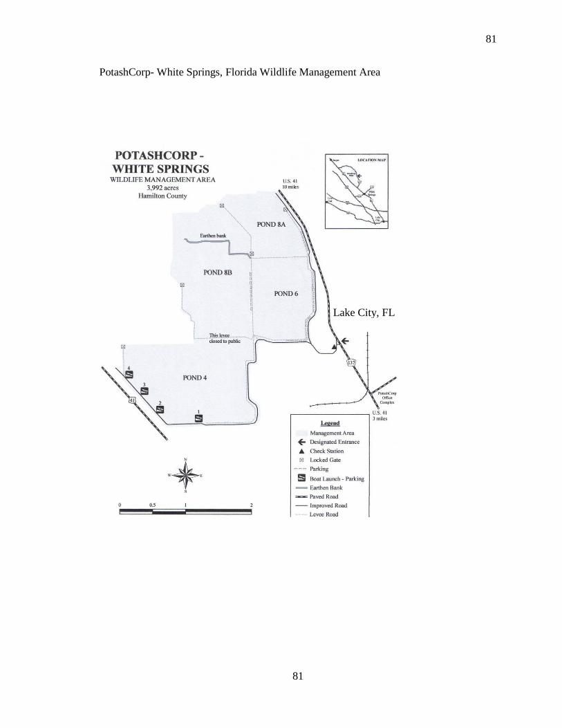
81
81
PotashCorp- White Springs, Florida Wildlife Management Area
Lake City, FL