detection of dentinal cracks using contrast-enhanced micro
TRANSCRIPT
J O U R N A L O F T H E M E C H A N I C A L B E H AV I O R O F B I O M E D I C A L M A T E R I A L S 3 ( 2 0 1 0 ) 2 2 3 – 2 2 7
available at www.sciencedirect.com
journal homepage: www.elsevier.com/locate/jmbbm
Technical note
Detection of dentinal cracks using contrast-enhancedmicro-computed tomography
Matthew D. Landrigana, John C. Flatleya, Travis L. Turnbulla, Jamie J. Kruzicb,Jack L. Ferracanec, Thomas J. Hiltonc, Ryan K. Roedera,∗
aDepartment of Aerospace and Mechanical Engineering, University of Notre Dame, Notre Dame, IN 46556, USAb School of Mechanical, Industrial, and Manufacturing Engineering, Oregon State University, Corvallis, OR 97331, USAcDivision of Biomaterials and Biomechanics, Department of Restorative Dentistry, School of Dentistry, Oregon Health and Science University,Portland, OR 97239, USA
A R T I C L E I N F O
Article history:
Received 12 June 2009
Received in revised form
17 September 2009
Accepted 1 October 2009
Published online 13 October 2009
A B S T R A C T
A new technique using contrast enhanced micro-computed tomography (micro-CT) was
developed to improve the ability to detect dentinal cracks in teeth and assess associated
risks to oral health. Extracted, whole human molars that exhibited visual evidence of
external cracks following extraction and machined, partially fractured elephant dentin
specimens were labeled by BaSO4 precipitation and imaged by micro-CT. Contrast-
enhanced micro-CT was demonstrated in vitro to enable non-destructive, 3-D imaging of
the presence, morphology and spatial location of dentinal cracks in whole human molars
and machined specimens. BaSO4 staining provided enhanced contrast for the detection of
cracks that could not be detected prior to staining. Backscattered SEM micrographs showed
that BaSO4 was precipitated on the surfaces of dentinal cracks and within adjacent tubules.
The new methods demonstrated in this study are expected to be useful for clinical and
scientific studies investigating the etiology and treatment of dentinal cracks in teeth.c© 2009 Elsevier Ltd. All rights reserved.
c
d
1. Introduction
Cracked teeth are commonly observed in dental practice
and are potentially symptomatic (Clark et al., 2003; Kahler,
2008; Snyder, 1976). Cracked teeth are the third most
common cause of tooth loss after caries and periodontal
disease (Braly and Maxwell, 1981; Hiatt, 1973). Treatment
options are typically lengthy, expensive and include a
risk of morbidity (Liu and Sidhu, 1995; Homewood, 1998;
∗ Corresponding address: Department of Aerospace and MechaniResearch Building, Notre Dame, IN 46556, USA. Tel.: +1 574 631 7003;
E-mail address: [email protected] (R.K. Roeder).
1751-6161/$ - see front matter c© 2009 Elsevier Ltd. All rights reservedoi:10.1016/j.jmbbm.2009.10.003
al Engineering, University of Notre Dame, 148 Multidisciplinaryfax: +1 574 631 2144.
Geurtsen and García-Godoy, 1999; Kahler, 2008), yet there islittle definitive evidence to support the diagnosis of cracksrequiring intervention or the effectiveness of interventions(e.g., Bader et al., 1996). Therefore, treatments may be eitherover-prescribed or prescribed too late.
Current methods for the diagnosis of cracks that maycompromise teeth are all based on optical assessment, withor without the aid of surgical loupes, microscopes, dyesand/or transillumination (Clark et al., 2003; Kahler, 2008).Improvements in the detection and classification of crackshave been demonstrated using enhanced magnification
.
224 J O U R N A L O F T H E M E C H A N I C A L B E H AV I O R O F B I O M E D I C A L M A T E R I A L S 3 ( 2 0 1 0 ) 2 2 3 – 2 2 7
(Clark et al., 2003; Slaton et al., 2003) and transillumination(Wright et al., 2004). However, optical methods suffer from aninherent inability to assess the severity of cracks (e.g., crackdepth), particularly sub-surface dentinal cracks. Moreover,dentinal cracks are generally not able to be detected in plainradiographs.
Micro-CT is useful for non-invasive, three-dimensional(3-D) imaging of the internal structure of mineralized tissues,due to relative differences in x-ray attenuation. Cone beamvolumetric micro-CT was recently demonstrated to providemore accurate assessment of proximal caries lesion depthcompared to conventional radiographs (Kalathingal et al.,2007). Intra-oral systems limit the volume irradiated and thusachieve a low radiation dose to the patient; however, thespatial resolution is currently limited to 40 µm and the imagecontrast is low relative to conventional CT (Kalathingal et al.,2007). High-resolution micro-CT systems are commerciallyavailable for both in vivo imaging of small animals and invitro imaging of tissue specimens. The spatial resolution istypically on the order of 10 µm and systems with 1 µmresolution are now available. However, the detection of smallcracks (∼1 µm in width) in relatively large specimens (e.g.,a molar) is limited by the resolution, maximum specimensize, and data collection time of the above instruments. X-raytomography using high energy, monochromatic synchrotronradiation is able to image cracks in dentin (Kruzic et al., 2003),but is neither readily available nor amenable to imaging largenumbers of tissue specimens that are relatively large in size.
Non-destructive, 3-D imaging of the presence, spatiallocation and accumulation of microdamage in bone tissuewas recently demonstrated for the first time using contrast-enhanced micro-CT with a precipitated BaSO4 stain (Lenget al., 2008; Wang et al., 2007). Although imaging of cracksand damage in bone had been previously demonstratedusing high energy, monochromatic synchrotron radiation(Nalla et al., 2005), this approach enabled the use ofcommercially available bench top micro-CT instruments.BaSO4 was precipitated within damaged tissue, cracks andvasculature, as verified by backscattered electron imaging andenergy dispersive spectroscopy (Leng et al., 2008; Wang et al.,2007). Moreover, precipitated BaSO4 enhanced the intensityof voxels in micro-CT due to the higher x-ray attenuation ofBaSO4 relative to bone tissue. The micro-CT scanner (10 µmresolution) was unable to detect microcracks or fatigue crackswithout the use of the contrast agent. Therefore, contrast-enhanced micro-CT is expected to find increased use inthe study of mineralized tissue mechanics, complementingor replacing methods of damage/crack detection that areinherently destructive, two-dimensional and tedious.
The objective of this study was to non-destructively andthree-dimensionally image dentinal cracks using contrast-enhanced micro-CT. Extracted, whole human molars thatexhibited visual evidence of external cracks followingextraction and machined, partially fractured elephant dentinspecimens were labeled by BaSO4 precipitation and imagedby micro-CT using methods adapted from previous studiesinvestigating microdamage in bone tissue (Leng et al., 2008;Wang et al., 2007).
2. Materials and methods
2.1. Elephant dentin
Two partially fractured, dehydrated compact-tension speci-mens of dentin from tusk shards of a single adult male ele-phant (Loxodonta africana) used in a previous study (Kruzicet al., 2003) provided well-characterized and highly controlledcracks for evaluating feasibility of the technique. Elephantdentin is similar to human dentin in containing tubuleswithin an extracellular matrix of mineralized collagen fibersand avoids the specimen size limitations of human dentin.Compact-tension specimens were machined while hydratedto a thickness of ∼2.5 mm and width of ∼15 mm. Specimenswere polished to a 1200 grit finish, followed by 1 and 0.05 µmalumina suspensions. An initial notch was introduced witha low speed diamond wafer saw such that the crack growthdirection was oriented perpendicular to the long axis of thetubules and the crack plane was in the plane of the tubules. Afinal notch with root radius ∼15 µm was achieved by repeat-edly sliding a razor blade over the initial notch. Fracture resis-tance (R-curve) testing was conducted in vacuo (∼10−4 Pa) ina scanning electron microscope (SEM). Specimens were rehy-drated and stored in phosphate buffered saline upon receiptand during all interim periods before staining and micro-CT.
2.2. Whole human molars
Ten extracted human molars from different donors wereprovided by the Practice-based Research in Oral Health (PROH)practitioner network through the Oregon Health and ScienceUniversity. Teeth were screened after extraction for evidenceof external cracks upon inspection with 3.5X surgical loupes.All teeth were maintained hydrated in a 0.05% solution ofchloramine T during all interim periods before staining andmicro-CT.
2.3. Histological staining and imaging
Elephant dentin specimens and whole human molars wereimaged before and after histological staining by micro-CT (µCT-80, Scanco Medical AG, Bassersdorf, Switzerland)at 10 µm resolution, 70 kVp voltage, 113 µA current, and400 ms integration time. Grayscale images were smoothedby Gaussian filtering. High intensity voxels representativeof BaSO4 were segmented to highlight cracks in 3-Dreconstructions. An image subregion was used to removethe effect of non-specific BaSO4 staining on specimenfree surfaces which would have otherwise obscured thevisualization of internal features in 3-D reconstructions.
All specimens were stained by BaSO4 precipitation,soaking in an equal parts mixture of 0.5 M barium chloride(Certified ACS crystal, Fisher Scientific, Fair Lawn, NJ) inde-ionized water, buffered saline, and acetone for 3 days,followed by an equal parts mixture of 0.5 M sodium sulfate(Anhydrous powder, Fisher Scientific, Fair Lawn, NJ) in de-ionized water, buffered saline, and acetone for 3 days, bothunder vacuum (∼50 mm Hg). The staining mechanism wasa precipitation reaction where BaCl2(aq) + Na2SO4(aq) →
BaSO4(s) + 2NaCl(aq). Barium and sulfate ions diffused into

J O U R N A L O F T H E M E C H A N I C A L B E H AV I O R O F B I O M E D I C A L M A T E R I A L S 3 ( 2 0 1 0 ) 2 2 3 – 2 2 7 225
and concentrated within void space in the dentinal tissue –e.g., tubules and cracks – which acted as a “micro-reactor” andprovided an abundance of heterogeneous nucleation sites ontissue surfaces (Leng et al., 2004, 2008).
After staining and micro-CT, specimens were dehydratedin a graded series of alcohol solutions, dried overnight inan oven at 90 ◦C, embedded in poly(methyl methacrylate),sectioned with a low-speed diamond wafer saw, polishedwith a series of diamond compounds to a 1 µm finish,and coated with Au–Pd by sputter deposition for scanningelectron microscopy (SEM). Specimens were imaged usingbackscattered electrons (Evo 50, LEO Electron Microscopy Ltd.,Cambridge, UK) at an accelerating voltage of 20 kV and aworking distance of 9–12 mm. Note that image contrast frombackscattered electrons is primarily due to compositionaldifferences in atomic number, with an increasing atomicnumber resulting in increased intensity. The elementalcomposition of the stain was verified by electron probemicroanalysis using energy dispersive spectroscopy (EDS)(INCA x-sight model 7636, Oxford Instruments America,Concord, MA).
3. Results
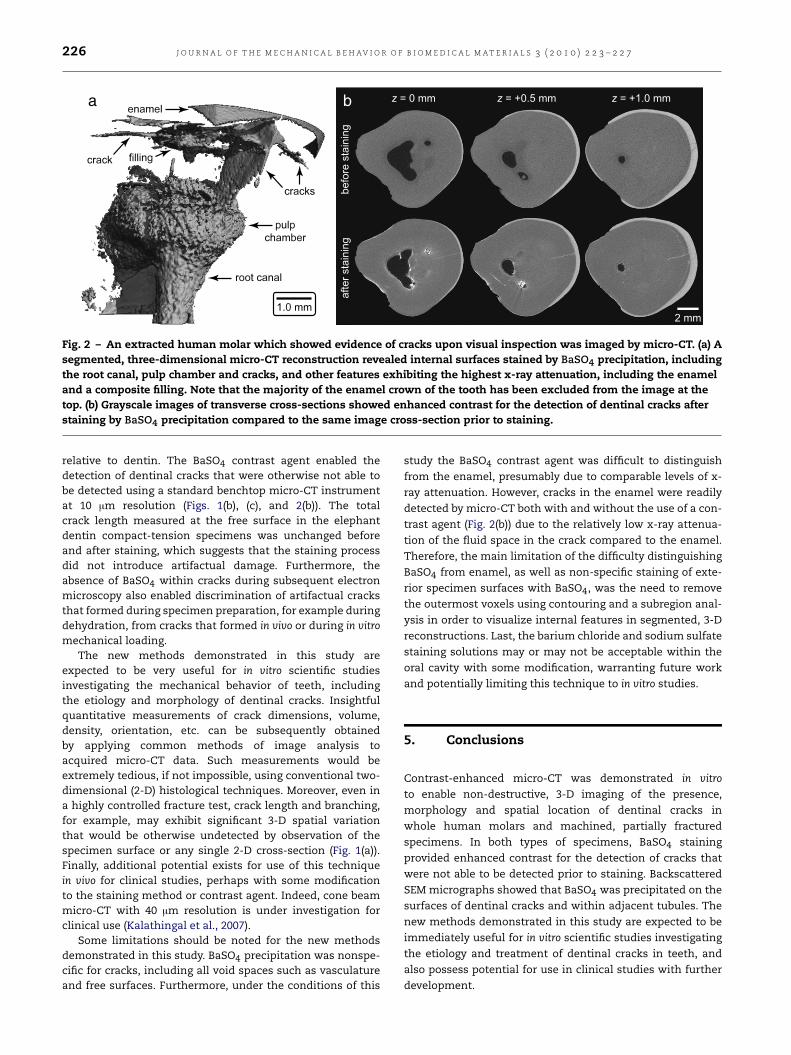
Contrast-enhanced micro-CT enabled non-destructive, 3-Dimaging of the presence, morphology and spatial location ofdentinal cracks in machined specimens (Fig. 1) and wholehuman molars (Fig. 2). In both types of specimens, BaSO4staining provided enhanced contrast for the detection ofcracks that were not able to be detected prior to staining(Figs. 1(b), (c), and 2(b)). Note that the entire 3-D volume ofgrayscale image slices for the specimens shown in Figs. 1and 2 are available as movie files (.avi) in Electronic Annex1 and 2, respectively, of the online version of this article.Backscattered SEM micrographs showed that BaSO4 wasprecipitated on the surfaces of dentinal cracks and withinadjacent tubules (Fig. 1(e)). The highest levels of imageintensity for both micro-CT and backscattered SEM wereshown to correspond to the presence of elemental bariumand sulfur measured using EDS. At higher magnification,SEM revealed the presence of BaSO4 crystals and aggregates,typically submicron but up to 5 µm in size, on the surfacesof propagating cracks and within adjacent tubules. BaSO4was also precipitated on all free surfaces, including the rootcanal and pulp chamber (Fig. 2(a)). Therefore, a subregion wasused to remove staining of the outer specimen surfaces andreveal internal features, including cracks, in segmented 3-Dreconstructions (Figs. 1(a), and 2(a)).
4. Discussion
The feasibility of non-destructive, 3-D imaging of dentinalcracks was demonstrated in vitro in machined, partiallyfractured specimens (Fig. 1) and whole human molars (Fig. 2)using contrast-enhanced micro-CT with a precipitated BaSO4stain. BaSO4 precipitation on the surfaces of dentinal cracksand within adjacent tubules enhanced the intensity of voxelsin micro-CT due to the higher x-ray attenuation of BaSO4
a
b
c
d
e
Fig. 1 – A branched crack that propagated from a notch(left) in a compact tension specimen of elephant dentinwas imaged by micro-CT before and after staining byBaSO4 precipitation, and by backscattered SEM afterstaining by BaSO4 precipitation. (a) A segmented,three-dimensional micro-CT reconstruction showed cracksurfaces stained with BaSO4. Cross-sectional grayscalemicro-CT images at the same depth approximately midwaythrough the specimen thickness (b) before and (c) afterstaining by BaSO4 precipitation showed that the crack wasunable to be detected at this location without the use of thecontrast agent. The contrast-enhanced micro-CT image in(c) was compared to (d) a backscattered SEM micrograph forthe same specimen at approximately the samecross-sectional depth and magnification. (e) Abackscattered SEM micrograph of the crack tip in (d) athigher magnification showed BaSO4 penetration intomicrotubules. Note that the presence of elemental Ba and Swas verified by EDS (not shown).
226 J O U R N A L O F T H E M E C H A N I C A L B E H AV I O R O F B I O M E D I C A L M A T E R I A L S 3 ( 2 0 1 0 ) 2 2 3 – 2 2 7
ba
Fig. 2 – An extracted human molar which showed evidence of cracks upon visual inspection was imaged by micro-CT. (a) Asegmented, three-dimensional micro-CT reconstruction revealed internal surfaces stained by BaSO4 precipitation, includingthe root canal, pulp chamber and cracks, and other features exhibiting the highest x-ray attenuation, including the enameland a composite filling. Note that the majority of the enamel crown of the tooth has been excluded from the image at thetop. (b) Grayscale images of transverse cross-sections showed enhanced contrast for the detection of dentinal cracks afterstaining by BaSO4 precipitation compared to the same image cross-section prior to staining.
relative to dentin. The BaSO4 contrast agent enabled thedetection of dentinal cracks that were otherwise not able tobe detected using a standard benchtop micro-CT instrumentat 10 µm resolution (Figs. 1(b), (c), and 2(b)). The totalcrack length measured at the free surface in the elephantdentin compact-tension specimens was unchanged beforeand after staining, which suggests that the staining processdid not introduce artifactual damage. Furthermore, theabsence of BaSO4 within cracks during subsequent electronmicroscopy also enabled discrimination of artifactual cracksthat formed during specimen preparation, for example duringdehydration, from cracks that formed in vivo or during in vitromechanical loading.
The new methods demonstrated in this study areexpected to be very useful for in vitro scientific studiesinvestigating the mechanical behavior of teeth, includingthe etiology and morphology of dentinal cracks. Insightfulquantitative measurements of crack dimensions, volume,density, orientation, etc. can be subsequently obtainedby applying common methods of image analysis toacquired micro-CT data. Such measurements would beextremely tedious, if not impossible, using conventional two-dimensional (2-D) histological techniques. Moreover, even ina highly controlled fracture test, crack length and branching,for example, may exhibit significant 3-D spatial variationthat would be otherwise undetected by observation of thespecimen surface or any single 2-D cross-section (Fig. 1(a)).Finally, additional potential exists for use of this techniquein vivo for clinical studies, perhaps with some modificationto the staining method or contrast agent. Indeed, cone beammicro-CT with 40 µm resolution is under investigation forclinical use (Kalathingal et al., 2007).
Some limitations should be noted for the new methodsdemonstrated in this study. BaSO4 precipitation was nonspe-cific for cracks, including all void spaces such as vasculatureand free surfaces. Furthermore, under the conditions of this
study the BaSO4 contrast agent was difficult to distinguish
from the enamel, presumably due to comparable levels of x-
ray attenuation. However, cracks in the enamel were readily
detected by micro-CT both with and without the use of a con-
trast agent (Fig. 2(b)) due to the relatively low x-ray attenua-
tion of the fluid space in the crack compared to the enamel.
Therefore, the main limitation of the difficulty distinguishing
BaSO4 from enamel, as well as non-specific staining of exte-
rior specimen surfaces with BaSO4, was the need to remove
the outermost voxels using contouring and a subregion anal-
ysis in order to visualize internal features in segmented, 3-D
reconstructions. Last, the barium chloride and sodium sulfate
staining solutions may or may not be acceptable within the
oral cavity with some modification, warranting future work
and potentially limiting this technique to in vitro studies.
5. Conclusions
Contrast-enhanced micro-CT was demonstrated in vitro
to enable non-destructive, 3-D imaging of the presence,
morphology and spatial location of dentinal cracks in
whole human molars and machined, partially fractured
specimens. In both types of specimens, BaSO4 staining
provided enhanced contrast for the detection of cracks that
were not able to be detected prior to staining. Backscattered
SEM micrographs showed that BaSO4 was precipitated on the
surfaces of dentinal cracks and within adjacent tubules. The
new methods demonstrated in this study are expected to be
immediately useful for in vitro scientific studies investigating
the etiology and treatment of dentinal cracks in teeth, and
also possess potential for use in clinical studies with further
development.
J O U R N A L O F T H E M E C H A N I C A L B E H AV I O R O F B I O M E D I C A L M A T E R I A L S 3 ( 2 0 1 0 ) 2 2 3 – 2 2 7 227
Acknowledgments
This research was partially supported by the U.S. ArmyMedical Research and Materiel Command (W81XWH-06-1-0196) through the Peer Reviewed Medical Research Program(PR054672).
Appendix. Supplementary data
Supplementary data associated with this article can be found,in the online version, at doi:10.1016/j.jmbbm.2009.10.003.
R E F E R E N C E S
Bader, J.D., Shugars, D.A., Roberson, T.M., 1996. Using crowns toprevent tooth fracture. Community Dent. Oral Epidemiol. 24(1), 47–51.
Braly, B.V., Maxwell, E.H., 1981. Potential for tooth fracture inrestorative dentistry. J. Prosthet. Dent. 45 (4), 411–414.
Clark, D.J., Sheets, C.G., Parquette, J.M., 2003. Definitive diagnosisof early enamel and dentin cracks based on microscopicevaluation. J. Esthet. Restor. Dent. 15 (7), 391–401.
Geurtsen, W., García-Godoy, F., 1999. Bonded restorations for theprevention and treatment of the cracked tooth syndrome. Am.J. Dent. 13 (6), 266–270.
Hiatt, W.H., 1973. Incomplete crown-root fracture in pulpal-periodontal disease. J. Periodontol. 44 (6), 369–379.
Homewood, C.I., 1998. Cracked tooth syndrome—Incidence,clinical findings and treatment. Aust. Dent. J. 43 (4), 217–222.
Kahler, W., 2008. The cracked tooth conundrum: Terminology,classification, diagnosis, and management. Am. J. Dent. 21,275–282.
Kalathingal, S.M., Mol, A., Tyndall, D.A., Caplan, D.J., 2007. Invitro assessment of cone beam local computed tomographyfor proximal caries detection. Oral Surg. Oral Med. Oral Pathol.Oral Radiol. Endod. 104, 699–704.
Kruzic, J.J., Nalla, R.K., Kinney, J.H., Ritchie, R.O., 2003. Crack blunt-ing, crack bridging and resistance-curve fracture mechanics indentin: Effect of hydration. Biomaterials 24 (28), 5209–5221.
Leng, H., Wang, X., Niebur, G.L., Roeder, R.K., 2004. Synthesisof a barium sulfate nanoparticle contrast agent for micro-computed tomography of bone microstructure. Ceram. Trans.159, 219–229.
Leng, H., Wang, X., Ross, R.D., Niebur, G.L., Roeder, R.K.,2008. Micro-computed tomography of fatigue microdamage incortical bone using a barium sulfate contrast agent. J. Mech.Behav. Biomed. Mater. 1 (1), 68–75.
Liu, H.H., Sidhu, S.K., 1995. Cracked teeth–treatment rationaleand case management: Case reports. Quintessence Int. 26 (7),485–492.
Nalla, R.K., Kruzic, J.J., Kinney, J.H., Ritchie, R.O., 2005. Mechanisticaspects of fracture and R-curve behavior in human corticalbone. Biomaterials 26 (2), 217–231.
Slaton, C.C., Loushine, R.J., Weller, R.N., Parker, M.H., Kimbrough,W.F., Pashley, D.H., 2003. Identification of resected root-enddentinal cracks: A comparative study of visual magnification.J. Endod. 29 (8), 519–522.
Snyder, D.E., 1976. The cracked-tooth syndrome and fracturedposterior cusp. Oral Surg. Oral Med. Oral Pathol. 41 (6), 698–704.
Wang, X., Masse, D.B., Leng, H., Hess, K.P., Ross, R.D., Roeder,R.K., Niebur, G.L., 2007. Detection of trabecular bonemicrodamage by micro-computed tomography. J. Biomech. 40(15), 3397–3403.
Wright, H.M., Loushine, R.J., Weller, R.N., Kimbrough, W.F., Waller,J., Pashley, D.H., 2004. Identification of resected root-enddentinal cracks: A comparative study of transillumination anddyes. J. Endod. 30 (10), 712–715.