detection of fetal anemia secondary to red blood cell ... · abnormal. umbilical artery ......
TRANSCRIPT

IUGRDiagnosis and Management
39th Annual Perinatal ConferenceVanderbilt UniversityDecember 6, 2013
Giancarlo Mari, M.D., M.B.A.Professor and Chair
Department of Obstetrics and GynecologyUniversity of Tennessee Health Science Center
Memphis, TNUSA

At the end of this presentation the participants will be able:
• To classify IUGR fetuses• To understand the cardiovascular changes
that occur in IUGR fetuses• To understand the issues in the management
of IUGR fetuses

IUGR

Pubmed: IUGR
15,000 papers

IUGR-Definition
•Memphis•Nashville•Sidney

Fetus who fails to reach its growth potential
IUGR

•EFW < 10th percentile (USA) •EFW < 5th percentile (USA) •EFW < 3rd percentile (USA)•EFW < 15th percentile (USA) •EFW > 2 SD below mean (Europe)•AC 10th 2.5th percentile (Europe)
Definitions:IUGR

•Each fetus is its own control (Deter-Rossavik)
•Ponderal index (Pediatricians)
•Population growth curves (Gardosi)
Definitions:
IUGR

Modified from Manning F. Fetal Medicine, 1995;7:307
1
26
51
76
101
126
151
176
>10 10 9 8 7 6 5 4 3 2 1 0.5Birth weight (%)
Peri
nata
l mor
talit
y (/1
000)
Perinatal Mortality

EFW < 10th percentile
Normal Pathologic80% ? 20% ?
IUGR

Placental Insufficiency
“Umbrella that covers our ignorance in terms of etiology and pathogenesis of the utero-placental chronic dysfunction”
Assali, Eur J Obstet Gynecol Reprod Biol 1975;5:87-91

Placental Insufficiency
It is not the cause of IUGR but is rather the consequence of a disease process that often we do not understand

Doppler in AGA and IUGR Fetuses

Doppler Indices

Umbilical Artery
Fitzgerald was the first to obtain a Doppler signal in pregnancy in 1977
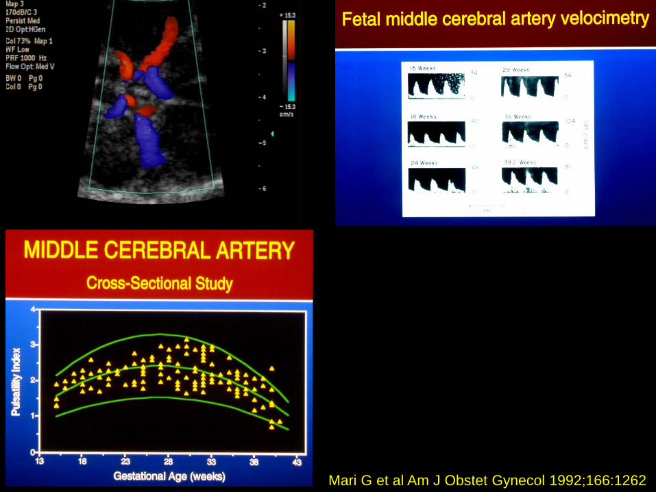
Mari G et al Am J Obstet Gynecol 1992;166:1262

Umbilical Artery:High placental vascular resistance

7.5
1.5Normal Abnormal
Giles WB, et al. Br J Obstet Gynecol 1985;92:31
Plac
enta
l arte
ries
/ Hig
h Po
wer
Fie
ld
Umbilical Doppler Waveform
Normal Abnormal
Umbilical Artery Doppler and Placental Vascular Histology

Umbilical Artery Doppler and Outcome
• Reduce perinatal death and unnecessary induction of labor in the preterm growth restricted fetus
• A meta-analysis use of Doppler ultrasonography reduced the odds of perinatal death by 38 percent (95% CI 15-55)
Alfirevic Z et al Am J Obstet Gynecol 1995

Brain Circulation

MCA Waveforms at 24 weeks
A = Normal
B = “Brain sparing effect”
MCA Waveforms at 24 Weeks

Normal pregnancy Preeclamptic and/orIUGR pregnancy
Khong TJ et al, Br J Obstet Gynaecol, 1986;93:1049
Utero-Placental Vessels
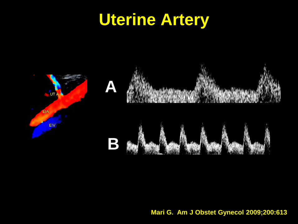
A
B
Uterine Artery
Mari G. Am J Obstet Gynecol 2009;200:613

SGA fetuses with normal umbilical artery
• Uterine arteries
• Middle cerebral arteries
Severi FM, et al. Ultrasound Obstet Gynecol 2002;19:225-8

Venous System

Umbilical Vein
Quantitative assessment: Velocity
Qualitative assessment: Pulsation
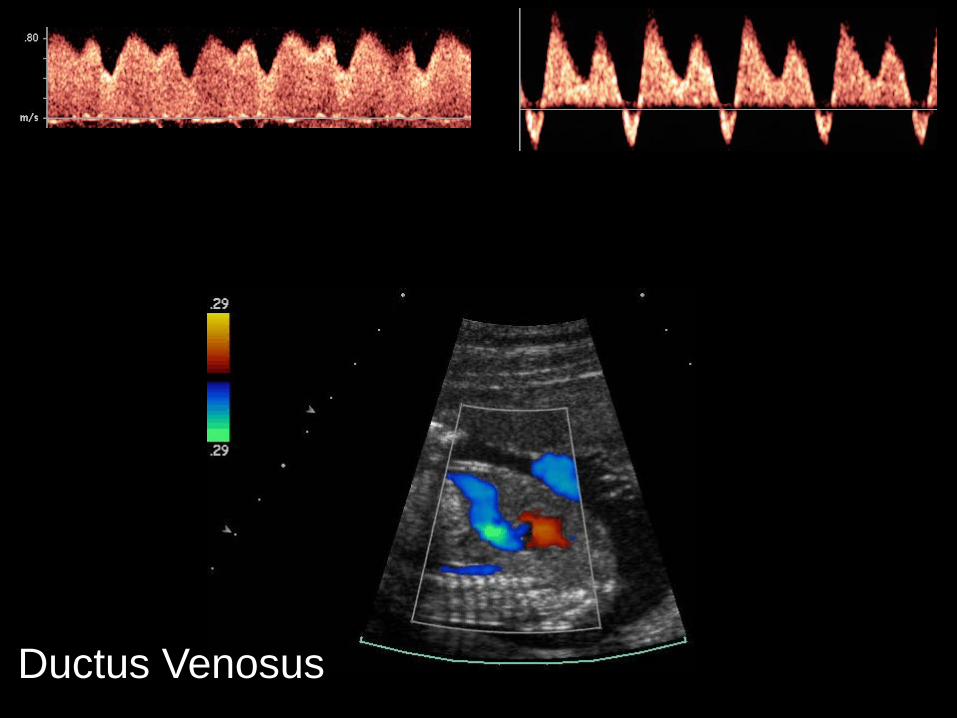
Ductus Venosus

Is Ductus venosus reversed flow an indication for delivery?
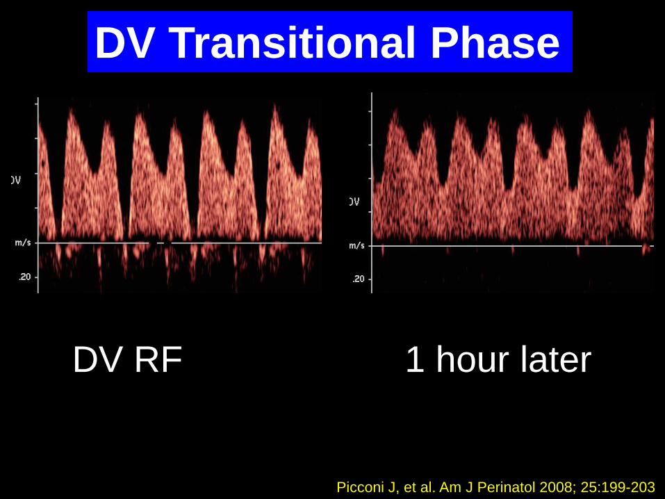
DV RF 1 hour later
Picconi J, et al. Am J Perinatol 2008; 25:199-203
DV Transitional Phase
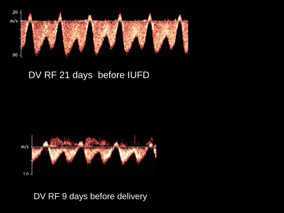
DV RF 21 days before IUFD
DV RF 9 days before delivery

Ductus venosus at < 28 weeks and EFW < 1000 g
Is the biophysical profile normal or abnormal?
A
B
C
Kaur et al. Am J Obstet Gynecol 2008;199:264

a. What is the SIA index and b. What does it indicate?
a. Peak systolic velocity Isovolumetric relaxation
+a-wave
b. Myocardial function
Picconi et al J Ultrasound Med 2008;27:1283

Picconi et al J Ultrasound Med 2008;27:1283
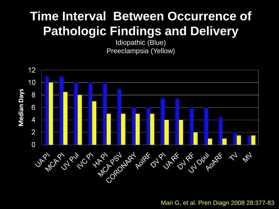
Time Interval Between Occurrence of Pathologic Findings and Delivery
Idiopathic (Blue)Preeclampsia (Yellow)
Mari G, et al. Pren Diagn 2008 28:377-83

Percent Parameters Abnormal in the Two Groups
Mari G, et al. Pren Diagn 2008 28:377-83

Staging and Classification of IUGR
Fetuses
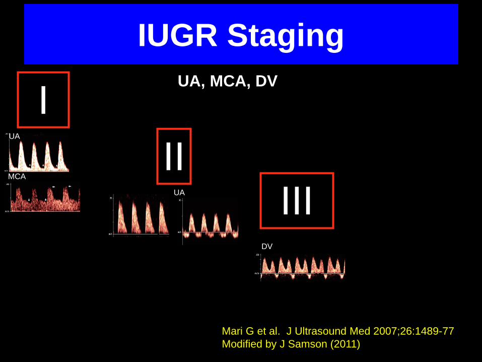
IUGR Staging
Mari G et al. J Ultrasound Med 2007;26:1489-77Modified by J Samson (2011)
IUA
MCAII
UA IIIDV
UA, MCA, DV
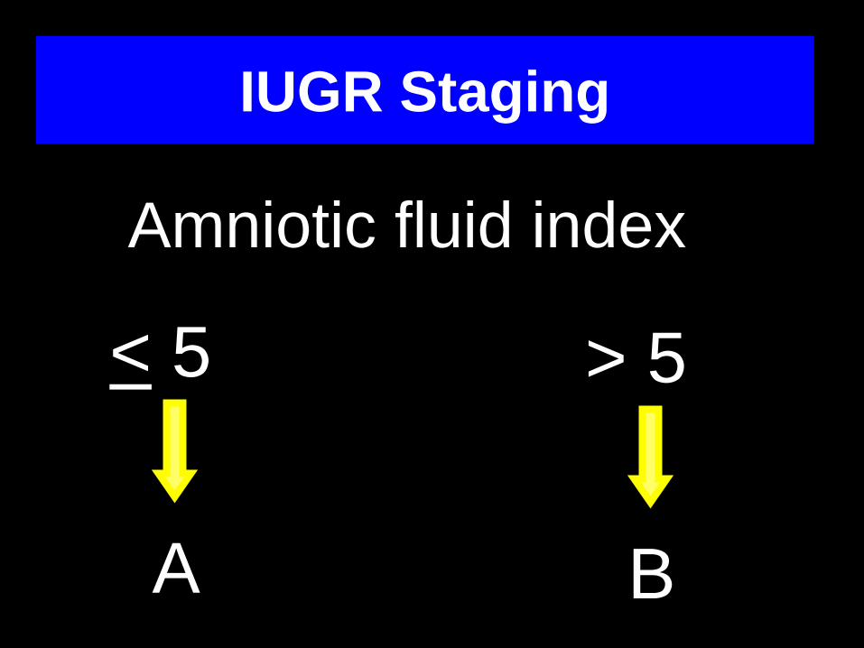
IUGR Staging
Amniotic fluid index
A
< 5
A
> 5
B

IUGR Classification• EFW < 10th percentile
• Abnormal UA PI
• Amniotic fluid index >5 cm
• No maternal-fetal pathology
• 25 weeks
IUGR Stage IB idiopathic 25 weeks

IUGR Staging
Mari G et al. J Ultrasound Med 2007;26:1489-77

IUGR Staging
Mari G et al. J Ultrasound Med 2007;26:1489-77

When should IUGR fetuses be delivered?

IUGR Delivery?
• GRIT• DIGITAT• TRUFFLE

IUGR and Gestational Age at Delivery
Between 25 and 29 weeks (“vital weeks”), for each week the IUGR fetus remains in utero the mortality decreases by 48%
Mari G et al. J Ultrasound Med 2007; 26:555-59
IUGR and Timing of deliveryDoppler/NST/BPP
Stage 0 IUGR Stage I IUGR Stage II IUGR Stage III IUGR

Infant Mortality Rates(per 1000 births)
Source: TN Dept. of Health, Office of Policy, Planning and Assessment, Division of Health Statistics