detection of hemolysin bl gene of bacillus cereus isolates · keyword: bacillus cereus, hemolysin...
TRANSCRIPT
Detection of Hemolysin BL Gene of Bacillus cereus IsolatesC. Wiwat* and R. ThiramanasDepartment of Microbiology, Faculty of Pharmacy, Mahidol University.447 Sri-Ayudhya Rd., Bangkok 10400, Thailand
Abstract Hemolysin BL (HBL) is the multi-component enterotoxin causing diarrheal type of Bacillus cereus food poisoning. The hbl operon of B. cereus was divided into 4 coding sequences (CDS) and identified as hblC, hblD, hblA and hblB genes consisting of 1,319, 1,221, 1,128 and 1,401 nucleotides encoding proteins of 439, 406, 375 and 466 amino acids with predicted sizes of 49.3, 43.8, 41.7 and 52.3 kDa, respectively. One hundred of B. cereus isolates were detected for the presence of each hbl gene by multiplex PCR and HBL production by discontinuous hemolysis on HBL sheep blood agar and Bacillus cereus Enterotoxin-Reverse Passive Latex Agglutination (BCET-RPLA) test kit. The results showed that 47 of 100 B. cereus isolates (47%) were detected by multiplex PCR with all hbl genes and gave positive result with RPLA test. Forty-one of 47 isolates (87.2%) produced discontinuous hemolysis on HBL sheep blood agar. However, this pattern was not observed clearly in 6 of 47 isolates (12.8%). In addition, B. cereus isolate E82/44 gave multiplex PCR positive with only 3 hbl genes, hblA, hblC and hblD, also showed positive result with RPLA test and produced discontinuous hemolysis.
Keyword: Bacillus cereus, Hemolysin BL, Enterotoxin, Food poisoning.
INTRODUCTION Bacillus cereus is a Gram-positive, facultatively anaerobic, spore-forming rod normally present in soil, air, dust, water and common contaminant in many raw and processed foods including rice, spices, eggs, vegetables, meat and dairy products1. Vegetative cells of B. cereus are eliminated by heat treatments but their spores can survive and are extremely resistant to different environmental stresses2. They should be considered as a critical point of hygienic importance for processes involving heat treatments that may activate the spores and kill the competitive microflora, since B. cereus vegetative cells grow well in the absence of competing organisms3. Consumption of food contaminated with B. cereus or their toxins can lead to food poisoning illnesses. B. cereus is the causative agent of two different types of food poisoning, the emetic and diarrheal syndromes4. The emetic syndrome is occurred
by directly ingestion of heat stable toxin, the emetic toxin (cereulide), produced by growing cells in food resulting in vomiting a few hours after ingestion5. The diarrheal syndrome is probably occurred by several heat labile enterotoxins produced during vegetative growth of B. cereus in small intestine resulting in enterotoxicosis, abdominal pain and diarrhea after incubation for 8-16 h6. At the present, only three enterotoxins have been shown to be involved in food poisoning including hemolysin BL (HBL), non-hemolytic enterotoxin (NHE) and cytotoxin K (CytK). While there are no any report about this symptom for the remaining two, enterotoxin T (BceT) and enterotoxin FM (EntFM)7. Hemolysin BL (HBL), a three-component enterotoxin consists of a binding component B and two lytic components L1 and L2, encoded by hblA, hblD and hblC genes, respectively, and is considered as a primary virulence factor in diarrheal type
*Corresponding author: Department of Microbiology, Faculty of Pharmacy, Mahidol University.Email: [email protected]
Original Article Mahidol University Journal of Pharmaceutical Sciences 2014; 41 (2), 22-30

C. Wiwat and R. Thiramanas 23
for its ability to cause fluid accumulation in rabbit ileal loops. All three components are required for enterotoxic and hemolytic activity. Function of B´ protein encoded by hblB gene, which shows high sequence ho-mology to hblA gene, is still unclear. HBL toxin exhibits hemolytic and dermonecrotic activities, increases vascular permeability and is toxic to Chinese hamster ovary cells8. Beta hemolysis surrounding colonies on blood agar is a diagnostic character of B. cereus9. On specific medium designated HBL agar, the hemolytic pattern surrounding HBL-producing strains is discontinuous and therefore diagnostic of hemolysin BL10. There are two major molecular techniques applied in the detection of HBL-producing B. cereus isolates, PCR-based and hybridizaton-based techniques. PCR-based method has been used extensively for all of the HBL-encoding genes. However, the presence of these toxin genes does not necessarily indicate that whether or not this bacterium is able to produce HBL enterotoxin and cause disease11. Detection of B. cereus with hybridization is mainly performed to confirm the results obtained with specific PCR, as well as, in the detection of HBL enterotoxic B. cereus12. The entire HBL enterotoxin complex can be demonstrated by HBL sheep blood agar13. However, this method is not widely used and a correlation between the results of the HBL plate and the PCR tests could not be observed for the strains that contained hbl genes14. Furthermore, there is only one available commercial Bacillus cereus Enterotoxin-Reverse Passive Latex Agglutination (BCET-RPLA) test kit of Oxoid, which commonly used to detect L2 part of the HBL toxin15. This is noticeable that this method performed based upon the specificity of some part of HBL. Applica-tion of this assay in detection of B. cereus HBL in Thailand may be result in false-negative, due to the DNA sequence varia-tion among B. cereus species between local isolates and standard strain. Therefore, the hbl genes from B. cereus isolated in Thailand
should be investigated and compared with B. cereus ATCC 14579. Objectives of this study were to detect HBL encoding gene profiles and HBL production by B. cereus isolates. The results from this study are useful for further study to develop a rapid test system for detection of HBL-producing B. cereus isolates.
MATERIALS AND METHODS
Bacillus cereus
All of B. cereus isolates used in this study were used for detection the presence of hbl genes by multiplex polymerase chain reaction and the production of HBL enterotoxin by HBL blood plates and BCET-RPLA test kit. Eight B. cereus isolates were isolated by Boonchaisuk16. Ninety B. cereus isolates were kindly provided by Division of Food Analysis, National Institute of Health (Department of Medical Science (DMSC), Ministry of Public Health, Thailand). B. cereus ATCC 14579 which was kindly provided by Dr. Daniel Zeigler (Bacillus Genetic Stock Center, The Ohio State University, Columbus, USA) was used as a positive control. B. cereus NC 1291 clinical isolate , which was reported that it gave cereulide positive results with Hep-2 cell vacuolation test and mitochondria respiratory assay, was kindly provided by Prof. Norio Agata (Nagoya City Public Health Research Institute, Japan). B. cereus INRA C15 food borne isolate, which was reported to give positive results in PCR reaction with hbl, nhe and cytK genes, was kindly provided by Marie-Helene Guinebretie`re (Institute National de la Recherche Agronomique, France).
Culture condition
For DNA preparation, B. cereus isolates were grown in Luria-Bertani (LB) broth at 37oC for overnight with continuous shaking. Then 1% of the overnight culture was inoculated in LB broth and further incubated at 37oC for 3 h with shaking to collect cells in mid-exponential phase.
Detection of Hemolysin BL Gene of Bacillus cereus Isolates24
Genomic DNA preparation
Genomic DNA of B. cereus used as template for polymerase chain reaction (PCR) was isolated by phenol-chloroform extraction method17.
Detection of Hemolysin BL genes by mul-tiplex PCR
The hbl enterotoxin gene profiles of one hundred B. cereus isolates were detected by multiplex PCR. The primers HD1 F - HD1 R, HB1 F - HB1 R, HC2 F - HC2 R and HA3 F - HA3 R gave a product size of 807, 986, 884 bp and 622 bp, were applied to detect hblD, hblB, hblC and hblA genes in multiplex PCR, respectively. The sequences of primers were shown in the Table 1. PCRprimers HD1 F - HD1 R and HA3 F - HA3 R were selected to amplify the fragment of hblD and hblA genes of B. cereus14. The other primers, HB1 F- HB1 R and HC2 F- HC2 R, were designed based on the known sequence of B. cereus A6 and the hbl operon sequence GenBank accession number AJ237785 of B. cereus ATCC 14579 reported by Økstad et al.18 Total DNA was extracted by boiling method as described by Hansen and
Hendriksen19. A 25 µl of PCR mixture consisted of 200 µM of each dNTP, 3 mM MgCl2, 200 nM of each primer, excepted for 400 nM of HC2 F-HC2 R primers, 1 unit of DyNazyme™ II DNA polymerase (Finnzymes, Finland) and 1X Mg2+-free DyNAzyme™ II buffer containing 10 mM Tris-HCl (pH 8.8 at 25°C), 50 mM KCl and 0.1% Triton® X-100. The volume of PCR mixture was adjusted with nuclease free water. Throughout the investigation, PCR analysis of non-hemolytic enterotoxin A (nheA) gene gave a 500 bp product with primers NheA F (5′- TACGCTAAGGAGGGGCA-3′) and NheA R (5′- GTTTTTATT-GCTTCATCGGCT -3′)19 was used as a control of DNA quality. Genomic DNAs of B. cereus ATCC 14579, prepared by boiling method and phenol-chloroform method, were also used as control of experimental procedure and reagent quality. The PCR reaction was carried out with a pre-heat at 94oC for 2 min, followed by 30 cycles of denaturation at 94oC for 1 min, annealing at 55oC for 1 min, extension at 72oC for 2 min and a final extension at 72oC for 5 min. Amplified products were analyzed by 1.5% agarose gel electrophoresis.
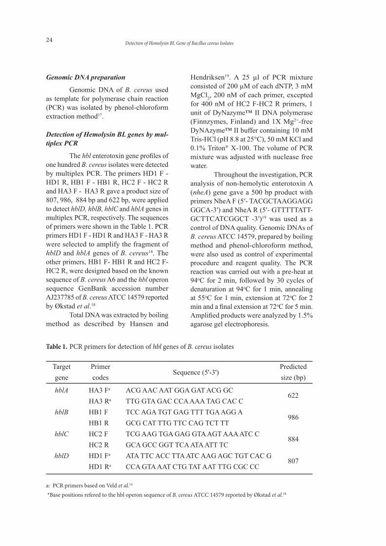
Table 1. PCR primers for detection of hbl genes of B. cereus isolates
Target Primer Predicted gene codes
Sequence (5'-3') size (bp)
hblA HA3 Fa ACG AAC AAT GGA GAT ACG GC 622
HA3 Ra TTG GTA GAC CCA AAA TAG CAC C hblB HB1 F TCC AGA TGT GAG TTT TGA AGG A
986 HB1 R GCG CAT TTG TTC CAG TCT TT hblC HC2 F TCG AAG TGA GAG GTA AGT AAA ATC C
884 HC2 R GCA GCC GGT TCA ATA ATT TC hblD HD1 Fa ATA TTC ACC TTA ATC AAG AGC TGT CAC G
807 HD1 Ra CCA GTA AAT CTG TAT AAT TTG CGC CC
a: PCR primers based on Veld et al.14
*Base positions refered to the hbl operon sequence of B. cereus ATCC 14579 reported by Økstad et al.18
C. Wiwat and R. Thiramanas 25
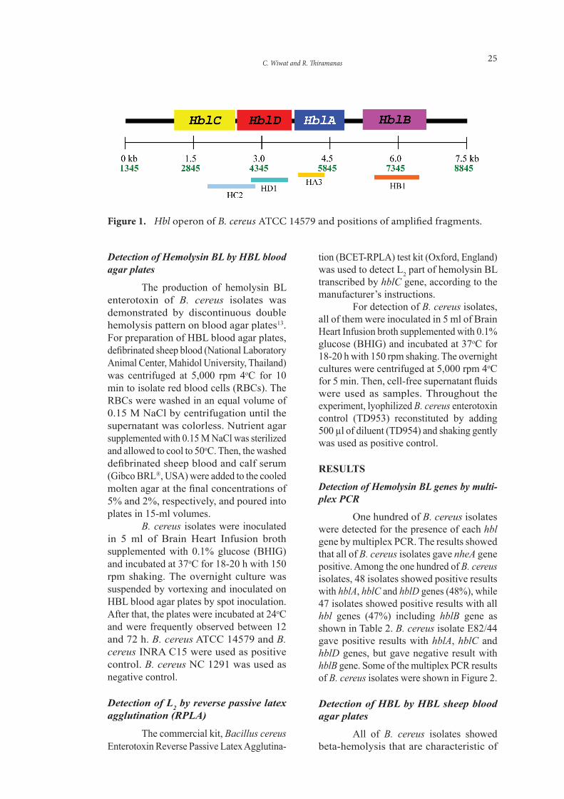
Figure 1. Hbl operon of B. cereus ATCC 14579 and positions of amplified fragments.
Detection of Hemolysin BL by HBL blood agar plates
The production of hemolysin BL enterotoxin of B. cereus isolates was demonstrated by discontinuous double hemolysis pattern on blood agar plates13. For preparation of HBL blood agar plates, defibrinated sheep blood (National Laboratory Animal Center, Mahidol University, Thailand) was centrifuged at 5,000 rpm 4oC for 10 min to isolate red blood cells (RBCs). The RBCs were washed in an equal volume of 0.15 M NaCl by centrifugation until the supernatant was colorless. Nutrient agar supplemented with 0.15 M NaCl was sterilized and allowed to cool to 50oC. Then, the washed defibrinated sheep blood and calf serum (Gibco BRL®, USA) were added to the cooled molten agar at the final concentrations of 5% and 2%, respectively, and poured into plates in 15-ml volumes. B. cereus isolates were inoculated in 5 ml of Brain Heart Infusion broth supplemented with 0.1% glucose (BHIG) and incubated at 37oC for 18-20 h with 150 rpm shaking. The overnight culture was suspended by vortexing and inoculated on HBL blood agar plates by spot inoculation. After that, the plates were incubated at 24oC and were frequently observed between 12 and 72 h. B. cereus ATCC 14579 and B. cereus INRA C15 were used as positive control. B. cereus NC 1291 was used as negative control.
Detection of L2 by reverse passive latex agglutination (RPLA)
The commercial kit, Bacillus cereus Enterotoxin Reverse Passive Latex Agglutina-
tion (BCET-RPLA) test kit (Oxford, England) was used to detect L2 part of hemolysin BL transcribed by hblC gene, according to the manufacturer’s instructions. For detection of B. cereus isolates, all of them were inoculated in 5 ml of Brain Heart Infusion broth supplemented with 0.1% glucose (BHIG) and incubated at 37oC for 18-20 h with 150 rpm shaking. The overnight cultures were centrifuged at 5,000 rpm 4oC for 5 min. Then, cell-free supernatant fluids were used as samples. Throughout the experiment, lyophilized B. cereus enterotoxin control (TD953) reconstituted by adding 500 µl of diluent (TD954) and shaking gently was used as positive control.
RESULTS
Detection of Hemolysin BL genes by multi-plex PCR
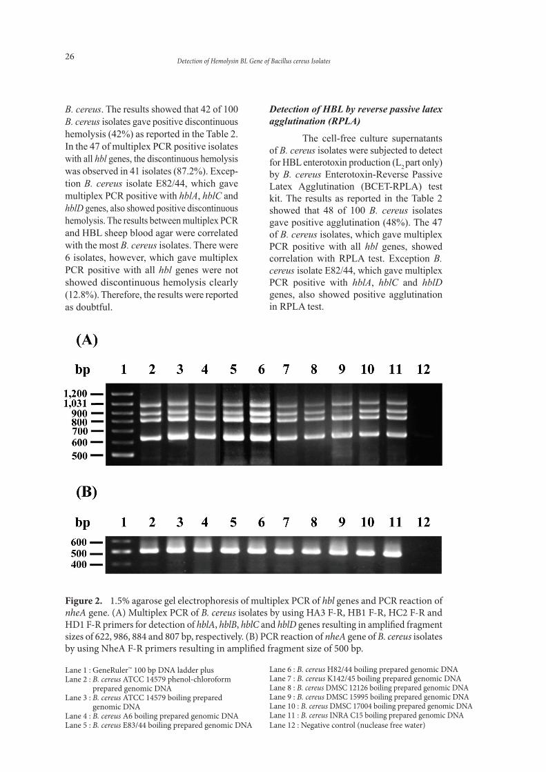
One hundred of B. cereus isolates were detected for the presence of each hbl gene by multiplex PCR. The results showed that all of B. cereus isolates gave nheA gene positive. Among the one hundred of B. cereus isolates, 48 isolates showed positive results with hblA, hblC and hblD genes (48%), while 47 isolates showed positive results with all hbl genes (47%) including hblB gene as shown in Table 2. B. cereus isolate E82/44 gave positive results with hblA, hblC and hblD genes, but gave negative result with hblB gene. Some of the multiplex PCR results of B. cereus isolates were shown in Figure 2.
Detection of HBL by HBL sheep blood agar plates
All of B. cereus isolates showed beta-hemolysis that are characteristic of
Detection of Hemolysin BL Gene of Bacillus cereus Isolates26
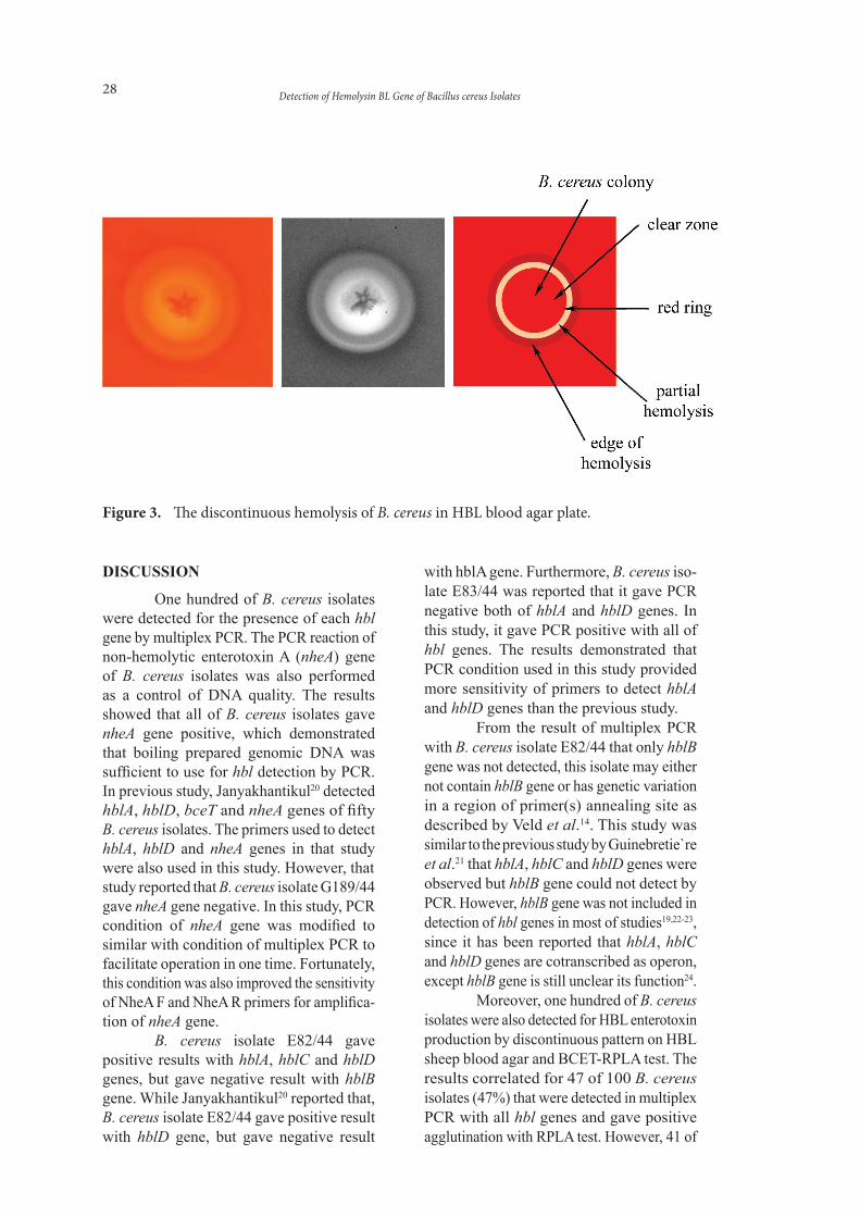
B. cereus. The results showed that 42 of 100 B. cereus isolates gave positive discontinuous hemolysis (42%) as reported in the Table 2. In the 47 of multiplex PCR positive isolates with all hbl genes, the discontinuous hemolysis was observed in 41 isolates (87.2%). Excep-tion B. cereus isolate E82/44, which gave multiplex PCR positive with hblA, hblC and hblD genes, also showed positive discontinuous hemolysis. The results between multiplex PCR and HBL sheep blood agar were correlated with the most B. cereus isolates. There were 6 isolates, however, which gave multiplex PCR positive with all hbl genes were not showed discontinuous hemolysis clearly (12.8%). Therefore, the results were reported as doubtful.
Detection of HBL by reverse passive latex agglutination (RPLA)
The cell-free culture supernatants of B. cereus isolates were subjected to detect for HBL enterotoxin production (L2 part only) by B. cereus Enterotoxin-Reverse Passive Latex Agglutination (BCET-RPLA) test kit. The results as reported in the Table 2 showed that 48 of 100 B. cereus isolates gave positive agglutination (48%). The 47 of B. cereus isolates, which gave multiplex PCR positive with all hbl genes, showed correlation with RPLA test. Exception B. cereus isolate E82/44, which gave multiplex PCR positive with hblA, hblC and hblD genes, also showed positive agglutination in RPLA test.
Figure 2. 1.5% agarose gel electrophoresis of multiplex PCR of hbl genes and PCR reaction of nheA gene. (A) Multiplex PCR of B. cereus isolates by using HA3 F-R, HB1 F-R, HC2 F-R and HD1 F-R primers for detection of hblA, hblB, hblC and hblD genes resulting in amplified fragment sizes of 622, 986, 884 and 807 bp, respectively. (B) PCR reaction of nheA gene of B. cereus isolates by using NheA F-R primers resulting in amplified fragment size of 500 bp.
Lane 1 : GeneRuler™ 100 bp DNA ladder plusLane 2 : B. cereus ATCC 14579 phenol-chloroform prepared genomic DNALane 3 : B. cereus ATCC 14579 boiling prepared genomic DNALane 4 : B. cereus A6 boiling prepared genomic DNALane 5 : B. cereus E83/44 boiling prepared genomic DNA
Lane 6 : B. cereus H82/44 boiling prepared genomic DNALane 7 : B. cereus K142/45 boiling prepared genomic DNALane 8 : B. cereus DMSC 12126 boiling prepared genomic DNALane 9 : B. cereus DMSC 15995 boiling prepared genomic DNALane 10 : B. cereus DMSC 17004 boiling prepared genomic DNALane 11 : B. cereus INRA C15 boiling prepared genomic DNALane 12 : Negative control (nuclease free water)
C. Wiwat and R. Thiramanas 27
HBL
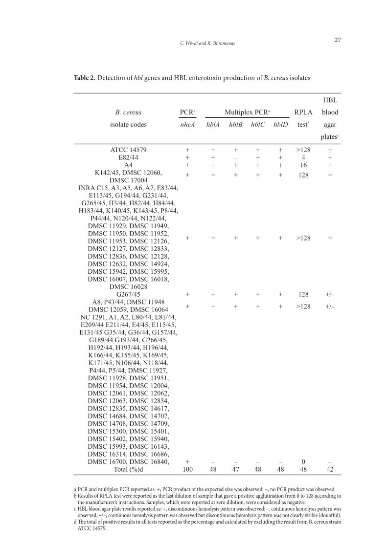
B. cereus PCRa Multiplex PCRa RPLA blood
isolate codes nheA hblA hblB hblC hblD testb agar
platesc
ATCC 14579 + + + + + >128 + E82/44 + + – + + 4 + A4 + + + + + 16 + K142/45, DMSC 12060, DMSC 17004
+ + + + + 128 +
INRA C15, A3, A5, A6, A7, E83/44, E113/45, G194/44, G231/44, G265/45, H3/44, H82/44, H84/44, H183/44, K140/45, K143/45, P8/44, P44/44, N120/44, N122/44, DMSC 11929, DMSC 11949, DMSC 11950, DMSC 11952,
+ + + + + >128 + DMSC 11953, DMSC 12126, DMSC 12127, DMSC 12833, DMSC 12836, DMSC 12128, DMSC 12632, DMSC 14924, DMSC 15942, DMSC 15995, DMSC 16007, DMSC 16018, DMSC 16028 G267/45 + + + + + 128 +/– A8, P43/44, DMSC 11948
+ + + + + >128 +/– DMSC 12059, DMSC 16064 NC 1291, A1, A2, E80/44, E81/44, E209/44 E211/44, E4/45, E115/45, E131/45 G35/44, G36/44, G157/44, G189/44 G193/44, G266/45, H192/44, H193/44, H196/44, K166/44, K155/45, K169/45, K171/45, N106/44, N118/44, P4/44, P5/44, DMSC 11927, DMSC 11928, DMSC 11951, DMSC 11954, DMSC 12004, DMSC 12061, DMSC 12062, DMSC 12063, DMSC 12834, DMSC 12835, DMSC 14617, DMSC 14684, DMSC 14707, DMSC 14708, DMSC 14709, DMSC 15300, DMSC 15401, DMSC 15402, DMSC 15940, DMSC 15993, DMSC 16143, DMSC 16314, DMSC 16686, DMSC 16700, DMSC 16840, + – – – – 0 – Total (%)d 100 48 47 48 48 48 42
Table 2. Detection of hbl genes and HBL enterotoxin production of B. cereus isolates
a PCR and multiplex PCR reported as: +, PCR product of the expected size was observed; –, no PCR product was observed.b Results of RPLA test were reported as the last dilution of sample that gave a positive agglutination from 0 to 128 according to the manufacturer’s instructions. Samples, which were reported at zero dilution, were considered as negative.c HBL blood agar plate results reported as: +, discontinuous hemolysis pattern was observed; –, continuous hemolysis pattern was observed; +/–, continuous hemolysis pattern was observed but discontinuous hemolysis pattern was not clearly visible (doubtful).d The total of positive results in all tests reported as the percentage and calculated by excluding the result from B. cereus strain ATCC 14579.
Detection of Hemolysin BL Gene of Bacillus cereus Isolates28
DISCUSSION
One hundred of B. cereus isolates were detected for the presence of each hbl gene by multiplex PCR. The PCR reaction of non-hemolytic enterotoxin A (nheA) gene of B. cereus isolates was also performed as a control of DNA quality. The results showed that all of B. cereus isolates gave nheA gene positive, which demonstrated that boiling prepared genomic DNA was sufficient to use for hbl detection by PCR. In previous study, Janyakhantikul20 detected hblA, hblD, bceT and nheA genes of fifty B. cereus isolates. The primers used to detect hblA, hblD and nheA genes in that study were also used in this study. However, that study reported that B. cereus isolate G189/44 gave nheA gene negative. In this study, PCR condition of nheA gene was modified to similar with condition of multiplex PCR to facilitate operation in one time. Fortunately, this condition was also improved the sensitivity of NheA F and NheA R primers for amplifica-tion of nheA gene. B. cereus isolate E82/44 gave positive results with hblA, hblC and hblD genes, but gave negative result with hblB gene. While Janyakhantikul20 reported that, B. cereus isolate E82/44 gave positive result with hblD gene, but gave negative result
with hblA gene. Furthermore, B. cereus iso-late E83/44 was reported that it gave PCR negative both of hblA and hblD genes. In this study, it gave PCR positive with all of hbl genes. The results demonstrated that PCR condition used in this study provided more sensitivity of primers to detect hblA and hblD genes than the previous study. From the result of multiplex PCR with B. cereus isolate E82/44 that only hblB gene was not detected, this isolate may either not contain hblB gene or has genetic variation in a region of primer(s) annealing site as described by Veld et al.14. This study was similar to the previous study by Guinebretie`re et al.21 that hblA, hblC and hblD genes were observed but hblB gene could not detect by PCR. However, hblB gene was not included in detection of hbl genes in most of studies19,22-23, since it has been reported that hblA, hblC and hblD genes are cotranscribed as operon, except hblB gene is still unclear its function24. Moreover, one hundred of B. cereus isolates were also detected for HBL enterotoxin production by discontinuous pattern on HBL sheep blood agar and BCET-RPLA test. The results correlated for 47 of 100 B. cereus isolates (47%) that were detected in multiplex PCR with all hbl genes and gave positive agglutination with RPLA test. However, 41 of
Figure 3. The discontinuous hemolysis of B. cereus in HBL blood agar plate.
C. Wiwat and R. Thiramanas 29
47 isolates (87.2%) produced discontinuous hemolysis on HBL sheep blood agar. The 52 of 100 isolates (52%) gave negative results in all of tests. Exception B. cereus isolate E82/44, which gave multiplex PCR positive with hblA, hblC and hblD genes and showed positive agglutination with RPLA test, also produced discontinuous hemolysis. This is possible according to the study indicated that only three hbl genes, hblA, hblC and hblD genes are required for the production of discontinuous hemolysis pattern on blood agar10. There were 6 of 47 isolates (12.8%), however, which gave multiplex PCR positive with all hbl genes and showed positive agglutination in RPLA test were not clearly produced discontinu-ous hemolysis. Therefore, the results were reported as doubtful. Beecher and Wong13 described that the discontinuous hemolysis pattern appeared transiently for most isolates, because of the central continuous pattern expanded with increasing time and obscured the discontinuous pattern. In addition, the discontinuous pattern was masked by other hemolysin generated by B. cereus. It has been reported that the discontinuous hemolysis pattern on blood agar was only visible at a limited of time and this method was not deserved as a reliable tool for HBL detection14. Therefore, the discontinuous pattern not observed in these 6 isolates may be resulted from one or all of those reasons. This study provided genetic and phenotypic informations of B. cereus isolates in Thailand including hbl gene profiles and HBL enterotoxin production. The potential multiplex PCR condition for detection all of hbl genes in B. cereus isolates by operation in one time was established. Suggestion for further study should be emphasized on the sequence, analyse the hemolysin BL operon of B. cereus local isolates and compare with other sequences reported in the database, characterization, cloning, expression, purification and production of these recombinant proteins, for development a rapid test system using specific antibodies toward the hemolysins.
ACKNOWLEDGEMENTS
This study was supported by the Center for Medical Biotechnology, Ministry of Public Health, Thailand.
REFERENCES 1. Kramer JM, Gilbert RJ. Bacillus cereus and other Bacillus species. In: Doyle MP, ed. Foodbome Bacterial Pathogens, New York: Marcel Dekker, 1989; 21-70. 2. Pirttijarvi TSM, Andersson MA, Salkinoja- Salonen MS. Properties of Bacillus cereus and other bacilli contaminating biomaterial- based industrial processes. Int J Food Microbiol 2000; 60:231-9. 3. Granum PE, Lund T. Bacillus cereus and its food poisoning toxins. FEMS Microbiol Lett 1997; 157:223-8. 4. Turnbull PCB, Kramer JM, Jorgensen KR, Gilbert J, Melling J. Properties and production characteristics of vomiting, diarrheal, and necrotizing toxins of Bacillus cereus. Am J Clin Nutr 1979; 32:219-28. 5. Agata N, Ohta M, Arakawa Y, Mori M. The bceT gene of Bacillus cereus encodes an enterotoxin protein. Microbiology 1995; 141:983-8. 6. Notermans S, Batt CA. A risk assessment approach for foodborne Bacillus cereus and its toxins. J Appl Microbiol Symp Suppl 1998; 84:51S-61S. 7. Choma C, Granum PE. The enterotoxin T (BceT) from Bacillus cereus can probably not contribute to food poisoning. FEMS Microbiol Lett 2002; 217:115-9. 8. Beecher DJ, Schoeni JL, Wong AC. Enterotoxic activity of hemolysin BL from Bacillus cereus. Infect Immun 1995; 63:4423-8. 9. Sneath PHA. Endospore-forming Gram- positive rods and cocci, In Sneath PH, Mair NS, Sharp ME, Holt JG, eds. Bergey’s manual of systematic bacteriology. Baltimore: Williams and Wilkins, 1986; 1104-207. 10. Beecher DJ, Wong AC. Improved purifi- cation and characterization of hemolysin BL, a hemolytic dermonecrotic vascular permeability factor from Bacillus cereus. Infect Immun 1994; 62:980-6.
Detection of Hemolysin BL Gene of Bacillus cereus Isolates30
11. Burgess G, Horwood P. Development of improved molecular detection methods for Bacillus cereus toxins. Research and Development Corporation, Australia, 2006. 12. Mantynen V, Lindstrom K. A rapid PCR- based DNA test for enterotoxic Bacillus cereus. Appl Environ Microbiol 1998; 64:1634-9. 13. Beecher DJ, Wong AC. Identification of hemolysin BL producing Bacillus cereus isolates by a discontinuous hemolytic pattern in blood agar. Appl Environ Microbiol 1994; 60:1646-51. 14. Veld PH, Ritmeester WS, Asch ED, Dufrenne JB, Wernars K, Smit E, Leusden FM. Detection of genes encoding for enterotoxins and determination of the production of enterotoxins by HBL blood plates and immunoassays of psychrotrophic strains of Bacillus cereus isolated from pasteurized milk. Int J Food Microbiol 2001; 64:63-70. 15. Beecher DJ, Wong AC. Identification and analysis of the antigens detected by two commercial Bacillus cereus diarrheal enterotoxin immunoassay kits. Appl Environ Microbiol 1994; 60:4614-6. 16. Boonchaisuk R. Development of DNA- probe for detection of enterotoxic Bacillus cereus. M.Sc. Thesis, Mahidol University. Thailand, 2004. 17. Ausubel FM, Brent R, Kingston RE, Moore DD, Scidman JD, Smith JA. Short Protocols in Molecular Biology, 4th ed. New York: John Wiley and Sons, 1999.
18. Økstad OA, Gominet M, Purnelle B, Rose M, Lereclus D, Kolstø AB. Sequence analysis of three Bacillus cereus loci carrying PIcR-regulated genes encoding degradative enzymes and enterotoxin. Microbiology 1999; 145:3129-38. 19. Hansen BM, Hendriksen NB. Detection of enterotoxic Bacillus cereus and Bacillus thuringiensis strains by PCR analysis. Appl Environ Microbiol 2001; 67:185-9. 20. Janyakhantikul S. PCR-based assay for detection of enterotoxin genes from Bacillus cereus. M.Sc. Thesis, Mahidol University. Thailand, 2003. 21. Guinebretière MH, Brousolle V, Nguyen C. The Enterotoxigenic profiles of food- poisoning and food-borne Bacillus cereus strains. J Clin Microbiol 2002; 40:3053-6. 22. Phelps RJ, Mckillip JL. Enterotoxin Production in Natural Isolates of Bacillaceae outside the Bacillus cereus Group. Appl Environ Microbiol 2002; 68:3147-51.23. Corona A, Fois MP, Mazzette R, De Santis EPL. A New Multiplex PCR for the Detection of hbl Genes in Strains of the ‘Bacillus cereus Group’. Vet Res Com 2003; 1:679-82. 24. Lund T, Granum PE. Comparison of biological effect of the two different enterotoxin complexes isolated from three different strains of Bacillus cereus. Microbiol 1997; 143:3329-36.