detection of ribosomal peptides in cyanobacterial strains · detection of ribosomal peptides in...
TRANSCRIPT
Detection of Ribosomal Peptides in
Cyanobacterial Strains
RAQUEL MARIA SANTOS CHAVES
TESE DE MESTRADO APRESENTADA
AO INSTITUTO DE CIÊNCIAS BIOMÉDICAS ABEL SALAZAR
DA UNIVERSIDADE DO PORTO EM
TOXICOLOGIA E CONTAMINAÇÃO AMBIENTAIS
M 2013

Raquel Maria Santos Chaves
Detection of Ribosomal Peptides
in Cyanobacterial Strains
Dissertação de Candidatura ao grau de Mestre em
Toxicologia e Contaminação Ambientais
submetida ao Instituto de Ciências
Biomédicas de Abel Salazar da Universidade
do Porto
Orientador - Doutor Pedro Leão
Categoria – Postdoctoral fellow
Afiliação - Centro Interdisciplinar de Investigação
Marinha e Ambiental
Co-orientador – Doutor Vitor Vasconcelos
Categoria – Professor Catedrático
Afiliação - Centro Interdisciplinar de Investigação
Marinha e Ambiental/ Faculdade de Ciências
da Universidade do Porto.


i
Acknowledgments
This work was carried out at LEGE- Laboratory of Ecotoxicology, Genomics and
Evolution, CIIMAR-Interdisciplinary Centre of Marine and Environmental Research,
University of Porto and at Division of Microbiology and Biotechnology, Department of
Food and Environmental Sciences, University of Helsinki. In that way I wish to warmly
thank my supervisors Vitor Vasconcelos, Kaarina Sivonen, Pedro Leão and David Fewer.
In particular, I want to express my gratitude to Vitor Vasconcelos and Kaarina Sivonen
that they took me in their research groups as Master’s student. Special thanks to Pedro
Leão and David Fewer for the practical supervision and advice.
I want to thank the cyanogroup , from University of Helsinki for all support and for
the good work atmosphere. Special thanks to Niina Leikoski for the guidance and advice
in molecular analysis, Lyudmila Saari, for the technical assistance, and Matti Wahlsten
and Juoni Jokela for guidance in the chemical methods. I want also to thank to Tania
Shishido, Sebastian Coloma, Saara Suominen and Kristel Panksep for all professional
and personal support, for helping me to integrate in a new city and for their friendship.
I would like to sincerely thank to all LEGE team, for the professional guidance, for all
good moments together and for the friendly work atmosphere. Special thanks to Joana
Martins, for the advice and their solicitude to help me; Micaela Vale, Joao Morais, Vitor
Ramos, Margarida Costa, Sofia Costa and Dina Gomes for the help and availability in the
daily work and also for the good mood each work day. I wish to thank to both teams for
allowing me to have such a great experience and to broaden up my views as a student
and as a person.
I’m very grateful to my family for all support, motivation and for always believe in me
and in my potential. Thank you so much for being present always.
Very special thanks to all my friends, for being there always, in good and hard
moments and for pushing me up, specially to Rita Vasconcelos, Sara Freitas, Tiago
Torres, João Pedro Pereira and Pedro Silva. For some friends that I met in Finland,
specially Nuria Puigpinós, Anna Yukhtenko and Marjolein Jozef for all good moments
travelling, as foreign students. I also wish to thank to my friends from Teatro Universitário
do Porto and dance academy for cheering me up and motivating me.

iii
Abstract
Cyanobacteria produce a variety of secondary metabolites of peptidic nature, many
of which with potent biological activities. These can be produced either ribosomally or
non- ribosomally. Cyanobactins are a group of recently described cyclic peptides with low
molecular weight, synthesized by ribosomal pathways that exhibit important bioactivities,
such as antitumor, cytotoxic or multi-drug-reversing activities.
Anacyclamides are a set of cyclic cyanobactins, which have been described from
strains belonging to the Anabaena genus.
The main objective of this work was the detection of anacyclamide- related genes in
cyanobacteria from the culture collection of LEGE (LEGE CC) in an attempt to expand the
diversity of anacyclamides know to date. In addition, we tried to characterize the
cyanobacterial strains using both morphological and molecular data, with the ultimate goal
of making phylogenetic inferences regarding cyanobactins production.
For this effect, we conducted a molecular screening among Anabaena strains, for a
gene involved in the synthesis of the ribosomal peptides cyanobactins (patE). As a result,
we detected a putative new anacyclamide in two of the ten Anabaena strains. Sequencing
of the patE gene allowed us to predict the amino acid sequence of a new anacyclamide.
The presence of the new compound in the cultures was checked using LC-MS, which
additionally suggested that the cyanobactin was post-translationally modified through two
prenylation events.
By expanding the known genetic and chemical diversity of anacyclamides this work can
help us to know more about the cyanobactins. In particular, this new anacyclamide can
aid in investigations on post-translational processing of cyclic peptides derived from these
pathways.
iv
Resumo
As cianobactérias produzem uma variedade de metabolitos secundários de natureza
peptídica, muitos dos quais apresentam forte bioactividade. Estes biocompostos podem
ser produzidos quer por via ribossomal quer por via não ribossomal. As cianobactinas são
um grupo de péptidos cíclicos recentemente descobertos, com baixo peso molecular,
sintetizados por ribossomas e que apresentam importantes bioactividades, tais como
actividade citotóxica, anti tumoral ou reversora de multi drogas.
Neste sentido, o objectivo principal deste trabalho foi a detecção dos genes relacionados
com a produção de anaciclamidas em cianobactérias provenientes da colecção de
culturas LEGE (LEGE CC), com a consequente identificação de novas anaciclamidas. Da
mesma forma, foi feita a caracterização morfológica e o rastreio molecular das estirpes de
cianobactérias, tendo como ultimo objectivo a análise filogenética, atendendo à produção
de cianobactinas por parte das mesmas.
Para este efeito, o rastreio molecular teve como alvo o gene envolvido na síntese dos
péptidos ribossomais (cianobactinas) – patE. Como resultado, detectámos uma nova
anaciclamida em duas das dez estirpes analisadas.
A sequenciação do gene patE permitiu-nos prever a presença da anaciclamida, bem
como a sua sequência de amino ácidos, em duas das dez estirpes em estudo. A
presença desta nova anaciclamida nas culturas foi confirmada usando a LC-MS. A
utilização desta técnica também demostrou que a nova cyanobactina apresentava
modificações pós tradução bem como dois grupos -prenil na sua constituição.
Assim, este trabalho ajudou a aumentar o conhecimento sobre este grupo de péptidos, de
baixo peso molecular e com grandes variações estruturais. O estudo da nova
anaciclamida pode estender-nos o conhecimento em relação às modificações pós
tradução, que caracterizam estes péptidos, particularmente sobre prenilação.
v
Contents
Acknowledgments ............................................................................................................... i
Abstract ............................................................................................................................ iii
Resumo ............................................................................................................................ iv
Contents ............................................................................................................................ v
Tables Index .................................................................................................................... vii
Figures Index .................................................................................................................. viii
Abbreviations .................................................................................................................... xi
1. Introduction ................................................................................................................ 1
1.2. Cyanobacteria .................................................................................................... 1
1.2.1. Cyanobacteria toxic effects .......................................................................... 2
1.3. Cyanobactins ...................................................................................................... 3
1.3.1. Analogous pathways .................................................................................... 4
1.3.2. Chemical diversity and Occurrence ............................................................. 5
1.3.3. Biosynthesis of cyanobactins ....................................................................... 7
1.3.4. Bioactivities ................................................................................................10
1.4. Anacyclamides ..................................................................................................11
1.5. Anabaena ..........................................................................................................12
1.6. Biotechnological Aspects ...................................................................................13
1.7. Objective ...........................................................................................................14
2. Material and Methods ..................................................................................................15
vi
2.1. Source of strains ...................................................................................................15
2.2. Cyanobacterial strains and culturing ..................................................................16
2.3. DNA extraction, PCR amplification, Cloning and sequencing .............................17
2.4. Cloning the acy biosynthetic gene cluster ..........................................................19
2.5. Biomass collection of LEGE 00248 and LEGE 00259 and preparation of extracts
20
2.5.1. Biomass extraction .....................................................................................21
2.6. Chemical analysis ..............................................................................................21
3. Results and Discussion ................................................................................................23
3.1. Morphological characterization ..............................................................................23
3.1.1. Morphometry ...................................................................................................25
3.2. Genomic DNA extraction, PCR amplification, cloning and sequencing ..................26
3.3. Chemical analysis .................................................................................................35
4. Conclusion and future work ..........................................................................................41
5. References ..................................................................................................................43
vii
Tables Index
Table 1- Identification of the ten strains in study, from the LEGE culture collection ..........16
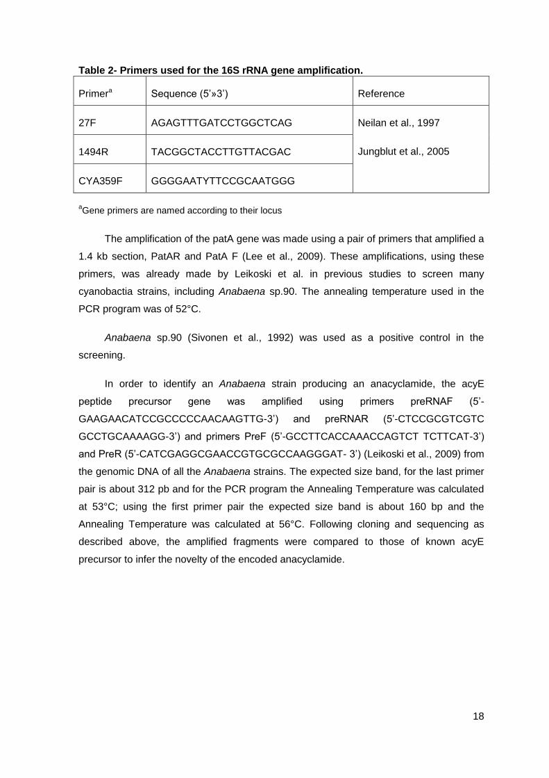
Table 2- Primers used for the 16S rRNA gene amplification. ...........................................18
Table 3- PCR reaction for each primers pair ....................................................................19
Table 4- Morphometric parameters for each strain ...........................................................25
Table 5- Anacyclamide predictions by each strain in study ..............................................33
Table 6- Anacyclamides and their detection by LC-MS, in two Anabaena strains ............35
Table 7- Extraction yields for LEGE 00248 and LEGE 00259 strains ...............................39
viii
Figures Index
Figure 1- Schematic of post-ribosomal peptide synthesis (PRPS) (Adapted from Arniston
et al., 2013 and Leikoski et al., 2013) ................................................................................ 4
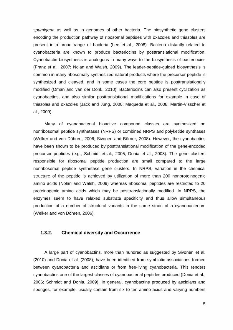
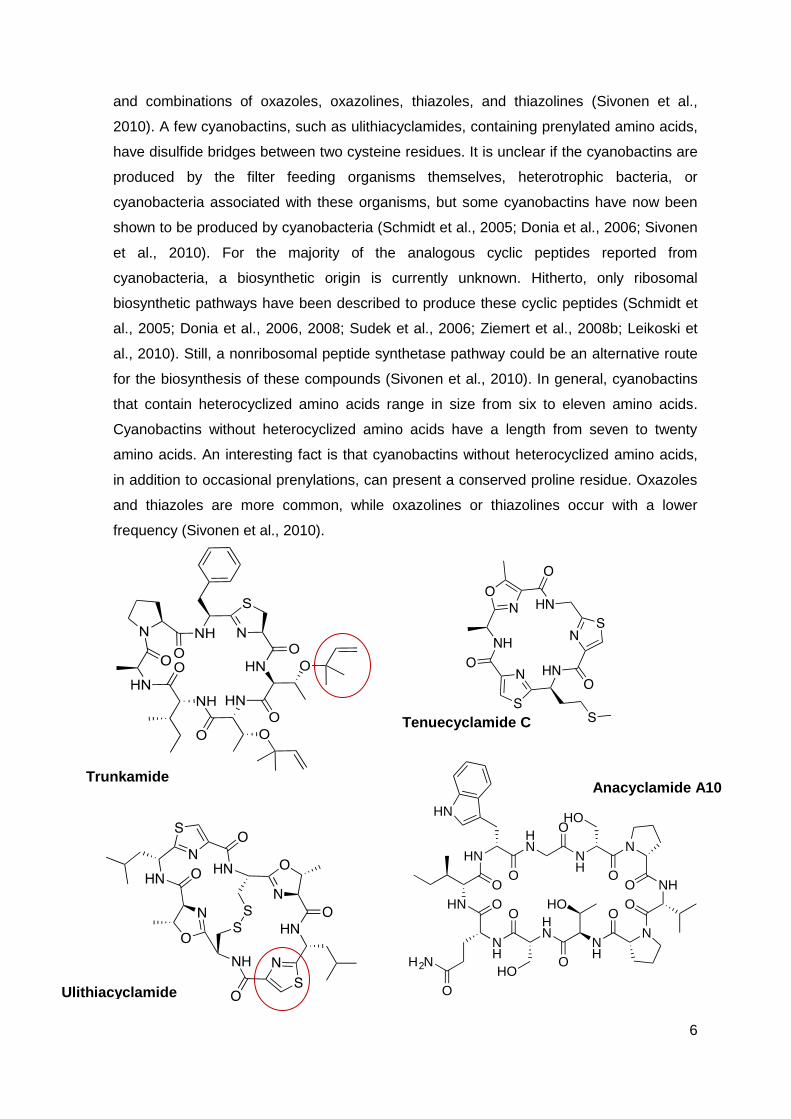
Figure 2- The chemical structures of a selection of cyanobactins. Trunkamide was isolated
from L. patella, tenuecyclamide from N. spongiaeforme, anacyclamide from Anabaena,
trichamide from T. erythraeum, ulithiacyclamide and patellamide from Prochloron(
Originally from L. Patella); Viridisamide from Oscillatoria viridis PCC7112.(Adapted from
Sivonen el al., 2010 and Leikoski et al., 2013). The post-translational modifications;
oxazoline, thiazole, and reverse O-prenyl are highlighted with circles. In the linear
cyanobactin, the identical prenylated N-termini and methylated C-termini bound to
thiazoles are in blue. (Adapted from Sivonen et al., 2010; Leikoski et al., 2013) ............... 7
Figure 3- Schematic figure of the cyanobactin gene cluster (homologous to the pat gene
cluster1) and the structure of the precursor peptide (E). The functions of the genes are
shown. The genes B and C are of unknown function. The cyanobactin structures are
formed from the core region shown in the precursor peptide. (Adapted from Houssen el
al., 2012; 1 Donia et al., 2008; 1Schmidt et al., 2005) ........................................................ 9
Figure 4- Cyanobactin gene clusters published from seven distantly related cyanobacteria.
These gene clusters are typified by genes encoding proteases (yellow), a short precursor
peptide (red), proteins involved in the maturation of the cyanobactin (black), as well as
conserved and hypothetical open reading frames (white). (Adapted from Sivonen et al.,
2010) ...............................................................................................................................10
Figure 5- Arquitecture of the anacyclamide gene cluster (Houssen el al., 2012). .............11
Figure 6- Heterocysts development (1) in Anabaena LEGE 00259, one of the strains used
in this work. ......................................................................................................................12
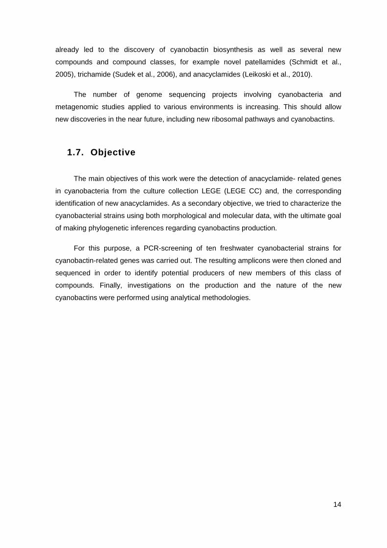
Figure 7- Schematic drawing of the workflow. ..................................................................15
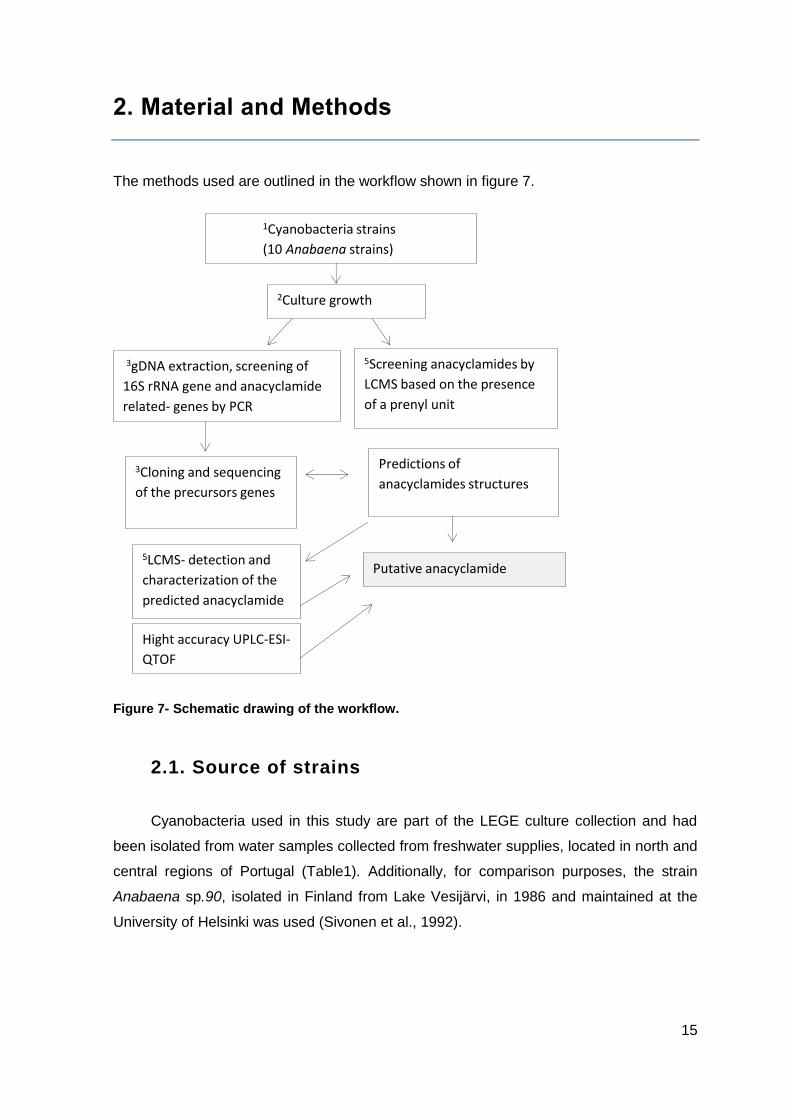
Figure 8- Cyanobacterial strains and Culturing Anabaena sp. Strains were grown in z8
medium, with aeration, under 14:10 light:dark at 25°C. ....................................................17
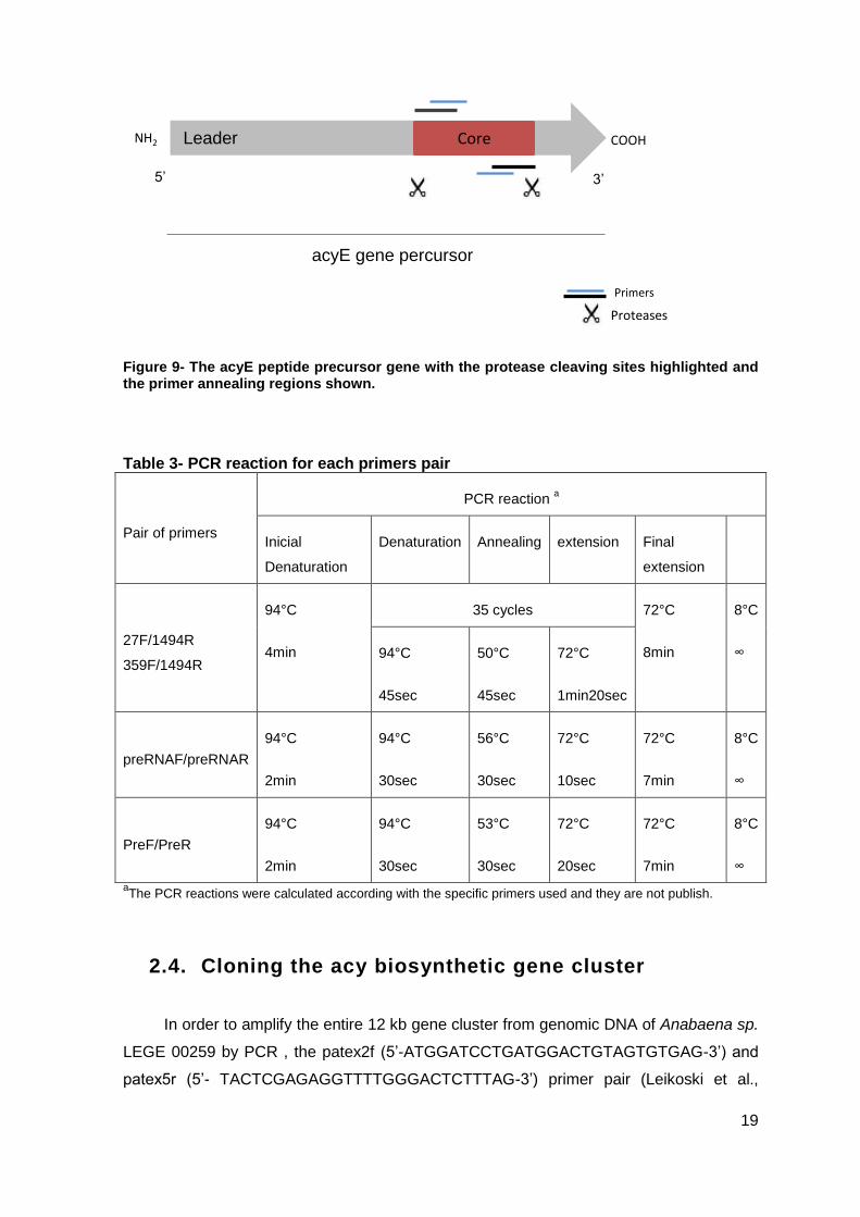
Figure 9- The acyE peptide precursor gene with the protease cleaving sites highlighted
and the primer annealing regions shown. .........................................................................19
ix
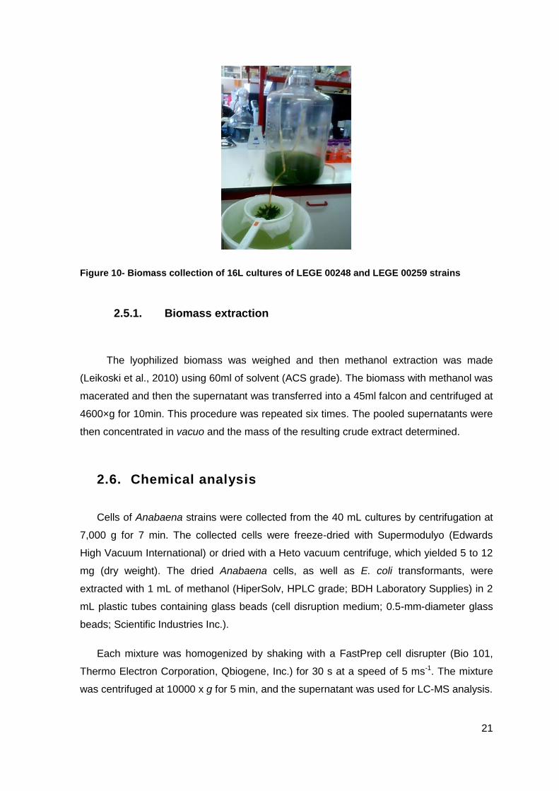
Figure 10- Biomass collection of 16L cultures of LEGE 00248 and LEGE 00259 strains .21
Figure 11- Morphological characterization of the Anabaena strains, using an optical
microscopy approach. 1- LEGE 00233, 2- LEGE 00241, 3- LEGE 00243, 4- LEGE 00245,
5- LEGE 00246, 6- LEGE 00248, 7- LEGE 00250, 8- LEGE 00253, 9-LEGE 00259, 10-
04289. .............................................................................................................................24
Figure 12- Amplification of the 16S rRNA gene with the primers 359F- 1494R(up); 27F-
1494R (down) for all ten strains. Samples: 3µl; 1kb plus DNA ladder: 1,5µl. 1,5% agarose
gel; 1- LEGE 00233, 2- LEGE 00241, 3- LEGE 00243, 4- LEGE 00245, 5- LEGE 00246,
6- LEGE 00248, 7- LEGE 00250, 8- LEGE 00253, 9-LEGE 00259, 10- 04289. ...............27
Figure 13- Amplification of the patA gene (1.4kb section) with the PatA pair of primers
designed for this effect, by Leikoski et al. All the ten strains are represented. Tann = 52°C.
Samples: 4µl; 10kb GeneRuler DNA ladder: 3µl. 0.7agarose gel. Positive control=
Anabaena sp. 90. .............................................................................................................28
Figure 14- Amplification of patE genes, using primers PreR and PreF, from nine strains.
Tann= 53ºC. Samples: 3µl ,1kb plus DNA ladder: 1.5µl. 1,5% agarose gel; 1- LEGE
00233, 2- LEGE 00241, 3- LEGE 00243, 4- LEGE 00245, 5- LEGE 00246, 6- LEGE
00248, 7- LEGE 00253, 8- LEGE 00259, 9-LEGE 04289 .................................................29
Figure 15- Amplification of the patE genes, using primers PreRNA R and PreRNA F, from
nine strians. Tann=56ºC. Samples: 3µl, 1kb plus DNA ladder: 1.5µl. 1,5% agarose gel; 1-
LEGE 00233, 2- LEGE 00241, 3- LEGE 00243, 4- LEGE 00245, 5- LEGE 00246, 6- LEGE
00248, 7- LEGE 00253, 8- LEGE 00259, 9-LEGE 04289. ................................................29
Figure 16- Alignment of the sequences of the acy precursor from all the strains with the
acy precursor of the Anabaena sp. 90 (acyE). The areas of the precursor protein which
form the mature cyanobactin are in grey ..........................................................................30
Figure 17- (A) AcyE peptide precursor from Anabaena sp. 90 with the hypervariable
region of the 49-aminoacid protein encoding the mature anacyclamide shaded, indicating
the position of cleavage and macrocyclization (grey). (B) Putative structure of the
decapeptide anacyclamide A10 from Anabaena sp. 90 (Leikoski et al., 2010) .................31
Figure 18- Putative structure of ADNEYGYKLDASDNYIP anacyclamide, using the
sequence of AcyE precursor peptide from strain LEGE 00259 (example) ........................32
x
Figure 19- Putative structure MLCDIQTRECTP of anacyclamide, using the sequence of
AcyE precursor peptide from strain LEGE 04289 (example) ............................................33
Figure 20- Ion chromatograms of the ten Anabaena strains filtered for Neutral losses of 68
Da. The two large clusters correspond to the two strains containing the diprenylated novel
anacyclamide ...................................................................................................................36
Figure 21- MS profile TIC to the LEGE 00248 strain ........................................................37
Figure 22- MS profile MS to the LEGE 00248 strain; Retention Time: 18,91min- 19,32min
........................................................................................................................................37
Figure 23- MS profile TIC to the LEGE 00259 strain ........................................................38
Figure 24- MS profile MS to the LEGE 00259 strain; Retention Time: 18.93min-19.38min
........................................................................................................................................38
xi
Abbreviations
Ala (A) Alanine
Arg (R) Arginine
Asn (N) Asparagine
Asp (D) Aspartic acid
ATP- Adenosine-5'-triphosphate
BLAST- Basic Local Alignment Search Tool
bp- base pair
Cys (C) Cysteine
DNA- Deoxyribonucleic acid
Gln (Q) Glutamine
Glu (E) Glutamic acid
Gly (G) Glycine
HIV- Human immunodeficiency vírus
Ile (I) Isoleusine
LC- liquid chromatography
LEGE- Laboratório de Ecologia, Genómica e Evolução
Leu (L) Leusine
Lys (K) Lysine
Met (M) Methionine
MS- mass spectrometry
xii
NMR- nuclear magnetic resonance
NRPS- non-ribosomal peptide synthetase
ORF- Open Reading frame
PCR- polymerase chain reaction
Phe(F) Phenylalanine
Pro (P) Proline
PRPS- post-ribosomal peptide synthesis
PS- photosystem
RiPP- ribosomally synthesized post-translationally modified peptide
rRNA- Ribosomal ribonucleic acid
S.D. - Standard Deviation
Ser (S) Serine
sp- species
Thr(T) Threonine
Trp (W) Tryptophan
Tyr(Y) Tyrosine
Val (V) Valine
1
1. Introduction
Many aquatic organisms are able to produce bioactive secondary metabolites, some
of which with the potential of being used in biotechnology (Arnison et al. 2013; Schmidt et
al. 2005; Sivonen et al, 2010). Cyanobacteria are a particularly rich source of bioactive
secondary metabolites (Sivonen et al., 2008). These include biomedically interesting
compounds, such as the anticancer drug lead cryptophycin (Magarvey et al., 2006) and
environmentally problematic hepatotoxic peptides, such as microcystins , nodularins and
many other toxins produced by bloom-forming cyanobacteria (Sivonen et al.,2008). Many
of these compounds are peptides containing nonproteinogenic amino acids, produced by
nonribosomal peptide synthesis (Sivonen et al., 2008; Welker et al., 2006). Additionally,
modified peptides produced by ribosomal pathways – cyanobactins - have recently been
found in cyanobacteria (Donia et al., 2008; Donia and Schmidt, 2010; Leikoski et al.,
2009, 2010, Schmidt et al., 2005; Sivonen et al., 2010). This study was carried out within
this topic and here we report a novel low-molecular-weight peptide produced ribosomally
by Anabaena strains.
1.2. Cyanobacteria
Cyanobacteria are a phylum of oxygenic photosynthetic prokaryotes that have two
photosystems (PSII and PSI) and use H2O as a photoreductant in photosynthesis. All
known cyanobacteria are photoautotrophic, using primarily CO2 as the carbon source.
(Castenholz, 2001). Many cyanobacterial species are also capable of atmospheric
nitrogen fixation.
Cyanobacteria have a gram-negative cell wall, which includes two distinct
membranes, the plasma membrane and an outer membrane, and a peptidoglycan layer,
which is thicker than in other Gram-negative bacteria, between these two membranes
(Hoiczyk and Hansel 2000). External to the cell wall are different layers that protect the
cells from desiccation and presumably from phages and predators. Cyanobacteria contain
extensive internal thylakoid membranes, which are the site of photosynthetic reactions
(Woese, 1987; Hoiczyk and Hansel, 2000; Liberton, 2011).
These organisms are morphologically very diverse, with unicellular, colonial, and
multicellular filamentous forms and can inhabit a wide range of environments including
2
extreme conditions such as hot springs, the poles and, desert soils. Cyanobacterial
species can occupy terrestrial habitats and, more commonly, aqueous environments
(Sivonen et al., 2010). According to the botanical system of classification, cyanobacterial
diversity has been traditionally grouped into five orders, Chroococcales, Pleurocapsales,
Oscillatoriales, Nostocales and Stigonematales, which generally correspond to the five
subsections proposed in the Bergey’s Manual of Systematic Bacteriology (Knoll, 2008;
Whitton, 2008). Cyanobacteria form a distinct group in bacterial tree of life, based on
bacterial 16S rRNA genes (Castenholz, 2001). They are responsible for the production of
many toxins, including the most studied group of cyanobacterial secondary metabolites,
the hepatotoxic microcystins (Sivonen and Jones, 1999). Cyanobacteria are also a rich
source of other secondary metabolites, some of which have interesting antimicrobial, anti-
HIV, anti-malaria and anticancer activities (Burja et al., 2001, Welker and von Döhren
2006, Linington et al., 2007). Of particular interest for the present work, we emphasize the
cyanobactins group of secondary metabolites, which are a group of cyclic peptides
recently found in cyanobacteria (Donia et al., 2008).
1.2.1. Cyanobacteria toxic effects
Some cyanobacteria frequently form mass occurrences (blooms) in aquatic systems
(freshwater as well as marine environments). This phenomenon occurs under favorable
environmental conditions, such as salinity, light, temperature and nutrient concentration
and usually results in the release of the cells’ constituents (which sometimes includes
potent toxins, termed cyanotoxins) to the water body (Béchemin et al., 1999; John and
Flynn, 2000). At this level, eutrophication (natural or anthropogenic) and global warming
have a strong influence in ecosystem health, but also in the economic development and in
public heath, because the population is a consumer of aquatic resources that can be
contaminated with cyanotoxins (John and Flynn, 2000; Paul, 2008).
Studies on planktonic cyanobacteria from freshwaters and marine ecosystems
became more common since the first report of a toxic episode caused by cyanobacteria in
1878 (Francis, 1878; Sivonen and Jones, 1999). Eventually, the study of toxic compounds
produced by cyanobacteria revealed the biotechnological potential of these organisms, in
terms of their production of bioactive compounds. These biocompounds can be of
different chemical families, including lipids, terpenes, glycosides, polyketides and peptides
3
or hybrids of some of these classes (Metting and Pyne, 1986, Rouhiainen et al., 2000;
Tan et al., 2001, Donia et al., 2008, Donia and Schmidt, 2010).
A significant fraction of these cyanobacterial compounds are linear and cyclic
peptides that are produced by either ribosomal or nonribosomal biosynthetic pathways.
The first, ribosomal pathway, was described in 2005 for the cyanobactin patellamide
(Schmidt et al., 2005) while the first nonribosomal gene cluster had been reported earlier
for the cyanotoxin microcystin (Tillet et al., 2000). Many cyanotoxins are partially or fully
produced by nonribosomal pathways and released to the environment, such as
microcystin; nodularin; saxitoxin and cylindrospermopsin. On the other hand,
cyanobactins have been discovered as novel peptides produced ribosomally and their
ecosystem-level impact is currently unknown (Schmidt et al., 2005; Arnison et al., 2013).
1.3. Cyanobactins
The cyanobactins are small peptides with low molecular weight, produced by
various strains of free-living or symbiotic cyanobacteria, in terrestrial, marine or freshwater
environments. (Schmidt et al., 2005; Donia et al., 2006; Sivonen., 2010). In this group are
included compounds with anti-malarial, anti-tumoral and multidrug reversing action (Burja
et al., 2001, Welker and von Döhren, 2006, Linington et al., 2007, Salvatella et al., 2003).
Therefore, cyanobactins are compounds of potential interest to the pharmaceutical
industry. These compounds possess versatile structures and are produced by proteolytic
cleavage and cyclization of hypervariable peptide precursors, coupled with other post-
translational modifications such as heterociclization, prenylation of amino acid or oxidation
(Sivonen et al., 2010; Leikoski et al., 2010). Cyanobactins are ribosomally synthesized
and post-translationally modified peptides (RiPPs), which are produced by a pathway now
designated as post-ribosomal peptide synthesis (PRPS) (Arnison et al., 2013). In PRPS,
an unmodified precursor peptide produced by normal translation on the ribosome,
includes the sequence that will correspond to the end-product peptide, termed core
sequence (Oman and van der Donk, 2010 and Arnison et al., 2013).Subsequently, the
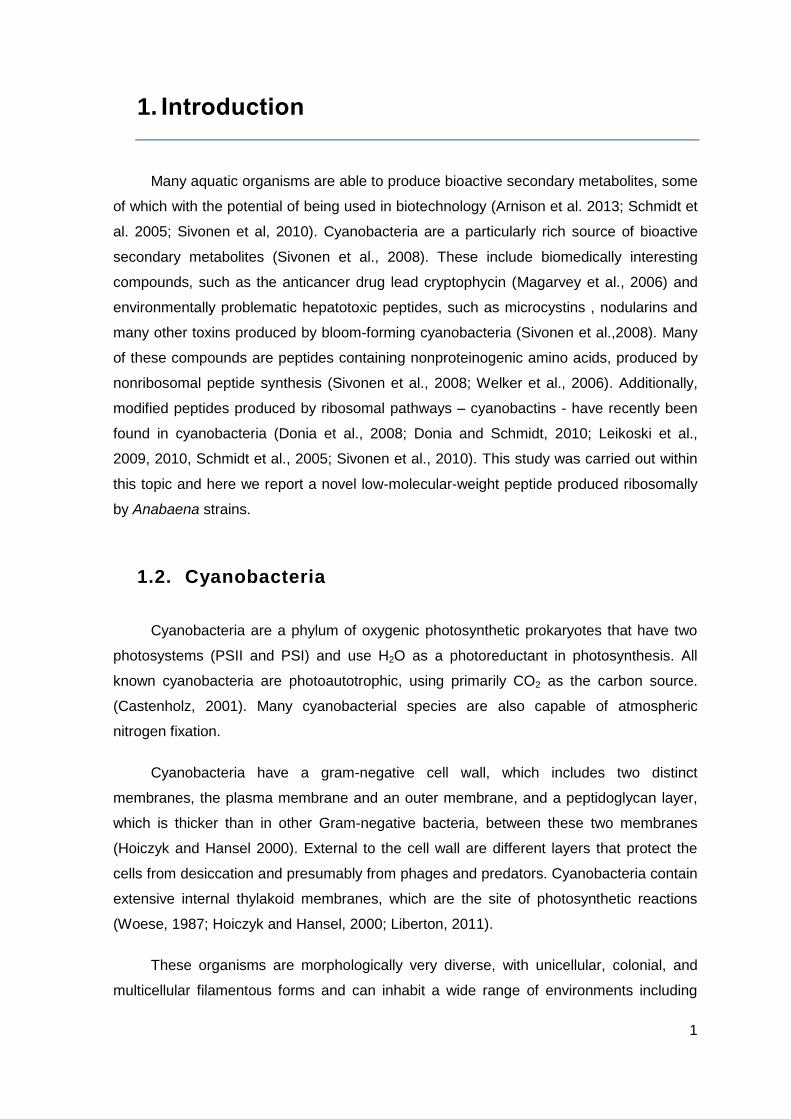
precursor peptide is cleaved and modified to form the final product (Figure1).
The cyanobactin gene cluster usually encodes two proteases responsible for the
cyclization and cleavage of the precursor peptide (Sivonen et al., 2010).
4
Figure 1- Schematic of post-ribosomal peptide synthesis (PRPS) (Adapted from Arniston et al., 2013 and Leikoski et al., 2013)
Initially, cyanobactins were proposed as a group of cyclic peptides containing
oxazolines, thiazolines, or their oxidized derivatives oxazoles and thiazoles (Sivonen et
al., 2010). This definition was modified to include cyclic peptides which consist solely in
proteinogenic amino acids (Leikoski et al., 2010). Sometimes, isoprenoid amino acids
derivatives are also found, for example in trunkamide, patellin, and anacyclamides,
although they are rare (Sivonen et al., 2010). Hence, cyanobactin was proposed as a
collective name for cyclic peptides which can contain heterocyclized amino acids or
isoprenoid amino acid derivatives (Donia et al., 2008; Schmidt and Donia, 2009). Very
recently it has been shown that some cyanobacteria produce short linear cyanobactins
with a chain length ranging from three to five amino acids (Leikoski et al., 2013).
According with the Leikoski et al. (2013) study, these linear cyanobactins were N-
prenylated and O-methylated on N and C termini, respectively.
1.3.1. Analogous pathways
Similarly to cyanobactins, another cyanobacterial peptide class, microviridins, was
recently shown to be ribosomally produced in Microcystis aeruginosa and Planktothrix
agardhii, but their biosynthetic machinery differs that of cyanobactins (Ziemert et al.,
2008a; Philmus et al., 2008; Sivonen et al., 2010). Initially, these compounds were
thought to be products of nonribosomal peptide biosynthesis. However, microviridins are
synthesized from precursor peptides that are converted into tricyclic depsipeptides
through the action of ATP grasp ligases and a transporter peptidase (Ziemert et al.,
2008a; Philmus et al., 2008). The work of Philmus et al. (2008) reported similar gene
clusters in the genomes of Anabaena variabilis, Nostoc punctiforme, and Nodularia
Translation of the precursor gene
Core peptide
NH2 - - COOH
Post-translational modifications
by the modifying enzymes
Percursor peptide
SRiPP
5
spumigena as well as in genomes of other bacteria. The biosynthetic gene clusters
encoding the production pathway of ribosomal peptides with oxazoles and thiazoles are
present in a broad range of bacteria (Lee et al., 2008). Bacteria distantly related to
cyanobacteria are known to produce bacteriocins by posttranslational modification.
Cyanobactin biosynthesis is analogous in many ways to the biosynthesis of bacteriocins
(Franz et al., 2007; Nolan and Walsh, 2009). The leader-peptide-guided biosynthesis is
common in many ribosomally synthesized natural products where the precursor peptide is
synthesized and cleaved, and in some cases the core peptide is posttranslationally
modified (Oman and van der Donk, 2010). Bacteriocins can also present cyclization as
cyanobactins, and also similar posttranslational modifications for example in case of
thiazoles and oxazoles (Jack and Jung, 2000; Maqueda et al., 2008; Martin-Visscher et
al., 2009).
Many of cyanobacterial bioactive compound classes are synthesized on
nonribosomal peptide synthetases (NRPS) or combined NRPS and polyketide synthases
(Welker and von Döhren, 2006; Sivonen and Börner, 2008). However, the cyanobactins
have been shown to be produced by posttranslational modification of the gene-encoded
precursor peptides (e.g., Schmidt et al., 2005; Donia et al., 2008). The gene clusters
responsible for ribosomal peptide production are small compared to the large
nonribosomal peptide synthetase gene clusters. In NRPS, variation in the chemical
structure of the peptide is achieved by utilization of more than 200 nonproteinogenic
amino acids (Nolan and Walsh, 2009) whereas ribosomal peptides are restricted to 20
proteinogenic amino acids which may be posttranslationally modified. In NRPS, the
enzymes seem to have relaxed substrate specificity and thus allow simultaneous
production of a number of structural variants in the same strain of a cyanobacterium
(Welker and von Döhren, 2006).
1.3.2. Chemical diversity and Occurrence
A large part of cyanobactins, more than hundred as suggested by Sivonen et al.
(2010) and Donia et al. (2008), have been identified from symbiotic associations formed
between cyanobacteria and ascidians or from free-living cyanobacteria. This renders
cyanobactins one of the largest classes of cyanobacterial peptides produced (Donia et al.,
2006; Schmidt and Donia, 2009). In general, cyanobactins produced by ascidians and
sponges, for example, usually contain from six to ten amino acids and varying numbers
6
NH
O
HO
HN
O
HO
NH
O
H2N
O
HN
O
HN
O
HN
HN
O
NH
O
HO
N
O NH
O
N
O
and combinations of oxazoles, oxazolines, thiazoles, and thiazolines (Sivonen et al.,
2010). A few cyanobactins, such as ulithiacyclamides, containing prenylated amino acids,
have disulfide bridges between two cysteine residues. It is unclear if the cyanobactins are
produced by the filter feeding organisms themselves, heterotrophic bacteria, or
cyanobacteria associated with these organisms, but some cyanobactins have now been
shown to be produced by cyanobacteria (Schmidt et al., 2005; Donia et al., 2006; Sivonen
et al., 2010). For the majority of the analogous cyclic peptides reported from
cyanobacteria, a biosynthetic origin is currently unknown. Hitherto, only ribosomal
biosynthetic pathways have been described to produce these cyclic peptides (Schmidt et
al., 2005; Donia et al., 2006, 2008; Sudek et al., 2006; Ziemert et al., 2008b; Leikoski et
al., 2010). Still, a nonribosomal peptide synthetase pathway could be an alternative route
for the biosynthesis of these compounds (Sivonen et al., 2010). In general, cyanobactins
that contain heterocyclized amino acids range in size from six to eleven amino acids.
Cyanobactins without heterocyclized amino acids have a length from seven to twenty
amino acids. An interesting fact is that cyanobactins without heterocyclized amino acids,
in addition to occasional prenylations, can present a conserved proline residue. Oxazoles
and thiazoles are more common, while oxazolines or thiazolines occur with a lower
frequency (Sivonen et al., 2010).
Trunkamide
Tenuecyclamide C
Ulithiacyclamide
Anacyclamide A10
7
1.3.3. Biosynthesis of cyanobactins
Cyanobactins are made through PRPS (Arnison et al., 2013). It means that a gene-
encoded precursor peptide is first transcribed. The precursor peptide is 50-150 amino
acids long and the final peptide is modified and cleaved (Donia and Schmidt, 2010;
Sivonen et al., 2010). The cleavage of the cyanobactin precursor peptide takes place at a
minimum of two sites. In adition to the cleavage, the cyanobactin precursor is N-to-C
macrocyclized and some amino acids can be modified (Donia and Schmidt, 2010; Oman
and van der Donk, 2010; Sivonen et al., 2010). A hypervariable amino acids core
sequence inside these will ultimately form the cyanobactin (Figure 3) (Donia et al., 2006,
Patellamide A
Trichamide
Viridisamide A
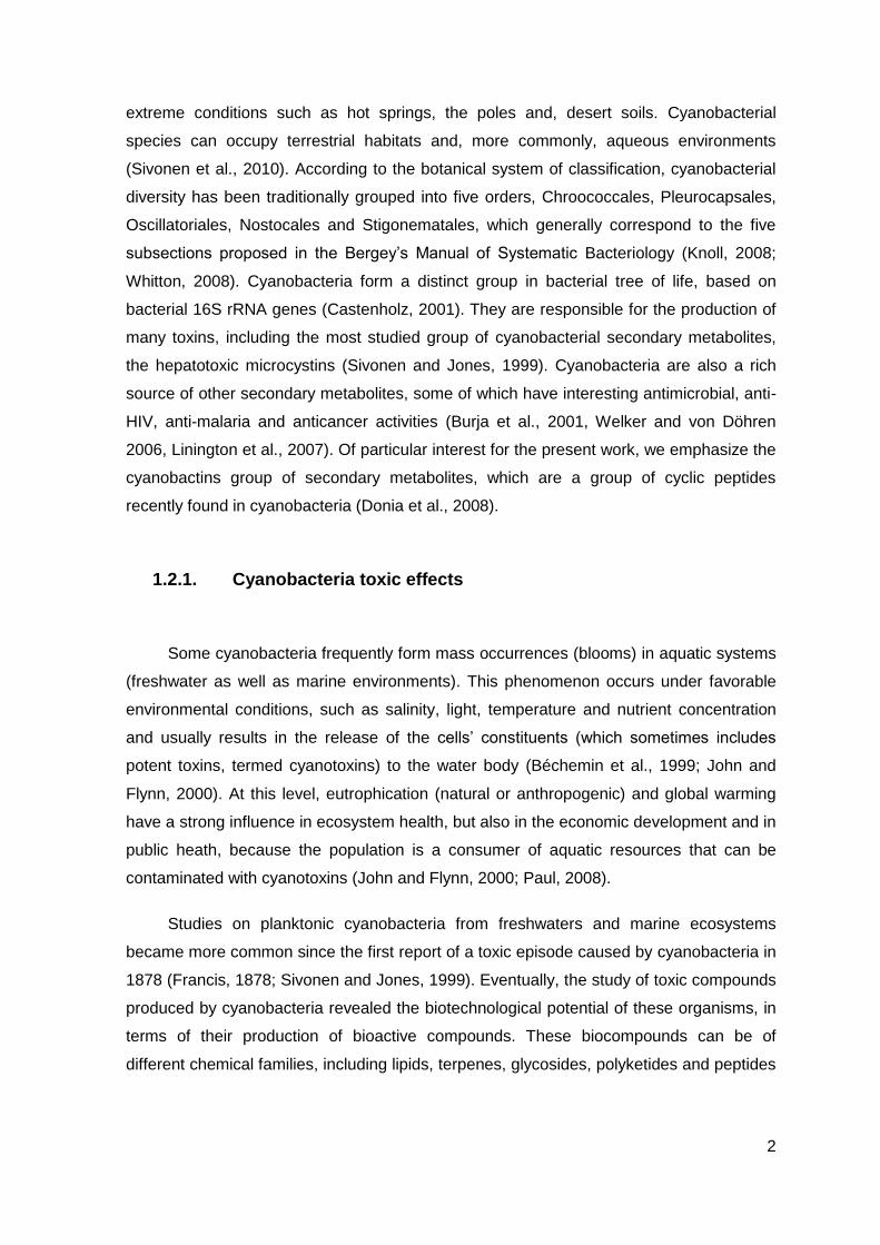
Figure 2- The chemical structures of a selection of cyanobactins. Trunkamide was isolated from L. patella, tenuecyclamide from N. spongiaeforme, anacyclamide from Anabaena, trichamide from T. erythraeum, ulithiacyclamide and patellamide from Prochloron( Originally from L. Patella); Viridisamide from Oscillatoria viridis PCC7112.(Adapted from Sivonen el al., 2010 and Leikoski et al., 2013). The post-translational modifications; oxazoline, thiazole, and reverse O-prenyl are highlighted with circles. In the linear cyanobactin, the identical prenylated N-termini and methylated C-termini bound to thiazoles are in blue. (Adapted from Sivonen et al., 2010; Leikoski et al., 2013)
8
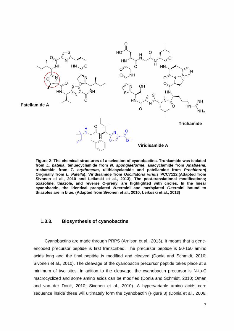
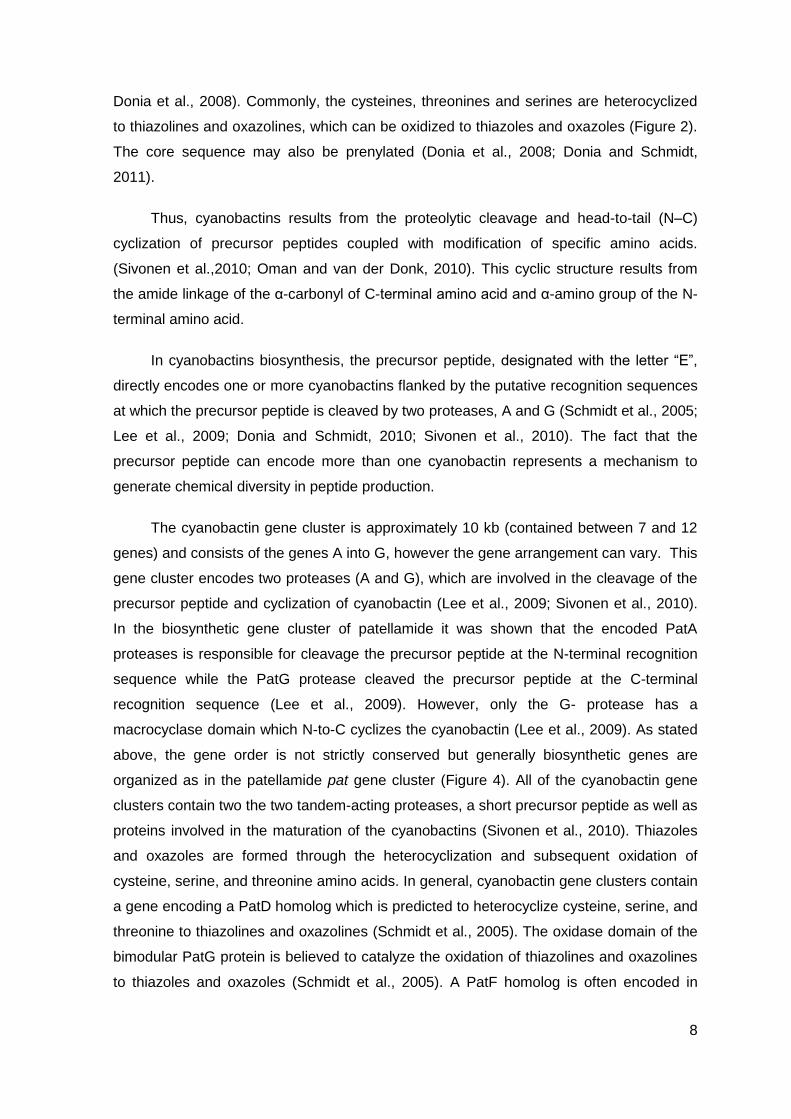
Donia et al., 2008). Commonly, the cysteines, threonines and serines are heterocyclized
to thiazolines and oxazolines, which can be oxidized to thiazoles and oxazoles (Figure 2).
The core sequence may also be prenylated (Donia et al., 2008; Donia and Schmidt,
2011).
Thus, cyanobactins results from the proteolytic cleavage and head-to-tail (N–C)
cyclization of precursor peptides coupled with modification of specific amino acids.
(Sivonen et al.,2010; Oman and van der Donk, 2010). This cyclic structure results from
the amide linkage of the α-carbonyl of C-terminal amino acid and α-amino group of the N-
terminal amino acid.
In cyanobactins biosynthesis, the precursor peptide, designated with the letter “E”,
directly encodes one or more cyanobactins flanked by the putative recognition sequences
at which the precursor peptide is cleaved by two proteases, A and G (Schmidt et al., 2005;
Lee et al., 2009; Donia and Schmidt, 2010; Sivonen et al., 2010). The fact that the
precursor peptide can encode more than one cyanobactin represents a mechanism to
generate chemical diversity in peptide production.
The cyanobactin gene cluster is approximately 10 kb (contained between 7 and 12
genes) and consists of the genes A into G, however the gene arrangement can vary. This
gene cluster encodes two proteases (A and G), which are involved in the cleavage of the
precursor peptide and cyclization of cyanobactin (Lee et al., 2009; Sivonen et al., 2010).
In the biosynthetic gene cluster of patellamide it was shown that the encoded PatA
proteases is responsible for cleavage the precursor peptide at the N-terminal recognition
sequence while the PatG protease cleaved the precursor peptide at the C-terminal
recognition sequence (Lee et al., 2009). However, only the G- protease has a
macrocyclase domain which N-to-C cyclizes the cyanobactin (Lee et al., 2009). As stated
above, the gene order is not strictly conserved but generally biosynthetic genes are
organized as in the patellamide pat gene cluster (Figure 4). All of the cyanobactin gene
clusters contain two the two tandem-acting proteases, a short precursor peptide as well as
proteins involved in the maturation of the cyanobactins (Sivonen et al., 2010). Thiazoles
and oxazoles are formed through the heterocyclization and subsequent oxidation of
cysteine, serine, and threonine amino acids. In general, cyanobactin gene clusters contain
a gene encoding a PatD homolog which is predicted to heterocyclize cysteine, serine, and
threonine to thiazolines and oxazolines (Schmidt et al., 2005). The oxidase domain of the
bimodular PatG protein is believed to catalyze the oxidation of thiazolines and oxazolines
to thiazoles and oxazoles (Schmidt et al., 2005). A PatF homolog is often encoded in
9
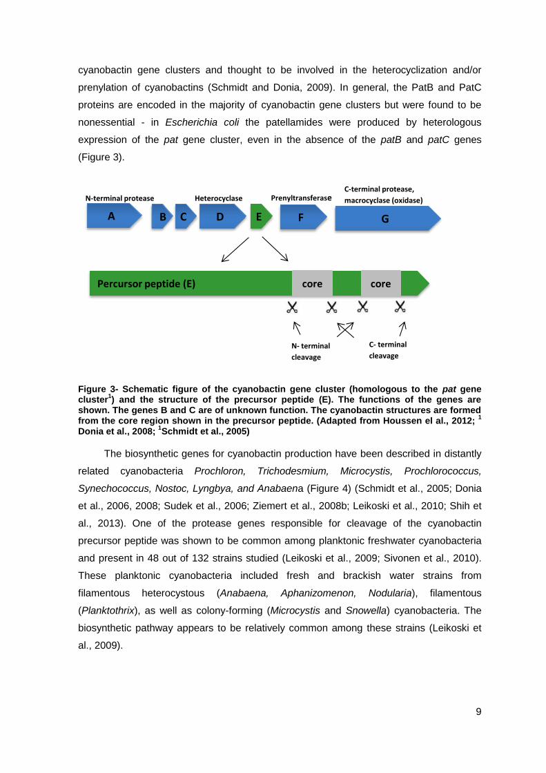
cyanobactin gene clusters and thought to be involved in the heterocyclization and/or
prenylation of cyanobactins (Schmidt and Donia, 2009). In general, the PatB and PatC
proteins are encoded in the majority of cyanobactin gene clusters but were found to be
nonessential - in Escherichia coli the patellamides were produced by heterologous
expression of the pat gene cluster, even in the absence of the patB and patC genes
(Figure 3).
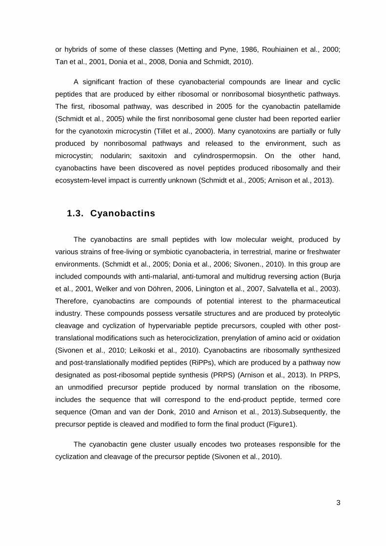
Figure 3- Schematic figure of the cyanobactin gene cluster (homologous to the pat gene cluster
1) and the structure of the precursor peptide (E). The functions of the genes are
shown. The genes B and C are of unknown function. The cyanobactin structures are formed from the core region shown in the precursor peptide. (Adapted from Houssen el al., 2012;
1
Donia et al., 2008; 1Schmidt et al., 2005)
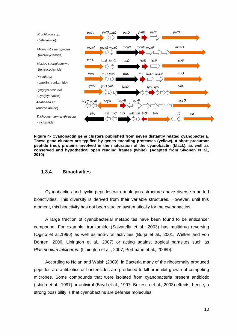
The biosynthetic genes for cyanobactin production have been described in distantly
related cyanobacteria Prochloron, Trichodesmium, Microcystis, Prochlorococcus,
Synechococcus, Nostoc, Lyngbya, and Anabaena (Figure 4) (Schmidt et al., 2005; Donia
et al., 2006, 2008; Sudek et al., 2006; Ziemert et al., 2008b; Leikoski et al., 2010; Shih et
al., 2013). One of the protease genes responsible for cleavage of the cyanobactin
precursor peptide was shown to be common among planktonic freshwater cyanobacteria
and present in 48 out of 132 strains studied (Leikoski et al., 2009; Sivonen et al., 2010).
These planktonic cyanobacteria included fresh and brackish water strains from
filamentous heterocystous (Anabaena, Aphanizomenon, Nodularia), filamentous
(Planktothrix), as well as colony-forming (Microcystis and Snowella) cyanobacteria. The
biosynthetic pathway appears to be relatively common among these strains (Leikoski et
al., 2009).
A B C D E F G
N-terminal protease Heterocyclase PrenyltransferaseC-terminal protease,
macrocyclase (oxidase)
N- terminal
cleavage
C- terminal
cleavage
Percursor peptide (E) core core
10
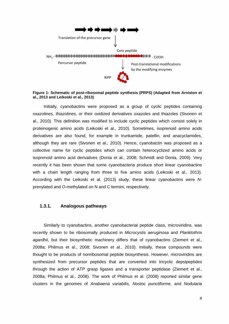
Figure 4- Cyanobactin gene clusters published from seven distantly related cyanobacteria. These gene clusters are typified by genes encoding proteases (yellow), a short precursor peptide (red), proteins involved in the maturation of the cyanobactin (black), as well as conserved and hypothetical open reading frames (white). (Adapted from Sivonen et al., 2010)
1.3.4. Bioactivities
Cyanobactins and cyclic peptides with analogous structures have diverse reported
bioactivities. This diversity is derived from their variable structures. However, until this
moment, this bioactivity has not been studied systematically for the cyanobactins.
A large fraction of cyanobacterial metabolites have been found to be anticancer
compound. For example, trunkamide (Salvatella et al., 2003) has multidrug reversing
(Ogino et al.,1996) as well as anti-viral activities (Burja et al., 2001, Welker and von
Döhren, 2006, Linington et al., 2007) or acting against tropical parasites such as
Plasmodium falciparum (Linington et al., 2007; Portmann et al., 2008b).
According to Nolan and Walsh (2009), in Bacteria many of the ribosomally produced
peptides are antibiotics or bactericides are produced to kill or inhibit growth of competing
microbes. Some compounds that were isolated from cyanobacteria present antibiotic
(Ishida et al., 1997) or antiviral (Boyd et al., 1997; Bokesch et al., 2003) effects; hence, a
strong possibility is that cyanobactins are defense molecules.
patA patB patC patD patE patF patG
mcaA mcaB mcaC mcaD mcaE mcaF mcaG
tenA tenB tenC tenD tenE tenF tenG
truA truB truC truD truE truF1 truF2 truG
lynA lynB lynC lynD lynE lynF lynG
acyC acyB acyA acyE acyF acyG
triEtriA triB triC triD triF triG triH triI triK
Prochloron spp.
(patellamide)
Microcystis aeruginosa
(microcyclamide)
Nostoc spongiaeforme
(teneucyclamide)
Prochloron
(patellin, trunkamide)
Lyngbya aestuarii
(Lyngbyabactin)
Anabaena sp.
(anacyclamide)
Trichodesmium erythraeum
(trichamide)
11
1.4. Anacyclamides
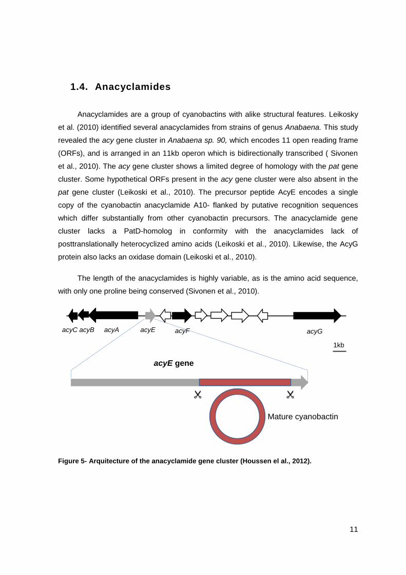
Anacyclamides are a group of cyanobactins with alike structural features. Leikosky
et al. (2010) identified several anacyclamides from strains of genus Anabaena. This study
revealed the acy gene cluster in Anabaena sp. 90, which encodes 11 open reading frame
(ORFs), and is arranged in an 11kb operon which is bidirectionally transcribed ( Sivonen
et al., 2010). The acy gene cluster shows a limited degree of homology with the pat gene
cluster. Some hypothetical ORFs present in the acy gene cluster were also absent in the
pat gene cluster (Leikoski et al., 2010). The precursor peptide AcyE encodes a single
copy of the cyanobactin anacyclamide A10- flanked by putative recognition sequences
which differ substantially from other cyanobactin precursors. The anacyclamide gene
cluster lacks a PatD-homolog in conformity with the anacyclamides lack of
posttranslationally heterocyclized amino acids (Leikoski et al., 2010). Likewise, the AcyG
protein also lacks an oxidase domain (Leikoski et al., 2010).
The length of the anacyclamides is highly variable, as is the amino acid sequence,
with only one proline being conserved (Sivonen et al., 2010).
Figure 5- Arquitecture of the anacyclamide gene cluster (Houssen el al., 2012).
Mature cyanobactin
acyE gene
acyC acyB acyA acyE acyF acyG
1kb
12
1.5. Anabaena
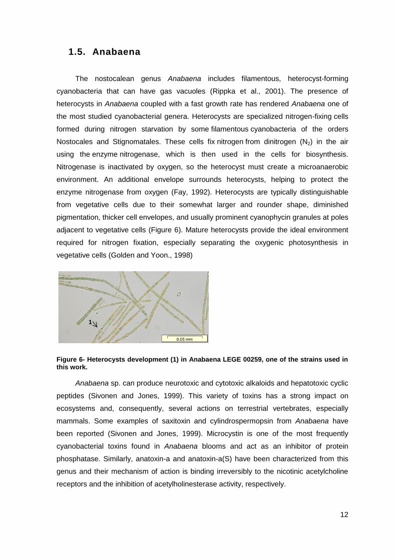
The nostocalean genus Anabaena includes filamentous, heterocyst-forming
cyanobacteria that can have gas vacuoles (Rippka et al., 2001). The presence of
heterocysts in Anabaena coupled with a fast growth rate has rendered Anabaena one of
the most studied cyanobacterial genera. Heterocysts are specialized nitrogen-fixing cells
formed during nitrogen starvation by some filamentous cyanobacteria of the orders
Nostocales and Stignomatales. These cells fix nitrogen from dinitrogen (N2) in the air
using the enzyme nitrogenase, which is then used in the cells for biosynthesis.
Nitrogenase is inactivated by oxygen, so the heterocyst must create a microanaerobic
environment. An additional envelope surrounds heterocysts, helping to protect the
enzyme nitrogenase from oxygen (Fay, 1992). Heterocysts are typically distinguishable
from vegetative cells due to their somewhat larger and rounder shape, diminished
pigmentation, thicker cell envelopes, and usually prominent cyanophycin granules at poles
adjacent to vegetative cells (Figure 6). Mature heterocysts provide the ideal environment
required for nitrogen fixation, especially separating the oxygenic photosynthesis in
vegetative cells (Golden and Yoon., 1998)
Figure 6- Heterocysts development (1) in Anabaena LEGE 00259, one of the strains used in this work.
Anabaena sp. can produce neurotoxic and cytotoxic alkaloids and hepatotoxic cyclic
peptides (Sivonen and Jones, 1999). This variety of toxins has a strong impact on
ecosystems and, consequently, several actions on terrestrial vertebrates, especially
mammals. Some examples of saxitoxin and cylindrospermopsin from Anabaena have
been reported (Sivonen and Jones, 1999). Microcystin is one of the most frequently
cyanobacterial toxins found in Anabaena blooms and act as an inhibitor of protein
phosphatase. Similarly, anatoxin-a and anatoxin-a(S) have been characterized from this
genus and their mechanism of action is binding irreversibly to the nicotinic acetylcholine
receptors and the inhibition of acetylholinesterase activity, respectively.
1
13
This study was carried out using only Anabaena strains.
1.6. Biotechnological Aspects
The biotechnological exploitation of cyanobactins will require detailed studies on the
enzymes involved in the biosynthesis as well as mechanisms of action of these peptides.
The small size of the cyanobactin gene clusters and their amenability to be fully
heterologously expressed (Schmidt et al., 2005; Donia et al., 2006, 2008; Leikoski et al.,
2010) will provide new possibilities to create compound libraries and novel compounds
(Donia et al., 2006, 2008). In the study of cyanobactins pathway, the heterologous
expression gives options to study the role of individual genes in biosynthesis as well as
produce novel peptides. The core sequence of the precursor peptide directly encodes the
resulting cyanobactin and this sequence can be changed easily by genetic engineering in
heterologous hosts and recombinant peptides can be produced (Donia et al., 2006;
Tianero et al., 2012).
According to Donia et al. (2006) the cyanobactin pathway was utilized in E.coli to
synthesize an engineered cyclic peptide (eptidemnamide) similar to an anticoagulant in
clinical use. This approach demonstrates a means to exploit cyanobacterial pathways and
produce novel compounds by the rational design of peptides (Donia et al., 2006; Sivonen
et al., 2010).
According to the work by Oman and van der Donk (2010) the peptide precursor-
directed synthesis allows manipulations directly to the precursor gene and enables
production of engineered peptides in heterologous hosts. In addition, the enzymes in the
cyanobactin pathways could be used as catalysts to assist the chemical synthesis of the
desired compounds.
The work by Lee et al. (2009), showcased not only the important role of proteases in
cyanobactin biosynthesis but also was demonstrated the potential of the enzymes as
general catalysts for cyclization of peptides. The PatG protease was shown to require no
exogenous energy for the cleavage and cyclization, and to be tolerant to different
substrate lengths and sequences as long as the C-terminal recognition sequence was
present (Sivonen et al., 2010; Lee et al., 2009). This is important, for example, in synthetic
peptide manufacture where the head-to-tail cyclization restricts peptide production in bulk
amounts (Sivonen et al., 2010). It is important to note that whole-genome information has
14
already led to the discovery of cyanobactin biosynthesis as well as several new
compounds and compound classes, for example novel patellamides (Schmidt et al.,
2005), trichamide (Sudek et al., 2006), and anacyclamides (Leikoski et al., 2010).
The number of genome sequencing projects involving cyanobacteria and
metagenomic studies applied to various environments is increasing. This should allow
new discoveries in the near future, including new ribosomal pathways and cyanobactins.
1.7. Objective
The main objectives of this work were the detection of anacyclamide- related genes
in cyanobacteria from the culture collection LEGE (LEGE CC) and, the corresponding
identification of new anacyclamides. As a secondary objective, we tried to characterize the
cyanobacterial strains using both morphological and molecular data, with the ultimate goal
of making phylogenetic inferences regarding cyanobactins production.
For this purpose, a PCR-screening of ten freshwater cyanobacterial strains for
cyanobactin-related genes was carried out. The resulting amplicons were then cloned and
sequenced in order to identify potential producers of new members of this class of
compounds. Finally, investigations on the production and the nature of the new
cyanobactins were performed using analytical methodologies.
15
2. Material and Methods
The methods used are outlined in the workflow shown in figure 7.
Figure 7- Schematic drawing of the workflow.
2.1. Source of strains
Cyanobacteria used in this study are part of the LEGE culture collection and had
been isolated from water samples collected from freshwater supplies, located in north and
central regions of Portugal (Table1). Additionally, for comparison purposes, the strain
Anabaena sp.90, isolated in Finland from Lake Vesijärvi, in 1986 and maintained at the
University of Helsinki was used (Sivonen et al., 1992).
1Cyanobacteria strains
(10 Anabaena strains)
2Culture growth
3gDNA extraction, screening of
16S rRNA gene and anacyclamide
related- genes by PCR
5Screening anacyclamides by
LCMS based on the presence
of a prenyl unit
3Cloning and sequencing
of the precursors genes
Predictions of
anacyclamides structures
Putative anacyclamide5LCMS- detection and
characterization of the
predicted anacyclamide
Hight accuracy UPLC-ESI-
QTOF

16
Table 1- Identification of the ten strains in study, from the LEGE culture collection
Anabaena
strain Origin/source Isolator
Year of
harvest
Co-
identification
LEGE00233 Maranhão reservoir Joana
Osswald 2000 J1
LEGE00241 Maranhão reservoir Joana
Osswald 2000 J14
LEGE00243 Maranhão reservoir Joana
Osswald 2000 J16
LEGE00245 Maranhão reservoir Joana
Osswald 2000 J18
LEGE00246 Maranhão reservoir Joana
Osswald 2000 J19
LEGE00248 Maranhão reservoir Joana
Osswald 2000 J21
LEGE00250 Maranhão reservoir Joana
Osswald 2000 J27
LEGE00253 Chaves reservoir Joana
Osswald 2000 J37
LEGE00259 Maranhão reservoir Joana
Osswald 2000 J46
LEGE04289 Marco de
Canaveses
Joana
Osswald 2004 J83
2.2. Cyanobacterial strains and culturing
The ten Anabaena sp. strains were grown in sterile Z8 medium (Kotai, 1972), in 40-
ml cultures under a 14:10 h light: dark cycle (Martins et al., 2005), with a photon irradiance
of 10-30 µmol m2 s-1 at 25°C.
17
Regarding large scale biomass production, for the chemical analyses, two isolates
were grown in sterile 16L culture vessels, with aeration, under 14:10h light: dark cycle,
with a photon irradiance of 10-30 µmol m2 s-1 at 25°C.
Figure 8- Cyanobacterial strains and Culturing Anabaena sp. Strains were grown in z8 medium, with aeration, under 14:10 light:dark at 25°C.
2.3. DNA extraction, PCR amplification, Cloning and
sequencing
Genomic DNA (gDNA) extraction was carried out either with the Purelink genomic
DNA mini kit (Invitrogen) or with the Dneasy Plant Mini kit (Qiagen). PCR amplification of
the 16S small ribosomal subunit gene (16S rRNA gene) was carried out using the gDNAs
as templates. Primers 27F, 359F and 1491R (Neilan et al., 1997; Jungblut et al., 2005),
targeting the 16S rRNA gene, were used.
All PCR reactions were prepared in a 20µl of volume containing 10x PCR buffer for
Super Taq (HT Biotechnology Ltd.). PCR conditions were as described previously.
Amplicons were cloned into a PGEM-T vector and transformed into Oneshot E.coli TOP10
cells (Invitrogen). Plasmid DNA was isolated from the transformed cells using the
GenElute Plasmid MiniPrep kit (Sigma-Aldrich) and sequenced (Macrogen) using M13
primers.
18
Table 2- Primers used for the 16S rRNA gene amplification.
Primera Sequence (5’»3’) Reference
27F AGAGTTTGATCCTGGCTCAG Neilan et al., 1997
Jungblut et al., 2005 1494R TACGGCTACCTTGTTACGAC
CYA359F GGGGAATYTTCCGCAATGGG
aGene primers are named according to their locus
The amplification of the patA gene was made using a pair of primers that amplified a
1.4 kb section, PatAR and PatA F (Lee et al., 2009). These amplifications, using these
primers, was already made by Leikoski et al. in previous studies to screen many
cyanobactia strains, including Anabaena sp.90. The annealing temperature used in the
PCR program was of 52°C.
Anabaena sp.90 (Sivonen et al., 1992) was used as a positive control in the
screening.
In order to identify an Anabaena strain producing an anacyclamide, the acyE
peptide precursor gene was amplified using primers preRNAF (5’-
GAAGAACATCCGCCCCCAACAAGTTG-3’) and preRNAR (5’-CTCCGCGTCGTC
GCCTGCAAAAGG-3’) and primers PreF (5’-GCCTTCACCAAACCAGTCT TCTTCAT-3’)
and PreR (5’-CATCGAGGCGAACCGTGCGCCAAGGGAT- 3’) (Leikoski et al., 2009) from
the genomic DNA of all the Anabaena strains. The expected size band, for the last primer
pair is about 312 pb and for the PCR program the Annealing Temperature was calculated
at 53°C; using the first primer pair the expected size band is about 160 bp and the
Annealing Temperature was calculated at 56°C. Following cloning and sequencing as
described above, the amplified fragments were compared to those of known acyE
precursor to infer the novelty of the encoded anacyclamide.
19
Figure 9- The acyE peptide precursor gene with the protease cleaving sites highlighted and the primer annealing regions shown.
Table 3- PCR reaction for each primers pair
Pair of primers
PCR reaction a
Inicial
Denaturation
Denaturation Annealing extension Final
extension
27F/1494R
359F/1494R
94°C
4min
35 cycles 72°C
8min
8°C
∞ 94°C
45sec
50°C
45sec
72°C
1min20sec
preRNAF/preRNAR
94°C
2min
94°C
30sec
56°C
30sec
72°C
10sec
72°C
7min
8°C
∞
PreF/PreR
94°C
2min
94°C
30sec
53°C
30sec
72°C
20sec
72°C
7min
8°C
∞
aThe PCR reactions were calculated according with the specific primers used and they are not publish.
2.4. Cloning the acy biosynthetic gene cluster
In order to amplify the entire 12 kb gene cluster from genomic DNA of Anabaena sp.
LEGE 00259 by PCR , the patex2f (5’-ATGGATCCTGATGGACTGTAGTGTGAG-3’) and
patex5r (5’- TACTCGAGAGGTTTTGGGACTCTTTAG-3’) primer pair (Leikoski et al.,
Primers
Leader
acyE gene percursor
5’ 3’
CoreNH2 COOH
Proteases
20
2009) was used, in three 60-µL reaction mixtures, containing 10× PCR buffer for Super
Taq Plus (HT Biotechnology Ltd.), 200 µmol of each nucleotide (Finnzymes), 0.75 µM of
each primer, 0.8 U Super Taq Plus proofreading polymerase (HT Biotechnology Ltd.), and
100 ng of Anabaena sp. LEGE 00259 gDNA. The thermocycling conditions were 94°C for
2 min, followed by 30 cycles of 94°C for 30 s, 56.4°C for 30 s, and 68°C for 9 min and
then a final extension at 68°C for 20 min. The PCR products were separated on a 0.7%
agarose gel containing 0.5× Tris-acetate-EDTA and run for 30 min at 100 V. The gel was
stained using SYBR Safe DNA gel stain (Invitrogen) and was visualized using a Dark
reader (Clare Chemical Research Inc.) to avoid DNA damage from UV light during gel
extraction. The 12-kb PCR product was gel extracted with a MinElute gel extraction kit
(Qiagen) and cloned into the PCR 2.1-TOPO vector using a TOPO TA cloning kit
(Invitrogen) with an insert-to-vector molar ratio of 3:1. The vector was used to transform
chemically competent E.coli One Shot TOP10 cells according to the manufacturer’s
instructions. The resultant RC_c1, RC_c2 and RC_c3 plasmids were analyzed by PCR
and restriction digestion to ensure that the integrity of the insert was maintained during the
cloning and amplification in E. coli. The transformants containing the 12-kb insert in the
plasmid were grown overnight with shaking at 120 rpm at 28°C in 50 ml of LB medium
containing 150 µg ml -1 of ampicillin (sodium salt; Sigma-Aldrich) for liquid chromatography
(LC)-mass spectrometry (MS) analysis. The cells were harvested by centrifugation at
10,000 × g for 10 min (Ep- pendorf 5804R centrifuge), dried with a Heto vacuum
centrifuge (Heto- Holten A/S), which yielded ca. 43 mg (dry weight) and sent to
sequencing (Macrogen; Seoul, Korea).
2.5. Biomass collection of LEGE 00248 and LEGE 00259
and preparation of extracts
The biomass of 16L cultures of LEGE 00248 and LEGE 00259 was collected following
approximately one month of growth under the conditions detailed in the “Cyanobacterial
strains and cultivation” section. The collection was carried out by filtration using a 41 µm
plankton net. The filtered cells were then centrifuged at 4600rpm for 10min and the pellet
was frozen and freeze-dried.
21
Figure 10- Biomass collection of 16L cultures of LEGE 00248 and LEGE 00259 strains
2.5.1. Biomass extraction
The lyophilized biomass was weighed and then methanol extraction was made
(Leikoski et al., 2010) using 60ml of solvent (ACS grade). The biomass with methanol was
macerated and then the supernatant was transferred into a 45ml falcon and centrifuged at
4600×g for 10min. This procedure was repeated six times. The pooled supernatants were
then concentrated in vacuo and the mass of the resulting crude extract determined.
2.6. Chemical analysis
Cells of Anabaena strains were collected from the 40 mL cultures by centrifugation at
7,000 g for 7 min. The collected cells were freeze-dried with Supermodulyo (Edwards
High Vacuum International) or dried with a Heto vacuum centrifuge, which yielded 5 to 12
mg (dry weight). The dried Anabaena cells, as well as E. coli transformants, were
extracted with 1 mL of methanol (HiperSolv, HPLC grade; BDH Laboratory Supplies) in 2
mL plastic tubes containing glass beads (cell disruption medium; 0.5-mm-diameter glass
beads; Scientific Industries Inc.).
Each mixture was homogenized by shaking with a FastPrep cell disrupter (Bio 101,
Thermo Electron Corporation, Qbiogene, Inc.) for 30 s at a speed of 5 ms-1. The mixture
was centrifuged at 10000 x g for 5 min, and the supernatant was used for LC-MS analysis.
22
The methanol extracts were analyzed with a high-performance liquid chromatograph
combined with a mass spectrometer (Agilent 1100 series LC/MSD with Ion Trap XCT Plus
and an electrospray ion source) in order to detect low-molecular-weight peptides.
Peptides were separated from the extracts by HPLC using a Phenomenex C18(2) column
(2.0 mm x 150 mm; particle size, 5µm). The mobile phase gradient consisted of 0.1%
aqueous (water purified with Milli-Q Plus; Millipore) formic acid (Fluka) (solvent A) and
0.1% formic acid in isopropyl alcohol (Sigma-Aldrich) (solvent B). Two different settings
were used; one setting was used for screening methanolic extracts of Anabaena cells,
and the other setting (values in parentheses below) was used for further structural
characterization of natural and synthetic peptides. The percentage of solvent B was
increased from 5 to 50% in 60 min. A flow rate of 0.15 mL min-1 was used, and the
columns were heated to 40°C during separation. The positive-ion mode of electrospray
ionization was used. The pressure of the nebulizer gas (N2) was 30 lb/in2 (35 lb/in2), the
drying gas flow rate was 8 L min-1, and the temperature was 350°C. The capillary voltage
was set at 5,000 V (4,500 V), and the capillary offset value was 300 V. A skimmer
potential of 85 V (100 V) and a trap drive value of 144 (111) were used. Spectra were
recorded using a scan range from m/z 100 to m/z 2200. Identification of the
anacyclamides was based on the molecular weights calculated from the predicted peptide
AcyE precursor amino acid sequences and the assigned fragment ion patterns of MSn (n=
1 to 3) spectra. A comparison with the retention time and MS data for a synthetic
reference was used in five cases (Beijing SBS Genetech Co., Ltd., China; anacyclamide
B7 from JPT Peptide Technologies GmbH, Germany).
23
3. Results and Discussion
In this study we proposed to detect new cyanobactins among ten Anabaena strains
from LEGE CC. At the same time, it was our aim to characterize the cyanobacterial strains
using both morphological and molecular data, with the final goal making phylogenetic
inferences. For this purpose were made a variety of procedures, including genomic DNA
extraction, PCR screening, in silico analysis and detection by LC-MS.
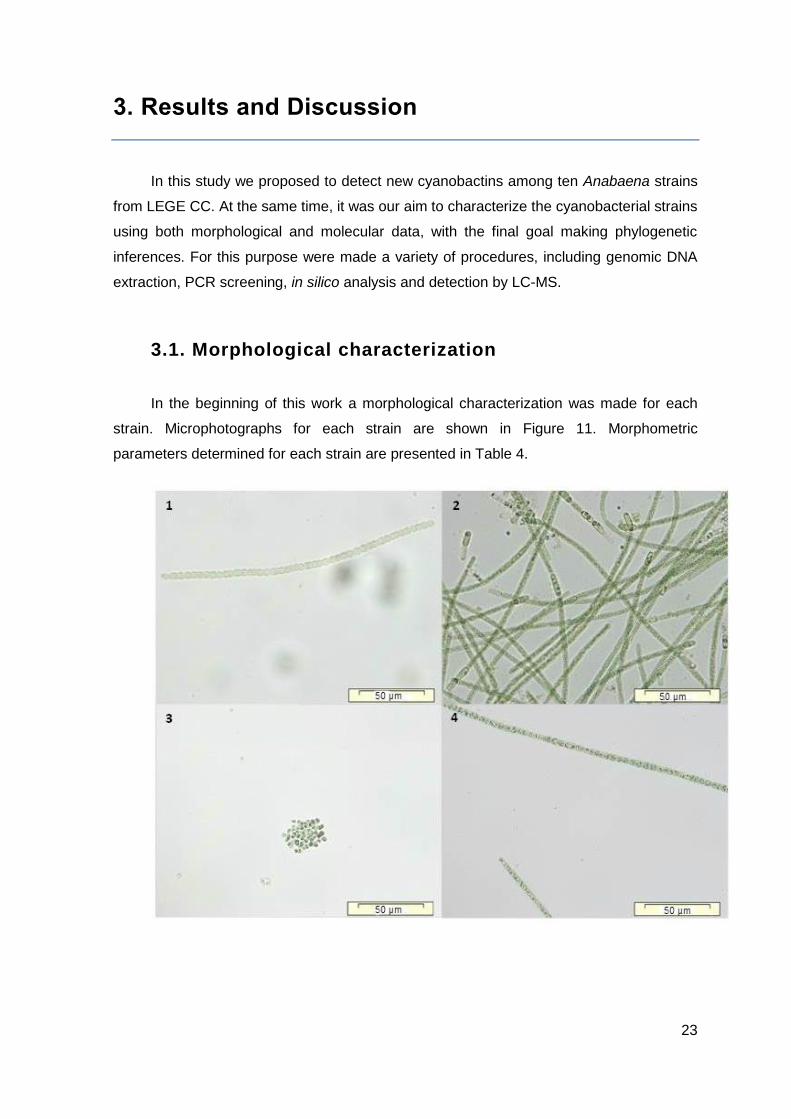
3.1. Morphological characterization
In the beginning of this work a morphological characterization was made for each
strain. Microphotographs for each strain are shown in Figure 11. Morphometric
parameters determined for each strain are presented in Table 4.
24
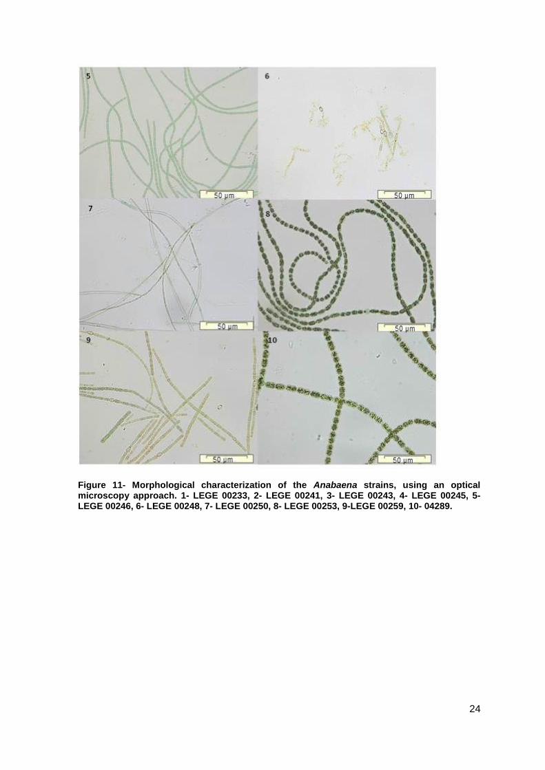
Figure 11- Morphological characterization of the Anabaena strains, using an optical microscopy approach. 1- LEGE 00233, 2- LEGE 00241, 3- LEGE 00243, 4- LEGE 00245, 5- LEGE 00246, 6- LEGE 00248, 7- LEGE 00250, 8- LEGE 00253, 9-LEGE 00259, 10- 04289.
25
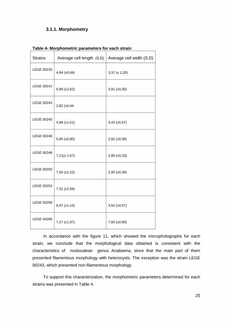
3.1.1. Morphometry
Table 4- Morphometric parameters for each strain
Strains Average cell length (S.D) Average cell width (S.D)
LEGE 00233
4,84 (±0,84) 3,37 (± 1,20)
LEGE 00241
6,99 (±2,62) 3,91 (±0,35)
LEGE 00243
2,82 (±0,44
LEGE 00245
4,98 (±1,01) 4,43 (±0,57)
LEGE 00246
5,85 (±0.80) 2,82 (±0,36)
LEGE 00248
7,21(± 1,67) 2,89 (±0,33)
LEGE 00250
7,55 (±1,22) 2,00 (±0,39)
LEGE 00253
7,31 (±2,59)
LEGE 00259
6,97 (±1,15) 3,52 (±0,57)
LEGE 04289
7,27 (±1,97) 7,60 (±0,90)
In accordance with the figure 11, which showed the microphotographs for each
strain, we conclude that the morphological data obtained is consistent with the
characteristics of nostocalean genus Anabaena, since that the main part of them
presented filamentous morphology with heterocysts. The exception was the strain LEGE
00243, which presented non-filamentous morphology.
To support this characterization, the morphometric parameters determined for each
strains was presented in Table 4.
5 6
7 8
9 10
26
3.2. Genomic DNA extraction, PCR amplification,
cloning and sequencing
To start the molecular screening of the ten strains, we performed the genomic DNA
extraction for each strain. Then, the amplification of the 16S rRNA gene was made, using
different primer pairs, 27F-1494R; 359F-1494R (Neilan et al., 1997; Jungblut et al.,2005).
The amplification of the 16S rRNA gene, using these primers, resulted in amplicons with
different size, since that with the 27F-1494R pair of primers all cyanobacteria strains
presented a 1467 bp amplicon, as expected. In the same way, with the 359F-1494R all
strains presented a 1135 bp amplicon. The organization of the results is performed
according to the following numerical correspondence (1-10):
Strain 1- LEGE 00233
Strain 2- LEGE 00241
Strain 3- LEGE 00243
Strain 4- LEGE 00245
Strain 5- LEGE 00246
Strain 6- LEGE 00248
Strain 7- LEGE 00250
Strain 8- LEGE 00253
Strain 9- LEGE 00259
Strain 10- LEGE 04289
27
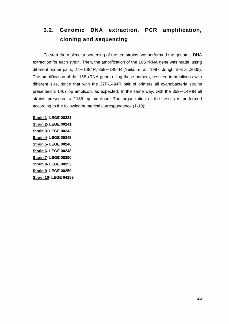
Figure 12- Amplification of the 16S rRNA gene with the primers 359F- 1494R(up); 27F- 1494R (down) for all ten strains. Samples: 3µl; 1kb plus DNA ladder: 1,5µl. 1,5% agarose gel; 1- LEGE 00233, 2- LEGE 00241, 3- LEGE 00243, 4- LEGE 00245, 5- LEGE 00246, 6- LEGE 00248, 7- LEGE 00250, 8- LEGE 00253, 9-LEGE 00259, 10- 04289.
All the strains showed amplicons of the expected size, using these sets of universal
primers. These results were consistent with the classification of the strains, as members
of cyanobacteria group.
These bands were excised and then the purified product was cloned into a pGEM-T
vector, according with the protocol and method described above. The amplicons resulting
from 27F-1494R was sent to sequencing but until the moment we haven’t obtained the
sequencing results. The sequencing results of the amplicons resulting from the 359F-
1494R will provide more specific information about the strains, since the primers 359F is a
cyanobacteria-specific primer.
Following, we performed a screening for the genes involved in the cyanobactins
production. In this work, was used a pair of primers, which were designed to amplify the
gene encoding PatA protease, which cleaves the precursor peptide at the N-terminal
recognition site (Lee et al., 2009). The primers amplified a 1.4 kb section of the patA gene.
According with the Leikoski et al. study (2013), the patA gene is especially common in
planktonic cyanobacteria. Furthermore, it’s also known that the cyanobactin pathway is
1 2 3 4 5 6 7 8 9 10
1 2 3 4 5 6 7 8 9 10
28
widespread and sporadically distributed in cyanobacteria and hinted at the potential
chemical diversity of cyanobactins encoded in this pathway.
These results were compared with the Anabaena sp.90, as a positive control (Leikoski et
al., 2010).
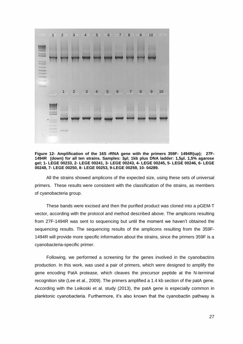
Figure 13- Amplification of the patA gene (1.4kb section) with the PatA pair of primers designed for this effect, by Leikoski et al. All the ten strains are represented. Tann = 52°C. Samples: 4µl; 10kb GeneRuler DNA ladder: 3µl. 0.7agarose gel. Positive control= Anabaena sp. 90.
According with Figure 13, showing the electrophoresis results of the patA gene
amplification, we can conclude that the strains with 1.4 kb amplicon were LEGE 00243,
LEGE 00245, LEGE 00248 and LEGE 00259.
We then proceeded to the amplification of the patE gene, using different pairs of
primers (PreF/PreR and PreRNAR/PreRNAR).
At this stage, we worked with only nine strains because one of them (LEGE 00250)
showed a deficit of growth. Due to the lag in growth, this strain was not included in
amplification of patE genes. We decided to increase the growth time and do the screening
for this strain later. When the strain achieved the proper growth the amplifications of the
patA genes was made and we verified that it did not result in an amplicon of the expected
size (Figure 13).
Regarding the patE genes, in the first screening the PreF/PreR pair of primers were
used. The results were in accordance with the patA genes amplification, since we
obtained positive result for the strains 3- LEGE 00243, 4-LEGE 00345, 6-LEGE 00248
and 9-LEGE 00259. The exception on these results was the strains 8-LEGE 00253 and
1 2 3 4 5 6 7 8 9 10 C+ C-
29
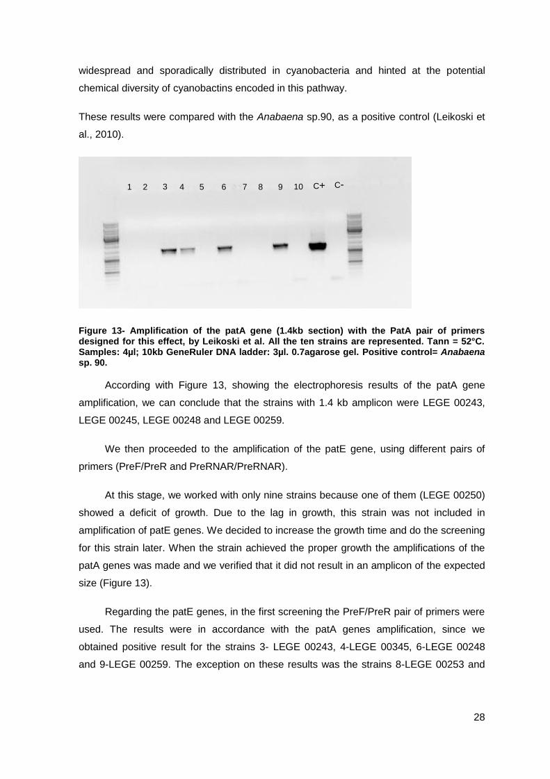
10- LEGE 04289, which also presented band in the expected size. The results are shown
in Figure 14 as an example.
Figure 14- Amplification of patE genes, using primers PreR and PreF, from nine strains. Tann= 53ºC. Samples: 3µl ,1kb plus DNA ladder: 1.5µl. 1,5% agarose gel; 1- LEGE 00233, 2- LEGE 00241, 3- LEGE 00243, 4- LEGE 00245, 5- LEGE 00246, 6- LEGE 00248, 7- LEGE 00253, 8- LEGE 00259, 9-LEGE 04289
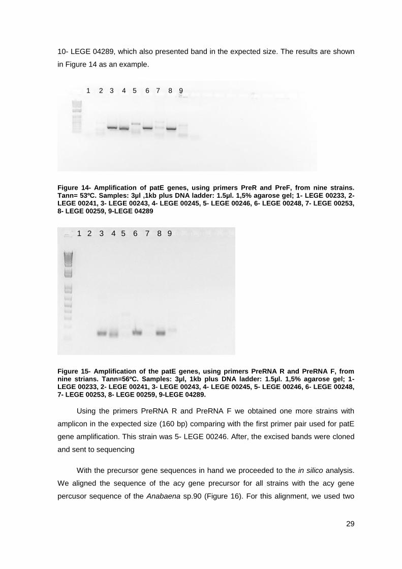
Figure 15- Amplification of the patE genes, using primers PreRNA R and PreRNA F, from nine strians. Tann=56ºC. Samples: 3µl, 1kb plus DNA ladder: 1.5µl. 1,5% agarose gel; 1- LEGE 00233, 2- LEGE 00241, 3- LEGE 00243, 4- LEGE 00245, 5- LEGE 00246, 6- LEGE 00248, 7- LEGE 00253, 8- LEGE 00259, 9-LEGE 04289.
Using the primers PreRNA R and PreRNA F we obtained one more strains with
amplicon in the expected size (160 bp) comparing with the first primer pair used for patE
gene amplification. This strain was 5- LEGE 00246. After, the excised bands were cloned
and sent to sequencing
With the precursor gene sequences in hand we proceeded to the in silico analysis.
We aligned the sequence of the acy gene precursor for all strains with the acy gene
percusor sequence of the Anabaena sp.90 (Figure 16). For this alignment, we used two
1 2 3 4 5 6 8 97
1 2 3 4 5 6 8 97
c-
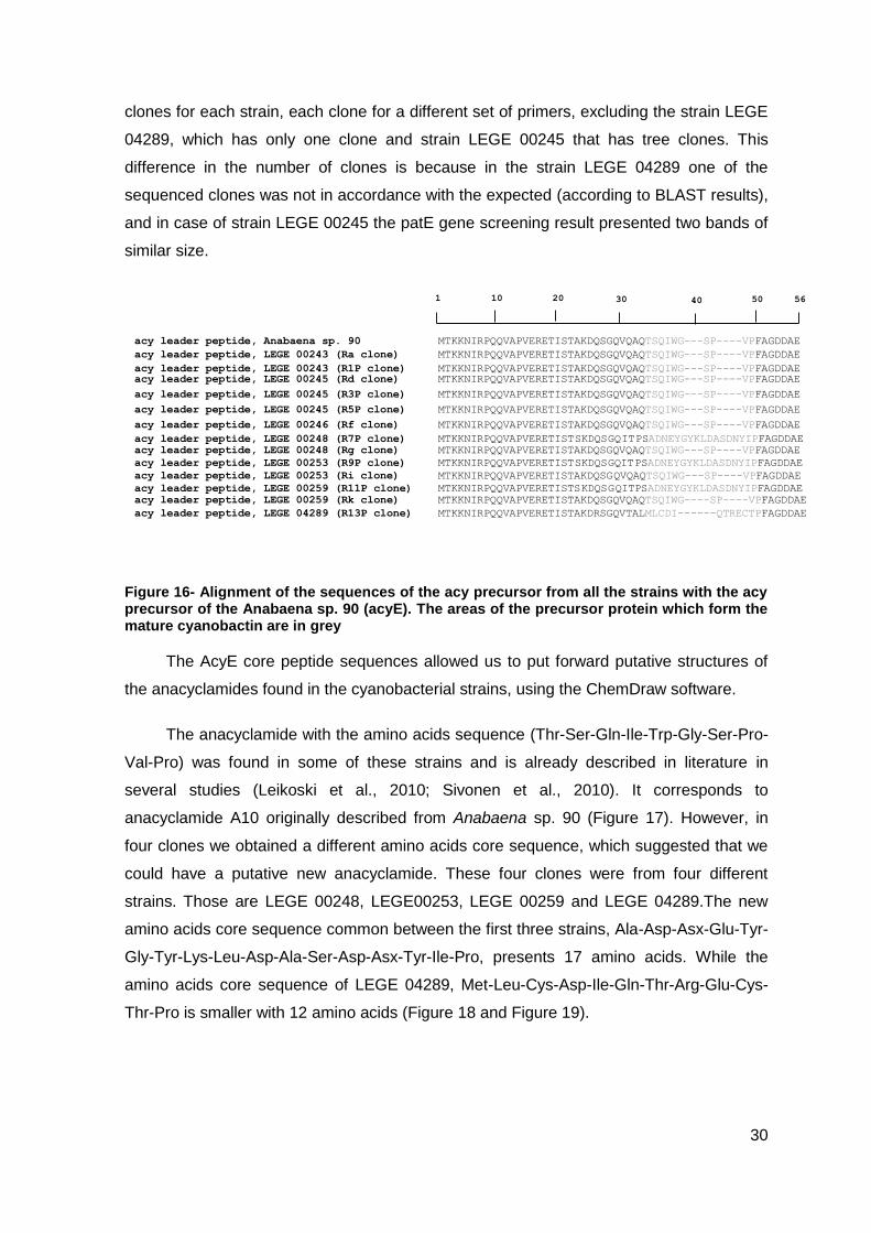
30
clones for each strain, each clone for a different set of primers, excluding the strain LEGE
04289, which has only one clone and strain LEGE 00245 that has tree clones. This
difference in the number of clones is because in the strain LEGE 04289 one of the
sequenced clones was not in accordance with the expected (according to BLAST results),
and in case of strain LEGE 00245 the patE gene screening result presented two bands of
similar size.
Figure 16- Alignment of the sequences of the acy precursor from all the strains with the acy precursor of the Anabaena sp. 90 (acyE). The areas of the precursor protein which form the mature cyanobactin are in grey
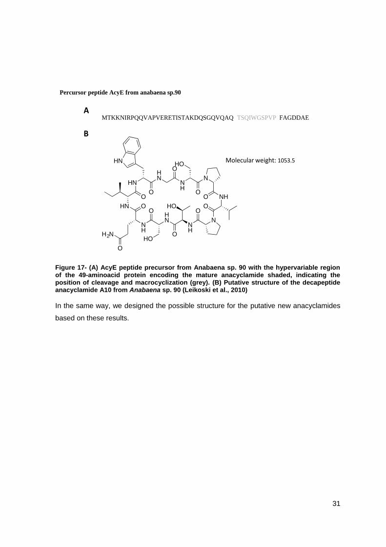
The AcyE core peptide sequences allowed us to put forward putative structures of
the anacyclamides found in the cyanobacterial strains, using the ChemDraw software.
The anacyclamide with the amino acids sequence (Thr-Ser-Gln-Ile-Trp-Gly-Ser-Pro-
Val-Pro) was found in some of these strains and is already described in literature in
several studies (Leikoski et al., 2010; Sivonen et al., 2010). It corresponds to
anacyclamide A10 originally described from Anabaena sp. 90 (Figure 17). However, in
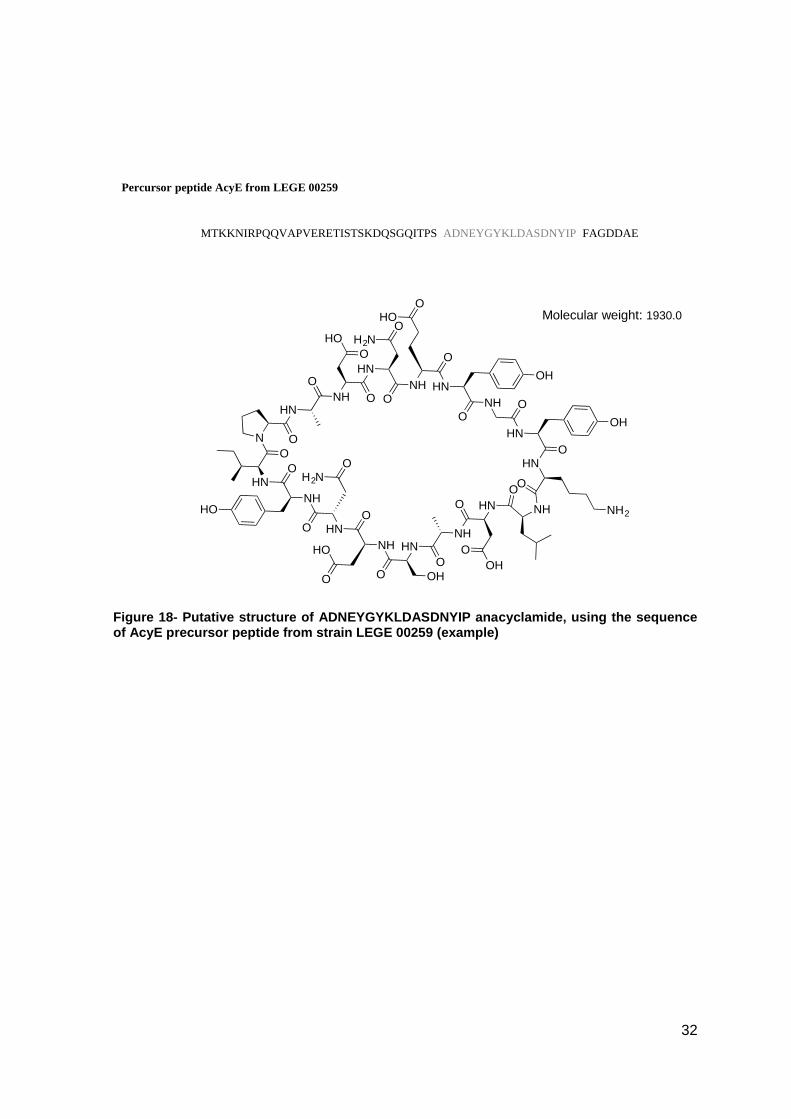
four clones we obtained a different amino acids core sequence, which suggested that we
could have a putative new anacyclamide. These four clones were from four different
strains. Those are LEGE 00248, LEGE00253, LEGE 00259 and LEGE 04289.The new
amino acids core sequence common between the first three strains, Ala-Asp-Asx-Glu-Tyr-
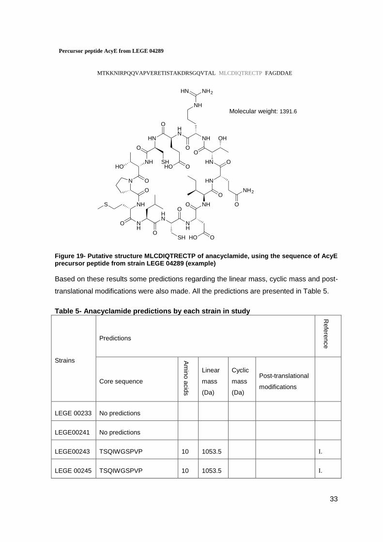
Gly-Tyr-Lys-Leu-Asp-Ala-Ser-Asp-Asx-Tyr-Ile-Pro, presents 17 amino acids. While the
amino acids core sequence of LEGE 04289, Met-Leu-Cys-Asp-Ile-Gln-Thr-Arg-Glu-Cys-
Thr-Pro is smaller with 12 amino acids (Figure 18 and Figure 19).
acy leader peptide, LEGE 00248 (R7P clone) MTKKNIRPQQVAPVERETISTSKDQSGQITPSADNEYGYKLDASDNYIPFAGDDAE
acy leader peptide, Anabaena sp. 90 MTKKNIRPQQVAPVERETISTAKDQSGQVQAQTSQIWG---SP----VPFAGDDAE
acy leader peptide, LEGE 00243 (R1P clone) MTKKNIRPQQVAPVERETISTAKDQSGQVQAQTSQIWG---SP----VPFAGDDAE
acy leader peptide, LEGE 00243 (Ra clone) MTKKNIRPQQVAPVERETISTAKDQSGQVQAQTSQIWG---SP----VPFAGDDAE
acy leader peptide, LEGE 00246 (Rf clone) MTKKNIRPQQVAPVERETISTAKDQSGQVQAQTSQIWG---SP----VPFAGDDAE
acy leader peptide, LEGE 00248 (Rg clone) MTKKNIRPQQVAPVERETISTAKDQSGQVQAQTSQIWG---SP----VPFAGDDAE
acy leader peptide, LEGE 00253 (Ri clone) MTKKNIRPQQVAPVERETISTAKDQSGQVQAQTSQIWG---SP----VPFAGDDAE
acy leader peptide, LEGE 00245 (Rd clone) MTKKNIRPQQVAPVERETISTAKDQSGQVQAQTSQIWG---SP----VPFAGDDAE
acy leader peptide, LEGE 00259 (R11P clone) MTKKNIRPQQVAPVERETISTSKDQSGQITPSADNEYGYKLDASDNYIPFAGDDAE
acy leader peptide, LEGE 00253 (R9P clone) MTKKNIRPQQVAPVERETISTSKDQSGQITPSADNEYGYKLDASDNYIPFAGDDAE
acy leader peptide, LEGE 00259 (Rk clone) MTKKNIRPQQVAPVERETISTAKDQSGQVQAQTSQIWG----SP----VPFAGDDAE
acy leader peptide, LEGE 04289 (R13P clone) MTKKNIRPQQVAPVERETISTAKDRSGQVTALMLCDI------QTRECTPFAGDDAE
acy leader peptide, LEGE 00245 (R3P clone) MTKKNIRPQQVAPVERETISTAKDQSGQVQAQTSQIWG---SP----VPFAGDDAE
acy leader peptide, LEGE 00245 (R5P clone) MTKKNIRPQQVAPVERETISTAKDQSGQVQAQTSQIWG---SP----VPFAGDDAE
1 10 20 30 40 50 56
31
Figure 17- (A) AcyE peptide precursor from Anabaena sp. 90 with the hypervariable region of the 49-aminoacid protein encoding the mature anacyclamide shaded, indicating the position of cleavage and macrocyclization (grey). (B) Putative structure of the decapeptide anacyclamide A10 from Anabaena sp. 90 (Leikoski et al., 2010)
In the same way, we designed the possible structure for the putative new anacyclamides
based on these results.
Percursor peptide AcyE from anabaena sp.90
MTKKNIRPQQVAPVERETISTAKDQSGQVQAQ TSQIWGSPVP FAGDDAEA
NH
O
HO
HN
O
HO
NH
O
H2N
O
HN
O
HN
O
HN
HN
O
NH
O
HO
N
O NH
O
N
O
Molecular weight: 1053.5
B
32
Figure 18- Putative structure of ADNEYGYKLDASDNYIP anacyclamide, using the sequence of AcyE precursor peptide from strain LEGE 00259 (example)
HN
O
NH O
HO
O
HN
O
OH2N
NH
O
HOO
HN
O
OH
NH O
HN
O
OH
HN
O
NH2NH
O
HNO
OH
O
NH
O
HN
O OH
NH
O
HO
O
HNO
OH2N
NH
O
HO
HN
O
N O
Percursor peptide AcyE from LEGE 00259
MTKKNIRPQQVAPVERETISTSKDQSGQITPS ADNEYGYKLDASDNYIP FAGDDAE
Molecular weight: 1930.0
33
Figure 19- Putative structure MLCDIQTRECTP of anacyclamide, using the sequence of AcyE precursor peptide from strain LEGE 04289 (example)
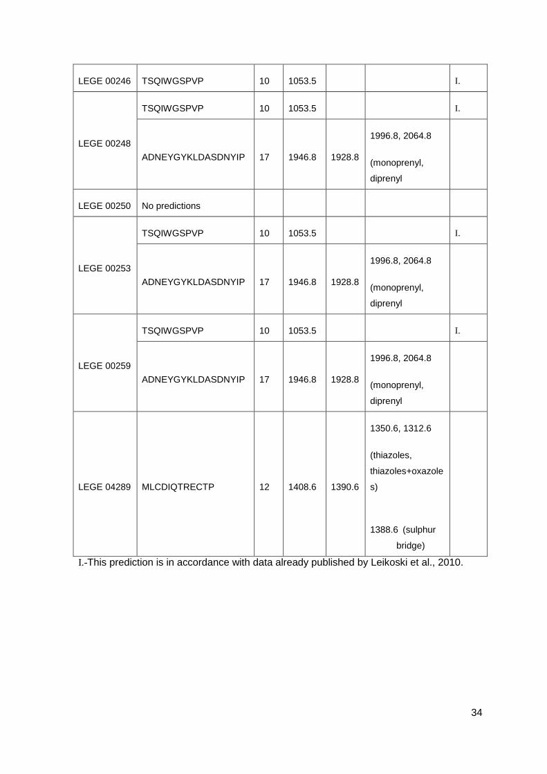
Based on these results some predictions regarding the linear mass, cyclic mass and post-
translational modifications were also made. All the predictions are presented in Table 5.
Table 5- Anacyclamide predictions by each strain in study
Strains
Predictions
Refe
rence
Core sequence
Am
ino a
cid
s
Linear
mass
(Da)
Cyclic
mass
(Da)
Post-translational
modifications
LEGE 00233 No predictions
LEGE00241 No predictions
LEGE00243 TSQIWGSPVP 10 1053.5 I.
LEGE 00245 TSQIWGSPVP 10 1053.5 I.
Percursor peptide AcyE from LEGE 04289
MTKKNIRPQQVAPVERETISTAKDRSGQVTAL MLCDIQTRECTP FAGDDAE
NH
O
S
NH
O
HN
O
SH
NH
O
HO O
NH
O
HN
O
NH2
O
HN
O
OHNH
O
NH2HN
NH
HN
O
HO O
HN
O
SHNH
O
HO
N
O
Molecular weight: 1391.6
34
LEGE 00246 TSQIWGSPVP 10 1053.5 I.
LEGE 00248
TSQIWGSPVP 10 1053.5 I.
ADNEYGYKLDASDNYIP 17 1946.8 1928.8
1996.8, 2064.8
(monoprenyl,
diprenyl
LEGE 00250 No predictions
LEGE 00253
TSQIWGSPVP 10 1053.5 I.
ADNEYGYKLDASDNYIP 17 1946.8 1928.8
1996.8, 2064.8
(monoprenyl,
diprenyl
LEGE 00259
TSQIWGSPVP 10 1053.5 I.
ADNEYGYKLDASDNYIP 17 1946.8 1928.8
1996.8, 2064.8
(monoprenyl,
diprenyl
LEGE 04289 MLCDIQTRECTP 12 1408.6 1390.6
1350.6, 1312.6
(thiazoles,
thiazoles+oxazole
s)
1388.6 (sulphur
bridge)
I.-This prediction is in accordance with data already published by Leikoski et al., 2010.
35
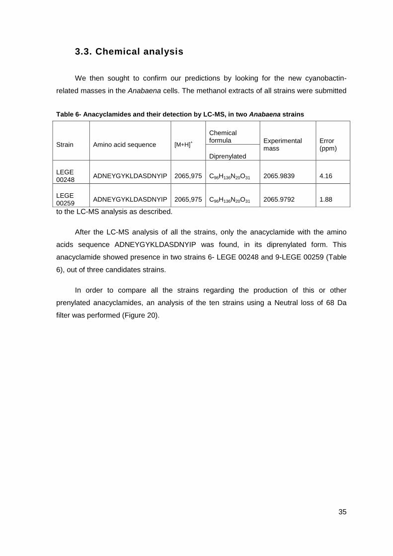
3.3. Chemical analysis
We then sought to confirm our predictions by looking for the new cyanobactin-
related masses in the Anabaena cells. The methanol extracts of all strains were submitted
to the LC-MS analysis as described.
After the LC-MS analysis of all the strains, only the anacyclamide with the amino
acids sequence ADNEYGYKLDASDNYIP was found, in its diprenylated form. This
anacyclamide showed presence in two strains 6- LEGE 00248 and 9-LEGE 00259 (Table
6), out of three candidates strains.
In order to compare all the strains regarding the production of this or other
prenylated anacyclamides, an analysis of the ten strains using a Neutral loss of 68 Da
filter was performed (Figure 20).
Table 6- Anacyclamides and their detection by LC-MS, in two Anabaena strains
Strain Amino acid sequence [M+H]+
Chemical formula Experimental
mass Error (ppm)
Diprenylated
LEGE 00248
ADNEYGYKLDASDNYIP 2065,975 C96H136N20O31 2065.9839 4.16
LEGE 00259
ADNEYGYKLDASDNYIP 2065,975 C96H136N20O31 2065.9792 1.88
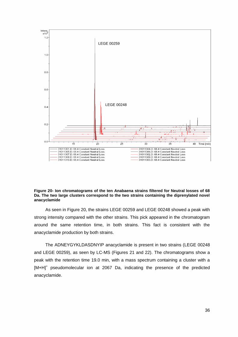
36
LEGE 00259
LEGE 00248
Figure 20- Ion chromatograms of the ten Anabaena strains filtered for Neutral losses of 68 Da. The two large clusters correspond to the two strains containing the diprenylated novel anacyclamide
As seen in Figure 20, the strains LEGE 00259 and LEGE 00248 showed a peak with
strong intensity compared with the other strains. This pick appeared in the chromatogram
around the same retention time, in both strains. This fact is consistent with the
anacyclamide production by both strains.
The ADNEYGYKLDASDNYIP anacyclamide is present in two strains (LEGE 00248
and LEGE 00259), as seen by LC-MS (Figures 21 and 22). The chromatograms show a
peak with the retention time 19.0 min, with a mass spectrum containing a cluster with a
[M+H]+ pseudomolecular ion at 2067 Da, indicating the presence of the predicted
anacyclamide.
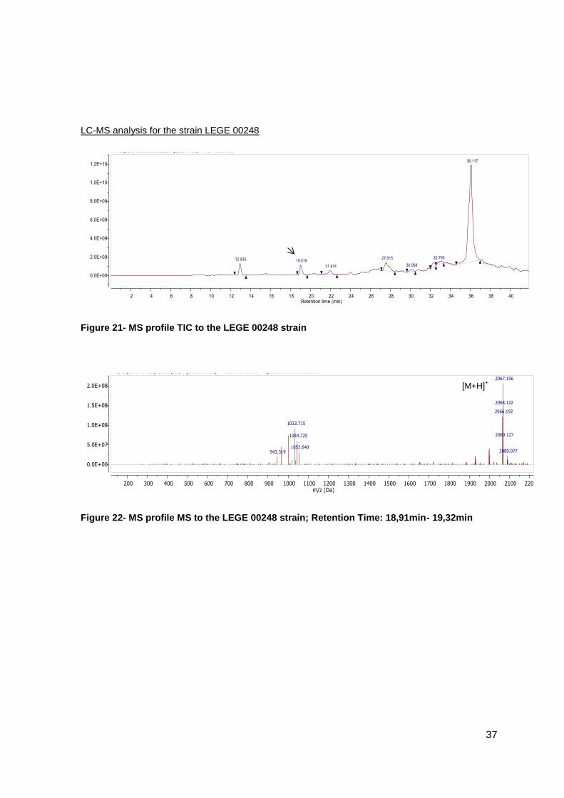
37
LC-MS analysis for the strain LEGE 00248
Figure 21- MS profile TIC to the LEGE 00248 strain
Figure 22- MS profile MS to the LEGE 00248 strain; Retention Time: 18,91min- 19,32min
[M+H]+
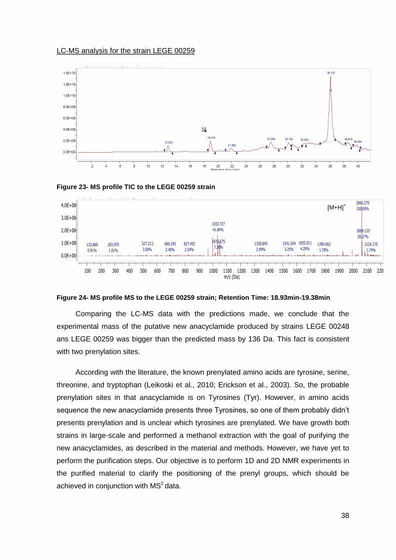
38
LC-MS analysis for the strain LEGE 00259
Figure 23- MS profile TIC to the LEGE 00259 strain
Figure 24- MS profile MS to the LEGE 00259 strain; Retention Time: 18.93min-19.38min
Comparing the LC-MS data with the predictions made, we conclude that the
experimental mass of the putative new anacyclamide produced by strains LEGE 00248
ans LEGE 00259 was bigger than the predicted mass by 136 Da. This fact is consistent
with two prenylation sites.
According with the literature, the known prenylated amino acids are tyrosine, serine,
threonine, and tryptophan (Leikoski et al., 2010; Erickson et al., 2003). So, the probable
prenylation sites in that anacyclamide is on Tyrosines (Tyr). However, in amino acids
sequence the new anacyclamide presents three Tyrosines, so one of them probably didn’t
presents prenylation and is unclear which tyrosines are prenylated. We have growth both
strains in large-scale and performed a methanol extraction with the goal of purifying the
new anacyclamides, as described in the material and methods. However, we have yet to
perform the purification steps. Our objective is to perform 1D and 2D NMR experiments in
the purified material to clarify the positioning of the prenyl groups, which should be
achieved in conjunction with MS2 data.
[M+H]+
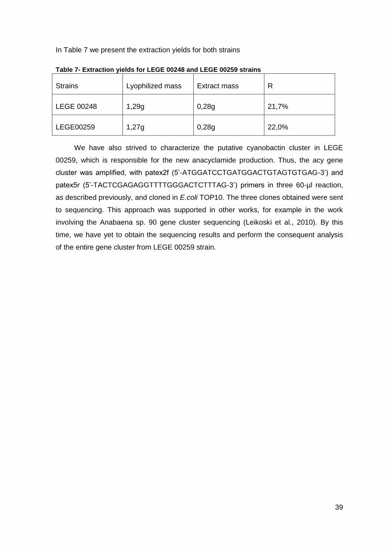
39
In Table 7 we present the extraction yields for both strains
Table 7- Extraction yields for LEGE 00248 and LEGE 00259 strains
Strains Lyophilized mass Extract mass R
LEGE 00248 1,29g 0,28g 21,7%
LEGE00259 1,27g 0,28g 22,0%
We have also strived to characterize the putative cyanobactin cluster in LEGE
00259, which is responsible for the new anacyclamide production. Thus, the acy gene
cluster was amplified, with patex2f (5’-ATGGATCCTGATGGACTGTAGTGTGAG-3’) and
patex5r (5’-TACTCGAGAGGTTTTGGGACTCTTTAG-3’) primers in three 60-µl reaction,
as described previously, and cloned in E.coli TOP10. The three clones obtained were sent
to sequencing. This approach was supported in other works, for example in the work
involving the Anabaena sp. 90 gene cluster sequencing (Leikoski et al., 2010). By this
time, we have yet to obtain the sequencing results and perform the consequent analysis
of the entire gene cluster from LEGE 00259 strain.

41
4. Conclusion and future work
In this study we reported a novel low-molecular-weight peptide produced
ribosomally by Anabaena strains. The new post-translationally modified anacyclamide,
with two prenyl groups, was detected in two of ten Anabaena strains screened for. The
probable prenylation sites are on Tyrosines. However the anacyclamide presented, in the
amino acids sequence, three Tyrosines. It is unclear which Tyrosines are prenylated and
further investigations on the prenylation mechanisms may help us gain more insight into
these post-translational tailoring events, for example, the basis for the non-prenylation of
one of the tyrosine side-chains.
The next step will be to carry out nuclear magnetic resonance (NMR) to fully
characterize this new compound, identifying in which Tyrosines the prenylation occurs to
make a phylogenetic inference regarding anacyclamides production in these and other
strains.
Overall, this study reports a new cyanobacterial secondary metabolite, which can
expand our knowledge about this family of compounds and their biological activity.

43
5. References
Arnison, P.G., Bibb, M.J., Bierbaum, G., Bowers, A.A., Bugni, T.S. Bulaj, G.,
Camarero, J.A., Campopiano, D.J., Challis, GL, Clardy, J., Cotter, P.D., Craik, D.J.,
Dawson, M., Dittmann, E., Donadio, S., Dorrestein, P.C., Entian, K.D., Fischbach, M.A.,
Garavelli, J.S., Göransson, U., Gruber, C.W., Haft, D.H., Hemscheidt, T.K., Hertweck, C.,
Hill, C., Horswill, A.R., Jaspars, M., Kelly, W.L., Klinman, J.P., Kuipers, O.P., Link, A.J.,
Liu, W., Marahiel, M.A., Mitchell, D.A., Moll, G.N., Moore, B.S., Müller, R., Nair, S.K., Nes,
I.F., Norris, G.E., Olivera, B.M., Onaka, H., Patchett, M.L., Piel, J., Reaney, M.J.,
Rebuffat, S., Ross, R.P., Sahl, H.G., Schmidt, E.W., Selsted, M.E., Severinov, K., Shen,
B., Sivonen, K., Smith, L., Stein, T., Süssmuth, R.D., Tagg, J.R., Tang, G.L., Truman,
A.W., Vederas, J.C., Walsh, C.T., Walton, J.D., Wenzel, S.C., Willey, J.M., van der Donk.
W.A. (2013) Ribosomally synthesized and posttranslationally modified peptide natural
products: overview and recommendations for a universal nomenclature. Nat. Prod. Rep.
30: 108-60.
Béchemin, C., Grzebyk, D., Hachame, F., Hummert, C., Maestrini, S.Y.
(1999).Effect of different nitrogen/phosphorus nutrient ratios on the toxin content in
Alexandrium minutum. Aquat Microb Ecol 20: 157–165.
Bokesch, H.R., O'Keefe, B.R., McKee, T.C., Pannell, L.K., Patterson, G.M.L.,
Gardellina, R.S. II, Sowder, R.C., Turpin, J., Watson, K., Buckheit, R.W. Jr, Boyd, M.R.
(2003).A potent novel anti-HIV protein from the cultured cyanobacteriumScytonema
varium. Biochemistry 42: 2578–2584.
Boyd, M.R., Gustafson, K.R., McMahon, J.B., Shoemaker, R.H., O'Keefe, B.R.,
Mori, T., Gulakowski, R.J., Wu, L., Rivera, M.I., Laurencot, C.M., Currens, M.J.,
Cardellina, J.H. 2nd, Buckheit, R.W. Jr., Nara, P.L., Pannell, L.K., Sowder, R.C. 2nd,
Henderson, L.E. (1997). Discovery of cyanovirin-N, a novel human immunodeficiency
virus-inactivating protein that binds viral surface envelope glycoprotein gp 120: potential
applications to microbicide development. Antimicrob Agents Chemother 42:1521–1530.
Burja, A.M., Banaigs, B., Abou-Mansour, E., Burgess, J.G., Wright, P.C. (2001).
Marine cyanobacteria – a prolific source of natural products. Tetrahedron 57:9347–9377.
44
Castenholz, R.W. (2001) Phylum BX. Cyanobacteria, oxygenic photosynthetic
bacteria. p. 473–599. In: Boone DR, Castenholz RW (ed.) Bergey’s Manual of Systematic
Bacteriology Part 1, 2nd edition. SpringerVerlag, New York, USA.
Donia, M. S., Ravel, J. and Schmidt, E.W. (2008). A global assembly line for
cyanobactins, Nat. Chem. Biol.4:341–343.
Donia, M.S., and Schmidt, E.W. (2010). Cyanobactins—ubiquitous cyanobacterial
ribosomal peptide metabolites. In Comprehensive Natural Products II Chemistry and
Biology (Oxford: Elsevier), 539–558.
Donia, M.S., Hathaway, B.J., Sudek, S., Haygood, M.G., Rosovitz, M.J., Ravel, J.,
Schmidt, E.W. (2006). Natural combinatorial peptide libraries in cyanobacterial symbionts
of marine ascidians. Nat Chem Biol 2:729–735.
Donia, M.S., Schmidt, E.W.(2011). Linking chemistry and genetics in the growing
cyanobactin natural products family. Chem Biol 18:508-19.
Fay, P. (1992). Oxygen relations of nitrogen fixation in cyanobacteria. Microbiol.
Rev. 56: 340-373
Francis, G. (1878). Poisonous Australian lake. Nature 18, 11e12. Garcia-Pichel, F.,
2008. Molecular ecology and environmental genomics of cyanobacteria. In: Herrero, A.,
Flores, E. (Eds.), The Cyanobacteria-Molecular Biology, Genomics and Evolution. Caister
Academic Press, Norfolk, UK, 484.
Franz, C.M.A.P., van Belkum M.J., Holzapfel W.H., Abriouel H., Galvez A. (2007)
Diversity of enterococcal bacteriocins and their grouping in a new classification scheme.
FEMS Microbiol. Rev. 31:293–310
Golden, J.W., Yoon H.S. (1998). Heterocyst formation in Anabaena. Curr. Opin.
Microbiol. 1:623–629
Harada, K.I. (2004). Production of secondary metabolites by freshwater
cyanobacteria. Chem. Pharm. Bull. 35: 889-899.
Hoiczyk, E., Hansel,A. (2000). Cyanobacterial Cell Walls: News from an Unusual
Prokaryotic Envelop. Journal of Bacte. 182(5), 1191-1199.
45
Houssen, W.E.; Koehnke, J.; Zollman, D.; Vendome, V.; Raab, A.; Smith, M.C.M.;
Naismith, J.H.; Jaspars, M.(2012) The Discovery of New Cyanobactins from Cyanothece
PCC 7425 Defines a New Signature for Processing of Patellamides. ChemBioChem, 13:
2625–2776.
Ishida, K., Matsuda H., Murakami M., Yamaguchi K. (1997). Kawaguchipeptin B and
antibacterial cyclic undekapeptide from the cyanobacterium Microcystis aeruginosa. J Nat
Prod 60:724–726.
Jack, R.W., Jung G. (2000) Lantibiotics and microcins: polypeptides with unusual
chemical diversity. Curr. Opin. Chem. Biol. 4:310–317.
John, E.H., Flynn K.J. (2000). Growth dynamics and toxicity of Alexandrium
fundyense (Dinophyceae): the effect of changing N:P supply ratios on the internal toxin
nutrient levels. Eur J Phycol 35:11–23.
Jungblut, A.D., Hawes, I., Mountfort, D., Hitzfeld, B., Dietrich, D.R., Burns, B.P.,
Neilan, B.A., (2005). Diversity within cyanobacterial mat communities in variable salinity
meltwater ponds of McMurdo Ice Shelf, Antarctica. Environ. Microbiol.7: 519-529.
Knoll, A.H., (2008). Cyanobacteria and earth history. In: Herrero, A., Flores, E.
(Eds.), The Cyanobacteria: Molecular Biology, Genomics and Evolution. Caister Academic
Press, Norfolk, UK,484.
Kotai, J. (1972). Instructions for preparation of modified nutrient solution Z8 for
algae. Norwegian Institute for Water Research, Oslo, Norway.13.
Lee, J., McIntosh J., Hathaway B.J., Schmidt E.W. (2009). Using marine natural
products to discover a protease that catalyses peptide macrocyclization of diverse
substrates. J. Am. Chem. Soc. 131:2122–2124.
Lee, S.W., Mitchell D.A., Markley A.L., Hensler M.E., Gonzalez D., Wohlrab A.,
Dorrestein P.C., Nizet V., Dixon J.E. (2008). Discovery of a widely distributed toxin
biosynthetic gene cluster. Proc Natl Acad Sci USA 105: 5879–5884.
Leikoski, N. (2013). Cyanobactins - ribosomally synthesized and posttranslationally
modified peptides produced by cyanobacteria. Academic dissertation. University of
Helsinki. 10-50.
46
Leikoski, N., Fewer, D.P., Jokela, J., Wahlsten, M., Rouhiainen, L., Sivonen, K.
(2010). Highly diverse cyanobactins in strains of the genus Anabaena. Appl Environ
Microbiol 76:701–709.
Leikoski, N., Fewer, D.P., Sivonen, K. (2009). Widespread occurrence and lateral
transfer of the cyanobactin biosynthesis gene cluster in cyanobacteria. Appl. Environ.
Microbiol.75:853–857.
Leikoski, N., Liu, L., Jokela J., Wahlsten, M., Gugger, M., Calteau, A., Permi, P.,
Kerfeld, C. A., Sivonen, K., Fewer, D.P. (2013). Genome Mining Expands the Chemical
Diversity of the Cyanobactin Family to Include Highly Modified Linear Peptides. Chem. &
Bio. 20: 1-11.
Liberton, M., Austin II, J.R.; Berg, R.H.; Pakrasi, H.B. (2011). Unique Thylakoid
Membrane Architecture of a Unicellular N2-Fixing Cyanobacterium Revealed by Electron
Tomography. 155(4): 1656-1666.
Linington, R.G., Gonzàles, J., Ureña L.D., Romero L.I., Ortega-Barria E., Gerwick
W.H. (2007). Venturamides A and B: Antimalarial constituents of the Panamanian marine
cyanobacterium Oscillatoriasp. J Nat Prod 70:397-401.
Magarvey, N. A., Z. Q. Beck, T. Golakoti, Y. Ding, U. Huber, T. K. Hemscheidt, D.
Abelson, R. E. Moore, and D. H. Sherman. (2006). Biosynthetic characterization and
chemoenzymatic assembly of the cryptophycins. Potent anticancer agents from
cyanobionts. ACS Chem. Biol.1:766–779.
Maqueda, M., Sanchez-Hidalgo, M., Fernandez M., Montalban-Lopez M., Valdivia
E., Martinez-Bueno M. (2008). Genetic features of circular bacteriocins produced by
Gram-positive bacteria. FEMS Microbiol Rev 32: 2–22.
Martins, M.R.F., (2005). Ecotoxicology of Marine Cyanobacteria from de Portuguese
Coast. Dissertação de Doutoramento, Faculdade de Ciências da Universidade do Porto,
106.
Martin-Visscher, L.A., Gong, X.D., Duszyk, M., Vederas, J.C. (2009). The three-
dimensional structure of carnocyclin A reveals that many circular bacteriocins share a
common structural motif. J Biol. Chem. 284:28674–28681.
47
Metting, B., Pyne, J.W. (1986). Biologically active compounds from microalgae.
Elsevier 8(7): 386-394.
Neilan, B.A., Jacobs, D., Therese, D.D., Blackall, L.L., Hawkins, P.R., Cox, P.T.,
Goodman, A.E., (1997). rRNA sequences and evolutionary relationships among toxic and
nontoxic cyanobacteria of the genus Microcystis. Int. J. Syst. Bacteriol.47: 693-697.
Nolan, E.M., Walsh, C.T. (2009). How nature morphs peptide scaffolds into
antibiotics. ChemBioChem 10: 34–53.
Ogino, J., Moore, R.E., Patterson, G.M.L., Smith, C.D. (1996). Dendroamides, new
cyclic hexapeptides from blue-green alga. Multidrug-resistance reversing activity of
dendroamide A. J Nat Prod 59: 581–586
Oman, T.J., van der Donk, W.A. (2010). Follow the leader: the use of leader
peptides to guide natural product biosynthesis. Nat. Chem. Biol. 6, 9–18.
Paul, V.J. (2008). Global Warming and cyanobacteria harmful algal blooms in
Cyanobacterial Harmful Algal Blooms: State of the Science and Research Needs, H.K.
Hudnell, Editor. 2008, U.S.Environmental Protection Agency: Springer US., 239-257.
Philmus, B., Christiansen, G., Yoshida, W.Y., Hemscheidt, T.K. (2008).
Posttranslational modification in microviridin biosynthesis. Chem.BioChem. 9:3066–3073.
Portmann, C., Blom, J.F., Kaiser, M., Brun, R., Jüttner F., Gademann, K. (2008b)
Isolation of aerucyclamides C and D and structure revision of microcyclamide 7806A:
Heterocyclic ribosomal peptides from Microcystis aeruginosa PCC 7806 and their
antiparasite evaluation. J Nat. Prod. 71:1891-1896.
Rippka, R., Castenholz, R. W. & Herdman, M. (2001). Subsection IV. In Bergey's
Manual of Systematic Bacteriology, 2nd edn, vol. 1, pp. 562–589. Edited by D. R. Boone &
R. W. Castenholz. New York: Springer.
Rouhiainen, L., Paulin, L., Suomalainen, S., Hyytiäinen, H., Buikema, W.,
Haselkorn, H., Sivonen, K. (2000). Genes encoding synthetases of cyclic depsipeptides,
anabaenopeptilides, in Anabaena strain 90. Molecular Microbiol. 37:156–167.
Salvatella, X., Caba, J.M., Albericio, F., Giralt, E. (2003). Solution structure of the
antitumor candidate trunkamide A by 2D NMR and restrained simulated annealing
methods. J Org. Chem. 68:211-215.
48
Schmidt, E.W., Donia, M.S. (2009). Cyanobactin ribosomally synthesized peptides—
a case of deep metagenome mining. Meth. Enzymol. 458:575–596.
Schmidt, E.W., Nelson, J.T., Rasko, D.A., Sudek, S., Eisen, J.A., Haygood, M.G.,
Ravel, J. (2005). Patellamide A and C biosynthesis by a microcin-like pathway in
Prochloron didemni, the cyanobacterial symbiont of Lissoclinum patella. Proc Natl Acad
Sci USA 102:7315-7320.
Shih, P.M., et al. (2013) Improving the coverage of the cyanobacterial phylum using
diversity-driven genome sequencing. PNAS. 110, 1053-1058.
Sivonen, K. , Namikoshi,M., Evans, W.R., Carmichael,W.W., Sun,F., Rouhiainen,L.
Luukkainen, R., Rinehart, K.L.(1992). Isolation and characterization of a variety of
microcystins from seven strains of the cyanobacterial genus Anabaena. Appl. Environ.
Microbiol. 58: 2495-2500.
Sivonen, K., and T. Börner.(2008). Bioactive compounds produced by
cyanobacteria, p. 159–197.InA. Herraro and E. Flores (ed.), The cyanobacteria: molecular
biology, genomics and evolution. Caister Academic Press,Norfolk, United Kingdom.
Sivonen, K., Jones, G. (1999). Cyanobacterial toxins, p. 41–111. In: Chorus, I. and
Bartram, J (ed.), Toxic cyanobacteria in water. A guide to their public health
consequences, monitoring and management, E & FN Spon, London, England.
Sivonen, K., Leikoski, N.,Fewer, D.P., and Jokela, J. (2010). Cyanobactinsribosomal
cyclic peptides produced by cyanobacteria. Appl. Microbiol. Biotechnol.86: 1213–1225.
Sudek, S., Haygood, M.G., Youssef,D.T.A, Schmidt, E.W. (2006). Structure of
trichamide, a cyclic peptide from the bloom-forming cyanobacterium Trichodesmium
erythraeum, predicted from the genome sequence. Appl. Environ. Microbiol. 72:4382–
4387.
Tan, L.T., Márquez, B.L., Gerwick, W.H. (2001). Lyngbouilloside, a Novel Glycosidic
Macrolide from the Marine Cyanobacterium Lyngbya bouillonii 65: 925- 928.
Tianero, M.D., Donia, M.S., Young, T.S., Schultz, P.G., Schmidt, E.W. (2012).
Ribosomal route to small-molecule diversity. J Am Chem. Soc. 134:418-25.
49
Tillett, D., Dittmann, E., Erhard, M., von Döhren H., Börner, T., Neilan, B.A. (2000).
Structural organization of microcystin biosynthesis in Microcystin aeruginosaPCC7806: an
integrated peptidepolyketide synthetase system. Chem. Biol .7:753–764
van der Donk, W.A, (2013). Ribosomally synthesized and posttranslationally
modified peptide natural products: overview and recommendations for a universal
nomenclature. Nat Prod Rep 30:108-60.
Welker, M., von Döhren, H. (2006). Cyanobacterial peptides – Nature’s own
combinatorial synthesis. FEMS Microbial Reviews 30:530–563.
Whitton, B.A., (2008). Cyanobacterial Diversity in Relation to the Environment. In:
Valtere Evangelista, L.B., Anna, Maria Frassanito, Vincenzo, Passarelli, Paolo, Gualtieri
(Eds.), Algal Toxins: Nature, Occurrence, Effect and Detection. Springer, Netherlands, pp.
17- 43.
Woese, C.R. (1987).Bacterial Evolution.Microbio.Rev.51: 221-71.
Ziemert, N., Ishida, K., Liaimer, A., Hertweck, C., Dittmann, E. (2008a). Ribosomal
synthesis of tricyclic depsipeptides in bloom-formingcyanobacteria. Angew Chem Int Ed
47:7756–7759.
Ziemert, N., Ishida, K., Quillardet, P., Bouchier, C., Hertweck, C., Tandeau de
Marsac, N., Dittmann, E. (2008b). Microcyclamide biosynthesis in two strains of
Microcystis aeruginosa: from structure to genes and vice versa. Appl. Environ. Microbiol.
74:1791–1797.