detection of some virulence factors of enterococcus...
TRANSCRIPT
Detection of some virulence factors of Enterococcus faecalis isolated from raw milk by Multiplex PCR
Dr. Azhar Noory Hussein Al-Qadisiyah - Pharmacy college- Clinical pathology department Aubstract
Since milk is a high nutrition food , it is extremely liable to enterococcal contamination , as this widely distributed in the environment . This study evaluated 40 raw milk samples ( 10 samples for each cows , sheep , goats and buffalo ) for enteroccal contamination . Raw milk samples were collected from different farms in Diwaniyah city /Al-Qadisiya governorate between May and July 2012. This objective of this study was numerate the number of enterococci in the raw milk samples . Also to find out the out the distribution of major virulence factors asa1 , gelE , cylA , esp and hyl genes by multiplex PCR among Enterococcus faecalis isolates and the production of gelatinase and hemolytic activity were also determined . The antibiotics susceptibity of the strains was determined using the disc diffusion method . The results of the current study showed that the raw milk had a total viable count in the range from 1*102 – 34*106 CFU/ml and all raw milk samples were positive for the presence of enterococci . Significant difference was not exist between the types of raw milk samples . twenty strains of E. faecalis were isolated from 40 raw milk samples . These strains were identified by conventional biochemical tests and the HiStrep system . Tn addition , E. faecalis specific 16S rDNA gene (amplicon size 290 bp ) primers were included as a control when the biochemical identification was doubtful . Multiplex PCR for virulence factors showed that E. faecalis isolates carried one or more virulence encoding genes . The asa1 gene was the predominant (75%) of the E. faecalis strains investigated followed by hyl gene (70%) . The esp , gelE and cylA genes were detected with percentages of 65% , 50% and 25% respectively . Antibiotics susceptibility testing showed that of the total 20 isolates 12(60%) were resistant to erythromycin and 9(45%) to ampicillin . Lower antibiotics resistance was seen with gentamycin , tetracycline , norfloxacin , penicillin and rifampicin ( 20% , 15% , 10% , 15% and 10% respectively ) . None of the isolates was found to be resistant to be chloramphenicol . Introduction:
Milk is a complex biological fluid and by its nature, a good growth medium for many microorganisms. Because of the specific production it is impossible to avoid contamination of milk with microorganisms therefore the microbial content of milk is a major feature in determining is quality (31). Microorganisms may contaminate milk from the animals udder , barn , milk collection materials , poor storage conditions, various ingredients added to dairy product and dairy farm workers (19).
Microbial load in fresh milk is although very low less than 10-3 CFU/L but this level may increase up to 100 fold if this milk is stored for many days of normal temperature (32) . Preventing the growth of contaminating microorganisms in milk involves limiting contamination levels , cooling immediately after milking and maintenance of cold storage temperature (18).

Enterococci are represent normal components of the raw milk microbiota. There no standards set for the minimum and maximum count of enterococci because they are not normally counted in microbiological analysis . A study of the levels of enterococci in raw cows milk from 10 New Zealand farm in 1997 revealed an enterococcal minimum a count of < 101 and maximum of 1.2* 104 CFU/ml , though 95% of the samples of the same study had less than 1.9* 103 CFU/ml (22). While enterococcal counts in goats milks can reach higher levels : log levels of 1.7* 104 CFU/ml have been reported in goats milk in Czech Republic (26 ).
For many years Enterococcus species were believed to be harmless to human and considered unimportant medically (15). Recently enterococci have become one of the most common nosocomial pathogens , with patients having a high mortality rate of up to 61% ( 12). The genus Enterococcus consists of gram-positive , catalase-negative , non-spore forming , facultative anaerobic bacteria and that can occur both as single cocci and in chains ( 14).
They are commensal microorganisms that colonize in the gastrointestinal tract of humans and animals and are also found in several different food source such as milk and cheese (5). These bacteria are able to survive extreme environments , such as 6.5% NaCl , pH of 9.6 , high heat as well as being able to grow and survive under other harsh environmental conditions (3). Different Enterococcus species are found in raw milk by Enterococcus faecalis and Enterococcus faecium remain the species of the greatest importance (17).
E. faecalis is an opportunistic pathogen known to cause serious infections such as bacteraemia , septicamemia , urinary tract infections , wound infections , meningitis and endocarditis ( 23; 36). Also their increasing antibiotic resistance which spread rapidly among populations (1) and available antibiotics currently limits the therapeutic options (7).E. faecalis strains possess several putative virulence determinants , although the molecular mechanism of virulence is still not completely understood (20). E. faecalis may carry various genes directly or indirectly contributing to virulence (39).
Gene encoding virulence factors such as aggregation substance (asa1), gelatinase (gelE), cytolysin(CylA), enterococcal surface protein (esp), enterococcal surface adhesion (ace) , adhesion-associated protein(efaA)(also known endocarditis antigen A) and hyaloronidase (hyl) as secretary factors have been described in E. faecalis ( 24 ; 13).
Aggregation substance (AS) as surface protein adehesion encoded by asa1 which is carried on a plasmid ( 39). Gelatinase ia a zinc-endopeptidase produced by E. faecalis that is capable of hydrolyzing gelatin , collagen , casein , hemoglobin and other peptides . Gelatinase encoded by the chromosomal gelE (40). Cytolysin is a bacterial toxin with hemolytic and bacteriolytic activity . Cytocylin genes are carried on a plasmid or are integrated into the bacterial chromosome 8).The E. faecalis cytolysin operon has been characterized and the regulation of cytocylin expression described (20).
The enterococcal surface protein , adhesion-associated protein and enterococcal surface adhesion , encoded by the chromosomal esp , efaA and ace respectively , which are associated with increased virulence , colonization and have been shown to enhance the persistence of E. faecalis in the urinary bladder during experimental urinary tract infection (Shanker et al. 2001).Recently another virulence factors, hyaluronidase was described in E. faecalis which is expressed by the hyl chromosomal gene , acts on hyaluronic acid and increases bacterial invasion (39).

The purpose of this study were to define the antibiotic resistance pattern and to investigate the presence of genes encoding several virulence factors in E. faecalis strains isolated from raw milk samples in Diwaniyah city- Al-Qadisiya governorate .
Materals and methods: - Source and collection of milk samples:
This investigation was based on collection of 40 raw milk samples (10) samples for each cows , goats , buffalo and sheep) from different farms in Diwaniyah city /Al-Qadisiya governorate between May and July 2012 . The raw milk samples were collected into clean sterile bottles and transported in an ice box to the laboratory of the department Biology / College of Education / Al-Qadisiya university , where examinations of the milk samples were done after collection of samples . - Microbiological analyses :
For the enumeration of Enterococci , one ml of each samples was diluted in peptone diluting saline to obtain the dilutions of 10-1- 10-7 and 0.1 ml of each dilution was spread on Rapid Hi-Enterococci agar M1414 ( Himedia , India). The plates were incubated for 24 hrs. at 37 Co (16).Typical blue colonies with the appearance of E. faecalis were selected for farther study.
E. faecalis were presumptively identify by the following tests : observation of cell morphology , gram staining , catalase , growth at 15 and 45 Co in the presence of 6.5% NaCl and at pH 9.6 (7). HiStrep system (Himedia , India) was used for E. faecalis isolates identification according to the manufacture's instructions . All isolates were kept in Brain heart infusion broth with 15% glycerol at -20 Co until further analysis .
-Phenotype method for hemolytic activity and gelatinase production
The hemolytic activity of isolates was determined according to (3) on blood agar base plates containing 5% (V/V) of sheep blood . After incubating the plates at 37 Co for 24 hrs. , the hemolytic reaction was recorded by the observation of hydrolysis of red blood cells and appearance of clear zone around bacterial isolates .
Gelatinase production was detected by inoculating E. faecalis isolates onto trypticase soya agar containing 3% gelatin . The appearance of turbid halos or zones around the colonies after incubation at 37 Co for 24 hrs. was considered to be a positive indication of gelatinase production ( 30 ) .
-Resistance of selected isolates to antibiotics
Antibiotic resistance of the isolated strains was tested by agar disc diffusion method on muller hinton agar with 10% of sheep blood . The following antibiotic discs ( HiMedia , India ) were used gentamycin , tetracycline , norfloxacin , penicillin , nitrofurantoin , chloramphenicol , erythromycin and ampicillin . After incubation 37 Co for 24 hrs. Interpretation of results was according to the standards adopted from United States Committee for Standardization of Clinical Trials , 15to 20 mm zone of inhibition is sensitive , 10 to 14 mm is the medium sensitive and 10 mm below is resistant ( 42 ) . - DNA extraction
The total genomic DNA of the E. faecalis was isolated using the DNA extraction and purification kit ( Geneaid , Korea ) according to the manufacturer

instructions . DNA preparations were then analyzed by electrophoresis in 1.5% agarose gel . -genotype identification using 16S rDNA
Genetic identification to species level was performed by16S rDNA as described by(2) briefly , the PCR reaction mixture ( Bioneer , Korea ) contained 5 µl template DNA , 1.5 µl of reverse primer ( 10 pmol/ µl ) , 1.5µl of forward primer ( 10 pmol/ µl ) , 250µM of dNTP , 1.5 mM of MgCl2 , 30 mM of KCl and 1 unit of Taq DNA polymerase with PCR water added to obtain 20 µl final volume in the PCR tube . The primers used for amplification of 16S rDNA gene were described in table (1) with expected product size of 290 bp. Samples were amplified in a DNA AMP thermocycler system (TECHNE , USA) by heading for 5 min. at 95 Co , followed by 30 cycles of 95 Co for 60 s. , 58 Co for 60 s. and 72 Co for 6o s. and a final step of 72 Co for 10 min. Gel electrophoresis was carried out by using 0.8% agarose in TBE ( Tris – Borate – EDTA ) . The DNA samples were loaded on the loading dye ( BioBasic , Canada) and the voltage was then applied 100 v/cm after soaking in TBE buffer . The DNA was visualized using UV transilluminater afer staining the gel with ethidium bromide (10 mg/ml) for 20 min. . -Oligonucleotide primers and Multiplex PCR :
The five Oligonucleotideprimer pairs ( table 1) used to amplify the genes asa1 , gelE , cylA , esp and hyl . The expected amplicon sizes are listed in table (1) . The Oligonucleotide used in this sudy were previously reported elsewhere (39) . The Multiplex PCR assays were performed in a DNA AMP thermocycler system (TECHNE , USA) as a final volume of 20 µl total containing AccuPower Multiplex PCR premix (Bioneer , Korea ) , 0.2 µM of each primer and 5 of DNA template . PCR buffer added to obtain 20 µl final volume in the PCR tube . The temperature and time conditions of the PCR program as follows : An initial activation step at 95 Co for 15 min., during which the HotStar Taq DNA polymerase is activated , was followed by 30 cycles of denaturation 94 Co for 1 min. , annealing 56 Co for 1 min. and extention 72 Co for 1 min. followed by one cycle consisting of 10 min. at 72 Co . After amplification , 5 µl of the amplicon was added directly on 1.5% agarose without addind a loading dye to analyze the PCR products and electrophoresed for 1 hrs. at 100 v/cm in TBE buffer containing 0.05 mg of ethidium promide per liter . A 100 bp DNA ladder ( Bioneer , Korea ) was used as a molecular size marker.

Table 1 PCR primers and products for detection of E. faecalis virulence determinants
Gene Sequence (5'-3') Product size (bp) References
16S rRNA
TGGCATAAGAGTGAAAGGCGC GGGGACGTTCAGTTACTAACGT
290 (2 )
asa1
ASA11- GCACGCTATTACGAACTATGA ASA12- TAAGAAAGAACATCACCACGA
375
gelE
GEL 11- TATGACAATGCTTTTTGGGAT GEL 12- AGATGCACCCGAAATAATATA
213
cylA CYT I- ACTCGGGGATTGATAGGC CYT IIb- GCTGCTAAAGCTGCGCTT
688
esp
ESP 14F- AGATTTCATCTTTGATTCTTGG ESP 12R- AATTGATTCTTTAGCATCTGG
510
hyl HYL n1- ACAGAAGAGCTGCAGGAAATG HYL n2- GACTGACGTCCAAGTTTCCAA
276
(39)
- Statistical Analysis The results were analysed statistically by Chi- square ( X2 ) test at the level of significant when P- Value ≤ 0.05 (29) . Results and discussion:
Enterococci naturally occur in raw milk as part of it′s microbial population , in Iraq no standards are set for enterococci either , given that these bacteria are not routinely counted in raw milk .
The results showed ( table 2) , the raw milk had a total viable count in the range from 1*102 – 34*106 CFU/ml and all raw milk samples were positive for the presence of enterococci . There was not significant difference ( P≤ 0.05 ) among the milk samples collected from different animals ( cows , goats , sheep and buffalo ) . These findings are similar with those of ( 18 ; 16 ; 26 ) .
Table (2): Numerate of Enterococci in raw milk samples ( CFU/ml ) No. of sample Cows Goats Sheep Buffalo
Sam.1 2*105 14*104 17*106 4*102 Sam.2 1*102 2*104 47*103 28*105 Sam.3 12*102 92*103 52*104 37*103 Sam.4 3*104 39*103 14*102 25*103 Sam.5 14*103 24*104 20*103 34*106 Sam.6 15*102 8*103 6*104 18*104 Sam.7 25*102 9*105 6*105 8*104 Sam.8 13*103 5*105 24*105 9*102 Sam.9 5*104 3*103 22*103 32*104 Sam.10 23*103 7*105 21*105 29*103

Enterococci primarily inhabit the gastrointestinal tract of mammals (26) . However , recent ecologic study have demonstrated that Enterococci were spread in the environment . According to (22) enterococci come to milk primarily while grazing in the grass and secondarily from environment where milk is handled . Considering that enterococci are predominantly ubiquitous , it may be in appropriate to regard them as indicators of faecal contamination (25) because of the ability to colonize a diverse of niches , enterococci can be found in raw milk as part of its microbial population (18 ) .
Higher level of enterococci were considered a result of rears unclean environmental conditions ,therefore , it is likely that raw milk might be contaminated from manure , soiled bedding and soil (19 ) . In addition , water used for cleaning the milking equipment and washing hands has been associated with potential source of enterococci ( 5) , therefore, (33) suggested that prior to using detergents , it is essential that the equipment be washed with cold water to remove as much previous milk and dirt as possible followed by washing with warm water to remove fatty deposits . Afterwards , the equipment has to be washed again with warm water and stored in clean , dry and dust free area .
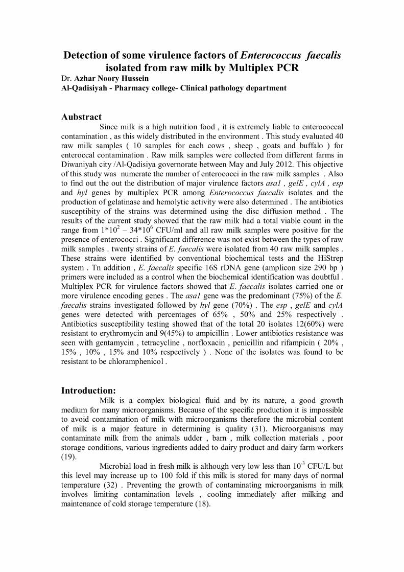
From the forty collected raw milk samples , a total of 20 E. faecalis isolates randomly picked from Rapid Hi-Enterococci agar medium ( figure 1) .
Figure (1): E. faecalis on Rapid Hi-Enterococci agar medium ( blue color )
For identification of E. faecalis used standard conventional and commercial tests . Positive gram stain , growth in the presence of 6.5% NaCl , absence of catalase , growth at 15-45 Co and pH 9.6 in brain heart infusion broth were observed . Hi strep. system was used for identification to species level. These bacterial isolates were further identified by partial sequencing of the gene coding for the 16S rDNA (2). The confirmation of identified isolates were E. faecalis (figure 2 )

Figure (2) : Gel electrophoresis of PCR amplification products 16S rDNA ( 290 bp) -Detection of virulence genes by multiplex PCR :
The multiplex PCR described is a fast , specific and reliable method which can be routinely used as an alternative to time consuming traditional tests (39). The results of the PCR amplification of the putative virulence genes tested are reported in table (3) anf figure (3). All genes were always detected in E. faecalis isolates. These isolates possessed between one (10%) and four (20%) determinants with the majority (55%) having three virulence factors . none of the isolates examined showed all of the virulence genes investigated in this study . Table (3) virulence factors encoding in E. faecalis strains
Phenotypic assay Virulence genes Strains of E.faecalis Gelatinase
activity β- hemolytic activity
gelE hyl asa1 cylA esp No.of virulence factors
1 2 3 4 5 6 7 8 9 10 11 12 13 14 15 16 17 18 19 20
+ve -ve -ve -ve -ve -ve +ve -ve +ve +ve -ve -ve +ve -ve +ve -ve +ve -ve -ve +ve
-ve -ve +ve -ve -ve -ve -ve -ve -ve -ve -ve -ve -ve -ve +ve -ve -ve -ve -ve -ve
+ - - - - - + - + + - + + - + + + - - +
+ + + + + - + + + + + + + + - - + - - -
+ + - + - + + + + - + + + + - + + + + -
- - + - - - - - - - - - - + + - + - + -
- - + + - + + + - + + + + + + - - - + +
3 2 3 3 1 2 4 3 3 3 3 4 4 4 3 2 3 1 3 2
Total (%)
8 (40%)
2 (10%)
10 (50%)
14 (70%)
15 (75%)
5 (25%)
13 (65%)
+: presence virulence factor gene +ve : positive results - : absence virulence factor gene -ve : negative results

Figure (3) agarose gel electrophoresis of amplified asa1(375bp), esp(510bp), gelE(213bp), cylA(688bp) and hyl (276bp) by multiplex PCR . 1.2 kbp DNA ladder .
Furthermore , the results showed (table 4) . that multiplex virulence gene co-existed in the E. faecalis isolates . three genes were found to co-exist predominantly in the strains , generally the genotype hyl , esp and asa1 were predominantly found in 4 (20%) of the strains study . (11) suggested that presence of determinants in combination possible synergistically activity in causes of infections . According to the type factor , no distinct pattern could be observed but this study found that 75% of positive asa1 strains ( 10). Table (4) : Genetic linkage of virulence factors-related gene clusters among E. faecalis strains isolated from raw milk.
Virulence markers No. (n=18) (%)
hyl , asa1 gelE, hyl,asa1 hyl,cylA,esp hyl,asa1,esp asa1,esp gelE,hyl,asa1,esp gel,hyl,esp hyl,asa1,cylA,esp gelE,cylA,esp asa1,cylAesp gelE,esp
1(5%) 3(15%) 1(5%)
4(20%) 1(5%)
3(15%) 1(5%) 1(5%) 1(5%) 1(5%) 1(5%)
The present study( table 3) revealed higher frequency of the asa1 gene (coding aggregation substance) among E. faecalis . in agreement with other research that revealed higher incidence this gene in E. faecalis ( 11 ; 41). The results showed that hyl gene was found in 14 E. faecalis isolates (70% of 20) table 3. more often hyl gene was combined with the other virulence factors , alone it was present in only one strain . According to some investigation points out its role in infection (38). Up until now , the hyl gene seemed to be specific for E. faecium (39). However , in this study it

was also detected in E. faecalis . it can be concluded that E. faecalis can code for hyl gene . this agreement with other study by (38).
The present study revealed 65% of the esp gene ( coding enterococcal surface protein ) among E. faecalis isolates as for adhesion . the percentage of strains bearing esp genes was similar to that reported in other studies for E. faecalis (40 ; 28). Since the first study on esp gene and its role in bacterial adhesion has been published ( 37), several conflicting results have been reported (23) demonstrated that in vitro adherence to urinary tract catheters was independent by esp.
Gelatinase and hemolytic activity producing strains of E. faecalis have been shown to be virulent in animals models of enterococcal infection ( Cosention et al. , 2010). The gelE gene ( coding for gelatinase is an extracellular zinc metallo endopeptidase ) was present in (10) 50% of the E. faecalis isolates (table 3). This results of present study are similar to that shown by (28) Londrina , Brazil .
Cytolysin gene ( coding hemolysin causes rapture of variety of the target membranes , including bacterial cells , erythrocytes and other mammalian cells , with a hemolytic activity on some types of blood agar (9) was found in 5 E. faecalis strains (25% of all ) . such results didn’t agree with study of (43) who wasn’t found any E. faecalis isolates had this gene.
As the correlation between phenotypic and genotypic tests , in this study the detection of some factors by PCR did not always correlate with phenotypic expression . the gelatinase gene was found in 10 isolates in all tested species . Moreover, 2 of the gel positive isolates didn’t express gelatinase activity (table 3). Two silent gene not showing phenotypic activity despite the presence of the gel gene were found in E. faecalis . these silent genes were also described by (38) and (10) in their studies.
The cylA gene was also observed only in 5 isolates in all tested species but 3 of the gel positive isolates didn’t express hemolytic activity on blood agar (table 3). That mean three silent genes didn’t show phenotypic activity in spite of presence of the cylA gene . these silent genes were described by (34) and (10). Therefore both genotypic and phenotypic assays seem necessary for a better characterization of the strain.
- Antibiotic resistance
Overuse and misuse of antimicrobial in food animals represent a public health risk as they contribute to the emergence of resistant forms of disease-causing bacteria . such as resistant bacteria can be transmitted from those food animals to humans, primarily via food . then , infections can results that are difficult to cure since the resistant bacteria don’t respond to treatment with covenantal antimicrobials ( 7).
Antibiotic resistance showed that (100%) E. faecalis isolates were
susceptible to chloramphenicol . whereas 12(60%) E. faecalis isolates were resistant to the erythromycin , 9(45%) isolates were resistant to ampicillin , 6(30%) isolates were resistant to the nitrofurantoin , 4(20% ) isolates resistant to gentamycin . 3(15%) isolates were resistant to the teteacycline and pencillin and 2(10%) isolates were resistant to norfloxacin and rifampicin. Multi drug resistance was observed in 13(65%) isolates of E. fecalis for 2 or more antibiotics ( table 5) .
In the present study of antibiotic resistance, the strains were seen to be highly sensitive to the more commonly used antibiotics ( chloramphenicol , rifampicin , tetracycline , and norfloxacin (100% , 90% , 70% and 70% respectively ) . Whereas resistance was observed in E. faecalis isolates against erythromycin , ampicillin and

nitrofurantoin ( table 5 ) . These results agreed with the study (7) and (24) who found that the rate of resistance to ampicillin in E. faecalis isolates were ( 42% and 41%) respectively . on the other hand these results disagreed with (6) who found that the rate of resistance to ampicillin was 88.2% . The study on antibiotic resistance among E. fecalis revealed that although many of these strains showed resistance to one or more of the antibiotics , the majority of the isolates were still sensitive to the clinically relevant antibiotics such as chloramphenicol and rifampicin . This results agreed with the study (27) who found that rate of susceptible was high to a large number of antibiotics Table (5) : percentage of susceptible , intermediate and resistant to 9 commonly used
antibiotic among E. faecalis strains. E. faecalis isolates
Antibiotic category G
enta
myc
in
tete
acyc
line
norf
loxa
cin
penc
illin
nitr
ofur
anto
in
chlo
ram
phen
icol
rifa
mpi
cin
eryt
hrom
ycin
ampi
cilli
n
No. of antibiotic resistance
1 2 3 4 5 6 7 8 9 10 11 12 13 14 15 16 17 18 19 20
R S S R S S S S R S S S I I S I S S R S
R S S S I S S S S R S I S S S I S R S S
S R S S I I I S S S I S S S S S S S S R
S S S S S I S S I R S S R S S I I S R S
R S S R S S S R R S S I I R S I S S S R
S S S S S S S S S S S S S S S S S S S S
S R S S S R R S S S S S S S R S R S S S
S R I I S R R S R R R R R S R R S R S R
S S R R R S S R S S R S R S R S R S R S
3 3 1 3 1 2 2 2 3 3 2 1 3 1 3 1 2 2 3 3
Resistance (%) 4 (20) 3(15) 2(10) 3(15) 6(30) 0 2(10) 12(60) 9(45) Susceptible(%) 13(65) 14(70) 14(70) 13(65) 11(55) 20(100) 18(90) 6(30) 11(55) Intermediate(%) 3 (15) 3(15) 4 (20) 4 (20) 3(15) 0 0 2(10) 0
*R: Resistance , S: Susceptible , I: Intermediate

References : 1- Akra, A. ; Vebq, H. ; Snipen, L. and Hirt, H. ; Astveit, A. ; Kapur, V. ; Dunny, G. ; Murray, B. and Nes, I. F. (2005). Transcriptional response of Enterococcus faecalis V583 to erythromycin . J. Antimicrob. Chemother., 49 : 2246 – 2259 . 2- Al-Ruwaili, M. A. ; Khalil, O. M. and Selim, S. A. ( 2012) . Phenotypic and genotypic differences in the expression of virulence factors in antimicrobial resistance of Enterococcus faecalis clinical strains Bios. Res., 9 (1) : 50 – 58 . 3- Banwo, K. ; Songlia, S, L. O. ; Frrreira, M. j. ; Rodnques, M. J. and Branco , M. P. C. (2012) Technological properties and probiotic potential of Enterococcus faecium stains isolated from cow milk . J. Appl. Microbiol., 14 : 229 – 241 . 4- Barros L. S. ; Sóglia S. L. O.; Ferreira, M. J. ; Rodrigues, M. J. and Branco, M. P. C. ( 2011) Aerobic and anaerobic bacteria and Candida species in crude milk .J. Microbiol. Antimicrob., 3(8): 206-212 . 5-Burgos, M. J. G. ;Lopez, R. L. ; Abriouel, H. ; Omar, N. B. and Galverz, A. ( 2009). Multilocus sequence typing of Enterococcus faecalis from vegetable food reveals tow new sequence types. Food borne Pathog. Dis. , 6 : 321 – 327 . 6- Castillo – Rojas, G. ; Mazar – Hiriart, M. ; Leon, S.P. ; Rosa, I. and et al.(2013). Comperson of Enterococcus faecium and Enterococcus faecalis strains isolated from water and clinical samples : antimicrobial susceptibility and genetic relationships . PLOSONE., 8(4) : 1-10. 7- Citak , S. ; Yucel,N. and Mendi, A.(2005).Antibiotic resistance of enterococcal isolates in raw milk. J. Food Pro. Pres.Vol. 29, (3-4) : 183–195. 8- Coburn, P.S. and Gilmore, M.S.( 2003). The Enterococcus faecalis cytolysin : a novel toxin active against eukaryotic and prokaryotic cells . Cell Microbiol., 5 : 661 – 575 . 9-Coburn, P.S.; Pillar, C.M.; Jett, B.D.; Haas, W. and Gilmore, M.S.( 2004). Enterococcus faecalis senses target cells and in response expresses cytolysin. Science. Dec., 24:306(5705):2270-2272. 10- Consentino, S. ; Podda, G . S. ; Coda, A. and et al. (2010) . Molecular detection of virulence factors and antibiotic resistance in clinical Enterococcus faecalis strains in Sardinia. J. Prev. Hyg., 51 : 31 – 36 . 11- Creti, R. ; Imperi, M. ; Bertuccini, L. ; Fabretti, F. ; Orefici, G. ; Di Rosa, R. and Baldassarri, L. (2004) . Survey for virulence determinants among Enterococcus faecalis isolated from different sources . J. Med. Microbiol., 53 : 13 – 20 . 12- De Fatima Sila Lopes, M. ; Ribeiro, T. ; Abrantes, M. ; Figueiredo Marques, J. J. ; Tenreiro, R. and Crespo, M. T. B. (2005) . Antimicrobial resistance profiles of dairy and clinical isolates and type strains of enterococci . Int. Food Microbiol. , 103 : 191 – 198 . 13-Dogru, A. K. ; Gencay, Y. E. and Ayaz, N. D. (2010) . Comparison of virulence gene profiles of Enterococcus faecium and Enterococcus faecalis chicken neck skin and faeces isolates . Kafkas Unv. Vet. Fak. Derg., 16 ( A ) : 129 – 133 . 14- Fisher, K. and Phillips, C. (2009). The ecology , epidermiology and virulence of Enterococcus . Microbiol., 155: 1749 – 1757. 15- Foulquie Moreno, M. R. ; Sarantinopoulos, P. ; Tsakalidou, E. and De Rivetti, L. (2006) . The role and application of enterococci in food and health . Int. Food Microbiol. , 106 :1-24. 16- Frahan, M. and Salik, S. (2007) Evaluation of bacteriological contamination in raw milk (un – processed milk sold in different regions of Lahore ( Pakistan ) . J. Agri. Soci. Sci. 3(3) : 104 – 106 .

17- Gelsomno, R. ; Vancanneyt, M. ; Condon, S. ; Swing, J. and Cogan, T. M. (2001) Enterococci diversity in the cheese – making environment of an Irish cheddar type cheese – making factory . Int. J . Food Microbiol. , 71 : 177 – 188 . 18- Gimenez-Pereira, M. L. (2005).Enterococci in Milk Products . thesis . Massey University Palmerston North, New Zealand. 19- Grimand, P. ; Sserunjogi, M. ; Wesuta, M. ; Grillet, N. ; Kato, M. and Faye, B. (2009) . Effects of season and agro – ecological zone on the microbial quality of raw milk along the various levels of the value chainin Uganda. Trop. Ani. Heal. Prod. , 41: 883 - 890. 20- Godefay, B. and Molla, B. (2000) . Bacteriological quality of raw milk from four dairy farms and milk collection center in and around addis ababa , Berl. Munch Tierarztl. Wschi., 113 : 1-3. 21- Gossens, H. D. ; Jabes , R. ; Rossi, C. ;Lammens, G. ; Privilera, and Courralin, P. (2003) . European survey of vancomycin – resistant enterococci in at- risk hospital wards and in vitro susceptibility testing of ramoplanin against these isolates . J. Antimicrob. Chemother., 51(3) :5-12. 22- Hill, B. M. and Smythe , B. W. (1997) . A study of the microbiological quality of raw milk from individual farms . New Zland Dai. Res. Ins. , 1-35. 23- Höllgren, A. ; Claesson, C, ; Saeedi, B. ; Hans – Jurg, I. ; Hanberger, H. and Nilsson, L. E. ( 2009). Molecular detection of aggregation substance , enterococcal surface protein an. Med. Microb. , 299 : 323 – 332 . 24- Jankoska, G. ; Trajkovska-Dokic, E. ; Panovski, N. ; Popovska-Jovanovska, K. and Petrovska, M. (2008).Virulence factors and antibiotic resistance in Enterococcus faecalis isolated from urine samples. Prilozi. Jul;29(1):57-66. 25- Kalhotka, L. ; Manga, I. ; Prichystalova, J. ; Hulova, M. ; Vyletelova, M. and Sustova, K.( 2012) . Decarboxylase activity test of the genus Enterococcus isolated from goat milk and cheese . Acta. Velerinaria Brno. , 81(2) : 145 – 151. 26- Kalhotka, L. ; Sustova, K. ; Hulova, M. and Prichystalova, J. (2013). Important groups of microorganisms in raw goat milk and fresh goat cheeses determined during lactation. J. Microbiol. Biotechnol. Food Sci., 2(5) : 2314 – 2317. 27- Mannu, L.;Paba, A.;Daga, E.;Comunian, R.;Zanetti, S.;Duprè, I.and Sechi, L.A. (2003) . Comparison of the incidence of virulence determinants and antibiotic resistance between Enterococcus faecium strains of dairy, animal and clinical origin. Int. J.of food microbiol. 88(2-3): 291-304. 28- Marques, E. and Suzart, S. (2004) . Occurrence of virulence associated genes in clinical Enterococcus faecalis strains isolated in Londrina, Brazil. J. Med. Microb., 53 : 1069 – 1073 . 29- Niazi, A. K. (2001). Statical analysis in medical research . Republic of Iraq . Al – Nehrein university . p : 148 . 30- Patidar, R. K. ; Gupta. M. K. and Singh, V.(2013). Phenotypic Detection of Virulence Traits and Antibiotic Susceptibility of Endodontic Enterococcus faecalis Isolates. Amer. J. Microbiol.Res., 1 (1) : 4-9. 31- Queslati, S. ; Ennouri, H. ; Bamri, M. ; Benothren, E.T. and Queslati, R. (2011) .Differential distribution of pathogens from raw milk and place of Shigella by mode of milking . Afr. J. Food Sci. Technol., 2(8) : 179 – 183 . 32- Rizwan, M. ; Pervez, A. and Khan, J. (2011). Bacterial quality of raw and packed milk . Candian J. Sci. Ind. Res., 2(2) : 86 – 94 . 33- Robinson, R. K. (2002).Dairy microbiology Handbook: The microbiology of milk and milk products.3rd edition .Jhon wiley and Sons Inc. , USA.

34- Semedo, T. ; Santos, M. A. ; Martins, P. et al. (2003) .Comperative study using type strains and food isolates to examine hemolytic activist and occurrence of the cyl operon in enterococci . J. Clin. Microbiol. , 41 :2569 – 2579 . 35- Shankar, N. ; Lockatell, C. V. ; Baghdayan , A. S. ; Drachenberg, C.; Gilmove, M. S.and Johnson, D. E. (2001) . Role of Enterococcus faecalis surface protein Esp in the pathogenesis of ascending urinary tract infection . Infect. Immun., 69 : 4366 – 4372 . 36-Sharifi, Y.; Hasani, A.; Ghotaslou, R.; Naghili, B.; Aghazadeh, M.; Milani, M. and Bazmani, A.( 2013 ).Virulence and Antimicrobial Resistance in Enterococci Isolated from Urinary Tract Infections. Adva. Pharm. Bull., 3(1), 197-201. 37- Toledo – Arana, A. ; Valle, J. ;Solano, C. and et al. (2001). 38- Trivedi, K. ; Cupakova, S. and Karpiskova, R.. ( 2011). Virulence factors and antibiotic resistance in enterococci isolsted from food – stuffs. Veter. Med. , 56(7) :352 – 357 . 39- Vankerckhoven, V. ; Autgaerden , T. V. ; Vael, C. ; Lammens, C. ;Chapelle, S. Rossi, R. ; Jabes , D, and Goosens , H. (2004). Development of a multiplex PCR for the detection of asa1 , gelE , cylA , esp and hyl genes in Enterococcus and survey for virulence determinants among European hospital isolates of Enterococcus faecium . J. Clin. Microb., 42 (10) : 4473 – 4479. 40- Vergis, E. N. ; Shankar, N. ; Chow, J. W. ;Hayden, M. K. et al . (2002). Assaciation between the presence of enterococcal virulence factors gelatinase , hemolysin and enterococcal surface protein and mortality among patients with bacteremia due to Enterococcus faecalis . C.I.D., 35 (1) : 570 – 575 . 41- Waar1, K. ; Muscholl-Silberhorn , A. B.; Willems, R. J. L. ; Slooff, M. J. H. ; Harmsen, H. J. M. and Degener, J. E. (2002). Genogrouping and Incidence of Virulence Factors of Enterococcus faecalis in Liver Transplant Patients Differ from Blood Culture and Fecal Isolates. J. Infect. Dis., 185 (8): 1121-1127. 42- Xia, Z. ; Dong, W. and Xiao – Lan , W. (2011). Characterization of pathogenic or non- pathogenic Enterococcus faecalis isolated from lambs from xinjiang , a remove north – west province of china . Afr. J. Microbiol. Res. , 5(18) ; 2827 – 2833 . 43- Zou, L. ; Wang, H. ; Zeng, B. ; Li, J. et al. (2011). Erythromycin resistance and virulence genes in Enterococcus faecalis from swine in china . New Microbiol., 34: 73 – 80 .

التحري عن بعض عوامل الضراوة في المكورات المعویة البرازیة المعزولة من الحلیب الخام باستخدام تفاعل الكوثرة المتعدد
جامعة القادسیة – كلیة الصیدلة-أزھار نوري حسین قسم االمراض السریریة . د
:الخالصة لـوث بـالمكورات المعویـة والتـي تنتـشر الحلیب غذاء عـالي التغذیـة فانـه عرضـة للتأن بما
عینــات لكــل مــن 10( عینــة مــن الحلیــب الخــام40قیمــت هــذه الدراســة . شكل واســع فــي البیئــة بــجمعـت عینـات الحلیـب الخـام . للتلـوث بـالمكورات المعویـة ) األبقار واألغنـام والمـاعز والجـاموس
2012 تمــوز للعــام إلــىللمــدة مــن أیــار محافظــة القادســیة \مــن مــزارع مختلفــة فــي مدینــة الدیوانیــة كمــــا تــــضمن ، تــــضمنت الدراســــة إحــــصاء عــــدد المكــــورات المعویــــة فــــي عینــــات الحلیــــب الخــــام .
موضـــوع هـــذه الدراســـة الكـــشف عـــن جینـــات بعـــض عوامـــل الـــضراوة الرئیـــسیة للمكـــورات المعویـــة تـم تحدیـد . المتعـدد البلمـرةبواسـطة تفاعـل hyl و esp و cylA و gelE و asa1البرازیة وهـي
. أنتاج هذه العزالت إلنزیم الجیالتنیز والفعالیة التحللیة للدم مظهریا أظهرت نتائج هذه الدراسـة احتـواء عینـات الحلیـب علـى المكـورات المعویـة بعـدد تـراوح بـین
خــام ایجابیــة كمــا كانــت جمیــع عینــات الحلیــب ال. مــل / وحــدة حقــل الخلیــة 106* 34 – 102*1. لم تسجل فروق معنویة بـین عینـات الحلیـب الخـام المختلفـة المـصدر . التواجد للمكورات المعویة
ــــــت ــــــة البرازیــــــة باســــــتخدام االختبــــــارات ی عزلــــــة بكت20عزل ــــــدیها للمكــــــورات المعوی ــــــة اثبــــــت عائ ری 16S للتحـري عـن ألجـین PCR ت تقنیـة كمـا اسـتخدم . HiStrepالكیموحیویـة التقلیـدي ونظـام
rDNA كعامـــل أثبـــات علـــى أن البكتریـــا المشخـــصة هـــي المكـــورات المعویـــة البرازیـــة بحجـــم قطعـــة . زوج قاعدي 290 بلغت مبلمرةوراثیة
المتعـدد امـتالك المكــورات المعویـة البرازیـة لواحـد او أكثـر مـن جینــات البلمـرة اظهـر تفاعـل وبنــسبة hylیلیــه الجـین ، %) 75( وبنـسبة الــسائدasa1كــان الجـین . عوامـل الــضراوة المدروسـة
% 50و % 65نـسب مئویـة بلغـت cylA و gelEو espفـي حـین أظهـرت الجینـات %) . 70( 20اظهــر اختبــار فحــص الحــساسیة للمــضادات الحیویــة ان مــن مجمــوع . علــى التــوالي % 25و
اومـــــة عـــــزالت مق%) 45(9عزلـــــة مقاومـــــة لمـــــضاد االریثرومایـــــسین و %) 60 (12عزلـــــة كانـــــت ظهــر انخفــاض واضــح للمقاومــة بــین عــزالت المكــورات المعویــة البرازیــة للمــضادات . لالمبــسلین
% 15، % 20وبنــسب الجنتامایــسین و التتراســایكلین و النورفلوكــساسین و البنــسلین والریفامبــسین ــــــــوالي % 10، % 15، % 10، ــــــــى الت ــــــــم تظهــــــــر أي مــــــــن العــــــــزالت مقاومتهــــــــا لمــــــــضاد . عل ل
.لهول اذ كانت جمیعها حساسة الكلورامفینیك