detection of the insulin gene hph i polymorphism using ... · the role of the insulin region in...
TRANSCRIPT
1
Detection of the Insulin Gene Hph I Polymorphism using Sequence Specific Oligonucleotides (SSO)
Table of Contents Page number
1. Summary of Test Principle and Clinical Relevance 2 2. Safety Precautions 2 3. Computerization; Data System Management 3 4. Specimen collection, Storage, and Handling Procedures 3 5. Procedures for Microscopic Examination; Criteria for Specimen Rejection 3 6. Preparation of Reagents, Calibrators (standards), Controls, and All Other 4
Materials; Equipment and Instrumentation a. Reagents 4 b. Reagent preparation 4 c. Standards 4 d. Controls 4 e. Equipment and materials 4 f. Instrumentation 5
7. Calibration and Calibration verification Procedures 6 8. Procedure Operating Instructions; Calculations; Interpretation of Results 6
a. Procedure 6 b. Calculations 6 c. Interpretation of Results 6
9. Reportable Range of Results 7 10. Quality Control (QC) Procedures 7
Quality Control Principles 7 Preparation of Quality Control Materials 7
11. Remedial Action if Calibration or QC Systems fail to meet acceptable criteria 8 12. Limitation of Method; Interfering Substances and Conditions 9 13. Reference Ranges (Normal Values ) 9 14. Critical Call Results 9 15. Specimen Storage and Handling During Testing 9 16. Alternate Methods for Performing Test or Storing Specimens if Test System Fails 9 17. Test Result Reporting System; Protocol for Reporting Critical Calls (if applicable) 10 18. Transfer or Referral of Specimens; Procedures for Specimen Accountability and Tracking 10 19. References 11 20. Appendix A: Puregene Method for DNA Isolation from Whole Blood and Cell Culture for PCR using the Puregene Genomic DNA Isolation Kit. 12 21. Appendix B: Human Identification with Short Tandem Repeat Loci using the AmpFlSTR Green I PCR Amplification Kit. 16 22. Appendix C: Identification of the Amelogenin and TH01 Markers. 18 23. Appendix D: Detection of the Insulin Gene –23 Hph I Polymorphism using the ABI PRISM 7700 Sequence Detection System. 21 24. Appendix E: Detection of the Insulin Gene –23 Hph I Polymorphism using sequence
Specific Oligonucleotides (SSO). 23
2
1. Summary of Test Principle and Clinical Relevance
Type 1 diabetes mellitus is a chronic autoimmune disease that involves a T cell-mediated destruction of the pancreatic beta cells, the body’s sole source for insulin1. This disorder is the most common chronic disease among children and young adults2. Complications include kidney failure, blindness, amputations, nerve damage as well as an increased risk for heart attacks and strokes3. Type 1 diabetes has been shown to involve a genetic component and an environmental component4. Thus, an environmental trigger in a susceptible genetic background results in type 1 diabetes development. This genetic component is the earliest predictor of type 1 diabetes and may eventually allow prediction in the prenatal phase leading to early prevention and/or treatment. The genes that are known to play a role in the genetic susceptibility include those in the Human Leukocyte Antigen (HLA) complex on chromosome 6p21, and the insulin gene on chromosome 11p155. The role of the insulin region in type 1 diabetes has been identified by various laboratories using association studies6-11. This region, named IDDM2, has several polymorphisms as well as a variable nucleotide repeat (VNTR) in the 5' upstream region12-14. The Hph I -23 polymorphism has been shown to segregate with the class I VNTR, which has been implicated in disease susceptibility14. Additionally, Halminen et. al has found in the Finnish population, that the Hph I polymorphism appears to be associated with type 1 diabetics, especially in the absence of the DR4 susceptibility haplotype13. This study will allow confirmation of these results as well as provide a comprehensive study of the role of the insulin gene in type 1 diabetes. Moreover, it will allow us to identify the minor risk factors in diabetes by controlling for the major genetic risk factors. Two assays have been developed by this laboratory to detect the –23 Hph I polymorphism. The first assay involves an allelic discrimination assay that has been developed for the ABI PRISM® 7700 Sequence Detection instrument. This assay utilizes two probes, one with sequence complementary to the T polymorphism (referred to as + since it has been shown to be associated with type 1 diabetes) and one with sequence complementary to the A polymorphism (referred to as – since it is not associated with type 1 diabetes). These probes will only bind to an individual’s DNA when there is an exact match at the –23 polymorphism. During the PCR amplification process, the Taq Polymerase enzyme will encounter the bound probe and displace and destroy the probe. This process releases a bound fluorescent molecule that is then detectable by the ABI PRISM® 7700 Sequence Detector. Each of the two probes has a unique fluorescent reporter molecule indicating the state of the polymorphism on each of the individual’s two chromosome 11s. The second assay to detect the –23 polymorphism is a sequence specific oligonucletide (SSO) PCR based assay. Two PCR primers are developed which are complementary to either the A or T polymorphism at the 3’ end such that each primer will only recognize one of the two possible variants at the –23 polymorphism. Each SSO primer is paired with a common forward primer to produce an amplified product. For each specimen, two PCR reactions are set up, one for each primer set. Only the reactions that find complementary sequence in the individual’s DNA will give an amplification product that can be visually identified by gel electrophoresis. Thus, for homozygous TT (+/+) and homozygous AA (-/-) genotypes, only one of the two reactions will give a successful amplification, while for heterozygous genotypes (+/-) both reactions will be successful. The ABI 7700 allelic discrimination assay will be used as the primary assay for insulin genotyping, however the SSO-PCR assay will be used in cases where it can resolve discrepancies.
2. Safety Precautions Standard safety precautions should be observed including wearing safety glasses, lab coats and gloves during the preparation of blood specimens. Follow Universal Precautions when handling all blood and blood products. Vaccination for hepatitis B is strongly encouraged. Laboratory items exposed to blood or blood products should be disposed of or decontaminated in compliance with guidelines from the Office of Health and Safety, CDC. The following chemicals are used in this genotyping process: Ethidium Bromide (EtBr)
Ethidium Bromide is used to visualize double-stranded DNA that has been separated by size on an agarose/acrylamide gel matrix. The EtBr intercolates into double-stranded DNA, and will
3
fluoresce when visualized on a UV transilluminator. EtBr is a potential carcinogen, and extreme caution should be taken when working with this chemical. Observe all safety precautions such as wearing a lab coat, fresh gloves, and protective eyewear.
HiDi Formamide
HiDi Formamide is used in small amounts within the laboratory to resuspend DNA for automated sequencing (also found in Template Suppression Reagent). Formamide is a teratogen, which can affect fetal development. Always exhibit extreme caution while in contact with formamide, and observe all safety precautions such as wearing a lab coat, fresh gloves, and protective eyewear. *Note: women who are or plan to become pregnant should not work with formamide due to its adverse effects on fetal development. Performance Optimized Polymer 4 (POP 4) This polymer is used within the automated sequencers and acts as medium through which DNA samples are transported through capillaries. This polymer contains high amounts of urea, which is a potential mutagen and has been shown to have reproductive and tumorigenic effects. Observe all safety precautions such as lab coat, fresh gloves, and protective eyewear. Acrylamide Acrylamide is used to pour the large sequencing gel used on the Applied Biosystems 377 Genetic analyzer. Acrylamide is a poison, neurotoxin, irritant, carcinogen, and possible teratogen. The effects of this chemical are cumulative, so always use it with the upmost caution. Observe all safety precautions such as lab coat, fresh gloves, and protective eyewear.
3. Computerization; Data System Management Integrity of specimen data generated by this method is maintained by proofreading all transcribed data by the analyst. All data is copied to a CD-R for transfer to a Microsoft Access database created to store all raw data generated in the GoKinD study. Only authorized personnel from the Molecular Biology Branch (as determined by the supervisor) have access to this database. Analyzed genotype results are recorded by the analyst in a Microsoft Access database located on CDC’s LAN, and only authorized personnel from the Molecular Biology Branch (as determined by the supervisor) have access to the data. 4. Specimen Collection, Storage, and Handling Procedures; Criteria for Specimen Rejection
a. Specimen collection: Whole blood obtained with EDTA as an anticoagulant may be used. All 10 ml of the venous blood collected will be processed for DNA.
b. Specimen storage: Blood samples which have been processed by the Puregene method through the cell
lysis step (see Appendix A) can be stored at room temperature for up to eighteen months. Extracted DNA can also be stored at -20EC indefinitely until assayed.
c. Freeze-Thaw effect: Repeated freeze-thaws may cause slight fragmentation of DNA. However,
the size DNA targeted for amplification is very small (<400bp) and there is no documented deleterious effect of freeze-thaw on this test.
5. Procedures For Microscopic Examination; Criteria for Rejection of Inadequately prepared slides
Not applicable for this procedure 6. Preparation of Reagents, Calibrators (standards), Controls, and All Other Materials
a. Reagents
The Puregene DNA Isolation Kit (Gentra Systems) contains the Red Blood Cell Lysis Solution (RBC Lysis Solution), Cell Lysis Solution, RNase Solution, Proteins Precipitation Solution, and DNA
4
Hybridization Solution. All reagents except the RNase solution are stable at room temperature until the expiration date indicated by the manufacturer. The RNase Solution is stable at 4°C until the expiration date. The AmpFlSTR Green I PCR Amplification Kit is stored at 4°C until the manufacture’s expiration date. Check the expiration date before each use and discard the kit after the expiration date. The HiDi Formamide is aliquoted into separate tubes and stored at -20°C. QIAGEN HotStarTaq Master Mix. This reagent should be stored at -20°C and is stable until the expiration date indicated by the manufacturer. TaqMan Universal PCR Mastermix. This reagent should be stored at 4°C and is stable until the expiration date indicated by the manufacturer. PCR primers and probes synthesized by Biotechnology Core Facility, Scientific Resources Program, NCID, CDC. Primers should be stored at - 20oC and labeled probes should be stored at 4oC.
b. Preparation of Reagents
See Appendix A, B and C.
c. Standards
This is a qualitative assay and calibration standards are not used. See Part 7. d. Controls
For the AmpFlSTR Green I Human Identification Assay a positive control of a previously genotyped DNA, and a negative “no template control” control are tested with each PCR amplification run. With each identification run the control DNA and a Green I Allelic Ladder is always tested. For the insulin gene –23 Hph I Polymorphism SSO Detection Assay and ABI PRISM 7700 Assay, in house control DNA samples of known genotypes are used as controls for each of the possible genotypes: homozygous +/+, homozygous -/- and heterozygous +/-. No template reactions are used as negative control. Identification of Amelogenin and TH01 Markers An in house control DNA of a known genotype and an in house allelic ladder are prepared at a set concentration. The in house control DNA is stored at -20C in a “clean” laboratory and the in house allelic ladder is stored at 4C in a “dirty” laboratory where amplified DNA is in use.
e. Equipment and Materials AmpFlSTR Green I PCR Amplification Kit, Applied Biosystems, Foster City, CA AmpFlSTR Green I PCR Amplification Kit User’s Manual, Applied Biosystems Performance Optimized Polymer 4 (POP-4), Applied Biosystems Puregene DNA Isolation Kit (Gentra Systrems, Minneapolis, MN) Glycogen (Gentra Systems, Minneapolis, MN) Qiagen Sigma centrifuge (Qiagen Inc., Chatsworth, CA) Stratalinker UV Crosslinker 2400 (Stratagene, La Jolla, CA) GeneAmp PCR System 9700 thermal cycler (Applied Biosystems, Foster City, CA) ABI PRISM 310 Genetic Analyzer, Applied Biosystems
5
Computer and software for analysis with GeneScan and Genotyper Software, Applied Biosystems, Electrophoresis unit with power supply (any vendor) Gel documentation system (any vendor) Incubator (any vendor) Vortex Genie, Daigger, Lincolnshire, IL Heating block (any vendor) Balance (any vendor) PipetAid Drummond (Daigger, Lincolnshire, IL) Dispensette III volume dispenser for reagents (Daigger, Lincolnshire, IL) Micropipettors (any vendor) Micropipettes with filter (aerosol) tips (any vendor) Sterile, individually wrapped, transfer pipettes (any vendor) 50 ml Falcon tubes (any vendor) MicroAmp 8-Strip Reaction Tubes (0.2 ml), Applied Biosystems MicroAmp Caps (8 caps/strip), Applied Biosystems MicroAmp Reaction tubes with caps, tube trays and bases, Applied Biosystems, Foster City, CA MicroAmp 96-Well Tray/Retainer, Applied Biosystems Racks for centrifuge tubes and blood tubes (any vendor) 0.5 ml Genetic Analyzer sample tubes and septa, Applied Biosystems 1.5 ml microfuge tubes (any vendor) Deionized water Ultra-pure agarose (Gibco/BRL, Rockville, MD) GeneScan-350[ROX] Internal Lane Size Standard, Applied Biosystems, Foster City, CA Hi Di formamide, Applied Biosystems
ABI PRISM® 7700 Sequence Detection System, Applied Biosystems. TaqMan® Universal PCR Mater Mix Applied Biosystems, Foster City, CA.
10 X TBE Buffer (any vendor) 10x Genetic Analyzer Buffer with EDTA, Applied Biosystems 1xTE (10mM Tris-HCl, 0.1mM EDTA, pH 8.0) (any vendor) DNA ladder (any vendor) Gel loading dye (any vendor) Ethidium Bromide (any vendor) 70% ethanol (any vendor) 100% isopropanol (any vendor) Glass flasks (any vendor) Nitrile gloves (any vendor) Disposable towels and tissues (any vendor) Disposable bench-top covers (any vendor) Boekel Orbital Rocker (Boekel Scientific, Inc. Feasterville, PA)
f. Instrumentation
The GeneAmp PCR System 9700 is an automated thermal cycler with interchangeable sample blocks, used to carry out PCR amplification reactions. Methods, instructions that specify how the instrument should heat or cool samples in a PCR thermal profile, are programmed and stored in the instrument software. The GeneAmp PCR System 9700 offers greater speed, oil free operation, lower reaction volumes, and cycle time reproducibility. The Perkin Elmer ABI Prism 310 and 3100 Genetic Analyzers can be used for both fragment analysis as well as for sequencing applications. All instruments utilize electrophoresis, laser excitation and detection via a charged-coupled device (CCD) camera which provides simultaneous detection of all four colors from a single sample run.
The ABI PRISM® 7700 Sequence Detection System contains a built-in 96-well thermal cycler. To
induce fluorescence during PCR, laser light is distributed to the 96 wells via a multiplexed array of
6
optical fibers. The resulting fluorescent emission returns via the fibers and is directed to a spectrograph with a charge-coupled device (CCD) camera.
7. Calibration and Calibration Verification Procedures
The GeneAmp 9700 thermal cycler, the ABI PRISM® 7700 Sequence Detection System and the ABI Prism 310 and 3100 Genetic analyzers are pre-calibrated by the respective manufacturer and annual preventive maintenance is preformed by the manufacturer’s authorized service representative. This PCR-based genotyping assay is a qualitative test. There are only three possible genotypes in the test population, i.e., homozygous AA, homozygous TT and heterozygous AT, thus we obtain a yes/no answer for each genotype. If the three in house controls fail to yield correct results in a run, then the test is repeated (see section 11).
8. Procedure Operating Instructions; Calculations; Interpretation of Results
a. Procedure: See Appendix A, B and C for DNA extraction, amplification and detection. b. Calculations
This is a qualitative assay and calculation is not used. (See item 7 for details.)
c. Interpretation of results
AmpFlSTR Green I Human Identification Assay. The AmpFlSTR Green I PCR Amplification Kit amplifies the TH01, TPOX, CSF1PO short tandem repeat loci. In addition, the primers included in the AmpFlSTR Green I Primer set amplify the Amelogenin locus, which can be used for gender determination. The amplification products are run on an ABI PRISM instrument and the collected multicolor fluorescent data is analyzed using GeneScan Analysis Software. The Genotyper Software converts fragment sizes to genotypes which can then be uploaded into a database. Identification of Amelogenin and TH01 Markers This PCR based method amplifies the Amelogenin locus and the TH01 short tandem repeat loci. These markers are used for quality control purposes for samples that are not trios. Insulin gene -23 Hph I Polymorphism SSO Detection Assay. For each sample two sets of primers are used for PCR amplification. Each set amplifies only one of the two variants of the -23 Hph I site. The genotype of each sample can be determined by the presence (seen as a band on an agarose gel) or absence of the 231 base pair amplified DNA fragment with either given set of primers. Insulin gene -23 Hph I Polymorphism Detection using the ABI PRISM® 7700 Sequence Detection System. The two alleles of the single nucleotide polymorphism at the –23 Hph site are designated as allele 1 and 2 (A1 & A2). Data collected by the instrument is then analyzed according to the specifications in the user’s manual. The analyzes samples will be genotyped as A1 for samples homozygous for that allele, A2 for samples homozygous for the other and A1 and A2 for heterozygous samples. The genotypes can be confirmed by examining the Real Time data collected. The data is viewed in the multicomponent view, where emission intensity is plotted over time. Distinct spectral patterns are produced by each of the control reactions:
a. spectral pattern of the “no-template controls” will have little or no increase in emission intensity of either one of the reporter dyes (TET and FAM).
b.spectral pattern of the homozygous controls should have a significant increase in emission intensity of the reporter dye present on the probe specific to each allele, relative to the “no-template controls”.
c.Homozygous test samples should have a similar increase in emission intensity of the respective reporter dye, relative to the “no-template controls”. Heterozygous test samples
7
should display a significant increase in emission of both reporter dyes, relative to the “no-template controls”.
9. Reportable Range of Results
Not applicable see item 7 for details.
10. Quality Control (QC) Procedures a. Quality Control Principles
The type I diabetes genotyping method described in this protocol has been well established in the Division of Environmental Health and Laboratory Sciences. These methods have proven to be accurate, precise, and reliable. Reliability of all test results should be monitored by routinely using multiple controls. The AmpFlSTR Green I human identification assay is considered out of control if: 1. The electropherogram for the fragment analysis contains less than 4 or more than 8 peaks. 2. The GeneScan-350 ROX Internal Lane standard does not appear on the electropherogram. 3. The signal is too weak on the electropherogram. If the run is declared “out of control,” the fragment analysis should be repeated. If the run is “out of control” again, the PCR as well as the fragment analysis is invalidated and repeated. The assay for the Idenfication of Amelogenin and TH01 Markers is considered out of control if:
1. The electropherogram for the fragment analysis contains less than 2 or greater than 4 peaks 2. The GeneScan-350 ROX Internal Lane standard does not show up on the electropherogram, or 3. The signal is too weak on the electropherogram.
If the run is declared “out of control,” the fragment analysis should be repeated. If the run is “out of control” again, the PCR as well as the fragment analysis is repeated. The Insulin gene -23 Hph I Polymorphism ABI PRISM® 7700 Sequence Detection Assay is considered out of control if:
1. “No-template controls” are assigned a genotype by allelic discrimination or if they display and increase in fluorescence intensity in either reporter dye.
2. Positive control homozyougs replicas of either allele fail to show an increase in emission intensity of the reporter dye specifically associated with each allele relative to the “no-template controls.”
The insulin gene –23 Hph I Polymorphism SSO Detection Assay is considered out of control if:
1. No amplified DNA can be detected from the PCR amplification product. 2. Nny of the in house control DNA amplification products give unexpected genotypes.
If the assay is declared “out of control” the experiment is invalidated and repeated. b. Preparation of controls.
For the AmpFlSTR Green I human identification assay, Control DNA of a known Genotype and a Green I Allelic Ladder are supplied in the AmpFl STR Green I PCR Amplification Kit at a set concentration. The control DNA is stored at 4°C in a designated Reagent Preparation Area and the Allelic Ladder is stored at 4°C in an Amplification/Detection Area of the laboratory where amplified DNA is in use. Identification of Amelogenin and TH01 Markers
An in house control DNA of a known genotype and an in house allelic ladder are prepared at a set concentration. The in house control DNA is stored at -20C in a “clean” laboratory and the in house allelic ladder is stored at 4C in a “dirty” laboratory where amplified DNA is in use.

8
For the insulin gene –23 Hph I Polymorphism SSO detection assay in house control DNA samples with known genotypes, homozygous AA, homozygous TT and heterozygous AT, serve as both positive and negative controls for the PCR reaction. 200 ng of each DNA sample is used for the controls with every PCR reaction. Control DNA is labeled with a unique identification number and the date prepared. DNA is stored at -20°C. For the insulin gene –23 Hph I Polymorphism ABI PRISM® 7700 Sequence Detection assay in house control DNA samples with known genotypes, homozygous AA, homozygous TT and heterozygous AT, are used as positive controls for each PCR reaction. 50 ng of each DNA sample is used for the controls. Control DNA is labeled with a unique identification number and the date prepared. DNA is stored at -20°C. “No-template Controls” are used as negative controls with each test.
11. Remedial Action if calibration or QC systems fail to meet acceptable criteria
There are several potential possibilities in a failed test and the analyst and the supervisor must use their scientific knowledge in solving the problems. As with any test procedure, good laboratory practices are essential for the proper performance of the assays. AmpFlSTR Green I human identification: a. If less than 4 or more than 8 peaks appear on the electropherogram, repeat the fragment analysis. If the
fragment analysis appears to be the same, repeat the test starting from PCR amplification. b. If the GeneScan-350 ROX Internal Lane standard does not appear on the electropherogram, repeat the
fragment analysis. c. If there is no/low green signal compared to the internal lane standard on the electropherogram, check
the concentration of the DNA and then repeat the experiment Identification of the Amelogenin and TH01 Markers a. If less than 2 or more than 4 peaks appear on the electropherogram, repeat the fragment analysis. b. If the fragment analysis appears to be the same, repeat the experiment from the beginning. c. If the GeneScan-350 ROX Internal Lane standard does not show up on the electropherogram, repeat
the fragment analysis. d. If there is no/low blue signal compared to the internal lane standard on the electropherogram, check the
concentration of the DNA again and repeat the experiment. Insulin gene –23 Hph I polymorphism ABI PRISM® 7700 Sequence Detection assay: a. If the Control DNA failed to amplify, repeat the test. If there is still no amplification try new reagents. b. If a given unknown specimen failed to amplify, repeat the test in the next available run. If it failed to
amplify again, re-isolate the DNA using the stored blood sample and repeat the test. c. If all samples including the controls fail to amplify, it is likely due to one of the following reasons. (1)
incorrect thermal cycle program, (2) interruption during the PCR run (i.e., power outage which will be registered in the history record of the instrument), or (3) an error in the PCR reaction mixture or (4) an error in the data analysis, such as not selecting “none” for the quencher in the Sample Type Pellet when using a dark quencher.
d. If any of the three control DNA samples give unexpected genotypes, the entire assay, including specimen preparation, amplification and detection should be repeated.
Insulin gene –23 Hph I polymorphism SSO genotyping: a. If the Control DNA failed to amplify, repeat the test. If there is still no amplification try new reagents. b. If a given unknown specimen failed to amplify, repeat the test in the next available run. If it failed to
amplify again, re-isolate the DNA using the stored blood sample and repeat the test. c. If all samples including the controls fail to amplify, it is likely due to one of the following reasons. (1)
incorrect thermal cycle program, (2) interruption during the PCR run (i.e., power outage which will be registered in the history record of the instrument), or (3) an error in the PCR reaction mixture.
d. If any of the three control DNA samples give unexpected genotypes, the entire assay, including specimen preparation, amplification and detection should be repeated.

9
12. Limitations of Method; interfering substances and conditions
This method is not labor-intensive as compared to other non-automated methods using restriction endonuclease digestions. Due to the high analytical sensitivity of the tests, extreme care should be taken to preserve the purity of the kit reagents, amplification mixtures and samples. All reagents should be closely monitored for purity. The following guidelines should be followed:
It is imperative that the work flow in the laboratory proceeds in a uni-directional manner, beginning in the Reagent Preparation Area and moving to the Specimen Preparation Area and then to the Amplification/Detection Area to avoid contamination. Supplies and equipment must be dedicated to each activity and not moved between areas. Gloves must be worn in each area and taken off before leaving the area. All sample tubes should be opened and closed carefully to avoid reagent or sample splashes. Positive displacement pipettes or air-displacement pipettors with filter-plugged tips should be used. Tips should be changed after each use.
13. Reference Ranges (Normal Values)
The role of the insulin region in type 1 diabetes has been identified by various laboratories using association studies6-11. This region, named IDDM2, has several polymorphisms as well as a variable nucleotide repeat (VNTR) in the 5' upstream region12-14. The –23 Hph I polymorphism has been shown to segregate with the class I VNTR, which has been implicated in disease susceptibility14. Additionally, Halminen et. al has found in the Finnish population, that the Hph I polymorphism appears to be associated with type 1 diabetics especially in the absence of the DR4 susceptibility haplotype13. There are only three possible genotypes in the test population so far, i.e., homozygous AA, homozygous TT and heterozygous AT, thus we obtain a yes/no answer for each genotype.
14. Critical Call Results (“Panic Values”) Not applicable.
15. Specimen storage and Handling during testing
The blood specimens are received by the laboratory in 50 ml Falcon tubes partially processed up to the cell lysis stage (see Appendix A). At this stage, the specimens can be stored at room temperature for 18 months. Fully processed DNA can be stored at -20°C indefinitely. Prior to testing, DNA can be thawed at room temperature for 10-30 minutes.
16. Alternative methods for performing test or storing specimens if test system fails The test presented here is the simplest method available. When a test fails, it generally falls into one of the reasons mentioned in item 10. If the instrument fails during a run, the test must be repeated using remaining DNA, which has been stored at 4°C short term or -20°C long term.
17. Test Result Reporting Systems; Protocol for reporting critical calls (If applicable) Each allele is reported according to standard HLA nomenclature (Anthony Nolan Web site www.anthonynolan.com). Results are proof read and entered into a common database, and given to the

10
supervisor for review. After review of raw data, the supervisor forwards the final report to the Molecular Biology Branch Chief and EHLS division director for final approval. The approved report is forwarded to requestor. Critical calls are not applicable.
18. Transfer or Referral of Specimens; Procedures for Specimen Accountability and Tracking Standard record keeping means (including the use of Excel and/or Access database software) should be used to track specimens. It is recommended that records be maintained for 2 years, including related QC data and that duplicate records be kept in electronic or hard copy format. Only numerical identifiers will be available (e.g. Patient/ Participant ID numbers).
11
References: 1. Eisenbarth, G. S. Type I diabetes mellitus. A chronic autoimmune disease. [Review] [81 refs].
New England Journal of Medicine 314, 1360-8 (1986). 2. LaPorte, R. & Cruickshanks, K. in Diabetes in America (eds. MI, H. & RF, H.) (NIH publication
no. 85-1468, National Diabetes Data Group, 1985). 3. Juvenile Diabetes Foundation International. (www.jdf.org/publications/diabetesfacts.html, 1999). 4. Todd, J. A. From genome to aetiology in a multifactorial disease, type 1 diabetes. Bioessays 21,
164-74 (1998). 5. She, J. X. & Marron, M. P. Genetic susceptibility factors in type 1 diabetes: linkage,
disequilibrium and functional analyses. Curr Opin Immunol 10, 682-9 (1998). 6. Lucassen, A. M. et al. Susceptibility to insulin dependent diabetes mellitus maps to a 4.1 kb
segment of DNA spanning the insulin gene and associated VNTR. Nature Genetics 4, 305-10 (1993).
7. Owerbach, D. & Gabbay, K. H. Localization of a type I diabetes susceptibility locus to the variable tandem repeat region flanking the insulin gene. Diabetes 42, 1708-14 (1993).
8. Bell, G. I., Horita, S. & Karam, J. H. A polymorphic locus near the human insulin gene is associated with insulin-dependent diabetes mellitus. Diabetes 33, 176-83 (1984).
9. Bain, S. C. et al. Insulin gene region-encoded susceptibility to type 1 diabetes is not restricted to HLA-DR4-positive individuals. Nature Genetics 2, 212-5 (1992).
10. Davies, J. L. et al. A genome-wide search for human type 1 diabetes susceptibility genes. Nature 371, 130-6 (1994).
11. Julier, C. et al. Multiple DNA variant association analysis: application to the insulin gene region in type I diabetes. American Journal of Human Genetics 55, 1247-54 (1994).
12. Undlien, D. E. et al. Insulin gene region-encoded susceptibility to IDDM maps upstream of the insulin gene. Diabetes 44, 620-5 (1995).
13. Halminen, M. et al. Effect of polymorphism in the insulin gene region on IDDM susceptibility and insulin secretion. The Childhood Diabetes in Finland (DiMe) Study Group [see comments]. European Journal of Clinical Investigation 26, 847-52 (1996).
14. Bennett, S. T. et al. Susceptibility to human type 1 diabetes at IDDM2 is determined by tandem repeat variation at the insulin gene minisatellite locus [see comments]. Nature Genetics 9, 284-92 (1995).
12
Appendix A
Puregene Method for DNA Isolation from Whole Blood for PCR Using the Puregene Genomic DNA Isolation Kit
Materials Puregene DNA Extraction Kit , catalog # D-50K, Gentra Systems, Minneapolis, 70% ethanol 100% isopropanol 50 ml Falcon centrifuge FinePointTM Aerosol Resistant Tips, Rainin Instrument Co., Emeryville, Sterile, individually wrapped transfer Racks for centrifuge tubes and blood tubes (bleach after each use)
Equipment PipetAid, Drummond, Daigger, Lincolnshire, Qiagen Sigma centrifuge, Sigma Co., St. Louis, Dispensett III volume dispenser for reagents, Daigger, Lincolnshire, IL Boekel Orbital Rocker, Boekle Scientific, Inc. Feasterville, PA Labeling Label all reagents and aliquots of reagents with the reagent name, concentration, date prepared and appropriate expiration date. Labels are created using a computer label making system, Label View Pro, and an Eltron printer. Preparation of reagents: NOTE: prepare all reagents and aliquots in a designated Reagent Area and record the date reagents were
opened. Purification Protocol NOTE: use universal precautions when working with blood, and perform all steps in a biological safety cabinet to avoid contamination or exposure to biological agents within the blood. Record the ID of samples to be extracted and assign a temporary ID number to each (for example 1-10). Label all processing tubes and columns with the temporary ID number. A. Cell Lysis
1. Label 50 ml Falcon tubes appropriately and fill each with 30 ml of Red Blood Cell Lysis solution (RBC).
2. Add 10 ml of whole blood to the appropriately labeled Falcon tube containing the RBC lysis solution. 3. Invert the tubes to mix and incubate for 10 minutes at room temperature. Invert the tubes at least once
during incubation. 4. Spin in a centrifuge at 2,000xg for 10 minutes. 5. Remove the supernatant, leaving behind the white pellet and approximately 200-400 ul of liquid. 6. Vortex each tube to resuspend cells. 7. Add 10 ml of the Cell Lysis Solution to the cells and pipette up and down to lyse the cells. Incubation
is usually not required, however, if cell clumps are visible, incubate at 37°C until the solution is homogenous and no clumps are detected.
NOTE: the samples are stable in the cell lysis solution for at least 18 months at room temperature.
B. RNase Treatement 1. Add 50 ul RNaseA Solution to the cell lysate solutions in the Falcon tubes. 2. Mix by inverting the tube 25 times and then incubate at 37°C for 15-60 minutes.

13
C. Protein Precipitation 1. Cool samples at room temperature.
2. Add 3.33 ml of the Protein Precipitation Solution to the cell lysate solutions. 3. Vortex for 20 seconds. 4. Centrifuge at 2,000xg for 11 minutes. The precipitated proteins should form a tight, dark brown pellet. If the protein pellet is diffuse, repeat step 3, followed by incubation on ice for 5 minutes and then
repeat step 4.
D. DNA Precipitation 1. Pour the supernatant containing the DNA (leaving the protein pellet behind) into a clean 50 ml tube
containing 10 ml of 100% isopropanol. 2. Add 16.7 ul of glycogen solution per 10 ml of isopropanol to increase the DNA yield. 3. Mix the sample by inverting the tubes gently 50 times until the white threads of DNA form a visible
clump. 4. Centrifuge at 2,000xg for 4 minutes. 5. Pour off supernatant and drain the tubes on a clean absorbent paper. 6. Add 10 ml of 70% ethanol and invert the tubes several times to wash the pellet. 7. Centrifuge at 2000xg for 2 minute. Carefully pour off the ethanol. 8. Allow to air dry for 10-15 minutes.
E. DNA Hydration 1. Add 1 ml DNA Hydration Solution. 2. Rehydrate DNA by incubating at 65°C for 1 hour and rock for seven days on Boekle orbital rocker at
room temperature. 3. For storage, samples may be centrifuged briefly and then transferred to a 1.5 ml tube. Store the DNA samples at 4°C, or at -20°C for long-term storage.
Puregene Method for DNA Isolation from Cell Culture for PCR Using the Puregene Genomic DNA Isolation Kit
Materials Puregene DNA Extraction Kit , catalog # D-50K, (Gentra Systems, Minneapolis), 70% ethanol 100% isopropanol 50 ml Falcon centrifuge tubes Ranin pipette tips with filters, (Rainin Instrument Co., Emeryville, CA) Sterile, individually wrapped transfer pipettes Racks for centrifuge tubes and blood tubes (bleach after each use) Equipment PipetAid, Drummond, Daigger, Lincolnshire, Qiagen Sigma centrifuge, Sigma Co., St. Louis, Dispensett III volume dispenser for reagents, Daigger, Lincolnshire, IL Boekel Orbital Rocker (Boekel Scientific, Inc.) Labeling Label all reagents and aliquots of reagents with the reagent name, concentration, date prepared and appropriate expiration date. Labels are created using a computer label making system, Label View Pro, and an Eltron printer. Preparation of reagents: NOTE: prepare all reagents and aliquots in clean lab and record the date reagents were opened.

14
Purification Protocol Record the ID of samples to be extracted and assign a temporary ID number to each (for example 1-10). Label all processing tubes and columns with the temporary ID number. NOTE: use universal precautions when working with blood, and perform all steps in a biological safety cabinet to avoid contamination or exposure to biological agents within the blood. NOTE: for appropriate amounts of reagents see table after section E. A. Cell Lysis
1. Add appropriate cell volume to a 50 ml Falcon tube. 2. Spin in a centrifuge at 2,000xg for 5 minutes. 3. Remove the supernatant, leaving behind the white pellet and a small volume of liquid. 4. Vortex each tube to resuspend cells. 5. Add appropriate volume of the Cell Lysis Solution to the cells and pipette up and down to lyse the
cells. Incubation is usually not required, however, if cell clumps are visible, incubate at 37°C until the solution is homogenous and no clumps are detected.
NOTE: The samples are stable in the cell lysis solution for at least 18 months at room temperature.
B. RNase Treatment
1. Add appropriate volume of RNaseA Solution to the cell lysate solutions in the Falcon tubes. 2. Mix by inverting the tube 25 times and then incubate at 37°C for 60 minutes.
D. Protein Precipitation 1. Cool samples at room temperature.
2. Add appropriate volume of the Protein Precipitation Solution to the cell lysate solutions. 3. Vortex for 20 seconds. 4. Centrifuge at 2,000xg for 12 minutes. The precipitated proteins should form a tight, dark brown pellet. If the protein pellet is not tight, repeat step 3, followed by incubation on ice for 5 minutes and then
repeat step 4.
E. DNA Precipitation 1. Pour the supernatant containing the DNA (leaving the protein pellet behind) into a clean 50 ml tube
containing appropriate volume of 100% isopropanol. 2. Add 16.7 ul of glycogen solution per 10 ml of isopropanol to increase the DNA yield. 3. Mix the sample by inverting the tubes gently 50 times until the white threads of DNA form a visible
clump. 4. Centrifuge at 2,000xg for 20 minutes. 5. Pour off supernatant and drain the tubes on a clean absorbent paper. 6. Add appropriate volume of 70% ethanol and invert the tubes several times to wash the pellet. 7. Centrifuge at 2,000xg for 12 minutes. Carefully pour off the ethanol. 8. Allow to air dry for 10-15 minutes.
E. DNA Hydration 1. Add appropriate volume of DNA Hydration Solution. 2. Rehydrate DNA by incubating at 65°C for 1 hour and place on Boekel orbital rocker for 7 days at
room temperature. 3. For storage, samples may be centrifuged briefly and then transferred to an appropriate tube. Store the DNA samples at 4°C, or at -20°C for long-term storage.
15
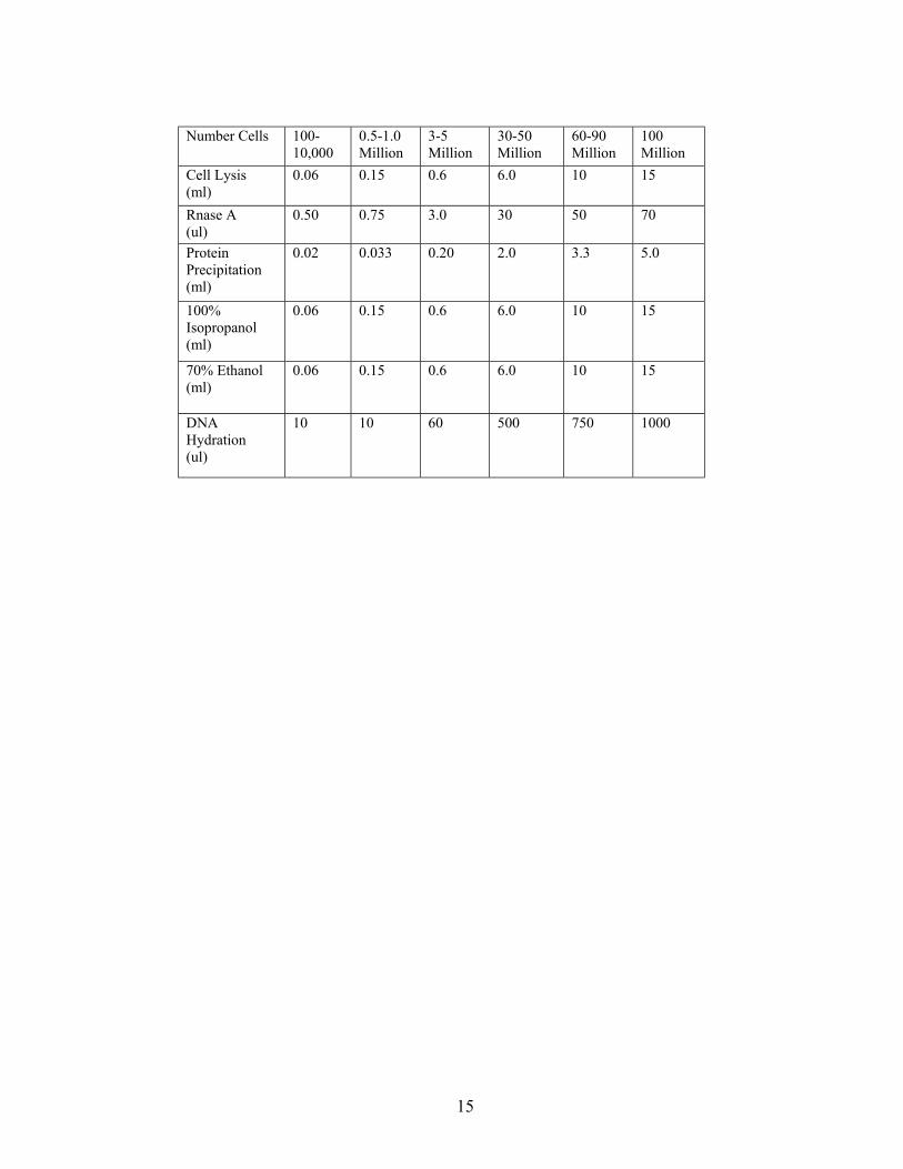
Number Cells 100-10,000
0.5-1.0 Million
3-5 Million
30-50 Million
60-90 Million
100 Million
Cell Lysis (ml)
0.06 0.15 0.6 6.0 10 15
Rnase A (ul)
0.50 0.75 3.0 30 50 70
Protein Precipitation (ml)
0.02
0.033 0.20 2.0 3.3 5.0
100% Isopropanol (ml)
0.06 0.15 0.6 6.0 10 15
70% Ethanol (ml)
0.06 0.15 0.6 6.0 10 15
DNA Hydration (ul)
10 10 60 500 750 1000
16
Appendix B Human Identification with Short Tandem Repeat Loci using
the AmpFlSTR Green I PCR Amplification Kit. Materials AmpFlSTR Green I PCR Amplification Kit, catalog # 402902, Applied Biosystems, Foster City, CA AmpFl STR Green I PCR Amplification Kit User’s Manual, catalog # 402944, Applied Biosystems Performance Optimized Polymer 4 (POP-4), catalog # 402838, Applied Biosystems MicroAmp 8-Strip Reaction Tubes (0.2ml), catalog #N801-0580, Applied Biosystems MicroAmp Caps(8 caps/strip), catalog # N801-0535, Applied Biosystems MicroAmp 96-Well Tray/Retainer, catalog # 403081, Applied Biosystems 0.5ml Genetic Analyzer sample tubes and septum, catalog # 401957 and 401956, Applied Biosystems Rainin pipette tips, Rainin, Emeryville, CA 1.5ml microfuge tubes, Marsh Biomedical Products, Rochester, NY 1xTE(10mM Tris-HCl, 0.1mM EDTA, pH 8.0) Hi Di formamide, catalog # 4311320, Applied Biosystems GeneScan-350[ROX] Internal Lane Size Standard, catalog # 401735, Applied Biosystems, Foster City, CA 10x Genetic Analyzer Buffer with EDTA, catalog # 402824, Applied Biosystems Performance Optimized Polymer-4 (POP-4), Applied Biosystems Equipment GeneAmp PCR System 9700, Applied Biosystems, Foster City, CA ABI PRISM 310 DNA sequencer, Applied Biosystems Rainin pipettors, Rainin, Emeryville, CA Stratalinker 2400 UV Crosslinker, Stratagene, La Jolla, CA Vortex Genie, Daigger, Lincolnshire, IL Heating block Computer and software for analysis with GeneScan and Genotyper Software, Applied Biosystems, Labeling Label all reagents and aliquots of reagents with the reagent name, concentration, date prepared and appropriate expiration date. Labels are created using computer label making system, Label View Pro, and Eltron printer. Procedures: A. PCR Amplification
1. Record the ID of the samples to be typed and assign an internal ID number to each sample. 2. Label all processing MicroAmp 8-Strip Reaction Tubes (0.2 ml) with the internal ID number and place
them into a MicroAmp 96-Well Tray/Retainer. There should be a tube for the samples, positive control and negative control.
3. Place the MicroAmp tubes/Tray and a 1.5 ml microcentrifuge tube into the Stratalinker (Stratagene) and UV crosslink twice at 120 joules (1200 on LED display) to sterilize the tubes. Remove the tubes from the Stratalinker.
4. Prepare a master mix of the following composition in a 1.5 ml microcentrifuge tube (all reagents are supplied in the AmpFlSTR Green I PCR Amplification Kit):
number of samples x 10.5 µl of AmpFlSTR PCR reaction Mix number of samples x 0.5 µl of AmpliTaq Gold DNA Polymerase number of samples x 5.5 µl of AmpFlSTR Green I Primer. number of samples x 9.9 µl of deionized water 5. Mix by vortexing. 6. Dispense 24 µl of the master mix into each of the MicroAmp Reaction Tubes.
17
7. Into each of the tubes containing the master mix, pipet 1 µl of genomic DNA (25 ng). 8. To the designated Positive Control tube add 1 µl (25ng) of Control DNA and to designated Negative
Control tube, add 1 µl of 1xTE buffer. NOTE: the final volume of the reaction is 25 µl. 9. Place the MicroAmp Caps on the tubes and seal tightly.
10. Place the tubes into the thermal cycler 11. Program the GeneAmp PCR System 9700 thermal cycler as follows (refer to the Geneamp PCR
System 9700 Users Manual for details): 1 Cycle : 11 min at 95°C 27 Cycles: 1 min at 94°C 1 min at 59°C 1 min at 72°C 1 Cycle: 45 min at 60°C Hold: at 25°C (Store amplified products away from light at 2-6°C for short periods and at -20°C for longer periods.)
B. Sample Preparation for the Genescan run using the 310 Genetic Analyzer 1. Following the instructions in the ABI PRISM 310 Genetic Analyzer User's Manual, clean the machine and
prepare the 310 Genetic Analyzer for running Genescan using Performance Optimized Polymer 4 (POP-4). 2. Open a new Genescan sample sheet in the 310 Data Collection Software. Fill in the sample names and
mark the red box as the standard. Save the sample sheet. 3. Open up a new injection list and select and open the sample sheet that was just created and select
Genescan-350-ROX as the internal lane standard. 4. Using the following formulas, calculate the needed amounts of Hi Di formamide and GeneScan-350 [ROX]
Internal Lane Standard and combine then into a 1.5 ml microcentrifuge tube (when determining the number of samples include the positive and negative controls):
(Number of samples + 2) x 24 ul of Hi Di formamide (Number of samples + 2) x 1.0 ul of genescan-350[ROX] size standard 5. Aliquot 25 µl of the Hi Di formamide/GneScan-350[ROX] mixture into 0.5 ml Genetic Analyzer tubes. 6. Add 1.0 µl of the AmpFl STR Green I PCR Product or 1.0 µl of AmpFl STR Green I Allelic Ladder per
tube and mix by pipetting up and down. 7. Seal each tube with a septum. 8. Denature each sample at 95°C for 3 minutes and chill the tubes for another 3 minutes in an ice water bath. 9. Place the tubes in the sampler tray of the 310 Genetic Analyzer and start the Genescan run. C. Data Analysis Analyze the data using the Genotyper Software and input all data into the database.

18
Appendix C Identification of the Amelogenin (sex) and THO1 Markers with the Short Tandem Repeat Loci
(This method will be used as Quality Control for indivduals who are not part of a Trio) Materials HotStarTaq Master Mix Kit, catalog # 203443, Qiagen, Valencia, CA. MicroAmp 8-Strip Reaction Tubes(0.2ml), catalog #N801-0580, Applied Biosystems. MicroAmp Caps(8 caps/strip), catalog # N801-0535, Applied Biosystems. MicroAMp 96-Well Tray/Retainer, catalog # 403081, Applied Biosystems. Rainin pipet tips, Rainin, Emeryville, CA. 1.5ml microfuge tubes, Marsh Biomedical Products, Rochester, NY. 10X TBE (Trizma Base, Boric Acid, EDTA) for Sequencing. GeneScan-350[ROX] Internal Lane Size Standard, catalog # 401735, Applied Biosystems. Hi Di Formamide, catalog # 4311320, Applied Biosystems. blue dextran/EDTA loading dye, Appled Biosystems. Long Ranger Singel Packs, catalog #50691, FMC BioProducts, Rockland, ME. Beakers. deionized water. Primers PCR primers synthesized by Biotechnology Core Facility, Scientific Resources Program, NCID, CDC are listed in the following table: FAM-AmeloF primer 5'-FAM-ccctgggctctgtaaagaatagtg-3’ AmeloR primer 5'-atcagagcttaaactgggaagctg-3' FAM-TH01R primer 5'-FAM-gtgggctaaaagctcccgattat-3’ T01F primer 5'-attcaaagggtatctgggctctgg-3' Equipment Geneamp PCR System 9700, Applied Biosystems, Foster City, CA. ABI PRISM 377 DNA sequencer, Applied Biosystems. Rainin pipettors, Rainin, Emeryville, CA. Stratalinker 2400 UV Crosslinker, Stratagene, La Jolla, CA. Vortex Genie, Diagger, Lincolnshire, IL. Heating block Computer and software for analysis with GeneScan and Genotyper Software, Applied Biosystems, Labeling Label all reagents and aliquots of reagents with the reagent name, concentration, date prepared and appropriate expiration date. Labels are created using computer label making system Label View Pro and Eltron printer. Reagent Preparation 10X TBE for sequencing: Final concentration g/L 890 mM Tris Base (Trizma Base) 108 g 890 mM Boric Acid 55 g 20 mM Disodium EDTA 7.44 g Add deionized water to a final volume of 1000 mL, mix thoroughly and filter through < 0.45mm membrane. Store at room temperature and do not use if precipitate forms. For 1X TBE for sequencing, dilute 150mL of the 10X TBE stock and bring the volume up to 1.5 L.
19
PCR Amplification 1. Record ID of samples to be typed and assign an internal ID number to each sample. 2. Label all processing MicroAmp 8-Strip Reaction Tubes (0.2ml tubes) with the internal ID number and
place them into a MicroAmp 96-Well Tray/Retainer. There will be a positive and negative control tube as well.
3. Place the MicroAmp tubes/Tray and a 1.5ml microcentrifuge tube into the Stratalinker (Stratagene) and UV crosslink twice at 120 joules (1200 on LED display) to sterilize the tubes prior to use to avoid contamination.
4. Make a master mix of the following contents in a 1.5ml microcentrifuge tube. #of samples x 12.5 µl of HotStarTaq Master Mix # of samples x 1.0 µl of FAM-AmeloF primer (3.6 pM/µl) # of samples x 1.0 µl of AmeloR primer (3.6 pM/µl) # of samples x 1.0 µl of TH01F primer (8 pM/µl) # of samples x 1.0 µl of FAM-TH01R primer (8 pM/µl) # of samples x 7.5 µl of water
5. Mix by Vortexing. 6. Dispence 24 µl of the master mix into each of the MicroAmp Reaction Tubes. 7. To each of the tubes containing master mix, pipet 1µl of Genomic DNA at a concentration of 25 ng/ml. 8. For the Positive Control tube, add 1µl of the selected control to the tube and for the Negative control
tube, add 1 µl of water. Note: The final volume for the PCR is 25 µl. 9. Place the MicroAmp Caps on the tubes and seal tightly.
10. Place the Tubes in the thermal cycler, Geneamp PCR System 9700, and program the following conditions into the machine and start the run under the reaction volume of 25ml (refer to the Geneamp PCR System 9700 Users Manual for details).
1 Cycle at 95°C for 10min, 27 Cycles at 94°C for 45sec, 60°C for 45sec, 72°C for 1min,
1 Cycle at 60°C -45min Hold at 4°C
(store the amplified products protected from light at 2-6°C for short periods and at –15°C to –25°C for longer periods.) GeneScan using the 377 DNA Sequencer A. Gel Preparation and casting using the Long Ranger Singel Pack.
i. Gel Preparation 1. Assemble glass plates and spacers in the cassette following the method described in the ABI PRISM
377 DNA Sequencer Users Manual. 2. Have the Long Ranger Singel pack at room temperature. 3. Remove the BLACK clip and mix the contents of the compartments by hand thoroughly but gently for
1 minute. 4. Place the pack on an orbital shaker for 5 minutes at medium speed. 5. Mix by hand thoroughly but gently for 1 minute 6. Place the pack on an orbital shaker for 5 minutes at medium speed. NOTE: Do not over mix. This may interfere with the polymerization of the gel.
ii. Gel Casting
NOTE: The following steps must be completed without delay. 1. Remove only the RED clip and mix the contents of the compartment well by hand for 1 minute. 2. Remove the WHITE clip to expose the filter to gel solution. 3. Hold the pack so the contents drain into the filter end. Fold the pack in half at the indicated line. 4. Hold the pack with the cut mark at the top and cut the corner within the space marked CUT. To avoid
introducing bubbles cut a large enough hole in the pouch to allow steady flow of the solution through the filter into a beaker.
5. Avoid introducing air into solution after mixing. Cast gel and insert comb according to your standard procedure.
20
6. Once the gel is polymerized(30 minutes), place paper towels soaked in electrophoresis buffer over the ends of the plates and then cover with plastic wrap. This will prevent moisture loss as the polymerization process continues.
7. Allow 2 hours for complete gel polymerization.
B. Preparing for Electrophoresis
1. Remove the comb, wash the plates and load the comb as described in the ABI PRISM 377 DNA Sequencer Users Manual.
2. Prepare a sufficient quantity of electrophoresis buffer to fill both anodal and cathodal chambers by diluting 10X TBE stock for sequencing with deionizd water to 1X.
3. Mount the gel cassette onto the sequencing apparatus and prepare the gel for the sequence run according to the 377 DNA Sequencers Users Manuals instructions.
4. Open a new GeneScan sample sheet in the 377-96 Data Collection Software, and input sample names to be run on the gel.
5. Save the sample sheet and open a new GeneScan sample run in the 377-96 Data Collection Software and open the new sample sheet that was just created in the previous step.
6. To assure plates and gel are clean, perform a plate check using the Plate Check module. 7. Pre-warm the acrylamide gel by running the GS PR 36A-2400 module. 8. Prepare the samples for the GeneScan run by combining 1.5 µl of the PCR product and 1.0 µl of
GeneScan Rox[350] with 5 µl of a 5:1 ratio of Hi Di formamide and blue dextran/EDTA loading dye (for example 5ml of the Hi Di formamide combined with 1ml of the blue dextran/EDTA loading dye).
9. Vortex the samples and centrifuge briefly. 10. Denature the samples by heating the samples at 95 + 5°C for 2 minutes. 11. Ice the samples immediately for 2 minutes and keep on ice until ready to use. 12. Stop the PRE-RUN when the temperature reaches 50°C and rinse out the top of the gel with 1XTBE
buffer. 13. Load 1.8 ml of the denatured samples on the gel. The odd lanes should be loaded first then run in for 1
minute before the even lanes are loaded. 14. Cancel the PRE-RUN and change the module to the GS Run 36A-2400 module and start the run. The
run will take 2.5 hours. 15. Analyze the Results using the GeneScan Analysis and Genotyper Software.
21
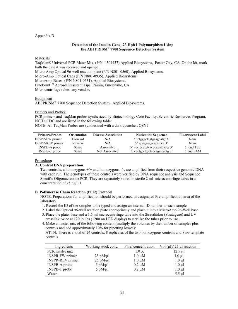
Appendix D
Detection of the Insulin Gene -23 Hph I Polymorphism Using the ABI PRISM® 7700 Sequence Detection System
Materials TaqMan® Universal PCR Mater Mix, (P/N 4304437) Applied Biosystems, Foster City, CA. On the kit, mark both the date it was received and opened. Micro-Amp Optical 96-well reaction plate (P/N N801-0560), Applied Biosystems. Micro-Amp Optical Caps (P/N N801-0935), Applied Biosystems. MicroAmp Bases, (P/N N801-0531), Applied Biosystems. FinePointTM Aerosol Resistant Tips, Rainin, Emeryville, CA Microcentrifuge tubes, any vendor. Equipment ABI PRISM® 7700 Sequence Detection System, Applied Biosystems. Primers and Probes: PCR primers and TaqMan probes synthesized by Biotechnology Core Facility, Scientific Resources Program, NCID, CDC and are listed in the following table: NOTE: All TaqMan Probes are synthesized with a dark quencher, QSY7.
Primers/Probes Orientation Disease Association Nucleotide Sequence Fluorescent Label INSPR-FW primer Forward N/A 5’ ctgggctcgtgaagcatgt 3’ None INSPR-REV primer Reverse N/A 5’ gcaggaggcgcatcca 3’ None
INSPB-A probe Sense Associated 5’ ccctgcctgtcacccagatcactg 3’ 5’ end TET INSPB-T probe Sense Not Associated 5’ ccctgcctgtctcccagatcactg 3’ 5’end FAM
Procedure: A. Control DNA preparation
Two controls, a homozygous +/+ and homozygous -/-, are amplified from their respective genomic DNA with each run. The genotypes of these controls were verified by DNA sequence analysis and Sequence Specific Oligonucleotide PCR. They are separately stored in sterile 2 ml microcentirfuge tubes in a concentration of 25 ng/ µl.
B. Polymerase Chain Reaction (PCR) Protocol NOTE: Preparations for amplification should be performed in designated Pre-amplification area of the laboratory. 1. Record the ID of the samples to be typed and assign an internal ID number to each sample. 2. Label the Optical 96-well reaction plate appropriately and place it into a MicroAmp 96-Well base. 3. Place the plate, base and a 1.5 ml microcentrifuge tube into the Stratalinker (Stratagene) and UV
crosslink twice at 120 joules (1200 on LED display) to sterilize the tubes prior to use. 4. Make a master mix of the following content (multiply the volumes by the number of samples plus
controls and add approximately 10% for pipetting losses): ATTN: There is a total of 24 controls: 8 replicates of the two homozygous controls and 8 no-template controls.
Ingredients Working stock conc. Final concentration Vol (µl)/ 25 µl reaction
PCR master mix 1.0 X 12.5 µl INSPR-FW primer 25 pM/µl 1.0 µM 1.0 µl INSPR-REV primer 25 pM/µl 1.0 µM 1.0 µl INSPB-A probe 5 pM/µl 0.2 µM 1.0 µl INSPB-T probe 5 pM/µl 0.2 µM 1.0 µl Water 5.5 µl

22
5. Mix by vortexing. 6. Dispense 24 µl of the master mix into those wells of the plate that will be used. 7. To the wells designated for controls, add 1 ul of genomic DNA of the controls to the master mix. 8. To the wells designated for no-template controls, add 1 µl of distilled water. 9. To the wells designated for samples, add 1 µl of the appropriate genomic DNA.
10. Cover the wells with MicroAmp Caps and seal tightly. 11. Place the Plate in the ABI PRISM® 7700 Sequence Detector.
C. PCR data collection: Real Time data collection
Create a Real Time plate document. (Refer to the ABI PRISM® 7700 Sequence Detection System User’s Manual for details). Perform a Real Time run under the following Thermal Cycler conditions: 1 cycle 10 min at 50°C
1 cycle 10 min at 95° 40 cycles 15 sec at 95°C, 1 min at 60°.
Perform analysis according to the user’s manual. Plate Read Data Collection – Allelic Discrimination Create an Allelic Discrimination plate document. (Refer to the ABI PRISM® 7700 Sequence Detection System User’s Manual for details). Perform a Post PCR Plate Read. Perform analysis according to the user’s manual. D. Genotype Determination
1. Open the Real Time plate document containing the collected data. Perform analysis according to the user’s manual and examine the mulitcomponent results. The +/+ homozygous controls will have an increase of fluorescence of TET relative to FAM, and the -/- homozaygous controls will have an increase in fluorescence of FAM relative to TET. No significant increase in fluorescence should be observed in the no-template controls.
2. After insuring that all the control results are correct inspect the multicomponent results of individual unknown specimens.
3. Open the Allelic Discrimination plate document containing the Plate Read data. Perform analysis according to the user’s manual. Open Allelic Discrimination from the Analysis menu. Examine the allelic calls made for the controls and make sure that they have been called correctly.
4. If here are any samples or controls that are not clustered with the other samples and controls of the same genotype on the graph, go back to the Real Time plate document and examine the mutlicomponent results to make you judgment.
5. Export Results from the Alleluia Discrimination document. This will create an Excel document with the results displayed in a table form. Proofread and save the results in the sample database.

23
Appendix E Detection of the Insulin Gene –23 Hph I Polymorphism using Sequence Specific Oligonucleotides (SSO)
Materials QIAGEN HotStarTaq Master Mix Kit, QIAGEN Inc., Valencia, CA. MicroAmp Reaction Tubes with Caps, Part No. N801-0540, Applied Biosystems, Foster City, CA. MicroAmp Bases, Part No. N801-0531, Applied Biosystems, Foster City, CA MicroAmp. Trays, Part No. N801-0541, Applied Biosystems, Foster City, CA. FinePointTM Aerosol Resistant Tips, Rainin, Emeryville, CAPCR primers synthesized by Biotechnology Core Facility, Scientific Resources Program, NCID, CDC, and listed in the table below:
Primer Name Orientation Genotype Nucleotide Sequence
INS FWD forward N/A tccaggacaggctgcatcag
INS REV-A reverse Hph I + cagaaggacagtgatctggga
INS REV-T reverse Hph I - cagaaggacagtgatctgggt
Deionized water Ultra-pure Agarose, Gibco/BRL, Rockville, MD. 1X TBE buffer made from 10X TBE. Low DNA Mass Ladder, Catalog No. 10068-013, Gibco/BRL, Rockville, MD Gel Loading Dye (Orange G, Ficoll 400, EDTA) Sigma, St. Luis, MO. Ethidium Bromide, Ameresco, Solon, OH. Equipment Rainin Pipettors, Rainin, Emeryville, CA. GeneAmp PCR System 9700 thermal cycler, Part No. N805-0001, Applied Biosystems, Foster City, CA. Electrophoresis unit, Owl Separation System, Portsmouth, NH. PowerPac 300 power supply, Catalog No. 165-5052, 165-5053, BioRad, Hercules, CA. AlphaImagerTM 2200 Documentation System, Alpha Innotech, San Leandro, GA. Micro-centrifuge Daigger, Linconshire, IL. Vortexer Genie, Daigger, Linconshire, IL. Glass flask. Incubator or water bath (at 37°C). Balance. Nitrile Gloves. Labeling: Label all reagents and aliquots of reagents with the reagent name, concentration, preparation date and expiration date. Labels can be hand-written or created using LabelView Pro software, Teklynx, Milwaukee, WC, for more permanent labels. Reagent specifications Primers used for PCR must be diluted with deionized water to a concentration of 50 pM/l using sterile techniques and stored at 4°C.

24
Procedure A. Polymerase Chain Reaction (PCR) Protocol
NOTE: Preparations for amplification should be performed in designated Pre-amplification area of the laboratory. 1. Determined the number of Macomb PCR tubes needed and label them appropriately. (There should be
two (2) PCR tubes for each sample and one (1) PCR tube for a negative control). 2. Place the labeled PCR tubes in the Strata linker and UV crosslink on the “auto crosslink” setting to
prevent contamination. Remove the tubes from the Strata linker. 3. Place tubes on a MicroAmp tube base and add the following to each tube:
Volume HotStarTaq Master Mix 12 :l INS FWD primer variable (0.4 pM final conc.) INS REV-A or INS REV-T variable (0.4 pM final conc.) Sample DNA variable (100-400 ng)
Deionized water enough to bring the total reaction volume to 25 µl Total Volume 25µl
For the negative control instead of Sample DNA add the same amount of deionized water.
4. Cap all PCR tubes, shake each tube briefly on the Vortexer and spin down in the microcentifuge. 5. Put the tubes on a MicroAmp tray and place them into the thermal cycler sample block. 6. Program the GeneAmp PCR System 9700 thermal cycler for amplification with HotStarTaq DNA
Polymerase as follows (consult the GeneAmp PCR System 9700 User’s Manual for additional information on programming and operation of the thermal cycler):
1. Initial Activation Step: 10 min at 95°C 2. 3-step cycling: 45 sec at 95°C 1 min at 63°C 1 min at 72°C Repeat 30 times (30 cycles)
3. Final Extension: 7 min at 72°C 4. Hold program: forever at 4°C
7. Run the program. (The program runs for approximately 1.6 hours.) Gel Electrophoresis to Visualize PCR products
1. Prepare a 2% agarose gel of the desired size using ultra-pure agarose and 1 x TBE as follows: weigh the appropriate amount of agarose and place it in a flask of a volume at least 1.5 times larger than the final volume of the agarose gel solution being prepared. Add the appropriate volume of 1x TBE to the agarose to get the right concentration.
2. Cover the flask with plastic wrap and poke a hole for ventilation. 3. Place the flask in a microwave oven and heat until all the agarose goes into solution. (ATTENTION:
Since the flask becomes hot in the microwave, use appropriate protection when handling it.) Take the flask out and visually inspect the solution to make sure that all the agarose has dissolved. If the viscous agarose is still visible return the flask into the microwave and heat for a few more seconds. Repeat if necessary.
4. Take out the flask from the microwave and set aside to cool to approximately 60°C or until it can be handled comfortably.
5. While the agarose mixture is cooling prepare a casting tray with the appropriate combs to give the desired number of wells. There should be one (1) well for each sample and one (1) additional well, per row of wells, for a marker DNA ladder
6. Pour cooled agarose gel mixture into the tray and leave gel to polymerize for at least 30 minutes. 7. Remove all combs and place the gel into an electrophoresis unit with enough 1x TBE buffer to cover.

25
8. Combine 4: 1 of the Low DNA Mass Ladder with 3: 1 of loading dye and pipette into the first well. Do this for each row of wells.
9. Combine 4: 1 of each sample and 3: 1 of loading dye and pipette into designated wells. 10. Start electrophoresis and run on 80 volts for 30-45 minutes. 11. Stain the gel in a staining solution (20: 1 Ethidium Bromide per 1L of 1 x TBE) for 5 minutes and then
de-stain it in 1 x TBE for 10 minutes. 12. Place the gel into the UV transilluminator of the AlphaImagerTM 2200 Documentation System,
photograph the gel and record the results. B. Interpretation of Results
The genotype of each sample can be determined by the presence (seen as a band on the gel) or absence of amplicons after PCR with a given pair of primers. The gel should have the following banding pattern for the three genotypes:
Genotype Primer Pairs
Homozygous A Homozygous T Heterozygous
INS FWD/ INS REV-A Band No band Band
INS FWD/ INS REV-T No band Band Band