determinants of metastatic competency in colorectal...
TRANSCRIPT

Acc
epte
d A
rtic
le
This article has been accepted for publication and undergone full peer review but has not been through the copyediting, typesetting, pagination and proofreading process, which may lead to differences between this version and the Version of Record. Please cite this article as doi: 10.1002/1878-0261.12018 Molecular Oncology (2016) © 2016 The Authors. Published by FEBS Press and John Wiley & Sons Ltd. This is an open access article under the terms of the Creative Commons Attribution License, which permits use, distribution and reproduction in any medium, provided the original work is properly cited.
Accepted Date: 21-Oct-2016
Article Type: Review
Determinants of metastatic competency in colorectal cancer
Daniele V.F. Tauriello1 #, Alexandre Calon2 #, Enza Lonardo1
and Eduard Batlle1,3 #
1. Institute for Research in Biomedicine (IRB Barcelona). The Barcelona Institute of Science
and Technology. Baldiri i Reixac 10, 08028 Barcelona, Spain
2. Hospital del Mar Medical Research Institute (IMIM). MAR Health Park Barcelona. Dr.
Aiguader, 88, 08003, Barcelona, Spain
3. ICREA, Pg. Lluís Companys 23, 08010 Barcelona, Spain.
# Correspondence: Daniele V.F. Tauriello [[email protected]], Alexandre
Calon [[email protected]] and Eduard Batlle [[email protected]]
Keywords
Heterogeneity, cancer stem cells, clonal diversity, tumour microenvironment, stroma,
dormancy, cancer immunology, combination therapy, immunotherapy, TGF-beta
Running title: Determinants of colorectal cancer metastasis

Acc
epte
d A
rtic
le
Molecular Oncology (2016) © 2016 The Authors. Published by FEBS Press and John Wiley & Sons Ltd.
Abbreviations:
AJCC: American Joint Committee on Cancer
CAF: cancer-associated fibroblast
CMS: consensus molecular subtype
CRC-SC: colorectal cancer stem cell
CRC: colorectal cancer
ctDNA: circulating tumour DNA
CTL: cytotoxic T lymphocyte
EMT: epithelial-mesenchymal transition
EndMT: endothelial-mesenchymal transition
HSC: hepatic stellate cell
ISC: intestinal stem cell
ITH: intratumoral heterogeneity
MDSC: myeloid-derived suppressor cell
Met-SC: Metastatic stem cell
MSC: mesenchymal stem cell
MSI: microsatellite instable
SC: stem cell
TAM: tumour-associated macrophage
Acc
epte
d A
rtic
le
Molecular Oncology (2016) © 2016 The Authors. Published by FEBS Press and John Wiley & Sons Ltd.
TAN: tumour-associated neutrophil
TME: tumour microenvironment
TNM: tumour, lymph node, metastasis
UPR: unfolded protein response
Abstract
Colorectal cancer (CRC) is one of the most common cancer types and
represents a major therapeutic challenge. Although initial events in colorectal
carcinogenesis are relatively well-characterized and treatment for early stage
disease has significantly improved over the last decades, the mechanisms
underlying metastasis – the main cause of death – remain poorly understood.
Correspondingly, no effective therapy is currently available for advanced or
metastatic disease. There is increasing evidence that colorectal cancer is
hierarchically organized and sustained by cancer stem cells, in concert with various
stromal cell types. Here we review the interplay between cancer stem cells and their
microenvironment in promoting metastasis and discuss recent insights relating to
both patient prognosis and novel targeted treatment strategies. A better
understanding of these topics may aid the prevention or reduction of metastatic
burden.

Acc
epte
d A
rtic
le
Molecular Oncology (2016) © 2016 The Authors. Published by FEBS Press and John Wiley & Sons Ltd.
1. Introduction
Colorectal cancer (CRC) is one of the most frequent types of cancer
worldwide, accounting for approximately 10% of all new cancer cases and 8.5% of
all cancer deaths (Torre et al., 2015). Whereas the vast majority of primary cancers
can be extirpated through surgical resection, only a fraction of the patients
diagnosed with overt metastatic disease can be cured by surgery. About 20% of the
CRC patients present with metastasis at the time of diagnosis (stage IV). In addition,
35-45% of the patients with localized disease (stage II and III) succomb to
recurrence within 5 years after surgery. Most of these relapses occur as metastases
and are caused by residual tumour cells that have spread to distant organs prior to
surgery. Clearly, current systemic therapies fail to eliminate latent disseminated
tumour cells and are similarly ineffective in treating growing metastases, offering
survival benefits of only a few months.
Metastasis is the spread of cancer to a distant organ, which in the case of
CRC patients involves mainly the liver and lungs. As described elsewhere
(Massague and Obenauf, 2016; Oskarsson et al., 2014; Valastyan and Weinberg,
2011), to gain metastatic competence, cancer cells require the capacity to invade the
surrounding tissues, survive in the circulation, colonize the foreign organ and
eventually resume growth. Metastasis is an inefficient process owing to the fact that
most tumour cells fail to acquire the necessary abilities to regenerate a tumour at a
distant site (Massague and Obenauf, 2016; Oskarsson et al., 2014). Over the past
few years, the main determinants of metastatic competence in CRC have begun to
be characterized. In the absence of mutations that associate to the process of
metastasis in CRC, it has become increasingly clear that the regeneration of the
tumour in a foreign organ is tightly bound to the acquisition of a stem-like phenotype
Acc
epte
d A
rtic
le
Molecular Oncology (2016) © 2016 The Authors. Published by FEBS Press and John Wiley & Sons Ltd.
by cancer cells. These metastatic stem cells adopt multiple phenotypes and
behaviours and critically depend on their interaction with the microenvironment to
migrate, survive in the circulation and thrive in a foreign organ.
In section 2 we will review the genetics of CRC development, to then discuss
both the evidence that supports the notion of hierarchical organization throughout
CRC progression and the ensuing implications in section 3. We will also focus on the
mechanisms involved in the multiple phenotypes and adaptations that tumour stem
cells go through in the metastatic process. In section 4 we will explore the current
knowledge about the role of the tumour microenvironment in promoting and
sustaining metastasis. This includes a dissection of the cell types and niches that
support the survival and maintenance of metastatic stem cells, and an analysis of the
ways that stromal features can improve disease prognosis. In section 5, we will
discuss the complexities and limitations imposed on clinical practice by the
heterogeneous nature of both epithelial and stromal compartments. Finally, we will
indicate how these emerging concepts are informing, and slowly transforming,
therapeutic strategies to treat patients with metastatic disease.
2. Colon carcinogenesis
2.1. From Normal Mucosa to Colorectal Cancer.
The epithelium of the normal colon undergoes continuous renewal. At the
base of glandular invaginations of the colonic mucosa, called crypts, a pool of rapidly
diving intestinal stem cells (ISCs) sustains the homeostatic regeneration of the
epithelium throughout a lifetime (Clevers, 2013). Over the past decade, signals that
regulate ISC renewal and proliferation have been extensively characterized: WNT,
Acc
epte
d A
rtic
le
Molecular Oncology (2016) © 2016 The Authors. Published by FEBS Press and John Wiley & Sons Ltd.
EGFR/MAPK and NOTCH signalling promote the undifferentiated proliferative state
of ISCs in the niche, whereas BMP and TGF-beta signalling induce cytostasis and
differentiation (Clevers, 2013).
The elevated division rate of ISCs increases their probability to acquire
mutations during DNA replication (Vogelstein et al., 2013). Additional environmental
factors such as life style, diet and microbiota can also greatly influence the
transformation of the epithelium (Bishehsari et al., 2014). The most common genetic
events in CRCs are alterations that inactivate the tumour suppressor gene APC. This
triggers the constitutive activation of WNT signalling and imposes a continuous stem-
like self-renewing state at the onset of tumorigenesis, giving rise to benign
outgrowths of the epithelium known as adenomas. Genetic experiments performed in
mouse models support the hypothesis that Apc mutation in ISCs represents the
origin of intestinal polyps (Barker et al., 2008; Tetteh et al., 2016), although chronic
inflammation or dysregulation of BMP signalling have been shown to help convert
non-stem cells into CRC initiating cells (Davis et al., 2014; Schwitalla et al., 2013).
A small fraction of adenomas become progressively aggressive through
acquisition of additional driver mutations, which mainly affect 3 additional signalling
pathways (Cancer Genome Atlas Network, 2012; Seshagiri et al., 2012): i. the MAPK
pathway is often hit by activating mutations in KRAS, BRAF or PIK3CA and provides
cell autonomous mitogenic and pro-survival stimuli to cancer cells; ii. the p53
pathway is inactivated by mutations in the eponymous protein, or less commonly in
ATM, facilitating acquisition of genomic instability; iii. the TGF-beta pathway is
frequently silenced by loss-of-function mutations in TGFBR2, SMAD4, SMAD2 or
SMAD3, which bypasses the suppressive effects of high TGF-beta levels present in
the tumour microenvironment (Fearon, 2011). Pioneer studies by Eric Fearon and

Acc
epte
d A
rtic
le
Molecular Oncology (2016) © 2016 The Authors. Published by FEBS Press and John Wiley & Sons Ltd.
Bert Vogelstein correlated these mutations with pathologically classifiable stages of
adenoma malignancy and suggested a linear progression model, in which the
compounding of the 4 mentioned pathway mutations associated with development of
aggressive adenocarcinomas (Fearon and Vogelstein, 1990).
Acquisition of these mutations is a slow process and consequently the
development of invasive CRC often takes decades (Jones et al., 2008; Vogelstein et
al., 2013). Of note, the linear progression model based on 4 stepwise genetic
alterations represents a simplification, as not every tumour carries genetic alterations
in these four pathways or develops through the equivalent sequence of events.
Moreover, full-blown CRCs have a riche and complex mutational landscape that
expands well beyond mutations in the 4 driver pathways (Cancer Genome Atlas
Network, 2012; Seshagiri et al., 2012). Due to the acquisition of chromosomal
instability or defects in the DNA mismatch repair system, tumours accumulate
hundreds or even thousands of genetic alterations. Some of these are passenger
mutations, as they do not confer advantages to tumour cells, but others drive the
biology of the cancer and therefore give selective advantage. Beyond the context of
the linear progression model, the role of many of these mutations remains poorly
understood. Together, these issues of complexity and heterogeneity impinge upon
the functional analysis of CRC and complicate the development and application of
therapeutic approaches.
2.2. Progression to Metastasis
As described above, the mutations that drive CRC progression affect the
signalling pathways that regulate ISC behaviour, endowing cancer cells with self-
renewal and growth capacity, independently of crypt niche signals. Evidence
obtained from the analysis of patient-derived and CRISPR-engineered CRC

Acc
epte
d A
rtic
le
Molecular Oncology (2016) © 2016 The Authors. Published by FEBS Press and John Wiley & Sons Ltd.
organoids have led to the hypothesis that acquisition of mutations in the 4 linear
progression model pathways may be sufficient to facilitate the growth of tumour cells
in unfavourable environments such as those encountered in foreign tissues (Drost et
al., 2015; Fujii et al., 2016; Matano et al., 2015). Yet, while most CRCs carry genetic
alterations in several of these 4 driver pathways, metastasis is a relatively inefficient
process. This suggests that additional bottlenecks and dependencies limit the extent
of tumour spread.
It is worth considering that most CRCs are invasive at the time of diagnosis
and, therefore, have had the opportunity to shed cells into circulation for months or
longer. When disseminating CRC cells enter the portal circulation, they are
transported to the liver sinusoids within minutes and can home into the liver
parenchyma because of vessel fenestration. In the case of pulmonary metastases,
CRC cells must first reach the general circulation and then infiltrate the lung
parenchyma. It has been reported that this process requires active killing of lung
capillary cells in a mechanism that involves the hormone PTHLH (Urosevic et al.,
2014) and possibly necroptosis (Strilic et al., 2016). The capacity of disseminated
CRC cells to infiltrate other organs such as the brain is less well-characterized and
may involve co-option of invasion mechanisms described for other tumour types
such as breast cancer (Valiente et al., 2014).
Work in experimental models has shown that the rate-limiting step in the
metastasis process is the capacity of circulating tumour cells to colonize the foreign
organ (Massague and Obenauf, 2016; Obenauf and Massagué, 2015). Most tumour
cells that survive in circulation and manage to infiltrate a distant organ will die for
reasons that remain incompletely understood but that may include recognition and
killing of tumour cells by the innate and adaptive immune system (Collignon et al.,

Acc
epte
d A
rtic
le
Molecular Oncology (2016) © 2016 The Authors. Published by FEBS Press and John Wiley & Sons Ltd.
2004; Strauss and Thomas, 2010). Those tumour cells that survive and adapt to the
new environment can generate an overt metastasis (Figure 1). However, not all
venturing cancer cells that have successfully pervaded a distant site necessarily
have the competency to establish a thriving colony in these hostile environments.
Frequently, they remain latent for months to years before resuming growth (Sosa et
al., 2014). Below we discuss tumour cell-intrinsic and extrinsic aspects that facilitate
metastatic colonization in CRC.
3. Colorectal Cancer Stem Cells and their Role in Metastasis
Besides (genetic) diversity between patients, no individual CRC is a uniform,
clonal mass of cancer cells: any given tumour consists of multiple cell populations
with varying levels of phenotypic and genetic heterogeneity. Intratumoral
heterogeneity (ITH) is a central concept to understand the phenomena of metastatic
progression and therapeutic resistance. Three factors contribute to ITH; the
hierarchical organization of cell lineages (sections 3.1 to 3.3), their clonal
diversification (section 3.4) and the microenvironment (section 4).
3.1. Hierarchical Organization of Cancer Cells
More than 30 years ago it was already proposed that the phenotypic diversity
in cancers could arise from spontaneous differentiation of tumour cells (Pierce and
Speers, 1988). The concept, originally developed by G. Barry Pierce in the 70s’,
states: “[carcinomas] are composed of a mixture of malignant stem cells, which have
a marked capacity for proliferation and a limited capacity for differentiation under
normal homeostatic conditions, and of the differentiated, possibly benign, progeny of
these malignant cells” (Pierce and Speers, 1988). This hypothesis was long ignored,

Acc
epte
d A
rtic
le
Molecular Oncology (2016) © 2016 The Authors. Published by FEBS Press and John Wiley & Sons Ltd.
yet several laboratories have recently put forward evidence to support that CRC
complies with this concept. Of note are studies by the groups of Ruggero de Maria
(Ricci-Vitiani et al., 2007), John Dick (O’Brien et al., 2007) and Michael Clarke
(Dalerba et al., 2007), wherein each identified a population of tumour cells within
human CRCs that has the rare capacity to propagate the disease upon inoculation
into immunodeficient mice. Re-launching the idea that CRCs are organized through
a hierarchy of cells with distinct tumorigenic potential, they named this population
“tumour initiating cells”.
Subsequent investigation showed that tumour-initiating cells (also referred to
as CRC Stem Cells or CRC-SCs) reside at the apex of a hierarchy of tumour cells.
They self-renew, display long-term proliferation potential and are capable of initiating
tumours when inoculated into mice. CRC-SCs cells express a gene programme that
to some extent overlaps with that of normal ISCs, and their progeny can undergo
differentiation towards a phenotype similar to that of the normal mucosa.
Differentiation coincides with loss of tumorigenic potential (Dalerba et al., 2011;
Kreso et al., 2014; Merlos-Suarez et al., 2011; Vermeulen et al., 2010, 2008).
Furthermore, the existence of stem cell hierarchy in CRC has been backed by
lineage tracing studies in adenomas (Kozar et al., 2013; Schepers et al., 2012) and
by studies of fate mapping analysis using lentiviral marking of individual tumour cells
(Dieter et al., 2011; Kreso et al., 2012).
The balance between stemness and differentiation in CRC depends upon the
same pathways that regulate normal ISCs, including WNT (Vermeulen et al., 2010),
BMP (Lombardo et al., 2011) and NOTCH (Lu et al., 2013) signalling. Many of these
signals are provided by cells of the tumour stroma, a finding that somewhat
contradicts the hypothesis that CRC progress through gain of ISC niche

Acc
epte
d A
rtic
le
Molecular Oncology (2016) © 2016 The Authors. Published by FEBS Press and John Wiley & Sons Ltd.
independency by acquired mutations. Yet, it is worth considering that not every CRC
carries alterations in all 4 driver pathways (WNT, EGFR, TGF-beta/BMP and p53)
and thus may still depend on stromal factors for further progression. As is discussed
below, additional stromal-derived cytokines and growth factors present at the sites of
invasion can further promote self-renewal of CRC-SCs.
3.2. Tumour Cell Phenotypes at the Invasion Front
Because CRC-SCs have both self-renewal and tumour-initiating capacity,
they likely represent (or give rise to) the so-called Metastatic Stem Cells (Met-SCs,
Figure 1), i.e. the cell of origin of metastasis (Oskarsson et al., 2014). Experimental
evidence supports this hypothesis: the abovementioned study by the group of Hanno
Glimm (Dieter et al., 2011) demonstrates that only cells that hold long-term self-
renewing ability are capable of generating metastasis.
Most CRCs are a relatively disorganized mixture of stem- and differentiated-
like cells that reside into glandular structures reminiscent of the normal crypts
(Merlos-Suarez et al., 2011). Metastases often have an equivalent appearance
(Merlos-Suarez et al., 2011). However, whereas CRC-SCs present in tumour glands
are tumorigenic if isolated and xenografted into mice, naturally they will only be
competent to generate metastasis if they first acquire phenotypic changes that
enable their migration and extravasation. This process is thought to occur when
cancer cells engage in communication with the adjacent tissues and the tumour
stroma. A key example is the interaction with endothelial cells, which induce NOTCH
signalling on cancer cells to facilitate transendothelial migration via the kinase ABL
and RHO activity at invasion fronts (Sonoshita et al., 2015, 2011) (Figure 2).

Acc
epte
d A
rtic
le
Molecular Oncology (2016) © 2016 The Authors. Published by FEBS Press and John Wiley & Sons Ltd.
In the interest of defining Met-SCs, there is a long-standing focus on cancer
cells at invasion fronts. A frequent finding in CRCs is the presence of tumour buds -
small clusters of detached cancer cells at the invasive border. These budding cells
typically possess attributes that help explain their invasive and migratory phenotype,
including increased expression of genes involved in extracellular matrix degradation
and in epithelial-mesenchymal transition (EMT) (Zlobec et al., 2010). Moreover,
cancer cells present at invasion fronts and in tumour buds display increased
accumulation of nuclear beta-catenin (Brabletz et al., 1998), possibly marking
stemness. Although most CRCs carry mutations that activate the WNT pathway
constitutively, a number of signalling molecules emanating from stromal cells such
as HGF (Vermeulen et al., 2010) and PGE2 (Li et al., 2012) have been shown to
further elevate WNT signalling in adjacent areas (Figure 2). In contrast, high levels
of nuclear beta-catenin at invasion fronts do not always correlate with tumour
budding and various stem cell markers have been found in only a small fraction of
budding cells, casting some doubt on their general representation of Met-SCs
(reviewed in (Dawson and Lugli, 2015)). Notwithstanding these caveats, tumour
budding has consistently been linked to poor survival and is a prognostic factor
poised to refine standard clinical risk assessment of early stage CRC (van Wyk et al.,
2015).
Additional factors derived from the tumour microenvironment that help sustain
invasiveness and self-renewal of CRC-SCs include IL-22, expressed by a subset of
T cells (Kryczek et al., 2014), and fibroblast-derived IL-17A (Lotti et al., 2013).
Perhaps the best example connecting invasion and stemness in CRC is the work by
Giorgio Stassi, de Maria, and colleagues, who found that a subpopulation of CRC-
SCs expressing the surface marker CD44-v6 is present at invasion fronts and gives

Acc
epte
d A
rtic
le
Molecular Oncology (2016) © 2016 The Authors. Published by FEBS Press and John Wiley & Sons Ltd.
rise to metastatic lesions in experimental models (Todaro et al., 2014). CD44-v6 is
required for cell migration and its expression is increased by factors secreted by
stromal cells such as HGF, OPN and SDF-1 (Todaro et al., 2014). Together with the
above-mentioned WNT stimulating mechanisms, these data imply that the
microenvironment instructs the formation of migratory CRC-SCs, or putative Met-
SCs (Figure 2).
Following local invasion, CRC cells that enter the vasculature are termed
circulating tumour cells (CTCs). From a theoretical perspective, both CRC-SCs and
non-SCs may be able to enter the circulation. Yet, few studies have assessed the
heterogeneity of CTCs in regard to their stem cell properties, such as CD133
expression (Iinuma et al., 2011). Still, CTCs have attracted considerable attention for
their diagnostic potential, although many challenges – including heterogeneity –
impede their robust detection and thus exploitation (reviewed in (Hardingham et al.,
2015)). In contrast, analysing mutations in circulating tumour DNA (ctDNA) appears
to be a more straightforward and robust method to detect residual disease and thus
to assess risk of relapse after therapy in patients with localized disease (Tie et al.,
2016).
3.3. Slow Proliferating and Dormant Tumour Cells
As described in the introductory section, metastasis often develops after a
period of latency, which in CRC typically ranges up to 5 years. Latency is the
consequence of disseminated cells remaining in a dormant state at distant sites. The
current therapeutic strategy to eliminate these residual cells is treatment with
standard chemotherapy, which has a limited benefit for the patients. Indeed, dormant
and slow proliferating cells are largely resistant to chemotherapy, as this typically
targets rapidly proliferating cells. In addition, dormant cells remain in a particular
Acc
epte
d A
rtic
le
Molecular Oncology (2016) © 2016 The Authors. Published by FEBS Press and John Wiley & Sons Ltd.
state of resilience against cell death-inducing signals and may even be protected
against attacks of the immune system (Ghajar, 2015; Malladi et al., 2016; Massague
and Obenauf, 2016). Metastatic latency likely includes two distinct mechanisms:
population dormancy, a condition in which tumour cell proliferation and death are
balanced, thus leading to micrometastatic lesions that do not expand; or a state of
cellular quiescence or temporary mitotic arrest (Ghajar, 2015; Massague and
Obenauf, 2016).
The characterization of these mechanisms is of key importance, as a better
understanding offers opportunities to cure patients by eliminating the residual
disease (or preventing its outgrowth) before appearance of overt metastasis.
Although progress in this area has been made for other cancer types (for excellent
reviews on this topic see (Ghajar, 2015; Massague and Obenauf, 2016)), these
processes remain poorly elucidated in CRC. Concerning the latter mechanism, fate-
mapping experiments with xenografts support the existence of slow proliferating cells
even in the tumour bulk. Specifically, not all tumour initiating cells responsible for
secondary and tertiary transplants had detectably contributed to primary xenografts,
implying the existence of dormant cells that can reactivate in tumour re-initiation
(Dieter et al., 2011). Through a similar strategy, such dormant or minor clones were
found to gain predominance upon chemotherapy treatment (Kreso et al., 2012).
Whereas the origin, identity and regulation of dormant cell populations in
CRCs remain unclear, advances in the understanding of cellular diversity of the
normal intestinal epithelium may provide some clues. In the crypts, most cells
positive for ISC marker Lgr5 proliferate at high rates and are therefore sensitive to
treatment with radio- and chemotherapy (Metcalfe et al., 2014; Yan et al., 2012).
Douglas Winton and colleagues showed that a subset of Lgr5+ cells that are
Acc
epte
d A
rtic
le
Molecular Oncology (2016) © 2016 The Authors. Published by FEBS Press and John Wiley & Sons Ltd.
differentiating towards secretory lineages have slow proliferation kinetics and are
relatively chemoresistant (Buczacki et al., 2013). Although these cells are not
clonogenic in homeostasis, they regain stemness and contribute to regenerating the
epithelium after ISC pool depletion by treatment with cytostatic drugs. Likewise,
lineage tracing experiments in similar settings have shown that differentiated cells of
the absorptive and secretory lineage can undergo a process of dedifferentiation and
repopulate the ISC niche, gaining self-renewal capacity (Tetteh et al., 2016, 2015;
van Es et al., 2012). Thus, differentiated cells constitute a reservoir of facultative
stem cells in normal mucosa. Given the other parallels with CRC, such as cellular
hierarchy, it would be clinically relevant to test if these concepts of (population)
dormancy and plasticity also apply to cancer and metastasis. For example, do latent
metastatic cells share characteristics – other than chemoresistance – with quiescent
crypt cells? And does dedifferentiation facilitate CRC recurrence after treatment?
3.4. Linking Clonal Diversity to Cancer Stem Cell Architecture
As a result of genomic instability, cancers acquire hundreds of genetic and
epigenetic alterations that impose distinct phenotypes and fates on tumour cells,
potentially leading to clonal expansion. This evolutionary phenomenon is the basis
for the striking capacity of cancer to adapt to different environments, colonize foreign
organs and resist therapy (Boland and Goel, 2005; Nowell, 1976; Swanton, 2012). In
CRC, a Big Bang-like model has been proposed in which tumours go through
extensive clonal diversification at early stages, with little indication for stringent
selection or clonal expansion. Additionally, malignant behaviour appears to be
determined early, and can be inferred from analysing subclone intermixing events
(Sottoriva et al., 2015). From this initial genetic diversity, artificial selection pressure
in the form of chemotherapy might select for the rise of pre-existing resistant clones

Acc
epte
d A
rtic
le
Molecular Oncology (2016) © 2016 The Authors. Published by FEBS Press and John Wiley & Sons Ltd.
(reviewed in (Greaves and Maley, 2012)). For instance, minor populations of KRAS
mutant cells already present in the primary CRC will expand upon anti-EGFR
therapy, leading to resistant relapses (Diaz Jr et al., 2012).
At the conceptual level, the phenotypic heterogeneity resulting from tumour
cell hierarchy and clonal diversity are not necessarily independent. CRC-SCs likely
represent the unit of clonal selection, as mutations occurring in more differentiated
cells may have a lower chance of being selected given the relatively short life-span
of this population (reviewed in (Greaves and Maley, 2012; Kreso and Dick, 2014;
Valent et al., 2012). A particularly interesting aspect that has been seldom explored
is how the genotype affects cellular hierarchy. The WNT (Vermeulen et al., 2010),
PI3K (Tenbaum et al., 2012), BMP/TGF-beta (Lombardo et al., 2011) signalling
pathways all affect self-renewal and differentiation capacity of CRC stem cells and
thus mutations in these pathways likely regulate the frequency of CRC-SCs. It is thus
possible that successive accumulation of genetic alterations in these pathways
increases frequencies of CRC-SCs, progressively increasing the probability of
mutation being selected and thus accelerating evolution, up to a point where full
compound mutant tumours contain abundant, variegated populations of CRC-SCs
with limited capacity to produce differentiated progeny. Testing this hypothesis will
require analysing how the tumour hierarchy changes in distinct mutational
backgrounds.
Although clonal diversity has been linked to therapeutic resistance, somatic
evolution and increased metastatic competency, sequencing of primary CRC and
metastases has revealed no specific genetic alterations associated to tumour
dissemination per se (Jones et al., 2008; Mlecnik et al., 2016b). While equivalent
studies analysing epigenetic marks have not been performed, the similarities
Acc
epte
d A
rtic
le
Molecular Oncology (2016) © 2016 The Authors. Published by FEBS Press and John Wiley & Sons Ltd.
between primary tumour and metastasis suggest that non-genetic factors, might be
of particular relevance in this process.
4. The Microenvironment During Progression to Metastasis
Heterogeneity between patients and within the CRC epithelial compartment
already raises significant challenges and opportunities for patient diagnosis and
treatment. Yet, further augmenting the complexity, ITH also includes the numerous
additional cell types that permeate the tumour and are collectively referred to as
stroma or tumour microenvironment (TME). Stromal-associated functions are linked
to all steps of cancer progression and metastasis, and a picture has emerged in
which cancer cells and the stroma cross-communicate and co-evolve during cancer
progression (Hanahan and Coussens, 2012; Quail and Joyce, 2013). Conceptually,
transformed cancer cells strongly change the nature and composition of the stroma,
to the point that this altered microenvironment forms an adapted niche, providing
protection and stimulation, essentially fostering cancer stem cells (Figure 3).
With the emergence of the TME as a critical player in cancer progression, an
increasing effort goes into the characterization of specific stromal cell populations
that are held responsible for tumour malignization and metastatic colonization.
Several mechanisms have emerged from this endeavour, providing potential targets
for the design of new anti-cancer therapies.
4.1. Cancer-Associated Fibroblasts (CAFs)
CAFs are a heterogeneous group of fibroblasts that are redirected toward
tumour promotion. As in other cancers types, CRC-associated fibroblasts differ from
neighbouring normal fibroblasts and express specific markers including alpha-
Acc
epte
d A
rtic
le
Molecular Oncology (2016) © 2016 The Authors. Published by FEBS Press and John Wiley & Sons Ltd.
smooth muscle actin (alpha-SMA), fibroblast surface protein (FSP-1) and fibroblast-
activated protein (FAP) (reviewed in (Calon et al., 2014)). CAFs provide CRC cells
with an array of cytokines that promote cancer cell survival and tumour initiation
(Kalluri, 2016). Mechanistically, co-inoculated CAFs enhance in vivo tumour growth
of cancer cells more than normal colon fibroblasts, and soluble factors secreted by
the former increase self-renewal and migration of epithelial cancer cells to a greater
extent than those secreted by the latter (Berdiel-Acer et al., 2014). In addition, gene
expression signatures derived from CAFs correlate with poor outcome in colorectal
cancer (Becht et al., 2016; Calon et al., 2015; Herrera et al., 2013; Isella et al., 2015).
Thus, CAFs constitute an important cell population in the TME and provide a
permissive niche for cancer progression (Figures 2 and 3).
TGF-beta has been linked to poor prognosis in CRC as an independent
biomarker across AJCC (American Joint Committee on Cancer) stages and ligand
expression levels correlate with TGF-beta-activated stromal cells that secrete a
cocktail of additional pro-metastatic factors (Calon et al., 2012) (Figure 3). Although
TGF-beta activates a wide range of tumour stroma cell types (Pickup et al., 2013),
CAFs are the main contributors to the association of stromal TGF-beta-driven
programmes with poor clinical outcome in CRC, suggesting a predominant role of
TGF-beta-activated CAFs during progression to metastasis (Calon et al., 2015).
These activated CAFs express several known pro-metastatic secreted factors
including angiopoietin-like-4 (ANGPTL-4) (Padua et al., 2008), connective tissue
growth factor (CTGF) (Kang et al., 2003), tenascin C (TNC) (Oskarsson et al., 2011)
– functionally described in breast cancer – and Periostin (POSTN), a promoter of
metastatic growth in colon cancer that increases cell survival via the AKT pathway
(Bao et al., 2004). In addition to these factors, we found that TGF-beta-activated
Acc
epte
d A
rtic
le
Molecular Oncology (2016) © 2016 The Authors. Published by FEBS Press and John Wiley & Sons Ltd.
CAFs secrete interleukin-11 (IL-11), which leads to enhanced STAT-3-dependent
survival and initiation of metastatic cancer cells (Calon et al., 2012). To support the
role of this cytokine in CRC progression, an IL-11 antagonist was shown to reduce
both proliferation and invasive capacity of CRC cells (Putoczki et al., 2013). A recent
study described an additional function for TGF-beta in promoting liver metastasis by
the adhesion of cancer cells to CAFs, followed by their subsequent co-dissemination
to the metastatic site (Gonzalez-Zubeldia et al., 2015).
Besides the above-mentioned TGF-beta targets, also modifiers of TGF-beta
signalling may be therapeutically relevant in the context of CAFs and liver metastasis.
Under physiological condition, hepatic stellate cells (HSCs) are maintained
unresponsive to TGF-beta through degradation of TGF-beta receptor type-2, by a
process involving IQGAP1 (IQ motif containing GTPase activating protein). During
cancer progression, paracrine signalling emanating from cancer cells decreases
IQGAP1 expression in HSCs, which promotes HSC activation into myofibroblasts as
well as metastatic outgrowth (Liu et al., 2013). These data indicate that CRC-SCs
are capable of initiating the formation of a metastatic niche by reprogramming
resident mesenchymal cells into CAFs (see also below). Furthermore, TGF-beta
signalling in HSCs was shown to be modulated by platelet-derived growth factor
receptor (PDGFR)-alpha, leading to paracrine effects on CRC cell proliferation and
migration (Liu et al., 2014). This adds to the rationale for the inhibition of the PDGFR
signalling pathway with imatinib, a compound targeting tyrosine kinases including
PDGFRs. This drug was previously proposed as a therapeutic agent in CRC for its
effects on impairing migration of bone marrow-derived mesenchymal stem cells
(MSCs) to the tumour site, resulting in decreased metastatic intake (Shinagawa et al.,
Acc
epte
d A
rtic
le
Molecular Oncology (2016) © 2016 The Authors. Published by FEBS Press and John Wiley & Sons Ltd.
2013). Incidentally, MSCs have also been reported to differentiated into CAF-like
cells in the TME (Shinagawa et al., 2010)
4.2. Endothelial Cells
The stromal niche surrounding cancer cells comprises a constantly
developing network of blood vessels, supplying the growing tumour with oxygen and
nutrients. Secreted factors produced in the tumour stimulate proliferation and
survival of endothelial cells, thus enhancing angiogenesis. Tumour-associated
angiogenesis gives rise to abnormal blood vessels characterized by a chaotic
network, excessive branching, decreased pericyte coverage, and leakiness (Dudley,
2012). The latter has been associated with metastasis and poor prognosis in patients
with CRC (Yonenaga et al., 2005).
VEGFA is a key regulator of endothelial cell proliferation in most human
tumours, inducing the MAPK/ERK signalling pathway. Tumoral VEGFA expression
correlates with invasiveness, increased vascular density and progression to
metastasis. Accordingly, Bevacizumab, a monoclonal antibody against VEGFA and
an inhibitor of angiogenesis, has been shown to increase survival of Stage IV CRC
patients when combined with chemotherapy (Ferrara and Adamis, 2016; Hurwitz et
al., 2004; Mathonnet, 2014) (Figures 2 and 3). Besides blocking neo-angiogenesis,
the clinical benefit of a dual targeting approach may involve the reprogramming of
tumour-associated blood vessels into normalized blood vessels by Bevacizumab,
decreasing vessel permeability, and improving anti-cancer drug bioavailability into
the tumour mass (Jain, 2005).
Interleukin-33 (IL-33), a cytokine secreted by endothelial and epithelial cells to
activate NF-κB and MAPK signalling, may also hold therapeutic interest for the

Acc
epte
d A
rtic
le
Molecular Oncology (2016) © 2016 The Authors. Published by FEBS Press and John Wiley & Sons Ltd.
reprogramming of endothelial cells and normalization of tumour vasculature in CRC.
Tumour-derived IL-33 dramatically enhances neo-angiogenesis by increasing
endothelial cell proliferation, migration and differentiation into blood vessels to
robustly increase metastatic spreading of CRC cells to the liver (Zhang et al., 2016).
Conversely, blockade of IL-33 signalling suppresses angiogenesis and reduces
tumorigenesis (Maywald et al., 2015).
Endothelial cells are a part of perivascular niches that have been reported to
foster cancer stem cells (Butler et al., 2010). A mechanism in which endothelial cells
directly promote the formation of CRC-SCs, involves a soluble form of Jagged-1 to
activate NOTCH signalling in cancer cells (Lu et al., 2013) (Figure 2). Another way in
which endothelial cells might participate more directly in niche formation is their
conversion into CAF-like cells, through a TGF-beta-driven mechanism called
endothelial-mesenchymal transition (EndMT). This process is associated with the up-
regulation of fibroblast-specific protein-1 (FSP-1) and down-regulation of the
endothelial marker CD31 (Zeisberg et al., 2007). Although the existence of EndMT in
CRC remains elusive, a similar mechanism was reported in response to
inflammation in the colon (Rieder et al., 2011).
4.3. Immune Cells
As the immune system can be a powerful weapon against tumorigenesis
through coordinated elimination of aberrant cells, successful cancers find a way to
circumvent tumour immunity; this is recognized as a hallmark of cancer (Hanahan
and Weinberg, 2011). In fact, it has become increasingly clear that the immune
system has a dual role in cancer, both able to suppress and promote cancer
progression (Schreiber et al., 2011). Even if a thriving cancer suggests inhibition of
the former and exploitation of the latter, there is evidence for a continued battle

Acc
epte
d A
rtic
le
Molecular Oncology (2016) © 2016 The Authors. Published by FEBS Press and John Wiley & Sons Ltd.
between the immune system and cancer cells. First, there is a positive prognostic
value of the presence of infiltrating immune cells such as T cells (Galon et al., 2006)
and dendritic cells (Gulubova et al., 2012), as well as high levels of gene signatures
of active immune responses (Galon et al., 2006). Conversely, the presence in blood
and tumour of immunosuppressive myeloid-derived suppressor cells (MDSCs)
(Solito et al., 2011; Sun et al., 2012) correlates with a poor prognosis. Second,
cancers show signs of immunoselection, where the number of neoantigens is lower
than expected based on mutation rates. This can be caused by T cell-mediated
killing of cells or clones expressing immunogenic neoantigens (Matsushita et al.,
2012) or by loss of antigen expression or presentation (DuPage et al., 2012; Rooney
et al., 2015).
Together, this indicates that evasion from anti-tumour immunity is only partial,
local, or reversible, and that immunity is likely a key hurdle to overcome in the
metastatic process. Relatedly, metastatic latency and population dormancy have
been linked to immunosurveillance in a melanoma model, where (micro-) metastasis
growth was equilibrated by immunologic cancer cell killing (Eyles et al., 2010),
(reviewed in (Giancotti, 2013; Sosa et al., 2014)). Although many of these important
concepts are emerging for cancer in general (Vinay et al., 2015), less is known about
the specific situation in CRC. Nevertheless, below are some recently described
mechanisms that together have an important impact on the prospects of
immunotherapies.
4.3.1. Innate Immune Cells
Besides their role in innate immunity and the coordination of adaptive immune
responses, several bone marrow-derived myeloid cells have been linked to tumour
progression (Taketo, 2009). Chemokine signalling may play an important role in their

Acc
epte
d A
rtic
le
Molecular Oncology (2016) © 2016 The Authors. Published by FEBS Press and John Wiley & Sons Ltd.
recruitment and subsequent communication with CRC cells and other TME residents,
often promoting both cancer progression and metastasis (Itatani et al., 2016) (Figure
3). Immature myeloid cells positive for C-C chemokine receptor type 1 (CCR1) have
been linked to CRC progression and liver metastasis (Kitamura et al., 2010, 2007), in
a mechanism that involves the loss of SMAD4 in CRC cells, triggering CCL15-
mediated recruitment of CCR1+ myeloid cells (Hirai et al., 2014; Itatani et al., 2013).
Similar mechanisms link MDSCs to CRC progression through the action of CCL15
(Inamoto et al., 2016) or CCL2 (Chun et al., 2015).
Macrophages, one of the most abundant tumour infiltrating cell types, have
been ascribed many tumour suppressor roles in CRC, ranging from direct
cytotoxicity to orchestrating and sustaining adaptive responses (Braster et al., 2015).
Besides this classical functional state (often called M1), alternative activation states
like M2 (also referred to as tumour-associated macrophages or TAMs) have been
described; these are characterized by mostly pro-tumorigenic potential (reviewed in
(Mantovani et al., 2009)). Polarization of macrophages is incompletely understood
but appears to be plastic and largely dependent on the TME (reviewed in (Braster et
al., 2015)). Recently, a lot of attention has gone towards the clinical implication of the
presence, plasticity and dual functions of TAMs (Braster et al., 2015; Mantovani and
Allavena, 2015). For example, their accumulation at the liver metastatic periphery
was recently shown to lend itself for exploitation, when a gene transfer strategy
delivered interferon alpha to the TME via Tie2+ monocytes/macrophages in a mouse
model for CRC metastasis, resulting in reduced tumour growth and enhanced
survival (Catarinella et al., 2016) (Figure 3).
Like macrophages, neutrophils (a class of granulocytes), have been found to
undergo functional repolarization in the TME, a process that involves TGF-beta

Acc
epte
d A
rtic
le
Molecular Oncology (2016) © 2016 The Authors. Published by FEBS Press and John Wiley & Sons Ltd.
(Fridlender et al., 2009) and converts them into a pro-tumorigenic state (N2 vs N1).
Tumour-associated neutrophils (TANs), like MDSCs, can be recruited to SMAD4-
mutant metastatic cancer cells by CCL15 and help promote lung colonization
(Yamamoto et al., 2016). In addition, a recent report links neutrophils to cancer
recurrence after surgery through a defence mechanism involving the release of
neutrophil chromatin strands: neutrophil extracellular traps (Tohme et al., 2016).
These may be possible explanations for the emergence of a high neutrophil to
lymphocyte ratio as a potential poor prognosis marker for CRC patients (Li et al.,
2014).
4.3.2. Adaptive Immune Cells
Specialized, adaptive immune responses heavily rely on cells from the
lymphoid lineage, including T cells and B cells. Central mediators of adaptive
responses are CD8+ cytotoxic T lymphocytes (CTLs) that recognize specific antigens.
For their function, they require assistance and reinforcement from CD4+ T helper
cells as well as from myeloid cells. CTL infiltration in CRC is a factor for good
prognosis and lack thereof indicates poor disease outcome (Galon et al., 2006; Naito
et al., 1998; Ropponen et al., 1997), indicating that T cell-mediated immune
surveillance plays an important role in CRC metastasis. Therefore, microsatellite
instable (MSI) cancers, bearing multiple mutations that can translate into aberrant
(peptide) antigens, may be linked to good prognosis – in large measure -- because
of effective adaptive immune responses (Kloor and von Knebel Doeberitz, 2016;
Rooney et al., 2015).
A growing number of recent therapeutic strategies aim to enhance existing T
cell responses, and early results point to successes mainly in MSI cancers (see
section 5.4). Besides reprogramming neutrophils, TGF-beta has been shown to
Acc
epte
d A
rtic
le
Molecular Oncology (2016) © 2016 The Authors. Published by FEBS Press and John Wiley & Sons Ltd.
repolarize various other immune cell types, including natural killer cells, dendritic
cells, macrophages and T cells (Flavell et al., 2010), and to directly inhibit T cell
mechanisms (Chen et al., 2005; Thomas and Massague, 2005; Yang et al., 2010;
Zhang et al., 2005). Moreover, dendritic cells, MDSCs and tumour-associate
macrophages are all, like CAFs, sources for stromal TGF-beta (Gabrilovich and
Nagaraj, 2009), indicating a mechanism in which TGF-beta reprograms a variety of
stromal cells, amplifying its own signal, and impeding anti-cancer immune responses
in a concerted way.
4.4. Heterogeneity of the TME as a Tool to Stratify CRC Patients
As described above, an understanding of the extent of intra- and inter-tumour
variegation helps frame the limited accuracy of the generic AJCC staging system in
predicting disease outcome, and has provided a number of additional parameters for
prognostic use. Numerous findings underscore the prognostic value of immune cells
in the tumour stroma during cancer progression. As mentioned above, the work by
Jérôme Galon and colleagues showed that an index evaluating the type and
localization of immune cells (Immunoscore) can predict disease outcome. Indeed,
they demonstrated a stronger prognostic power than the classical TNM (Tumour,
lymph Node, Metastasis) system (Galon et al., 2014, 2006, Mlecnik et al., 2016a,
2016b).
Furthermore, since the development of DNA microarray technology, there has
been a growing interest in refining CRC patient stratification with unbiased molecular
classification based on gene expression profiles (Blanco-Calvo et al., 2015; Golub et
al., 1999). A series of recent studies have identified between three and six molecular
subtypes of CRCs associated with distinct outcome and response to treatment
(Budinska et al., 2013; De Sousa E Melo et al., 2013; Marisa et al., 2013; Roepman

Acc
epte
d A
rtic
le
Molecular Oncology (2016) © 2016 The Authors. Published by FEBS Press and John Wiley & Sons Ltd.
et al., 2014; Sadanandam et al., 2013; Schlicker et al., 2012). To consolidate the
various classifiers, a consortium of experts in the field of molecular classification
integrated these molecular stratifications of CRC into four consensus molecular
subtypes (CMS) (Guinney et al., 2015). CMS1 includes most of the MSI and CpG
Island Methylation Phenotype (CIMP)-high CRCs whereas CMS2, CMS3 and CMS4
are chromosomal instable tumours displaying roughly equivalent genotypes yet
distinct expression profiles. CRCs belonging to CMS2 express a gene programme
that suggest elevated WNT/MYC activation and thus they may represent the
canonical class of CRC. CMS3 cancers express signatures that reflect particular
metabolic reprogramming, and those of CMS4 display elevated expression of
mesenchymal genes.
Importantly, CMS4 is associated with poor outcome. Whereas mesenchymal
gene expression was initially attributed to cancer cells undergoing EMT, two studies
revealed that the signatures that identified patients with the worst outcome were in
fact of stromal origin (Calon et al., 2015; Isella et al., 2015). Recent analysis also
suggest that the gene expression profile of the CMS4 subtype reflects
immunosuppression (Becht et al., 2016). Importantly, the power of stromal gene
signatures to predict disease relapse outperforms both the classical AJCC staging
system and the consensus molecular stratification of patients (Calon et al., 2015;
Isella et al., 2015). As mentioned in earlier sections, further investigation within these
prognostic stromal gene signatures identified a prominence of TGF-beta target
genes expressed by CAFs in the most aggressive tumours, which are promising
poor-prognosis biomarkers that can be assessed using either transcriptomic or
immunohistochemical techniques (Calon et al., 2015, 2012). Altogether, these data
suggest that stromal evaluation will greatly benefit upcoming patient classification

Acc
epte
d A
rtic
le
Molecular Oncology (2016) © 2016 The Authors. Published by FEBS Press and John Wiley & Sons Ltd.
systems and may translate to better clinical assessment of the disease as well as
provide new avenues for therapeutic intervention.
5. Specific Targeting of Cell Types to Treat Metastasis
For many years, the standard of care for advanced disease had been 5-
fluoruouracil – commonly supplemented with folinic acid – which only confered a
marginal survival advantage. Somewhat more encouraging results emerged from the
addition of oxaliplatin or irinotecan to the regimen (FOLFOX or FOLFIRI,
respectively) both in metastatic disease (de Gramont et al., 2000; Douillard et al.,
2000) and as adjuvant therapy in some stage II and most stage III CRC patients
(André et al., 2009; Van Cutsem et al., 2016). However, these systemic
chemotherapies in effect indiscriminately kill proliferative cells and their therapeutic
index is limited for many patients. Moreover, this approach neither targets dormant
CRC-SCs, nor offers an answer to resistance or stromal mitigation. Understanding
intratumoral heterogeneity and the biology of the different cell types that populate the
tumour is guiding the development of new therapeutic strategies to treat advanced
disease.
5.1. Targeted therapies
Standard systemic chemotherapy is increasingly combined with targeted
treatments that eliminate specific dysregulated pathways crucial for cancer growth or
survival. For example, inhibitors of EGFR signalling such as Cetuximab and
Panitumumab improve survival in CRC patients (Van Cutsem et al., 2016) (Figure 3).
Unfortunately, these therapies are met with both intrinsic and acquired resistance in
the vast majority of cases, often involving mutations downstream to EGFR (including
Acc
epte
d A
rtic
le
Molecular Oncology (2016) © 2016 The Authors. Published by FEBS Press and John Wiley & Sons Ltd.
in KRAS and BRAF genes), but also in other mitogenic protein kinase receptor
signalling pathways (Bertotti et al., 2015).
As additional targeted therapies become available, the development of
increasingly adding cancer genome and transcriptome sequencing to regular clinical
practice might help in this systematic approach of targeting signalling networks most
relevant to individual tumours. However, clonal diversity can severely complicate this
class of analysis (see section 3.4). Additionally, signalling networks perturbed in one
way can rewire elsewhere and confer resistance, indicating a challenge in providing
straight-forward biomarkers for response for targeted therapies (Prahallad et al.,
2012). Therapies combining multiple pathway inhibitors are being tested as way to
prevent resistance to individual drugs but these strategies may face important toxic
effects on normal tissues that limit their implementation.
5.2. Anti-Cancer Stem Cell Therapies.
The hierarchical organization of CRC has led to the hypothesis that the cause
of disease relapse is that standard chemotherapy eliminates the tumour bulk while
sparing CRC-SCs. While formal proof for this idea is still lacking, several initiatives to
develop anti-CRC-SC therapies are currently ongoing. There are three classes of
such therapies. First, targeting the key pathways that regulate the behaviour of CRC-
SCs – including WNT, NOTCH, EGFR and TGF-beta/BMP – is an obvious approach
to prevent the maintenance or expansion of this cell population. Whereas inhibitors
and agonist of these pathways exist, they are not always effective, given the fact that
CRCs carry genetic alterations that alter many or all of these pathways. For instance,
CRC-SCs depend on WNT signalling to sustain self-renewal and inhibitors of WNT
secretion and of WNT receptors are in advanced stages of testing. Yet, the vast
majority of CRCs carry mutations in the tumour suppressor gene APC, which
Acc
epte
d A
rtic
le
Molecular Oncology (2016) © 2016 The Authors. Published by FEBS Press and John Wiley & Sons Ltd.
constitutively activate the pathway downstream of the receptor. Unfortunately,
developing inhibitors that target pathway components downstream of APC has
proven to be tremendously challenging (Anastas and Moon, 2012; Kahn, 2014), until
two recent studies reported promising compounds. The first, called NCB-0846,
inhibits TNIK (an essential regulatory component of WNT/beta-catenin signalling
(Mahmoudi et al., 2009)) and effectively abrogates CRC stemness in vitro and polyp
formation in mice (Masuda et al., 2016). The second (called MSAB) binds to beta-
catenin and promotes its proteasomal degradation, inhibiting the growth of xenograft
tumours in mice (Hwang et al., 2016) (Figure 2). A drawback of this class of
strategies is the fact that normal ISCs also critically depend on the same pathways
as CRC-SCs, which may lead to strong side effects.
A second therapeutic strategy is to deplete CRC-SCs through the use of
antibodies-drug conjugates (ADCs) designed to bind surface antigens express by
this cell population. As recently shown, antibodies targeting Lgr5 coupled to different
toxins demonstrated potent anti-tumour efficacy in preclinical models of CRC
(Junttila et al., 2015) (Figure 3). Again, this strategy may also have significant side
effects given the fact that many surface makers of CRC-SCs are shared with normal
ISCs. Furthermore, as discussed in section 3.3, if stress-induced dedifferentiation in
the normal crypt has its parallel in cancer, regeneration of CRC-SCs pool by
conversion of non-SCs cells upon cessation of the treatment may also limit the
effectiveness of this strategy (Figure 3).
A third strategy relies on identifying molecules and dependencies specific for
CRC-SCs that can be targeted therapeutically, supposedly without major toxicities.
For instance, targeting self-renewal of CRC-SCs using an inhibitor of BMI1 (PTC-
209) has been shown to have robust therapeutic effects (Kreso et al., 2014). ER-

Acc
epte
d A
rtic
le
Molecular Oncology (2016) © 2016 The Authors. Published by FEBS Press and John Wiley & Sons Ltd.
stress-induced activation of the unfolded protein response (UPR) forces CRC-SCs to
differentiate and therefore drugs that induce UPR could have therapeutic activity
against this cell population (Wielenga et al., 2015). Furthermore, a low-dose of the
DNA-demethylating agent 5-AZA-CdR induces viral-like response in CRC-SCs by
triggering the expression of double stranded RNAs derived from endogenous
retroviral elements, which has an anti-tumoral effect (Roulois et al., 2015) (Figure 3).
5.3. Stromal Therapies
In line with the growing realization that a large number of stromal cells are
actively involved in driving CRC progression (including maintenance of CRC-SCs),
new strategies are taking shape wherein either the TME is the main target, or a
combination of agents attack both cancer cells and stromal cells, to improve
therapeutic response and prevent acquired resistance (Fang and Declerck, 2013).
For example, a recent study suggested that HGF secreted by fibroblasts might
decrease the response to irinotecan. The successful reversion of such resistance
using anti-HGF targeted therapy (Woo et al., 2015) emphasizes the promises of
multi-targeted treatments for patients (Figure 3). This strategy might also be of high
value for CRC patients with non-mutated KRAS gene where resistance to inhibitors
of EGFR is associated to increased levels of HGF (Liska et al., 2011; Takahashi et
al., 2014).
As mentioned in section 4.2, another group of widely applied agents for CRC
treatment in fact target the TME. These drugs inhibit VEGF signalling in endothelial
cells and thereby oppose tumour vascularization. Besides monoclonal antibodies,
also a recombinant fusion protein (aflibercept, blocking VEGFA, VEGFB and
placental growth factor signalling) was shown to improve survival in phase III trials
on selected metastatic CRC patients (Van Cutsem et al., 2012). However, the

Acc
epte
d A
rtic
le
Molecular Oncology (2016) © 2016 The Authors. Published by FEBS Press and John Wiley & Sons Ltd.
majority of responsive patients tend to develop resistance to anti-angiogenic
therapies over time, in part because the hypoxic conditions established during
treatment may independently cause further malignization of cancer cells (Ulivi et al.,
2016). Nevertheless, anti-angiogenic therapies are likely to continue to be part of
future combinatory treatment strategies.
Instead of targeting elements in the TME for depletion or destruction, an
attractive alternative is the repolarization of stromal cells into a non-permissive state,
for instance by blocking or reverting the corruptive signals from cancer cells. In this
perspective, stromal reprogramming might have a lower toxicity than destructive
therapies and can thus be a powerful tool to combine with more conventional
treatments. The many stromal pro-tumorigenic functions associated to TGF-beta in
CRC (and other cancers) suggest that its successful and safe inhibition would be an
invaluable therapeutic goal. In a mouse model of metastatic initiation, stromal TGF-
beta signalling enhanced metastatic spreading while therapeutic inhibition of this
pathway in the stroma with galunisertib abrogated liver metastasis initiation by CRC
cells (Calon et al., 2015, 2012) (Figure 3). Extensive discussion on the
implementation of anti-TGF-beta therapies for advanced CRC can be found
elsewhere (Tauriello and Batlle, 2016).
5.4. Immunotherapies
It has been proposed that conventional chemotherapy may in a large part rely
on immune components to be efficient, for instance by causing immunologic cell
death (Zitvogel et al., 2013). Consequently, T cells infiltration of both the primary
tumour and liver metastases has been associated with response to chemotherapy in
patients with metastatic CRC (Halama et al., 2011, 2009). Immunotherapies – which
aim to directly induce of immune responses, or to enhance pre-existing ones – have

Acc
epte
d A
rtic
le
Molecular Oncology (2016) © 2016 The Authors. Published by FEBS Press and John Wiley & Sons Ltd.
seen impressive efficacies in a growing number of cancer types (Palucka and
Coussens, 2016). Designed to activate tumour-specific CD8+ CTL immune
responses, cancer vaccines have demonstrated benefit in prostate cancer,
melanoma and other cancer types (Butterfield, 2015) and several strategies have
been developed for CRC (Xiang et al., 2013). Many such treatments focussed on
advanced (metastatic) disease have had disappointing responses, possibly in part
because of the progressively immunosuppressive TMEs of those tumours,
suggesting a higher benefit at earlier stages (Merika et al., 2010). Recently,
promising results were reported for a vaccine based on a Mucin 1 peptide in
prophylactic treatment of patients with precancerous adenomas (Kimura et al., 2013).
Even in this study, treatment failure was linked to the presence of high levels of
immunosuppressive MDSCs already at early this early stage, which might be a
useful biomarker for further exploration of similar strategies as well as indicate these
cells can be therapeutic target themselves. To circumvent both the requirement of in
situ activation and the problem of tumoral immune tolerance, a passive form of
immunotherapy can be used, where in vitro activated immune effectors (most often T
cells) are administered to the patient. However, early trials with adoptive cell therapy
resulted in severe toxicities and were not efficacious (Xiang et al., 2013).
Early clinical trials with a different type of immunotherapy, checkpoint
inhibition – which unblocks T cell-mediated adaptive anti-cancer responses, have
shown benefit in at least a subset of CRC patients (Puzzoni et al., 2016; Zumwalt
and Goel, 2015) (Figure 3). Notably responsive are hyper-mutated MSI tumours,
which commonly carry many neoantigens, are heavily infiltrated by T lymphocytes,
and express relatively higher levels of various checkpoints (Diaz Jr. and Le, 2015;
Kloor et al., 2010; Llosa et al., 2015). However, even in microsatellite-stable CRCs,

Acc
epte
d A
rtic
le
Molecular Oncology (2016) © 2016 The Authors. Published by FEBS Press and John Wiley & Sons Ltd.
there is a correlation between mutational/neoantigen load, immune infiltration and
survival (Giannakis et al., 2016), offering a perspective on successful future
exploitation of immunotherapies. Several clinical trials are ongoing, evaluating the
benefit of checkpoint inhibitors like anti-CTLA-4 or anti-PD-1 antibodies (Moehler et
al., 2016). In addition, combinations of multiple checkpoint inhibitors, or of such
agents with other strategies such as vaccines and/or chemotherapy, are likely to
increase the number of patients with good responses (Sharma and Allison, 2015).
Alternatively, immunotherapy can be designed to inhibit pro-tumorigenic
interactions between immune cells and neoplastic CRC cells. In a phase I trial,
cancer-stromal crosstalk through accumulating myeloid cells and T cells, and pro-
tumorigenic cytokine signalling was successfully targeted using anti-CCR5 therapy in
patients with advanced/metastatic CRC (Halama et al., 2016) (Figure 3). As TGF-
beta is a classical immune suppressor as well as a key modulator of cellular
crosstalk, the discovery that high levels of TGF-beta correlate with poor prognosis
may imply that colorectal cancer exploit this cytokine in tumoral immune evasion,
besides affecting CAF-mediated secretion of protumorigenic factors (Tauriello and
Batlle, 2016). It will be of great interest to study the effects of this therapeutic
strategy in immunocompetent models, as well as explore the putative role of CAFs
as immunosuppressors (Feig et al., 2013; Kraman et al., 2010). Indeed, TGF-beta
inhibition, for which several approaches are in clinical trials (Akhurst and Hata, 2012;
Neuzillet et al., 2015; Smith et al., 2012), might acts as or synergize with
immunotherapy.
Acc
epte
d A
rtic
le
Molecular Oncology (2016) © 2016 The Authors. Published by FEBS Press and John Wiley & Sons Ltd.
6. Concluding remarks
Taken together, recent data discussed here emphasize the importance of
tumour heterogeneity – in terms of cellular hierarchy, clonal diversity and tumour
microenvironment – in modulating CRC progression and metastasis (Figure 1).
These factors have strong implications for patient stratification as well as for the
development, optimization, and application of therapeutic strategies (Figures 2 and
3). While key mechanisms and dependencies in cancer progression and metastasis
are increasingly being translated into targeted therapies, it is vital to integrate these
emerging concepts both in the selection of patients for clinical testing of new agents,
and in combining approved therapies for the treatment of individual patients.
All types of heterogeneity play a role in generating and maintaining colorectal
cancer stem cells (CRC-SCs) with chemoresistance and metastatic competency,
and especially the TME supports metastatic colonization (Figures 2 and 3).
Therefore, therapies that target any or several of the mechanisms discussed here
may potentially be able to prevent metastasis from developing in the 40-50% of
patients with early stage disease at risk for distant recurrence. Moreover, several of
these therapies may be very beneficial for advanced/metastatic CRC patients.
Among these promising therapeutic strategies are treatments that target CRC-SCs
directly or through their dependency on the TME. In addition, a deeper
understanding of colorectal cancer immunity may lead to a better exploitation of
immunotherapeutic options, as well as offer opportunities in targeting
immunosuppressive mechanisms.
The fast pace of progress as well as the high number of open questions in
each of the research fields we have borrowed from assure that there will be
significant challenges ahead in our understanding of the full complexity of CRC as a
Acc
epte
d A
rtic
le
Molecular Oncology (2016) © 2016 The Authors. Published by FEBS Press and John Wiley & Sons Ltd.
heterogeneous disease. Breaking our inability to effectively treat metastasis requires
the concerted effort of a large number of researchers and clinicians, and will likely
involve patient-specific combination of therapies aimed at targeted elimination of
metastatic competency mechanisms.
Figure legends
Figure 1. Types of heterogeneity underlying the process of CRC metastasis.
Schematic representation of a primary tumour with clonal diversity (represented by
different colours), tumour microenvironment (different cell shapes) and cellular
hierarchy: colorectal cancer stem cells (CRC-SCs) are drawn with darker colours
than non-stem cells (non-SCs). In addition, metastatic stem cells (Met-SCs) are
represented by thicker outlines. There are distinct steps (blue arrows) along the
metastatic process, each with attrition rates: survival in the vasculature during
migration, overcoming mechanisms of latency and managing to establish an overt
metastatic colony. During these events, interactions with the microenvironment that
promote survival, immune evasion, dormancy/proliferation, and stemness are
thought to determine outcome. Below: legends depict a basic scheme of cellular
hierarchy and the wealth of stromal cell types.
Figure 2. Mechanisms and therapeutic opportunities in early dissemination. In
the centre is a cancer stem cell (CRC-SC) that is in the process of invasion (which
might involve EMT or tumour budding) or vascular migration. Depicted are various
recently described interactions with stromal cells: proteins expressed by stromal cells
are depicted in black, processes in bold, activated pathways in red and relevant
Acc
epte
d A
rtic
le
Molecular Oncology (2016) © 2016 The Authors. Published by FEBS Press and John Wiley & Sons Ltd.
cellular markers are written in blue (drawn inside grey labels). Agents or inhibitors
that target pathways or proteins are drawn in green. See the main text for more
details.
Figure 3. Mechanisms and therapeutic opportunities in the metastatic setting.
In the centre is a metastatic stem cell (Met-SC) that is in the process of colonizing
the liver. Depicted are various recently described interactions with stromal cells:
proteins expressed by stromal cells are depicted in black, processes in bold,
activated pathways in red and relevant cellular markers are written in blue (drawn
inside grey labels). Agents, inhibitors or therapeutic strategies that target pathways
or proteins are drawn in green. See the main text for more details.
Acknowledgements
Work by the authors has been supported by grant SAF2014-53784_R (EB) and Juan
de la Cierva fellowships (DVFT and AC) from the Spanish Ministry of Economy and
Competitiveness (MINECO), fellowships from Fundación Olga Torres (EL) and
Asociación Española contra el Cáncer (EL) and by the Dr. Josef Steiner Foundation
(EB). Work in the laboratory of EB is supported by Fundación Botín and Banco
Santander, through Santander Universities.
IRB Barcelona is the recipient of a Severo Ochoa Award of Excellence from the
MINECO. We would like to thank members of the lab of EB for fruitful discussions.

Acc
epte
d A
rtic
le
Molecular Oncology (2016) © 2016 The Authors. Published by FEBS Press and John Wiley & Sons Ltd.
References
Akhurst, R.J., Hata, A., 2012. Targeting the TGFbeta signalling pathway in disease.
Nat Rev Drug Discov 11, 790–811. doi:10.1038/nrd3810
Anastas, J.N., Moon, R.T., 2012. WNT signalling pathways as therapeutic targets in
cancer. Nat. Rev. Cancer 13, 11–26. doi:10.1038/nrc3419
André, T., Boni, C., Navarro, M., Tabernero, J., Hickish, T., Topham, C., Bonetti, A.,
Clingan, P., Bridgewater, J., Rivera, F., De Gramont, A., 2009. Improved overall
survival with oxaliplatin, fluorouracil, and leucovorin as adjuvant treatment in
stage II or III colon cancer in the MOSAIC trial. J. Clin. Oncol. 27, 3109–3116.
doi:10.1200/JCO.2008.20.6771
Bao, S., Ouyang, G., Bai, X., Huang, Z., Ma, C., Liu, M., Shao, R., Anderson, R.M.,
Rich, J.N., Wang, X.F., 2004. Periostin potently promotes metastatic growth of
colon cancer by augmenting cell survival via the Akt/PKB pathway. Cancer Cell
5, 329–339. doi:10.1016/S1535-6108(04)00081-9
Barker, N., Ridgway, R.A., van Es, J.H., van de Wetering, M., Begthel, H., van den
Born, M., Danenberg, E., Clarke, A.R., Sansom, O.J., Clevers, H., 2008. Crypt
stem cells as the cells-of-origin of intestinal cancer. Nature 457, 608–611.
doi:10.1038/nature07602
Becht, E., de Reyniès, A., Giraldo, N.A., Pilati, C., Buttard, B., Lacroix, L., Sèlves, J.,
Sautès-Fridman, C., Laurent-Puig, P., Fridman, W.-H., 2016. Immune and
stromal classification of colorectal cancer is associated with molecular subtypes
and relevant for precision immunotherapy. Clin. Cancer Res.
Berdiel-Acer, M., Sanz-Pamplona, R., Calon, A., Cuadras, D., Berenguer, A.,
Sanjuan, X., Paules, M.J., Salazar, R., Moreno, V., Batlle, E., Villanueva, A.,
Molleví, D.G., 2014. Differences between CAFs and their paired NCF from
adjacent colonic mucosa reveal functional heterogeneity of CAFs, providing
prognostic information. Mol. Oncol. 8, 1290–1305.
doi:10.1016/j.molonc.2014.04.006
Bertotti, A., Papp, E., Jones, S., Adleff, V., Anagnostou, V., Lupo, B., Sausen, M.,
Phallen, J., Hruban, C.A., Tokheim, C., Niknafs, N., Nesselbush, M., Lytle, K.,
Sassi, F., Cottino, F., Migliardi, G., Zanella, E.R., Ribero, D., Russolillo, N.,
Mellano, A., Muratore, A., Paraluppi, G., Salizzoni, M., Marsoni, S., Kragh, M.,
Lantto, J., Cassingena, A., Li, Q.K., Karchin, R., Scharpf, R., Sartore-Bianchi, A.,

Acc
epte
d A
rtic
le
Molecular Oncology (2016) © 2016 The Authors. Published by FEBS Press and John Wiley & Sons Ltd.
Siena, S., Diaz, L.A., Trusolino, L., Velculescu, V.E., 2015. The genomic
landscape of response to EGFR blockade in colorectal cancer. Nature 526,
263–7. doi:10.1038/nature14969
Bishehsari, F., Mahdavinia, M., Vacca, M., Malekzadeh, R., Mariani-Costantini, R.,
2014. Epidemiological transition of colorectal cancer in developing countries:
Environmental factors, molecular pathways, and opportunities for prevention.
World J. Gastroenterol. 20, 6055–6072. doi:10.3748/wjg.v20.i20.6055
Blanco-Calvo, M., Concha, Á., Figueroa, A., Garrido, F., Valladares-Ayerbes, M.,
2015. Colorectal Cancer Classification and Cell Heterogeneity: A Systems
Oncology Approach. Int. J. Mol. Sci. 16, 13610–13632.
doi:10.3390/ijms160613610
Boland, C.R., Goel, A., 2005. Somatic evolution of cancer cells. Semin Cancer Biol
15, 436–450. doi:10.1016/j.semcancer.2005.06.001
Brabletz, T., Jung, A., Hermann, K., Günther, K., Hohenberger, W., Kirchner, T.,
1998. Nuclear overexpression of the oncoprotein beta-catenin in colorectal
cancer is localized predominantly at the invasion front. Pathol. Res. Pract. 194,
701–4. doi:10.1016/S0344-0338(98)80129-5
Braster, R., Bögels, M., Beelen, R.H.J., van Egmond, M., 2015. The delicate balance
of macrophages in colorectal cancer; their role in tumour development and
therapeutic potential. Immunobiology. doi:10.1016/j.imbio.2015.08.011
Buczacki, S.J.A., Zecchini, H.I., Nicholson, A.M., Russell, R., Vermeulen, L., Kemp,
R., Winton, D.J., 2013. Intestinal label-retaining cells are secretory precursors
expressing Lgr5. Nature 495, 65–69. doi:10.1038/nature11965
Budinska, E., Popovici, V., Tejpar, S., D’Ario, G., Lapique, N., Sikora, K.O., Di Narzo,
A.F., Yan, P., Graeme Hodgson, J., Weinrich, S., Bosman, F., Roth, A.,
Delorenzi, M., 2013. Gene expression patterns unveil a new level of molecular
heterogeneity in colorectal cancer. J. Pathol. 231, 63–76. doi:10.1002/path.4212
Butler, J.M., Kobayashi, H., Rafii, S., 2010. Instructive role of the vascular niche in
promoting tumour growth and tissue repair by angiocrine factors. Nat. Rev.
Cancer 10, 138–46. doi:10.1038/nrc2791
Butterfield, L.H., 2015. Cancer vaccines. BMJ 350, h988. doi:10.1136/bmj.h988
Calon, A., Espinet, E., Palomo-Ponce, S., Tauriello, D.V.F., Iglesias, M., Céspedes,
M.V., Sevillano, M., Nadal, C., Jung, P., Zhang, X.H.-F.F., Byrom, D., Riera, A.,
Rossell, D., Mangues, R., Massagué, J., Sancho, E., Batlle, E., 2012.

Acc
epte
d A
rtic
le
Molecular Oncology (2016) © 2016 The Authors. Published by FEBS Press and John Wiley & Sons Ltd.
Dependency of Colorectal Cancer on a TGF-β-Driven Program in Stromal Cells
for Metastasis Initiation. Cancer Cell 22, 571–584. doi:10.1016/j.ccr.2012.08.013
Calon, A., Lonardo, E., Berenguer-llergo, A., Espinet, E., Hernando-momblona, X.,
Iglesias, M., Sevillano, M., Palomo-ponce, S., Tauriello, D.V.F., Byrom, D.,
Cortina, C., Morral, C., Barceló, C., Tosi, S., Riera, A., Attolini, C.S.-O., Rossell,
D., Sancho, E., Batlle, E., 2015. Stromal gene expression defines poor-
prognosis subtypes in colorectal cancer. Nat. Genet. 47, 320–329.
doi:10.1038/ng.3225
Calon, A., Tauriello, D.V.F., Batlle, E., 2014. TGF-beta in CAF-mediated tumor
growth and metastasis. Semin. Cancer Biol.
Cancer Genome Atlas Network, T., 2012. Comprehensive molecular characterization
of human colon and rectal cancer. Nature 487, 330–7. doi:10.1038/nature11252
Catarinella, M., Monestiroli, A., Escobar, G., Fiocchi, A., Tran, N.L., Aiolfi, R., Marra,
P., Esposito, A., Cipriani, F., Aldrighetti, L., Iannacone, M., Naldini, L., Guidotti,
L.G., Sitia, G., 2016. IFNα gene/cell therapy curbs colorectal cancer colonization
of the liver by acting on the hepatic microenvironment. EMBO Mol. Med. 8, 155–
70. doi:10.15252/emmm.201505395
Chen, M.L., Pittet, M.J., Gorelik, L., Flavell, R.A., Weissleder, R., von Boehmer, H.,
Khazaie, K., 2005. Regulatory T cells suppress tumor-specific CD8 T cell
cytotoxicity through TGF-beta signals in vivo. Proc Natl Acad Sci U S A 102,
419–424. doi:10.1073/pnas.0408197102
Chun, E., Lavoie, S., Michaud, M., Gallini, C.A., Kim, J., Soucy, G., Odze, R.,
Glickman, J.N., Garrett, W.S., 2015. CCL2 Promotes Colorectal Carcinogenesis
by Enhancing Polymorphonuclear Myeloid-Derived Suppressor Cell Population
and Function. Cell Rep. 12, 244–57. doi:10.1016/j.celrep.2015.06.024
Clevers, H., 2013. The intestinal crypt, a prototype stem cell compartment. Cell 154,
274–84. doi:10.1016/j.cell.2013.07.004
Collignon, F.P., Holland, E.C., Feng, S., 2004. Organ Donors with Malignant
Gliomas: An Update. Am. J. Transplant. doi:10.1046/j.1600-6143.2003.00289.x
Dalerba, P., Dylla, S.J., Park, I.-K., Liu, R., Wang, X., Cho, R.W., Hoey, T., Gurney,
A., Huang, E.H., Simeone, D.M., Shelton, A.A., Parmiani, G., Castelli, C., Clarke,
M.F., 2007. Phenotypic characterization of human colorectal cancer stem cells.
Proc. Natl. Acad. Sci. U. S. A. 104, 10158–10163.
doi:10.1073/pnas.0703478104

Acc
epte
d A
rtic
le
Molecular Oncology (2016) © 2016 The Authors. Published by FEBS Press and John Wiley & Sons Ltd.
Dalerba, P., Kalisky, T., Sahoo, D., Rajendran, P.S., Rothenberg, M.E., Leyrat, A.A.,
Sim, S., Okamoto, J., Johnston, D.M., Qian, D., Zabala, M., Bueno, J., Neff, N.F.,
Wang, J., Shelton, A.A., Visser, B., Hisamori, S., Shimono, Y., van de Wetering,
M., Clevers, H., Clarke, M.F., Quake, S.R., 2011. Single-cell dissection of
transcriptional heterogeneity in human colon tumors. Nat. Biotechnol. 29, 1120–
1127. doi:10.1038/nbt.2038
Davis, H., Irshad, S., Bansal, M., Rafferty, H., Boitsova, T., Bardella, C., Jaeger, E.,
Lewis, A., Freeman-Mills, L., Giner, F.C., Rodenas-Cuadrado, P., Mallappa, S.,
Clark, S., Thomas, H., Jeffery, R., Poulsom, R., Rodriguez-Justo, M., Novelli, M.,
Chetty, R., Silver, A., Sansom, O.J., Greten, F.R., Wang, L.M., East, J.E.,
Tomlinson, I., Leedham, S.J., 2014. Aberrant epithelial GREM1 expression
initiates colonic tumorigenesis from cells outside the stem cell niche. Nat. Med.
21, 62–70. doi:10.1038/nm.3750
Dawson, H., Lugli, A., 2015. Molecular and pathogenetic aspects of tumor budding in
colorectal cancer. Front. Med. 2, 11. doi:10.3389/fmed.2015.00011
de Gramont, A., Figer, A., Seymour, M., Homerin, M., Hmissi, A., Cassidy, J., Boni,
C., Cortes-Funes, H., Cervantes, A., Freyer, G., Papamichael, D., Le Bail, N.,
Louvet, C., Hendler, D., de Braud, F., Wilson, C., Morvan, F., Bonetti, A., 2000.
Leucovorin and fluorouracil with or without oxaliplatin as first-line treatment in
advanced colorectal cancer. J. Clin. Oncol. 18, 2938–47.
De Sousa E Melo, F., Wang, X., Jansen, M., Fessler, E., Trinh, A., de Rooij,
L.P.M.H., de Jong, J.H., de Boer, O.J., van Leersum, R., Bijlsma, M.F.,
Rodermond, H., van der Heijden, M., van Noesel, C.J.M., Tuynman, J.B.,
Dekker, E., Markowetz, F., Medema, J.P., Vermeulen, L., 2013. Poor-prognosis
colon cancer is defined by a molecularly distinct subtype and develops from
serrated precursor lesions. Nat. Med. 19, 614–8. doi:10.1038/nm.3174
Diaz Jr., L.A., Le, D.T., 2015. PD-1 Blockade in Tumors with Mismatch-Repair
Deficiency. N Engl J Med 373, 1979. doi:10.1056/NEJMc1510353
Diaz Jr, L.A., Williams, R.T., Wu, J., Kinde, I., Hecht, J.R., Berlin, J., Allen, B., Bozic,
I., Reiter, J.G., Nowak, M.A., Kinzler, K.W., Oliner, K.S., Vogelstein, B., 2012.
The molecular evolution of acquired resistance to targeted EGFR blockade in
colorectal cancers. Nature 486, 537. doi:10.1038/nature11219
Dieter, S.M., Ball, C.R., Hoffmann, C.M., Nowrouzi, A., Herbst, F., Zavidij, O., Abel,
U., Arens, A., Weichert, W., Brand, K., Koch, M., Weitz, J., Schmidt, M.,

Acc
epte
d A
rtic
le
Molecular Oncology (2016) © 2016 The Authors. Published by FEBS Press and John Wiley & Sons Ltd.
von Kalle, C., Glimm, H., 2011. Distinct Types of Tumor-Initiating Cells Form
Human Colon Cancer Tumors and Metastases, Cell Stem Cell.
doi:10.1016/j.stem.2011.08.010
Douillard, J., Cunningham, D., Roth, A., Navarro, M., James, R., Karasek, P., Jandik,
P., Iveson, T., Carmichael, J., Alakl, M., Gruia, G., Awad, L., Rougier, P., 2000.
Irinotecan combined with fluorouracil compared with fluorouracil alone as first-
line treatment for metastatic colorectal cancer: a multicentre randomised trial.
Lancet 355, 1041–1047. doi:10.1016/S0140-6736(00)02034-1
Drost, J., van Jaarsveld, R.H., Ponsioen, B., Zimberlin, C., van Boxtel, R., Buijs, A.,
Sachs, N., Overmeer, R.M., Offerhaus, G.J., Begthel, H., Korving, J., van de
Wetering, M., Schwank, G., Logtenberg, M., Cuppen, E., Snippert, H.J.,
Medema, J.P., Kops, G.J.P.L., Clevers, H., 2015. Sequential cancer mutations
in cultured human intestinal stem cells. Nature 521, 43–47.
Dudley, A.C., 2012. Tumor Endothelial Cells. Cold Spring Harb. Perspect. Med. 2,
a006536–a006536. doi:10.1101/cshperspect.a006536
DuPage, M., Mazumdar, C., Schmidt, L.M., Cheung, A.F., Jacks, T., 2012.
Expression of tumour-specific antigens underlies cancer immunoediting. Nature
482, 405–9. doi:10.1038/nature10803
Eyles, J., Puaux, A.L., Wang, X., Toh, B., Prakash, C., Hong, M., Tan, T.G., Zheng,
L., Ong, L.C., Jin, Y., Kato, M., Prévost-Blondel, A., Chow, P., Yang, H.,
Abastado, J.P., 2010. Tumor cells disseminate early, but immunosurveillance
limits metastatic outgrowth, in a mouse model of melanoma. J. Clin. Invest. 120,
2030–2039. doi:10.1172/JCI42002
Fang, H., Declerck, Y.A., 2013. Targeting the tumor microenvironment: from
understanding pathways to effective clinical trials. Cancer Res 73, 4965–4977.
doi:10.1158/0008-5472.CAN-13-0661
Fearon, E.R., 2011. Molecular genetics of colorectal cancer. Annu Rev Pathol 6,
479–507. doi:10.1146/annurev-pathol-011110-130235
Fearon, E.R., Vogelstein, B., 1990. A genetic model for colorectal tumorigenesis.
Cell 61, 759–767.
Feig, C., Jones, J.O., Kraman, M., Wells, R.J.B., Deonarine, A., Chan, D.S., Connell,
C.M., Roberts, E.W., Zhao, Q., Caballero, O.L., Teichmann, S.A., Janowitz, T.,
Jodrell, D.I., Tuveson, D.A., Fearon, D.T., 2013. Targeting CXCL12 from FAP-
expressing carcinoma-associated fibroblasts synergizes with anti-PD-L1
Acc
epte
d A
rtic
le
Molecular Oncology (2016) © 2016 The Authors. Published by FEBS Press and John Wiley & Sons Ltd.
immunotherapy in pancreatic cancer. Proc. Natl. Acad. Sci. U. S. A. 110,
20212–7. doi:10.1073/pnas.1320318110
Ferrara, N., Adamis, A.P., 2016. Ten years of anti-vascular endothelial growth factor
therapy. Nat. Rev. Drug Discov. 15, 385–403. doi:10.1038/nrd.2015.17
Flavell, R.A., Sanjabi, S., Wrzesinski, S.H., Licona-Limón, P., 2010. The polarization
of immune cells in the tumour environment by TGFbeta. Nat. Rev. Immunol. 10,
554–67. doi:10.1038/nri2808
Fridlender, Z.G., Sun, J., Kim, S., Kapoor, V., Cheng, G., Ling, L., Worthen, G.S.,
Albelda, S.M., 2009. Polarization of tumor-associated neutrophil phenotype by
TGF-beta: “N1” versus “N2” TAN. Cancer Cell 16, 183–194.
doi:10.1016/j.ccr.2009.06.017
Fujii, M., Shimokawa, M., Date, S., Takano, A., Matano, M., Nanki, K., Ohta, Y.,
Toshimitsu, K., Nakazato, Y., Kawasaki, K., Uraoka, T., Watanabe, T., Kanai, T.,
Sato, T., 2016. A Colorectal Tumor Organoid Library Demonstrates Progressive
Loss of Niche Factor Requirements during Tumorigenesis. Cell Stem Cell.
doi:10.1016/j.stem.2016.04.003
Gabrilovich, D.I., Nagaraj, S., 2009. Myeloid-derived suppressor cells as regulators
of the immune system. Nat. Rev. Immunol. 9, 162–74. doi:10.1038/nri2506
Galon, J., Costes, A., Sanchez-Cabo, F., Kirilovsky, A., Mlecnik, B., Lagorce-Pagès,
C., Tosolini, M., Camus, M., Berger, A., Wind, P., Zinzindohoué, F., Bruneval, P.,
Cugnenc, P.-H., Trajanoski, Z., Fridman, W.-H., Pagès, F., 2006. Type, density,
and location of immune cells within human colorectal tumors predict clinical
outcome. Science 313, 1960–4. doi:10.1126/science.1129139
Galon, J., Mlecnik, B., Bindea, G., Angell, H.K., Berger, A., Lagorce, C., Lugli, A.,
Zlobec, I., Hartmann, A., Bifulco, C., Nagtegaal, I.D., Palmqvist, R., Masucci, G.
V, Botti, G., Tatangelo, F., Delrio, P., Maio, M., Laghi, L., Grizzi, F., Asslaber, M.,
D’Arrigo, C., Vidal-Vanaclocha, F., Zavadova, E., Chouchane, L., Ohashi, P.S.,
Hafezi-Bakhtiari, S., Wouters, B.G., Roehrl, M., Nguyen, L., Kawakami, Y.,
Hazama, S., Okuno, K., Ogino, S., Gibbs, P., Waring, P., Sato, N., Torigoe, T.,
Itoh, K., Patel, P.S., Shukla, S.N., Wang, Y., Kopetz, S., Sinicrope, F.A.,
Scripcariu, V., Ascierto, P.A., Marincola, F.M., Fox, B.A., Pages, F., 2014.
Towards the introduction of the “Immunoscore” in the classification of malignant
tumours. J Pathol 232, 199–209. doi:10.1002/path.4287
Ghajar, C.M., 2015. Metastasis prevention by targeting the dormant niche. Nat. Rev.

Acc
epte
d A
rtic
le
Molecular Oncology (2016) © 2016 The Authors. Published by FEBS Press and John Wiley & Sons Ltd.
Cancer 15, 238–47. doi:10.1038/nrc3910
Giancotti, F.G., 2013. Mechanisms Governing Metastatic Dormancy and
Reactivation. Cell 155, 750–764. doi:10.1016/j.cell.2013.10.029
Giannakis, M., Mu, X.J., Shukla, S.A., Qian, Z.R., Cohen, O., Nishihara, R., Bahl, S.,
Cao, Y., Amin-Mansour, A., Yamauchi, M., Sukawa, Y., Stewart, C., Rosenberg,
M., Mima, K., Inamura, K., Nosho, K., Nowak, J.A., Lawrence, M.S.,
Giovannucci, E.L., Chan, A.T., Ng, K., Meyerhardt, J.A., Van Allen, E.M., Getz,
G., Gabriel, S.B., Lander, E.S., Wu, C.J., Fuchs, C.S., Ogino, S., Garraway, L.A.,
2016. Genomic Correlates of Immune-Cell Infiltrates in Colorectal Carcinoma.
Cell Rep. 15, 857–865.
Golub, T.R., Slonim, D.K., Tamayo, P., Huard, C., Gaasenbeek, M., Mesirov, J.P.,
Coller, H., Loh, M.L., Downing, J.R., Caligiuri, M.A., Bloomfield, C.D., Lander,
E.S., 1999. Molecular classification of cancer: class discovery and class
prediction by gene expression monitoring. Science 286, 531–537.
doi:10.1126/science.286.5439.531
Gonzalez-Zubeldia, I., Dotor, J., Redrado, M., Bleau, A.-M., Manrique, I., de
Aberasturi, A.L., Villalba, M., Calvo, A., 2015. Co-migration of colon cancer cells
and CAFs induced by TGFβ1 enhances liver metastasis. Cell Tissue Res.
doi:10.1007/s00441-014-2075-6
Greaves, M., Maley, C.C., 2012. Clonal evolution in cancer. Nature 481, 306–313.
doi:10.1038/nature10762
Guinney, J., Dienstmann, R., Wang, X., de Reyniès, A., Schlicker, A., Soneson, C.,
Marisa, L., Roepman, P., Nyamundanda, G., Angelino, P., Bot, B.M., Morris,
J.S., Simon, I.M., Gerster, S., Fessler, E., De Sousa E Melo, F., Missiaglia, E.,
Ramay, H., Barras, D., Homicsko, K., Maru, D., Manyam, G.C., Broom, B.,
Boige, V., Perez-Villamil, B., Laderas, T., Salazar, R., Gray, J.W., Hanahan, D.,
Tabernero, J., Bernards, R., Friend, S.H., Laurent-Puig, P., Medema, J.P.,
Sadanandam, A., Wessels, L., Delorenzi, M., Kopetz, S., Vermeulen, L., Tejpar,
S., 2015. The consensus molecular subtypes of colorectal cancer. Nat. Med. 21,
1350–6. doi:10.1038/nm.3967
Gulubova, M. V., Ananiev, J.R., Vlaykova, T.I., Yovchev, Y., Tsoneva, V., Manolova,
I.M., 2012. Role of dendritic cells in progression and clinical outcome of colon
cancer. Int. J. Colorectal Dis. 27, 159–169. doi:10.1007/s00384-011-1334-1
Halama, N., Michel, S., Kloor, M., Zoernig, I., Benner, A., Spille, A., Pommerencke,

Acc
epte
d A
rtic
le
Molecular Oncology (2016) © 2016 The Authors. Published by FEBS Press and John Wiley & Sons Ltd.
T., Von Knebel Doeberitz, M., Folprecht, G., Luber, B., Feyen, N., Martens, U.M.,
Beckhove, P., Gnjatic, S., Schirmacher, P., Herpel, E., Weitz, J., Grabe, N.,
Jaeger, D., 2011. Localization and density of immune cells in the invasive
margin of human colorectal cancer liver metastases are prognostic for response
to chemotherapy. Cancer Res. 71, 5670–5677. doi:10.1158/0008-5472.CAN-11-
0268
Halama, N., Michel, S., Kloor, M., Zoernig, I., Pommerencke, T., Doeberitz, M.V.K.,
Schirmacher, P., Weitz, J., Grabe, N., Jäger, D., 2009. The localization and
density of immune cells in primary tumors of human metastatic colorectal cancer
shows an association with response to chemotherapy. Cancer Immun. 9, 1.
doi:090102 [pii]
Halama, N., Zoernig, I., Berthel, A., Kahlert, C., Klupp, F., Suarez-Carmona, M.,
Suetterlin, T., Brand, K., Krauss, J., Lasitschka, F., Lerchl, T., Luckner-Minden,
C., Ulrich, A., Koch, M., Weitz, J., Schneider, M., Buechler, M.W., Zitvogel, L.,
Herrmann, T., Benner, A., Kunz, C., Luecke, S., Springfeld, C., Grabe, N., Falk,
C.S., Jaeger, D., 2016. Tumoral Immune Cell Exploitation in Colorectal Cancer
Metastases Can Be Targeted Effectively by Anti-CCR5 Therapy in Cancer
Patients. Cancer Cell 29, 587–601. doi:10.1016/j.ccell.2016.03.005
Hanahan, D., Coussens, L.M., 2012. Accessories to the crime: functions of cells
recruited to the tumor microenvironment. Cancer Cell 21, 309–322.
doi:10.1016/j.ccr.2012.02.022
Hanahan, D., Weinberg, R.A., 2011. Hallmarks of Cancer: The Next Generation. Cell
144, 646–674. doi:10.1016/j.cell.2011.02.013
Hardingham, J.E., Grover, P., Winter, M., Hewett, P.J., Price, T.J., Thierry, B., 2015.
Detection and Clinical Significance of Circulating Tumor Cells in Colorectal
Cancer--20 Years of Progress. Mol. Med. 21 Suppl 1, S25-31.
doi:10.2119/molmed.2015.00149
Herrera, M., Herrera, A., Domínguez, G., Silva, J., García, V., García, J.M., Gómez,
I., Soldevilla, B., Muñoz, C., Provencio, M., Campos-Martin, Y., García de
Herreros, A., Casal, I., Bonilla, F., Peña, C., 2013. Cancer-associated fibroblast
and M2 macrophage markers together predict outcome in colorectal cancer
patients. Cancer Sci. 104, 437–444. doi:10.1111/cas.12096
Hirai, H., Fujishita, T., Kurimoto, K., Miyachi, H., Kitano, S., Inamoto, S., Itatani, Y.,
Saitou, M., Maekawa, T., Taketo, M.M., 2014. CCR1-mediated accumulation of
Acc
epte
d A
rtic
le
Molecular Oncology (2016) © 2016 The Authors. Published by FEBS Press and John Wiley & Sons Ltd.
myeloid cells in the liver microenvironment promoting mouse colon cancer
metastasis. Clin. Exp. Metastasis 31, 977–89. doi:10.1007/s10585-014-9684-z
Hurwitz, H., Fehrenbacher, L., Novotny, W., Cartwright, T., Hainsworth, J., Heim, W.,
Berlin, J., Baron, A., Griffing, S., Holmgren, E., Ferrara, N., Fyfe, G., Rogers, B.,
Ross, R., Kabbinavar, F., 2004. Bevacizumab plus irinotecan, fluorouracil, and
leucovorin for metastatic colorectal cancer. N Engl J Med 350, 2335–2342.
doi:10.1056/NEJMoa032691\r350/23/2335 [pii]
Hwang, S.-Y., Deng, X., Byun, S., Lee, C., Lee, S.-J., Suh, H., Zhang, J., Kang, Q.,
Zhang, T., Westover, K.D., Mandinova, A., Lee, S.W., 2016. Direct Targeting of
β-Catenin by a Small Molecule Stimulates Proteasomal Degradation and
Suppresses Oncogenic Wnt/β-Catenin Signaling, Cell Reports. Elsevier.
doi:10.1016/j.celrep.2016.05.071
Iinuma, H., Watanabe, T., Mimori, K., Adachi, M., Hayashi, N., Tamura, J., Matsuda,
K., Fukushima, R., Okinaga, K., Sasako, M., Mori, M., 2011. Clinical significance
of circulating tumor cells, including cancer stem-like cells, in peripheral blood for
recurrence and prognosis in patients with dukes’ stage B and C colorectal
cancer. J. Clin. Oncol. 29, 1547–1555. doi:10.1200/JCO.2010.30.5151
Inamoto, S., Itatani, Y., Yamamoto, T., Minamiguchi, S., Hirai, H., Iwamoto, M.,
Hasegawa, S., Taketo, M.M., Sakai, Y., Kawada, K., 2016. Loss of SMAD4
Promotes Colorectal Cancer Progression by Accumulation of Myeloid-Derived
Suppressor Cells through the CCL15-CCR1 Chemokine Axis. Clin. Cancer Res.
22, 492–501. doi:10.1158/1078-0432.CCR-15-0726
Isella, C., Terrasi, A., Bellomo, S.E., Petti, C., Galatola, G., Muratore, A., Mellano, A.,
Senetta, R., Cassenti, A., Sonetto, C., Inghirami, G., Trusolino, L., Fekete, Z.,
De Ridder, M., Cassoni, P., Storme, G., Bertotti, A., Medico, E., 2015. Stromal
contribution to the colorectal cancer transcriptome. Nat. Genet. 47, 312–9.
doi:10.1038/ng.3224
Itatani, Y., Kawada, K., Fujishita, T., Kakizaki, F., Hirai, H., Matsumoto, T., Iwamoto,
M., Inamoto, S., Hatano, E., Hasegawa, S., Maekawa, T., Uemoto, S., Sakai, Y.,
Taketo, M.M., 2013. Loss of SMAD4 from colorectal cancer cells promotes
CCL15 expression to recruit CCR1+ myeloid cells and facilitate liver metastasis.
Gastroenterology 145, 1064–1075.e11. doi:10.1053/j.gastro.2013.07.033
Itatani, Y., Kawada, K., Inamoto, S., Yamamoto, T., Ogawa, R., Taketo, M.M., Sakai,
Y., 2016. The Role of Chemokines in Promoting Colorectal Cancer
Acc
epte
d A
rtic
le
Molecular Oncology (2016) © 2016 The Authors. Published by FEBS Press and John Wiley & Sons Ltd.
Invasion/Metastasis. Int. J. Mol. Sci. 17. doi:10.3390/ijms17050643
Jain, R.K., 2005. Antiangiogenic therapy for cancer: current and emerging concepts.
Oncology (Williston Park). 19, 7–16.
Jones, S., Chen, W.D., Parmigiani, G., Diehl, F., Beerenwinkel, N., Antal, T.,
Traulsen, A., Nowak, M.A., Siegel, C., Velculescu, V.E., Kinzler, K.W.,
Vogelstein, B., Willis, J., Markowitz, S.D., 2008. Comparative lesion sequencing
provides insights into tumor evolution. Proc Natl Acad Sci U S A 105, 4283–
4288. doi:10.1073/pnas.0712345105
Junttila, M.R., Mao, W., Wang, X., Wang, B.-E., Pham, T., Flygare, J., Yu, S.-F., Yee,
S., Goldenberg, D., Fields, C., Eastham-Anderson, J., Singh, M., Vij, R., Hongo,
J.-A., Firestein, R., Schutten, M., Flagella, K., Polakis, P., Polson, A.G., 2015.
Targeting LGR5+ cells with an antibody-drug conjugate for the treatment of
colon cancer. Sci. Transl. Med. 7, 314ra186. doi:10.1126/scitranslmed.aac7433
Kahn, M., 2014. Can we safely target the WNT pathway? Nat. Rev. Drug Discov. 13,
513–532. doi:10.1038/nrd4233
Kalluri, R., 2016. The biology and function of fibroblasts in cancer. Nat. Rev. Cancer
16, 582–598. doi:10.1038/nrc.2016.73
Kang, Y., Siegel, P.M., Shu, W., Drobnjak, M., Kakonen, S.M., Cordon-Cardo, C.,
Guise, T.A., Massague, J., 2003. A multigenic program mediating breast cancer
metastasis to bone. Cancer Cell 3, 537–549. doi:10.1016/S1535-
6108(03)00132-6
Kimura, T., McKolanis, J.R., Dzubinski, L.A., Islam, K., Potter, D.M., Salazar, A.M.,
Schoen, R.E., Finn, O.J., 2013. MUC1 vaccine for individuals with advanced
adenoma of the colon: a cancer immunoprevention feasibility study. Cancer
Prev. Res. (Phila). 6, 18–26. doi:10.1158/1940-6207.CAPR-12-0275
Kitamura, T., Fujishita, T., Loetscher, P., Revesz, L., Hashida, H., Kizaka-Kondoh, S.,
Aoki, M., Taketo, M.M., 2010. Inactivation of chemokine (C-C motif) receptor 1
(CCR1) suppresses colon cancer liver metastasis by blocking accumulation of
immature myeloid cells in a mouse model. Proc. Natl. Acad. Sci. U. S. A. 107,
13063–8. doi:10.1073/pnas.1002372107
Kitamura, T., Kometani, K., Hashida, H., Matsunaga, A., Miyoshi, H., Hosogi, H.,
Aoki, M., Oshima, M., Hattori, M., Takabayashi, A., Minato, N., Taketo, M.M.,
2007. SMAD4-deficient intestinal tumors recruit CCR1+ myeloid cells that
promote invasion. Nat. Genet. 39, 467–75. doi:10.1038/ng1997

Acc
epte
d A
rtic
le
Molecular Oncology (2016) © 2016 The Authors. Published by FEBS Press and John Wiley & Sons Ltd.
Kloor, M., Michel, S., von Knebel Doeberitz, M., 2010. Immune evasion of
microsatellite unstable colorectal cancers. Int J Cancer 127, 1001–1010.
doi:10.1002/ijc.25283
Kloor, M., von Knebel Doeberitz, M., 2016. The Immune Biology of Microsatellite-
Unstable Cancer. Trends in Cancer 2, 121–133.
doi:10.1016/j.trecan.2016.02.004
Kozar, S., Morrissey, E., Nicholson, A.M., Van der Heijden, M., Zecchini, H.I., Kemp,
R., Tavaré, S., Vermeulen, L., Winton, D.J., 2013. Continuous Clonal Labeling
Reveals Small Numbers of Functional Stem Cells in Intestinal Crypts and
Adenomas. Cell Stem Cell 13, 626–633. doi:10.1016/j.stem.2013.08.001
Kraman, M., Bambrough, P.J., Arnold, J.N., Roberts, E.W., Magiera, L., Jones, J.O.,
Gopinathan, A., Tuveson, D.A., Fearon, D.T., 2010. Suppression of antitumor
immunity by stromal cells expressing fibroblast activation protein-alpha. Science
(80-. ). 330, 827–830. doi:10.1126/science.1195300
Kreso, A., Brien, C.A.O., Galen, P. Van, Gan, O., Notta, F., Brown, A.M.K., Ng, K.,
Ma, J., Wienholds, E., Dunant, C., Pollett, A., Gallinger, S., Mcpherson, J.,
Mullighan, C.G., Shibata, D., Dick, J.E., 2012. Variable clonal repopulation
dynamics influence chemotherapy response in colorectal cancer. Science 339,
543–8. doi:10.1126/science.1227670
Kreso, A., Dick, J.E., 2014. Evolution of the cancer stem cell model. Cell Stem Cell
14, 275–291. doi:10.1016/j.stem.2014.02.006
Kreso, A., van Galen, P., Pedley, N.M., Lima-Fernandes, E., Frelin, C., Davis, T.,
Cao, L., Baiazitov, R., Du, W., Sydorenko, N., Moon, Y.-C., Gibson, L., Wang, Y.,
Leung, C., Iscove, N.N., Arrowsmith, C.H., Szentgyorgyi, E., Gallinger, S., Dick,
J.E., O’Brien, C.A., 2014. Self-renewal as a therapeutic target in human
colorectal cancer. Nat Med 20, 29–36. doi:10.1038/nm.3418
Kryczek, I., Lin, Y., Nagarsheth, N., Peng, D., Zhao, L., Zhao, E., Vatan, L., Szeliga,
W., Dou, Y., Owens, S., Zgodzinski, W., Majewski, M., Wallner, G., Fang, J.,
Huang, E., Zou, W., 2014. IL-22+CD4+ T cells promote colorectal cancer
stemness via STAT3 transcription factor activation and induction of the
methyltransferase DOT1L. Immunity 40, 772–784.
doi:10.1016/j.immuni.2014.03.010
Li, H.J., Reinhardt, F., Herschman, H.R., Weinberg, R.A., 2012. Cancer-stimulated
mesenchymal stem cells create a carcinoma stem cell niche via prostaglandin

Acc
epte
d A
rtic
le
Molecular Oncology (2016) © 2016 The Authors. Published by FEBS Press and John Wiley & Sons Ltd.
E2 Signaling. Cancer Discov. 2, 840–855. doi:10.1158/2159-8290.CD-12-0101
Li, M.-X., Liu, X.-M., Zhang, X.-F., Zhang, J.-F., Wang, W.-L., Zhu, Y., Dong, J.,
Cheng, J.-W., Liu, Z.-W., Ma, L., Lv, Y., 2014. Prognostic role of neutrophil-to-
lymphocyte ratio in colorectal cancer: a systematic review and meta-analysis. Int.
J. cancer 134, 2403–13. doi:10.1002/ijc.28536
Liu, C., Billadeau, D.D., Abdelhakim, H., Leof, E., Kaibuchi, K., Bernabeu, C., Bloom,
G.S., Yang, L., Boardman, L., Shah, V.H., Kang, N., 2013. IQGAP1 suppresses
TβRII-mediated myofibroblastic activation and metastatic growth in liver. J. Clin.
Invest. 123, 1138–56. doi:10.1172/JCI63836
Liu, C., Li, J., Xiang, X., Guo, L., Tu, K., Liu, Q., Shah, V.H., Kang, N., 2014. PDGF
receptor-α promotes TGF-β signaling in hepatic stellate cells via transcriptional
and posttranscriptional regulation of TGF-β receptors. Am. J. Physiol.
Gastrointest. Liver Physiol. 307, G749-59. doi:10.1152/ajpgi.00138.2014
Llosa, N.J., Cruise, M., Tam, A., Wicks, E.C., Hechenbleikner, E.M., Taube, J.M.,
Blosser, R.L., Fan, H., Wang, H., Luber, B.S., Zhang, M., Papadopoulos, N.,
Kinzler, K.W., Vogelstein, B., Sears, C.L., Anders, R.A., Pardoll, D.M.,
Housseau, F., 2015. The vigorous immune microenvironment of microsatellite
instable colon cancer is balanced by multiple counter-inhibitory checkpoints.
Cancer Discov 5, 43–51. doi:10.1158/2159-8290.CD-14-0863
Lombardo, Y., Scopelliti, A., Cammareri, P., Todaro, M., Iovino, F., Ricci-Vitiani, L.,
Gulotta, G., Dieli, F., De Maria, R., Stassi, G., 2011. Bone morphogenetic
protein 4 induces differentiation of colorectal cancer stem cells and increases
their response to chemotherapy in mice. Gastroenterology 140, 297–309.
doi:10.1053/j.gastro.2010.10.005
Lotti, F., Jarrar, A.M., Pai, R.K., Hitomi, M., Lathia, J., Mace, A., Gantt, G.A.,
Sukhdeo, K., DeVecchio, J., Vasanji, A., Leahy, P., Hjelmeland, A.B., Kalady,
M.F., Rich, J.N., 2013. Chemotherapy activates cancer-associated fibroblasts to
maintain colorectal cancer-initiating cells by IL-17A. J. Exp. Med. 210, 2851–72.
doi:10.1084/jem.20131195
Lu, J., Ye, X., Fan, F., Xia, L., Bhattacharya, R., Bellister, S., Tozzi, F., Sceusi, E.,
Zhou, Y., Tachibana, I., Maru, D.M., Hawke, D.H., Rak, J., Mani, S.A., Zweidler-
McKay, P., Ellis, L.M., 2013. Endothelial Cells Promote the Colorectal Cancer
Stem Cell Phenotype through a Soluble Form of Jagged-1. Cancer Cell 23,
171–185. doi:10.1016/j.ccr.2012.12.021
Acc
epte
d A
rtic
le
Molecular Oncology (2016) © 2016 The Authors. Published by FEBS Press and John Wiley & Sons Ltd.
Mahmoudi, T., Li, V.S., Ng, S.S., Taouatas, N., Vries, R.G., Mohammed, S., Heck,
A.J., Clevers, H., 2009. The kinase TNIK is an essential activator of Wnt target
genes. EMBO J. 28, 3329–3340.
Malladi, S., Macalinao, D.G., Jin, X., He, L., Basnet, H., Zou, Y., de Stanchina, E.,
Massagué, J., 2016. Metastatic Latency and Immune Evasion through Autocrine
Inhibition of WNT. Cell 165, 45–60. doi:10.1016/j.cell.2016.02.025
Mantovani, A., Allavena, P., 2015. The interaction of anticancer therapies with tumor-
associated macrophages. J. Exp. Med. 212, 435–445.
doi:10.1084/jem.20150295
Mantovani, A., Sica, A., Allavena, P., Garlanda, C., Locati, M., 2009. Tumor-
associated macrophages and the related myeloid-derived suppressor cells as a
paradigm of the diversity of macrophage activation. Hum. Immunol. 70, 325–330.
doi:10.1016/j.humimm.2009.02.008
Marisa, L., de Reynies, A., Duval, A., Selves, J., Gaub, M.P., Vescovo, L., Etienne-
Grimaldi, M.C., Schiappa, R., Guenot, D., Ayadi, M., Kirzin, S., Chazal, M.,
Flejou, J.F., Benchimol, D., Berger, A., Lagarde, A., Pencreach, E., Piard, F.,
Elias, D., Parc, Y., Olschwang, S., Milano, G., Laurent-Puig, P., Boige, V., 2013.
Gene Expression Classification of Colon Cancer into Molecular Subtypes:
Characterization, Validation, and Prognostic Value. PLoS Med. 10, e1001453.
doi:10.1371/journal.pmed.1001453
Massague, J., Obenauf, A.C., 2016. Metastatic colonization by circulating tumour
cells. Nature 529, 298–306. doi:10.1038/nature17038
Masuda, M., Uno, Y., Ohbayashi, N., Ohata, H., Mimata, A., Kukimoto-Niino, M.,
Moriyama, H., Kashimoto, S., Inoue, T., Goto, N., Okamoto, K., Shirouzu, M.,
Sawa, M., Yamada, T., 2016. TNIK inhibition abrogates colorectal cancer
stemness. Nat. Commun. 7, Published online: August 26 2016.
doi:10.1038/ncomms12586
Matano, M., Date, S., Shimokawa, M., Takano, A., Fujii, M., Ohta, Y., Watanabe, T.,
Kanai, T., Sato, T., 2015. Modeling colorectal cancer using CRISPR-Cas9–
mediated engineering of human intestinal organoids. Nat. Med. 21, 256–62.
doi:10.1038/nm.3802
Mathonnet, M., 2014. Hallmarks in colorectal cancer: Angiogenesis and cancer
stem-like cells. World J. Gastroenterol. 20, 4189. doi:10.3748/wjg.v20.i15.4189
Matsushita, H., Vesely, M.D., Koboldt, D.C., Rickert, C.G., Uppaluri, R., Magrini, V.J.,

Acc
epte
d A
rtic
le
Molecular Oncology (2016) © 2016 The Authors. Published by FEBS Press and John Wiley & Sons Ltd.
Arthur, C.D., White, J.M., Chen, Y.-S., Shea, L.K., Hundal, J., Wendl, M.C.,
Demeter, R., Wylie, T., Allison, J.P., Smyth, M.J., Old, L.J., Mardis, E.R.,
Schreiber, R.D., 2012. Cancer exome analysis reveals a T-cell-dependent
mechanism of cancer immunoediting. Nature 482, 400–404.
doi:10.1038/nature10755
Maywald, R.L., Doerner, S.K., Pastorelli, L., De Salvo, C., Benton, S.M., Dawson,
E.P., Lanza, D.G., Berger, N.A., Markowitz, S.D., Lenz, H.-J., Nadeau, J.H.,
Pizarro, T.T., Heaney, J.D., 2015. IL-33 activates tumor stroma to promote
intestinal polyposis. Proc. Natl. Acad. Sci. U. S. A. 112, E2487-96.
doi:10.1073/pnas.1422445112
Merika, E., Saif, M.W., Katz, A., Syrigos, K., Syrigos, C., Morse, M., 2010. Review.
Colon cancer vaccines: an update. In Vivo 24, 607–28.
Merlos-Suarez, A., Barriga, F.M., Jung, P., Iglesias, M., Cespedes, M. V, Rossell, D.,
Sevillano, M., Hernando-Momblona, X., da Silva-Diz, V., Munoz, P., Clevers, H.,
Sancho, E., Mangues, R., Batlle, E., 2011. The intestinal stem cell signature
identifies colorectal cancer stem cells and predicts disease relapse. Cell Stem
Cell 8, 511–524. doi:10.1016/j.stem.2011.02.020
Metcalfe, C., Kljavin, N.M., Ybarra, R., de Sauvage, F.J., 2014. Lgr5+ Stem Cells
Are Indispensable for Radiation-Induced Intestinal Regeneration. Cell Stem Cell
14, 149–159. doi:10.1016/j.stem.2013.11.008
Mlecnik, B., Bindea, G., Angell, H.K., Maby, P., Angelova, M., Tougeron, D., Church,
S.E., Lafontaine, L., Fischer, M., Fredriksen, T., Sasso, M., Bilocq, A.M.,
Kirilovsky, A., Obenauf, A.C., Hamieh, M., Berger, A., Bruneval, P., Tuech, J.-J.,
Sabourin, J.-C., Le Pessot, F., Mauillon, J., Rafii, A., Laurent-Puig, P., Speicher,
M.R., Trajanoski, Z., Michel, P., Sesboüe, R., Frebourg, T., Pagès, F., Valge-
Archer, V., Latouche, J.-B., Galon, J., 2016a. Integrative Analyses of Colorectal
Cancer Show Immunoscore Is a Stronger Predictor of Patient Survival Than
Microsatellite Instability. Immunity 44, 698–711.
doi:10.1016/j.immuni.2016.02.025
Mlecnik, B., Bindea, G., Kirilovsky, A., Angell, H.K., Obenauf, A.C., Tosolini, M.,
Church, S.E., Maby, P., Vasaturo, A., Angelova, M., Fredriksen, T., Mauger, S.,
Waldner, M., Berger, A., Speicher, M.R., Pagès, F., Valge-Archer, V., Galon, J.,
2016b. The tumor microenvironment and Immunoscore are critical determinants
of dissemination to distant metastasis. Sci. Transl. Med. 8, 327ra26.
Acc
epte
d A
rtic
le
Molecular Oncology (2016) © 2016 The Authors. Published by FEBS Press and John Wiley & Sons Ltd.
doi:10.1126/scitranslmed.aad6352
Moehler, M., Delic, M., Goepfert, K., Aust, D., Grabsch, H.I., Halama, N., Heinrich, B.,
Julie, C., Lordick, F., Lutz, M.P., Mauer, M., Alsina Maqueda, M., Schild, H.,
Schimanski, C.C., Wagner, A.D., Roth, A., Ducreux, M., 2016. Immunotherapy
in gastrointestinal cancer: Recent results, current studies and future
perspectives. Eur. J. Cancer. doi:10.1016/j.ejca.2016.02.020
Naito, Y., Saito, K., Shiiba, K., Ohuchi, A., Saigenji, K., Nagura, H., Ohtani, H., 1998.
CD8+ T cells infiltrated within cancer cell nests as a prognostic factor in human
colorectal cancer. Cancer Res. 58, 3491–4.
Neuzillet, C., Tijeras-Raballand, A., Cohen, R., Cros, J., Faivre, S., Raymond, E., de
Gramont, A., 2015. Targeting the TGFbeta pathway for cancer therapy.
Pharmacol Ther 147, 22–31. doi:10.1016/j.pharmthera.2014.11.001
Nowell, P.C., 1976. The clonal evolution of tumor cell populations. Science (80-. ).
194, 23–28.
O’Brien, C.A., Pollett, A., Gallinger, S., Dick, J.E., 2007. A human colon cancer cell
capable of initiating tumour growth in immunodeficient mice. Nature 445, 106–
110. doi:10.1038/nature05372
Obenauf, A.C., Massagué, J., 2015. Surviving at a Distance: Organ-Specific
Metastasis. Trends in Cancer. doi:10.1016/j.trecan.2015.07.009
Oskarsson, T., Acharyya, S., Zhang, X.H.-F., Vanharanta, S., Tavazoie, S.F., Morris,
P.G., Downey, R.J., Manova-Todorova, K., Brogi, E., Massagué, J., 2011.
Breast cancer cells produce tenascin C as a metastatic niche component to
colonize the lungs. Nat. Med. 17, 867–874. doi:10.1038/nm.2379
Oskarsson, T., Batlle, E., Massagué, J., 2014. Metastatic Stem Cells: Sources,
Niches, and Vital Pathways. Cell Stem Cell 14, 306–321.
doi:10.1016/j.stem.2014.02.002
Padua, D., Zhang, X.H., Wang, Q., Nadal, C., Gerald, W.L., Gomis, R.R., Massague,
J., 2008. TGFbeta primes breast tumors for lung metastasis seeding through
angiopoietin-like 4. Cell 133, 66–77. doi:10.1016/j.cell.2008.01.046
Palucka, A.K., Coussens, L.M., 2016. The Basis of Oncoimmunology. Cell 164,
1233–1247. doi:10.1016/j.cell.2016.01.049
Pickup, M., Novitskiy, S., Moses, H.L., 2013. The roles of TGFbeta in the tumour
microenvironment. Nat Rev Cancer 13, 788–799. doi:10.1038/nrc3603
Pierce, G.B., Speers, W.C., 1988. Tumors as caricatures of the process of tissue

Acc
epte
d A
rtic
le
Molecular Oncology (2016) © 2016 The Authors. Published by FEBS Press and John Wiley & Sons Ltd.
renewal: Prospects for therapy by directing differentiation. Cancer Res.
Prahallad, A., Sun, C., Huang, S., Di Nicolantonio, F., Salazar, R., Zecchin, D.,
Beijersbergen, R.L., Bardelli, A., Bernards, R., 2012. Unresponsiveness of colon
cancer to BRAF(V600E) inhibition through feedback activation of EGFR. Nature
483, 100–3. doi:10.1038/nature10868
Putoczki, T.L., Thiem, S., Loving, A., Busuttil, R.A., Wilson, N.J., Ziegler, P.K.,
Nguyen, P.M., Preaudet, A., Farid, R., Edwards, K.M., Boglev, Y., Luwor, R.B.,
Jarnicki, A., Horst, D., Boussioutas, A., Heath, J.K., Sieber, O.M., Pleines, I.,
Kile, B.T., Nash, A., Greten, F.R., McKenzie, B.S., Ernst, M., 2013. Interleukin-
11 is the dominant IL-6 family cytokine during gastrointestinal tumorigenesis and
can be targeted therapeutically. Cancer Cell 24, 257–271.
doi:10.1016/j.ccr.2013.06.017
Puzzoni, M., Silvestris, N., Leone, F., Giampieri, R., Faloppi, L., Demurtas, L.,
Dell’Aquila, E., Marino, D., Brunetti, O., Garattini, S.K., Ongaro, E., Astara, G.,
Orgiano, L., Aprile, G., Santini, D., Scartozzi, M., 2016. The Immune Revolution
in Gastrointestinal Tumours: Leading the Way or Just Following? Target. Oncol.
doi:10.1007/s11523-016-0437-6
Quail, D.F., Joyce, J.A., 2013. Microenvironmental regulation of tumor progression
and metastasis. Nat Med 19, 1423–1437. doi:10.1038/nm.3394
Ricci-Vitiani, L., Lombardi, D.G., Pilozzi, E., Biffoni, M., Todaro, M., Peschle, C., De
Maria, R., 2007. Identification and expansion of human colon-cancer-initiating
cells. Nature 445, 111–115. doi:10.1038/nature05384
Rieder, F., Kessler, S.P., West, G.A., Bhilocha, S., De La Motte, C., Sadler, T.M.,
Gopalan, B., Stylianou, E., Fiocchi, C., 2011. Inflammation-induced endothelial-
to-mesenchymal transition: A novel mechanism of intestinal fibrosis. Am. J.
Pathol. 179, 2660–2673. doi:10.1016/j.ajpath.2011.07.042
Roepman, P., Schlicker, A., Tabernero, J., Majewski, I., Tian, S., Moreno, V., Snel,
M.H., Chresta, C.M., Rosenberg, R., Nitsche, U., Macarulla, T., Capella, G.,
Salazar, R., Orphanides, G., Wessels, L.F., Bernards, R., Simon, I.M., 2014.
Colorectal cancer intrinsic subtypes predict chemotherapy benefit, deficient
mismatch repair and epithelial-to-mesenchymal transition. Int J Cancer 134,
552–562. doi:10.1002/ijc.28387
Rooney, M.S., Shukla, S.A., Wu, C.J., Getz, G., Hacohen, N., 2015. Molecular and
genetic properties of tumors associated with local immune cytolytic activity. Cell
Acc
epte
d A
rtic
le
Molecular Oncology (2016) © 2016 The Authors. Published by FEBS Press and John Wiley & Sons Ltd.
160, 48–61. doi:10.1016/j.cell.2014.12.033
Ropponen, K.M., Eskelinen, M.J., Lipponen, P.K., Alhava, E., Kosma, V.-M., 1997.
Prognostic value of tumour-infiltrating lymphocytes (TILs) in colorectal cancer. J.
Pathol. 182, 318–324. doi:10.1002/(SICI)1096-9896(199707)182:3<318::AID-
PATH862>3.0.CO;2-6
Roulois, D., Loo Yau, H., Singhania, R., Wang, Y., Danesh, A., Shen, S.Y., Han, H.,
Liang, G., Jones, P.A., Pugh, T.J., O’Brien, C., De Carvalho, D.D., 2015. DNA-
Demethylating Agents Target Colorectal Cancer Cells by Inducing Viral Mimicry
by Endogenous Transcripts. Cell 162, 961–973. doi:10.1016/j.cell.2015.07.056
Sadanandam, A., Lyssiotis, C.A., Homicsko, K., Collisson, E.A., Gibb, W.J.,
Wullschleger, S., Ostos, L.C.G., Lannon, W.A., Grotzinger, C., Del Rio, M.,
Lhermitte, B., Olshen, A.B., Wiedenmann, B., Cantley, L.C., Gray, J.W.,
Hanahan, D., 2013. A colorectal cancer classification system that associates
cellular phenotype and responses to therapy. Nat. Med. 19, 619–25.
doi:10.1038/nm.3175
Schepers, A.G., Snippert, H.J., Stange, D.E., Born, M. van den, Es, J.H. van,
Wetering, M. van de, Clevers, H., 2012. Lineage Tracing Reveals Lgr5+ Stem
Cell Activity in Mouse Intestinal Adenomas. Science (80-. ). 337, 730–735.
doi:10.1126/science.1224676
Schlicker, A., Beran, G., Chresta, C.M., McWalter, G., Pritchard, A., Weston, S.,
Runswick, S., Davenport, S., Heathcote, K., Castro, D.A., Orphanides, G.,
French, T., Wessels, L.F., 2012. Subtypes of primary colorectal tumors correlate
with response to targeted treatment in colorectal cell lines. BMC Med Genomics
5, 66. doi:10.1186/1755-8794-5-66
Schreiber, R.D., Old, L.J., Smyth, M.J., 2011. Cancer Immunoediting: Integrating
Immunity’s Roles in Cancer Suppression and Promotion. Science (80-. ). 331,
1565–1570. doi:10.1126/science.1203486
Schwitalla, S., Fingerle, A.A., Cammareri, P., Nebelsiek, T., Göktuna, S.I., Ziegler,
P.K., Canli, O., Heijmans, J., Huels, D.J., Moreaux, G., Rupec, R.A., Gerhard,
M., Schmid, R., Barker, N., Clevers, H., Lang, R., Neumann, J., Kirchner, T.,
Taketo, M.M., Van Den Brink, G.R., Sansom, O.J., Arkan, M.C., Greten, F.R.,
2013. Intestinal tumorigenesis initiated by dedifferentiation and acquisition of
stem-cell-like properties. Cell 152, 25–38. doi:10.1016/j.cell.2012.12.012
Seshagiri, S., Stawiski, E.W., Durinck, S., Modrusan, Z., Storm, E.E., Conboy, C.B.,
Acc
epte
d A
rtic
le
Molecular Oncology (2016) © 2016 The Authors. Published by FEBS Press and John Wiley & Sons Ltd.
Chaudhuri, S., Guan, Y., Janakiraman, V., Jaiswal, B.S., Guillory, J., Ha, C.,
Dijkgraaf, G.J.P., Stinson, J., Gnad, F., Huntley, M.A., Degenhardt, J.D.,
Haverty, P.M., Bourgon, R., Wang, W., Koeppen, H., Gentleman, R., Starr, T.K.,
Zhang, Z., Largaespada, D.A., Wu, T.D., de Sauvage, F.J., 2012. Recurrent R-
spondin fusions in colon cancer. Nature 488, 660–664. doi:10.1038/nature11282
Sharma, P., Allison, J.P., 2015. The future of immune checkpoint therapy. Science
(80-. ). 348, 56–61. doi:10.1126/science.aaa8172
Shinagawa, K., Kitadai, Y., Tanaka, M., Sumida, T., Kodama, M., Higashi, Y.,
Tanaka, S., Yasui, W., Chayama, K., 2010. Mesenchymal stem cells enhance
growth and metastasis of colon cancer. Int. J. Cancer. doi:10.1002/ijc.25440
Smith, A.L., Robin, T.P., Ford, H.L., 2012. Molecular pathways: targeting the TGF-
beta pathway for cancer therapy. Clin Cancer Res 18, 4514–4521.
doi:10.1158/1078-0432.CCR-11-3224
Solito, S., Falisi, E., Diaz-Montero, C.M., Doni, A., Pinton, L., Rosato, A.,
Francescato, S., Basso, G., Zanovello, P., Onicescu, G., Garrett-Mayer, E.,
Montero, A.J., Bronte, V., Mandruzzato, S., 2011. A human promyelocytic-like
population is responsible for the immune suppression mediated by myeloid-
derived suppressor cells. Blood 118, 2254–65. doi:10.1182/blood-2010-12-
325753
Sonoshita, M., Aoki, M., Fuwa, H., Aoki, K., Hosogi, H., Sakai, Y., Hashida, H.,
Takabayashi, A., Sasaki, M., Robine, S., Itoh, K., Yoshioka, K., Kakizaki, F.,
Kitamura, T., Oshima, M., Taketo, M.M., 2011. Suppression of colon cancer
metastasis by aes through inhibition of notch signaling. Cancer Cell 19, 125–137.
doi:10.1016/j.ccr.2010.11.008
Sonoshita, M., Itatani, Y., Kakizaki, F., Sakimura, K., Terashima, T., Katsuyama, Y.,
Sakai, Y., Taketo, M.M., 2015. Promotion of colorectal cancer invasion and
metastasis through activation of NOTCH–DAB1–ABL–RHOGEF protein TRIO.
Cancer Discov. 5, 198–211. doi:10.1158/2159-8290.CD-14-0595
Sosa, M.S., Bragado, P., Aguirre-Ghiso, J.A., 2014. Mechanisms of disseminated
cancer cell dormancy: an awakening field. Nat. Rev. Cancer 14, 611–622.
doi:10.1038/nrc3793
Sottoriva, A., Kang, H., Ma, Z., Graham, T.A., Salomon, M.P., Zhao, J., Marjoram, P.,
Siegmund, K., Press, M.F., Shibata, D., Curtis, C., 2015. A Big Bang model of
human colorectal tumor growth. Nat. Genet. 47, 209–16. doi:10.1038/ng.3214
Acc
epte
d A
rtic
le
Molecular Oncology (2016) © 2016 The Authors. Published by FEBS Press and John Wiley & Sons Ltd.
Strauss, D.C., Thomas, J.M., 2010. Transmission of donor melanoma by organ
transplantation. Lancet Oncol. doi:10.1016/S1470-2045(10)70024-3
Strilic, B., Yang, L., Albarrán-juárez, J., Wachsmuth, L., Han, K., Müller, U.C.,
Pasparakis, M., Offermanns, S., 2016. Tumour-cell-induced endothelial cell
necroptosis via death receptor 6 promotes metastasis. Nature 536, 215–8.
doi:10.1038/nature19076
Sun, H.-L., Zhou, X., Xue, Y.-F., Wang, K., Shen, Y.-F., Mao, J.-J., Guo, H.-F., Miao,
Z.-N., 2012. Increased frequency and clinical significance of myeloid-derived
suppressor cells in human colorectal carcinoma. World J. Gastroenterol. 18,
3303–9. doi:10.3748/wjg.v18.i25.3303
Swanton, C., 2012. Intratumor heterogeneity: evolution through space and time.
Cancer Res 72, 4875–4882. doi:10.1158/0008-5472.CAN-12-2217
Taketo, M.M., 2009. Role of bone marrow-derived cells in colon cancer: lessons from
mouse model studies. J. Gastroenterol. 44, 93–102. doi:10.1007/s00535-008-
2321-3
Tauriello, D.V.F., Batlle, E., 2016. Trends in Cancer Targeting the Microenvironment
in Advanced Colorectal Cancer. Trends in Cancer 2, 495–504.
doi:10.1016/j.trecan.2016.08.001
Tenbaum, S.P., Ordóñez-Morán, P., Puig, I., Chicote, I., Arqués, O., Landolfi, S.,
Fernández, Y., Herance, J.R., Gispert, J.D., Mendizabal, L., Aguilar, S., Ramón
y Cajal, S., Schwartz, S., Vivancos, A., Espín, E., Rojas, S., Baselga, J.,
Tabernero, J., Muñoz, A., Palmer, H.G., 2012. β-catenin confers resistance to
PI3K and AKT inhibitors and subverts FOXO3a to promote metastasis in colon
cancer. Nat. Med. 18, 892–901. doi:10.1038/nm.2772
Tetteh, P.W., Basak, O., Farin, H.F., Wiebrands, K., Kretzschmar, K., Begthel, H.,
Van Den Born, M., Korving, J., De Sauvage, F., Van Es, J.H., Van Oudenaarden,
A., Clevers, H., 2016. Replacement of Lost Lgr5-Positive Stem Cells through
Plasticity of Their Enterocyte-Lineage Daughters. Cell Stem Cell 18, 203–213.
doi:10.1016/j.stem.2016.01.001
Tetteh, P.W., Farin, H.F., Clevers, H., 2015. Plasticity within stem cell hierarchies in
mammalian epithelia. Trends Cell Biol. 25, 100–108.
doi:10.1016/j.tcb.2014.09.003
Thomas, D.A., Massague, J., 2005. TGF-beta directly targets cytotoxic T cell
functions during tumor evasion of immune surveillance. Cancer Cell 8, 369–380.

Acc
epte
d A
rtic
le
Molecular Oncology (2016) © 2016 The Authors. Published by FEBS Press and John Wiley & Sons Ltd.
doi:10.1016/j.ccr.2005.10.012
Tie, J., Wang, Y., Tomasetti, C., Li, L., Springer, S., Kinde, I., Silliman, N., Tacey, M.,
Wong, H.-L., Christie, M., Kosmider, S., Skinner, I., Wong, R., Steel, M., Tran,
B., Desai, J., Jones, I., Haydon, A., Hayes, T., Price, T.J., Strausberg, R.L., Diaz,
L.A., Papadopoulos, N., Kinzler, K.W., Vogelstein, B., Gibbs, P., 2016.
Circulating tumor DNA analysis detects minimal residual disease and predicts
recurrence in patients with stage II colon cancer. Sci. Transl. Med. 8, E359–
E386. doi:10.1126/scitranslmed.aaf6219
Todaro, M., Gaggianesi, M., Catalano, V., Benfante, A., Iovino, F., Biffoni, M.,
Apuzzo, T., Sperduti, I., Volpe, S., Cocorullo, G., Gulotta, G., Dieli, F., De Maria,
R., Stassi, G., 2014. CD44v6 is a marker of constitutive and reprogrammed
cancer stem cells driving colon cancer metastasis. Cell Stem Cell 14, 342–56.
doi:10.1016/j.stem.2014.01.009
Tohme, S., Yazdani, H.O., Al-Khafaji, A.B., Chidi, A.P., Loughran, P., Mowen, K.,
Wang, Y., Simmons, R.L., Huang, H., Tsung, A., 2016. Neutrophil Extracellular
Traps Promote the Development and Progression of Liver Metastases after
Surgical Stress. Cancer Res. 76, 1367–80. doi:10.1158/0008-5472.CAN-15-
1591
Torre, L.A., Bray, F., Siegel, R.L., Ferlay, J., Lortet-Tieulent, J., Jemal, A., 2015.
Global cancer statistics, 2012. CA. Cancer J. Clin. 65, 87–108.
doi:10.3322/caac.21262
Ulivi, P., Marisi, G., Passardi, A., Ulivi, P., Marisi, G., Passardi, A., 2016.
Relationship between hypoxia and response to antiangiogenic therapy in
metastatic colorectal cancer. Oncotarget 5.
Urosevic, J., Garcia-Albéniz, X., Planet, E., Real, S., Céspedes, M.V., Guiu, M.,
Fernandez, E., Bellmunt, A., Gawrzak, S., Pavlovic, M., Mangues, R., Dolado, I.,
Barriga, F.M., Nadal, C., Kemeny, N., Batlle, E., Nebreda, A.R., Gomis, R.R.,
2014. Colon cancer cells colonize the lung from established liver metastases
through p38 MAPK signalling and PTHLH. Nat. Cell Biol. 16, 685–94.
doi:10.1038/ncb2977
Valastyan, S., Weinberg, R.A., 2011. Tumor metastasis: molecular insights and
evolving paradigms. Cell 147, 275–292. doi:10.1016/j.cell.2011.09.024
Valent, P., Bonnet, D., De Maria, R., Lapidot, T., Copland, M., Melo, J. V,
Chomienne, C., Ishikawa, F., Schuringa, J.J., Stassi, G., Huntly, B., Herrmann,

Acc
epte
d A
rtic
le
Molecular Oncology (2016) © 2016 The Authors. Published by FEBS Press and John Wiley & Sons Ltd.
H., Soulier, J., Roesch, A., Schuurhuis, G.J., Wöhrer, S., Arock, M., Zuber, J.,
Cerny-Reiterer, S., Johnsen, H.E., Andreeff, M., Eaves, C., 2012. Cancer stem
cell definitions and terminology: the devil is in the details. Nat. Rev. Cancer 12,
767–75. doi:10.1038/nrc3368
Valiente, M., Obenauf, A.C., Jin, X., Chen, Q., Zhang, X.H.-F., Lee, D.J., Chaft, J.E.,
Kris, M.G., Huse, J.T., Brogi, E., Massagué, J., 2014. Serpins promote cancer
cell survival and vascular co-option in brain metastasis. Cell 156, 1002–16.
doi:10.1016/j.cell.2014.01.040
Van Cutsem, E., Cervantes, A., Adam, R., Sobrero, A., Van Krieken, J.H., Aderka, D.,
Aranda Aguilar, E., Bardelli, A., Benson, A., Bodoky, G., Ciardiello, F., D’Hoore,
A., Diaz-Rubio, E., Douillard, J.-Y., Ducreux, M., Falcone, A., Grothey, A.,
Gruenberger, T., Haustermans, K., Heinemann, V., Hoff, P., Köhne, C.-H.,
Labianca, R., Laurent-Puig, P., Ma, B., Maughan, T., Muro, K., Normanno, N.,
Österlund, P., Oyen, W.J.G., Papamichael, D., Pentheroudakis, G., Pfeiffer, P.,
Price, T.J., Punt, C., Ricke, J., Roth, A., Salazar, R., Scheithauer, W., Schmoll,
H.J., Tabernero, J., Taïeb, J., Tejpar, S., Wasan, H., Yoshino, T., Zaanan, A.,
Arnold, D., 2016. ESMO consensus guidelines for the management of patients
with metastatic colorectal cancer. Ann. Oncol. mdw235.
doi:10.1093/annonc/mdw235
Van Cutsem, E., Tabernero, J., Lakomy, R., Prenen, H., Prausova, J., Macarulla, T.,
Ruff, P., van Hazel, G.A., Moiseyenko, V., Ferry, D., McKendrick, J., Polikoff, J.,
Tellier, A., Castan, R., Allegra, C., 2012. Addition of Aflibercept to Fluorouracil,
Leucovorin, and Irinotecan Improves Survival in a Phase III Randomized Trial in
Patients With Metastatic Colorectal Cancer Previously Treated With an
Oxaliplatin-Based Regimen. J. Clin. Oncol. 30, 3499–3506.
doi:10.1200/JCO.2012.42.8201
van Es, J.H., Sato, T., van de Wetering, M., Lyubimova, A., Yee Nee, A.N.,
Gregorieff, A., Sasaki, N., Zeinstra, L., van den Born, M., Korving, J., Martens,
A.C.M., Barker, N., van Oudenaarden, A., Clevers, H., 2012. Dll1+ secretory
progenitor cells revert to stem cells upon crypt damage. Nat. Cell Biol. 14,
1099–1104. doi:10.1038/ncb2581
van Wyk, H.C., Park, J., Roxburgh, C., Horgan, P., Foulis, A., McMillan, D.C., 2015.
The role of tumour budding in predicting survival in patients with primary
operable colorectal cancer: a systematic review. Cancer Treat. Rev. 41, 151–9.
Acc
epte
d A
rtic
le
Molecular Oncology (2016) © 2016 The Authors. Published by FEBS Press and John Wiley & Sons Ltd.
doi:10.1016/j.ctrv.2014.12.007
Vermeulen, L., De Sousa E Melo, F., van der Heijden, M., Cameron, K., de Jong,
J.H., Borovski, T., Tuynman, J.B., Todaro, M., Merz, C., Rodermond, H., Sprick,
M.R., Kemper, K., Richel, D.J., Stassi, G., Medema, J.P., 2010. Wnt activity
defines colon cancer stem cells and is regulated by the microenvironment. Nat.
Cell Biol. 12, 468–476. doi:10.1038/ncb2048
Vermeulen, L., Todaro, M., de Sousa Mello, F., Sprick, M.R., Kemper, K., Perez Alea,
M., Richel, D.J., Stassi, G., Medema, J.P., 2008. Single-cell cloning of colon
cancer stem cells reveals a multi-lineage differentiation capacity. Proc. Natl.
Acad. Sci. U. S. A. 105, 13427–32. doi:10.1073/pnas.0805706105
Vinay, D.S., Ryan, E.P., Pawelec, G., Talib, W.H., Stagg, J., Elkord, E., Lichtor, T.,
Decker, W.K., Whelan, R.L., Kumara, H.M.C.S., Signori, E., Honoki, K.,
Georgakilas, A.G., Amin, A., Helferich, W.G., Boosani, C.S., Guha, G., Ciriolo,
M.R., Chen, S., Mohammed, S.I., Azmi, A.S., Keith, W.N., Bilsland, A., Bhakta,
D., Halicka, D., Fujii, H., Aquilano, K., Ashraf, S.S., Nowsheen, S., Yang, X.,
Choi, B.K., Kwon, B.S., 2015. Immune evasion in cancer: Mechanistic basis and
therapeutic strategies. Semin. Cancer Biol. 35, S185–S198.
doi:10.1016/j.semcancer.2015.03.004
Vogelstein, B., Papadopoulos, N., Velculescu, V.E., Zhou, S., Diaz Jr., L.A., Kinzler,
K.W., 2013. Cancer Genome Landscapes. Science (80-. ). 339, 1546–1558.
doi:10.1126/science.1235122
Wielenga, M.C.B., Colak, S., Heijmans, J., van Lidth de Jeude, J.F., Rodermond,
H.M., Paton, J.C., Paton, A.W., Vermeulen, L., Medema, J.P., van den Brink,
G.R., 2015. ER-Stress-Induced Differentiation Sensitizes Colon Cancer Stem
Cells to Chemotherapy. Cell Rep. 13, 490–4. doi:10.1016/j.celrep.2015.09.016
Xiang, B., Snook, A.E., Magee, M.S., Waldman, S.A., 2013. Colorectal cancer
immunotherapy. Discov. Med. 15, 301–8.
Yamamoto, T., Kawada, K., Itatani, Y., Inamoto, S., Okamura, R., Iwamoto, M.,
Miyamoto, E., Chen-Yoshikawa, T.F., Hirai, H., Hasegawa, S., Date, H., Taketo,
M.M., Sakai, Y., 2016. Loss of SMAD4 Promotes Lung Metastasis of Colorectal
Cancer by Accumulation of CCR1+ Tumor-associated Neutrophils through
CCL15-CCR1 Axis. Clin. Cancer Res. doi:10.1158/1078-0432.CCR-16-0520
Yan, K.S., Chia, L.A., Li, X., Ootani, A., Su, J., Lee, J.Y., Su, N., Luo, Y., Heilshorn,
S.C., Amieva, M.R., Sangiorgi, E., Capecchi, M.R., Kuo, C.J., 2012. The
Acc
epte
d A
rtic
le
Molecular Oncology (2016) © 2016 The Authors. Published by FEBS Press and John Wiley & Sons Ltd.
intestinal stem cell markers Bmi1 and Lgr5 identify two functionally distinct
populations. Proc. Natl. Acad. Sci. 109, 466–471. doi:10.1073/pnas.1118857109
Yang, L., Pang, Y., Moses, H.L., 2010. TGF-beta and immune cells: an important
regulatory axis in the tumor microenvironment and progression. Trends Immunol
31, 220–227. doi:10.1016/j.it.2010.04.002
Yonenaga, Y., Mori, A., Onodera, H., Yasuda, S., Oe, H., Fujimoto, A., Tachibana, T.,
Imamura, M., 2005. Absence of Smooth Muscle Actin-Positive Pericyte
Coverage of Tumor Vessels Correlates with Hematogenous Metastasis and
Prognosis of Colorectal Cancer Patients. Oncology 69, 159–166.
doi:10.1159/000087840
Zeisberg, E.M., Potenta, S., Xie, L., Zeisberg, M., Kalluri, R., 2007. Discovery of
endothelial to mesenchymal transition as a source for carcinoma-associated
fibroblasts. Cancer Res. 67, 10123–8. doi:10.1158/0008-5472.CAN-07-3127
Zhang, Q., Yang, X., Pins, M., Javonovic, B., Kuzel, T., Kim, S.-J., Parijs, L. Van,
Greenberg, N.M., Liu, V., Guo, Y., Lee, C., 2005. Adoptive transfer of tumor-
reactive transforming growth factor-beta-insensitive CD8+ T cells: eradication of
autologous mouse prostate cancer. Cancer Res. 65, 1761–1769.
doi:10.1158/0008-5472.CAN-04-3169
Zhang, Y., Davis, C., Shah, S., Hughes, D., Ryan, J.C., Altomare, D., Peña, M.M.O.,
2016. IL-33 promotes growth and liver metastasis of colorectal cancer in mice
by remodeling the tumor microenvironment and inducing angiogenesis. Mol.
Carcinog. doi:10.1002/mc.22491
Zitvogel, L., Galluzzi, L., Smyth, M.J., Kroemer, G., 2013. Mechanism of action of
conventional and targeted anticancer therapies: reinstating immunosurveillance.
Immunity 39, 74–88. doi:10.1016/j.immuni.2013.06.014
Zlobec, I., Lugli, A., Zlobec, I., Lugli, A., 2010. Epithelial mesenchymal transition and
tumor budding in aggressive colorectal cancer: Tumor budding as oncotarget.
Oncotarget 1, 651–661.
Zumwalt, T.J., Goel, A., 2015. Immunotherapy of Metastatic Colorectal Cancer:
Prevailing Challenges and New Perspectives. Curr. Colorectal Cancer Rep. 11,
125–140. doi:10.1007/s11888-015-0269-2
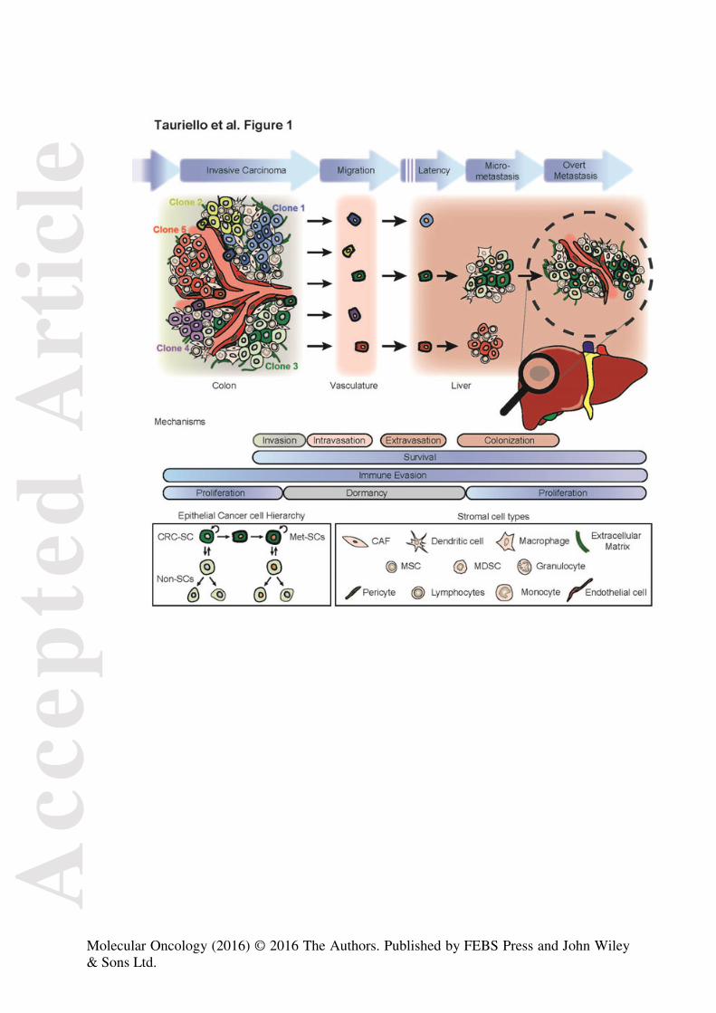
Acc
epte
d A
rtic
le
Molecular Oncology (2016) © 2016 The Authors. Published by FEBS Press and John Wiley & Sons Ltd.

Acc
epte
d A
rtic
le
Molecular Oncology (2016) © 2016 The Authors. Published by FEBS Press and John Wiley & Sons Ltd.

Acc
epte
d A
rtic
le
Molecular Oncology (2016) © 2016 The Authors. Published by FEBS Press and John Wiley & Sons Ltd.