determination of sex - san diego miramar...
TRANSCRIPT

1
Dead Men DO Tell Tales:An Introduction to Determining
Age and Sex of the Human Skeleton
Based on a lecture byValerie Dean O’Loughlin, Ph.D.Assistant Professor of Anatomy
Indiana UniversityPresented at HAPS 2002
Determination of Sex
• Male and female human skeletons differ both ingeneral shape and size. However, patterns ofsexual dimorphism can vary among groups.
• For example, male Asian skeletal remains can beless robust than female Native American skeletalremains.
• Thus, one cannot use size as the only factor todetermine sex. The most reliable indicator of sexis the os coxae (hip bones), followed by the skull.
Tips to keep in mind
• Multiple features on each bone can be used to determine sex.
• It is common for some of these features to appear more “male” and others“female” – if this happens, sex is determined by the greatest number offeatures. For example, if the skull demonstrates 4 male-like traits, and 7female-like traits, then you would classify the skull as females.
• It is difficult to impossible to determine the sex of infant and juvenileremains. Most infant/juvenile remains appear “female-like” until wellafter puberty.
• Sex determination should be made with respect to the generalsize/robusticity patterns of the population from which the skeletal remainscame.
• Like reading an EKG, determining sex of a skeleton is an art. It takesgreat practice to precisely determine sex.
Os Coxae Feature Male Characteristic Female CharacteristicSuperior Inlet Heart shaped Spacious, wide and ovalGeneral Size More robust and muscle-marked Less robust
Obturator Foramen Larger and oval Smaller and triangular
Acetabulum Larger, directed more forward/Anteriorly
Smaller, directed moreLaterally
Greater Sciatic Notch Narrow and deep Wide and shallow
Body of Pubis Short, triangular Longer, more rectangular
Subpubic Angle(area underneath the twopubic bones)
Narrow, V-shaped Broader, more convex
Preauricular Sulcus(depression between greatersciatic notch andsacroiliac articulation)
Usually absent Usually present
Sex Differences in Os Coxae
2
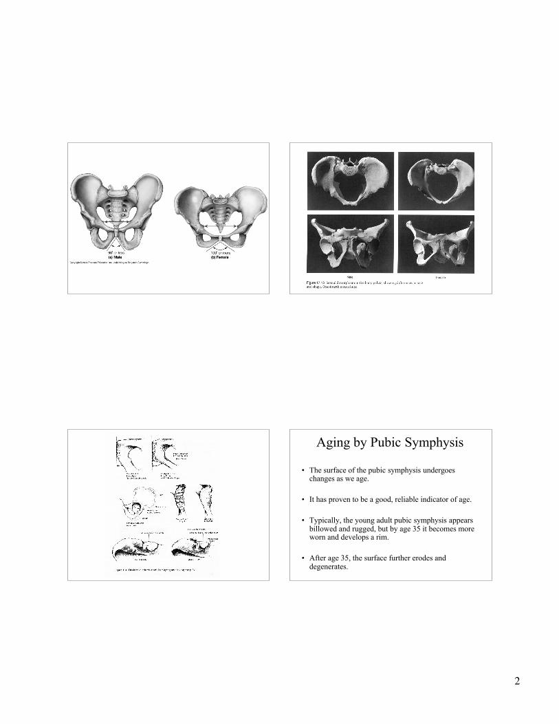
Aging by Pubic Symphysis
• The surface of the pubic symphysis undergoeschanges as we age.
• It has proven to be a good, reliable indicator of age.
• Typically, the young adult pubic symphysis appearsbillowed and rugged, but by age 35 it becomes moreworn and develops a rim.
• After age 35, the surface further erodes anddegenerates.

3
Skull Feature Male Characteristic Female Characteristic
General size More robust More gracile/delicate
Nuchal Crest(prominence on back ofskull, in occipital region)
Well-demarcated nuchallines and a prominent bumpor “hook”
External surface of occipitalbone is smooth, with nobony projections here
Mastoid Process Large, projects below theexternal auditory canal
Smaller
Supra-orbital margin(upper orbit rim)
Thick, rounded, bluntborder
Thin, sharp border
Supra-orbital ridge(“brow ridges”)
Prominent Little or no prominence
Mental Eminence(chin)
Squarish, greater forwardprojection
More pointed (versussquarish), little forwardprojection
Gonial Angle(side of jaw angle)
Flared, Less obtuse, <125degrees (typically, about 90degrees)
Typically > 125 degrees
Sex Differences in the Skull

4
Determination of Age• There are many methods to determine the age at death
of a skeleton.
• Some of these methods work best forjuvenile/immature remains, whereas others may workbest for adult skeletal remains.
• Classes commonly used for skeletal remains:1) fetal (before birth), 2) infant (birth – 3 yrs),3) child (4-12 yrs), 4) adolescent (13-19 yrs),5) young adult (20-34 yrs), 6) middle adult (35-49 yrs),7) old adult (50+ yrs)
Aging by the Skull• After the os coxae, the skull (cranium plus
mandible) is the next most reliable structure fromwhich to determine the sex.
• The skull exhibits a varying degree of sexualdimorphism.
• However, this dimorphism can vary frompopulation to population.
• Thus, one should realize that sex determinationfrom the skull is dependent upon the population.
Aging by Cranial Suture Closure• Cranial sutures fuse progressively as one ages.
• Typically, the anteriorly-placed sutures (coronal) fusefirst, followed by the more posteriorly-placed sutures(i.e., sagittal and lambdoidal, respectively).
• However, there is considerable variability in closurerates, so this aging method should be used inconjunction with another methods.
• One cranial feature that has a high reliability rate is thespheno-occipital synchondrosis (at the base of theskull), which fuses between 20 and 25 years for over95% of populations studied.
5
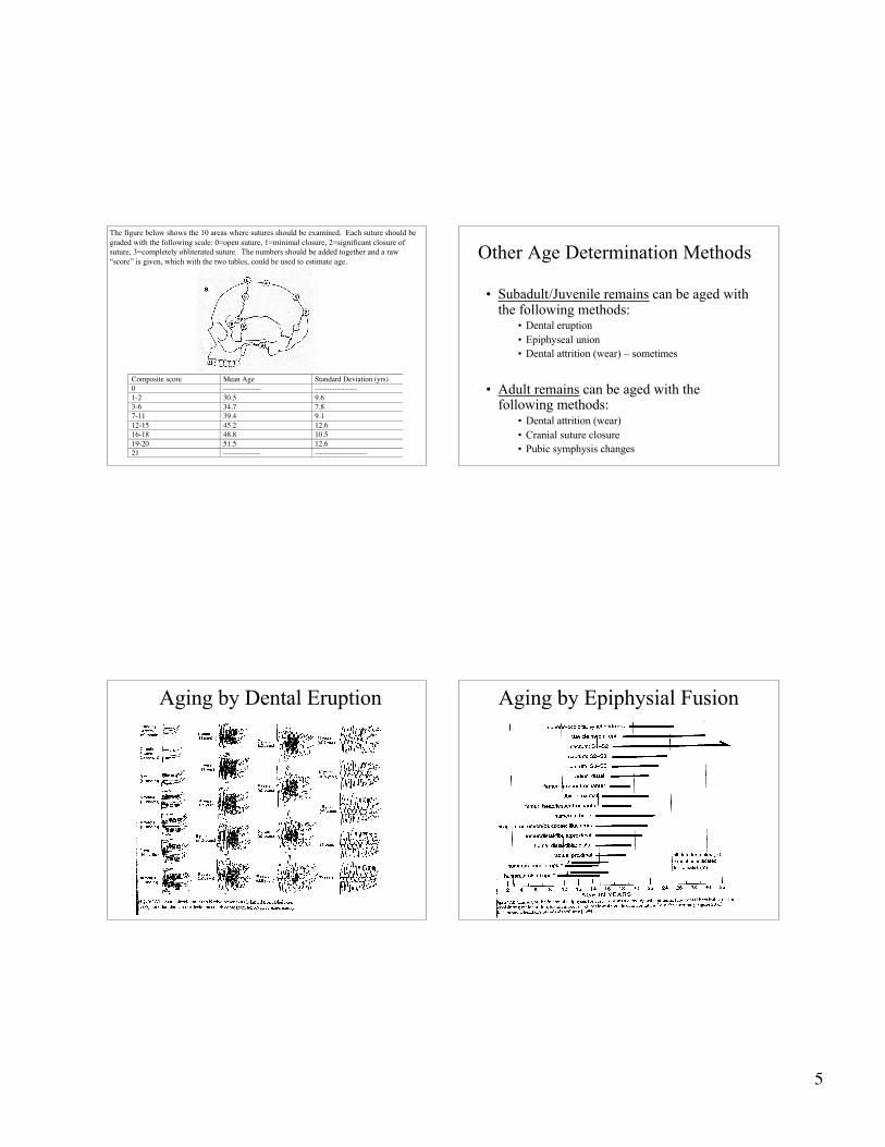
Composite score Mean Age Standard Deviation (yrs)0 --------------- -----------------1-2 30.5 9.63-6 34.7 7.87-11 39.4 9.112-15 45.2 12.616-18 48.8 10.519-20 51.5 12.621 --------------- ---------------------
The figure below shows the 10 areas where sutures should be examined. Each suture should begraded with the following scale: 0=open suture, 1=minimal closure, 2=significant closure ofsuture, 3=completely obliterated suture. The numbers should be added together and a raw“score” is given, which with the two tables, could be used to estimate age.
Other Age Determination Methods
• Subadult/Juvenile remains can be aged withthe following methods:
• Dental eruption• Epiphyseal union• Dental attrition (wear) – sometimes
• Adult remains can be aged with thefollowing methods:
• Dental attrition (wear)• Cranial suture closure• Pubic symphysis changes
Aging by Dental Eruption Aging by Epiphysial Fusion
6
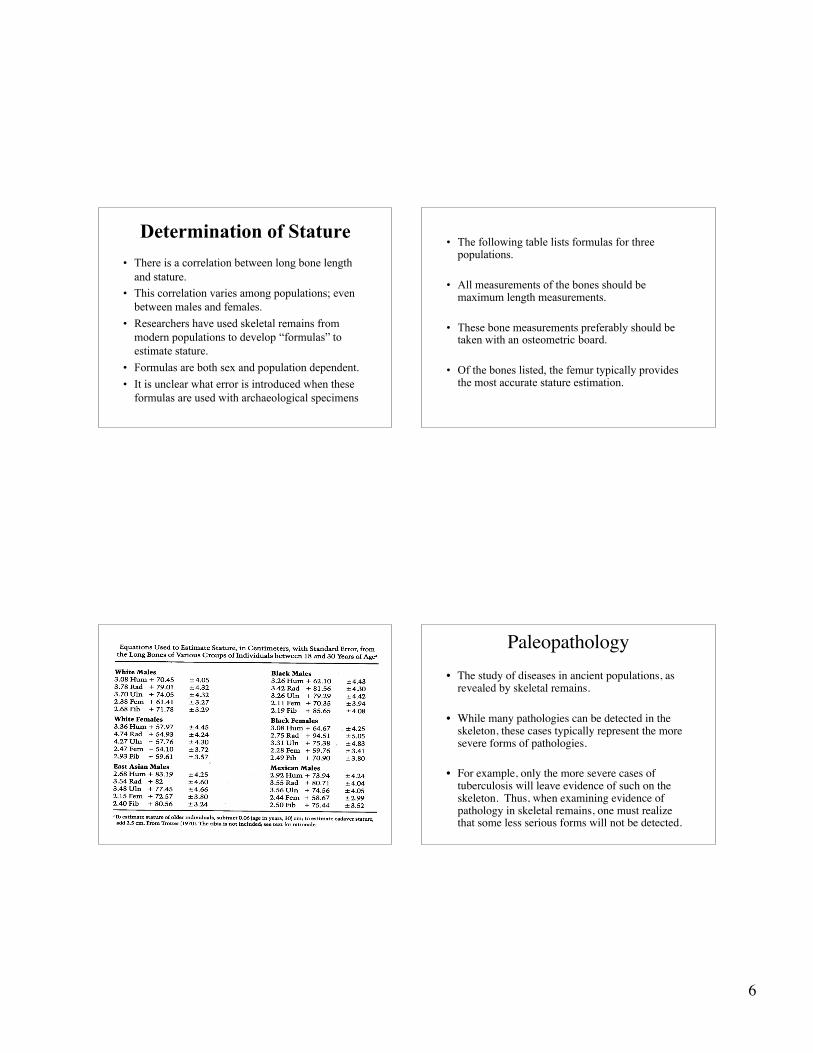
Determination of Stature
• There is a correlation between long bone lengthand stature.
• This correlation varies among populations; evenbetween males and females.
• Researchers have used skeletal remains frommodern populations to develop “formulas” toestimate stature.
• Formulas are both sex and population dependent.
• It is unclear what error is introduced when theseformulas are used with archaeological specimens
• The following table lists formulas for threepopulations.
• All measurements of the bones should bemaximum length measurements.
• These bone measurements preferably should betaken with an osteometric board.
• Of the bones listed, the femur typically providesthe most accurate stature estimation.
Paleopathology• The study of diseases in ancient populations, as
revealed by skeletal remains.
• While many pathologies can be detected in theskeleton, these cases typically represent the moresevere forms of pathologies.
• For example, only the more severe cases oftuberculosis will leave evidence of such on theskeleton. Thus, when examining evidence ofpathology in skeletal remains, one must realizethat some less serious forms will not be detected.
7
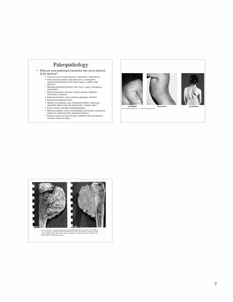
Paleopathology• What are some pathologies/anomalies that can be detected
in the skeleton?• Trauma (recent or healed fractures, dislocations, subluxations)• Some infectious diseases, like tuberculosis, osteomyelitis
(infection/inflammation of the bone), leprosy, syphilis, otherinfections
• Metabolic/nutritional disorders, like scurvy, rickets, osteoporosis,osteomalacia
• Blood (hemopoietic) disorders, such as anemias, leukemias(sometimes), myelomas
• Endocrine disorders, such as pituitary gigantism, dwarfism• Benign and malignant tumors• Arthritis (osteoarthritis, gout, rheumatoid arthritis, ankylosing
spondylitis which causes the characteristic ‘bamboo spine’)• Caries (cavities) and other dental pathologies• Skeletal dysplasias, such as achondroplasia (dwarfism), osteogenesis
imperfecta (characterized by numerous fractures)• Scoliosis (lateral curvature of spine), kyphosis (increased anterior
curvature in thoracic spine)