determination of the volume of the thyroid gland by a high resolutional ultrasonic scanner
TRANSCRIPT

surgery by the new apparatus were compared to theweights of the resected thyroid glands or nodules.
PATIENTS AND METHODS
PatientsStudies were performed on 100 patients with thyroid dis
orders (67 patients with Graves' disease and 33 patients withthyroid adenoma) to estimate the accuracy of the volumes ofthe thyroid glands and nodules by the ultrasonic scanner.Ultrasonic scanningofthe thyroid in these patients was carriedout prior to operation. The weights of the Graves' thyroidswere calculated by adding the surgeons' estimates ofwhat hadbeen left, to the actual weights of the thyroids which weresurgically removed. Thyroid adenomas were weighed afterremoving tissues surrounding them. The echography of thethyroid, the determination ofthyroid volumes from echogram,and the surgical measurement ofthyroid weights were carriedout by different doctors independently. Both results werecompared and analyzed at the end ofthe study.
Normal subjects were 57 healthy volutiteers (1 1 males and46 females, 37-74 yr). None of them had signs or symptomsof thyroid disorders and none had a history of thyroid disorders. Their serum levels of T3, T4, free T4, and TSH werewithin the normal range.
Volume 27 •Number 9 •September1986 1475
TechnicalNotes
Determination of the Volumeof the Thyroid Gland by a High ResolutionalUltrasonic ScannerN. Yokoyama, Y. Nagayama, F. Kakezono, T. Kiriyama, S. Morita, S. Ohtakara,S. Okamoto, I. Morimoto, M. Izumi, N. Ishikawa, K. Ito, and S. Nagataki
First Department oflnternal Medicine, Nagasaki University School ofMedicine, Nagasaki;and Ito Hospital, Tokyo, Japan
We developeda new ultrasonicscannerfor the thyroidand, in thisstudy,the estimatedvolumes of the thyrods by this scanner were compared with the weights of those Obtainedatoperation. In this ultrasonic scanner, an annular array transducer was employed instead of theconventional single element concave transducer. The distance of the focused area by thistransducer was as long as 5 cm compared to 1 cm by the conventional transducer; therefore,the image obtained by the new scanner was so clear that it was not difficult to drawaccurately the outlines of the [email protected] volumes of the thyroids were calculated by acomputerized d@zer. The estimated volumes of the thyroids by the uftrasonic scanner weredosely correlated with their weights calculated by adding the actual wsights of the thyr@dsremoved to the estimates of the thyroids left at operation. Their correlation coefficients wereas highas 0.99. This suggeststhat thisnew ultrasonicscanneris very usefulin thedeterminationof the volumesof the thyroids,sincethe measurementis veryaccurate,simple,and reproducible.
J NucI Med 27:1475—1479,1986
he accurate estimation of the size of the thyroid isvery important for the evaluation and management ofthyroid disorders (1,2). Changes in goiter size withtreatment is still one of the best indicators for theprognosis ofGraves' disease in spite ofthe developmentof many in vitro methods to predict the outcome of thedisease (3). The accurate estimation ofgoiter size is alsoessential in radioiodine therapy ofGraves' disease.
Although many methods for estimating thyroid sizehave been developed, some thyroid experts, especiallysurgeons, still believe their fingers are more accuratethan any instrument. Even in well-known journals, theexpressions of thyroid size have been quite differentaccording to various investigators (4—6).
We have developed a new ultrasonic apparatus forthe estimation of the size of the thyroid using a transducer ofthe Aloka Co. Ltd., Japan. In the present study,the estimated volumes ofthe thyroids obtained prior to
Received Oct. 3, 1985; revision accepted Mar. 21, 1986.For reprints contact: Shigenobu Nagataki, MD, First Depart
ment ofinternal Medicine, Nagasaki University School of Mcdicine, Nagasaki852, Japan.
by on April 3, 2019. For personal use only. jnm.snmjournals.org Downloaded from

$c.,v@ Ii•sd arch scannIng
\J @Li@O@@O
—— — : s'@gi. elem,@ conce@ed tT@nsduc*' (7561
:EIect@onLc@IIv focused bee., of s@'nuIor6U@4 WIDTH@ t104'S4uCST
1T@
/
1,2,5mmNAamiular array •@ .. -
probe ::.
water-bath -
( 2 sec. for a arch)
3) Annular array probe
T @.o@ biswc!@ (cm)
4-@ —Focal Zone
( 5 cm)
FIGURE 1Mechanism of ultrasonic scannerwith 7.5 MHz annular array transducer
MethodsUltrasonic apparatus. The new ultrasonic apparatus and
the computerized digitizer were developed recently for thisstudy. Our new ultrasonic apparatus was composed of anultrasonic scanner with a new transducer, an echo camera,and a computerized multiphoto camera. A computerized multiphoto camera' was connected to an echo camera to recordthe images of the cross-sections of the thyroid automatically.Aloka SSD-270 as an echo camera and Aloka ASU-46 as anultrasonic scanner were employed. Figure 1 shows the mechanism of the ultrasonic scanner in detail. In this ultrasonicscanner,a25-mmdiameter7.5MHzannulararraytransducerwas employed instead of the conventional single elementconcave transducer. The performance ofthe fixed focus transducer was optimized for both sensitivity and resolution in asmall region about the focal point. An annular array wasformed by creating five concentric rings of transducer materials that were electrically isolated from one another. Eacharray element had its own electronic amplifiers. A dynamicfocusing control system combined signals from each arrayelement creating an electronically variable lens. Therefore, thedistance of focused area by this transducer was as long as 5cm compared with 1 cm by the conventional transducer. Thewide focused area of this new transducer was very importantin obtaining clear images of the thyroid.
Procedure of echography. Patients were examined in thesupine position, with the neck hyperextended. The transducermoved over the neck transversally 140 mm in the water bath,
making an arch with the speed of 2 sec per arch for the echoangle of incidence to the thyroid to be vertical, yielding animage of a cross-section of the thyroid. After making eachimage of a cross-section, the transducer moved automaticallyeither 1, 2, or 5 mm longitudinally to produce many serialcross-sectionsofthethyroidswithvarioussizesandshapes.Inmost patients, all images could be recorded on one film.
Measurement ofihe volume ofihe thyroid. A computerizeddigitizer was used to calculate the volume ofthe thyroid fromthe images of serial cross-sections. At first, the outline of thethyroid gland on the film of each cross-section was tracedmanually with a white marker. Then, the outlines of thethyroid on every cross-section were traced on the computerized digitizer and the computer calculated the volume of thethyroid by multiplying the traced areas of the thyroid by agiven thickness such as 1, 2, or 5 mm. The thickness waschanged according to the size ofthe thyroid gland or adenoma.Usually the thickness was determined to produce at least eightcross-sections of thyroid glands or adenomas, but the maximum thickness was 5 mm.
RESULTS
Figure 2 shows an image of a cross-section of a normalthyroid recorded on film and its scheme. The image ofa crosssectionbythisscannershowedexcellentresolution(7—11).The skin, thyroid, common carotid artery, internal jugular
1476 Yokoyama,Nagayama,Kakezonoetal TheJournalof NuclearMedicine
1) Scanner 2) ScannIng m.thod
f@100;:.@
_/@k—l4Omm—)(
@jJtAnnulararray transducer
( 7.5MHz)
by on April 3, 2019. For personal use only. jnm.snmjournals.org Downloaded from

FIGURE 2Imageof cross-sectionof normalthyroidandits scheme
vein, sternocleidmastoid muscle, and longus colli musclecould be so clearly discriminated that it was easy to draw theoutline of the thyroid.
Figure 3A shows an image ofthe cross-section ofa Graves'thyroid. In this enlarged thyroid. the margins were clear andthe echo density was homogeneous. Figure 3B shows an imageof the cross-section of a thyroid adenoma.
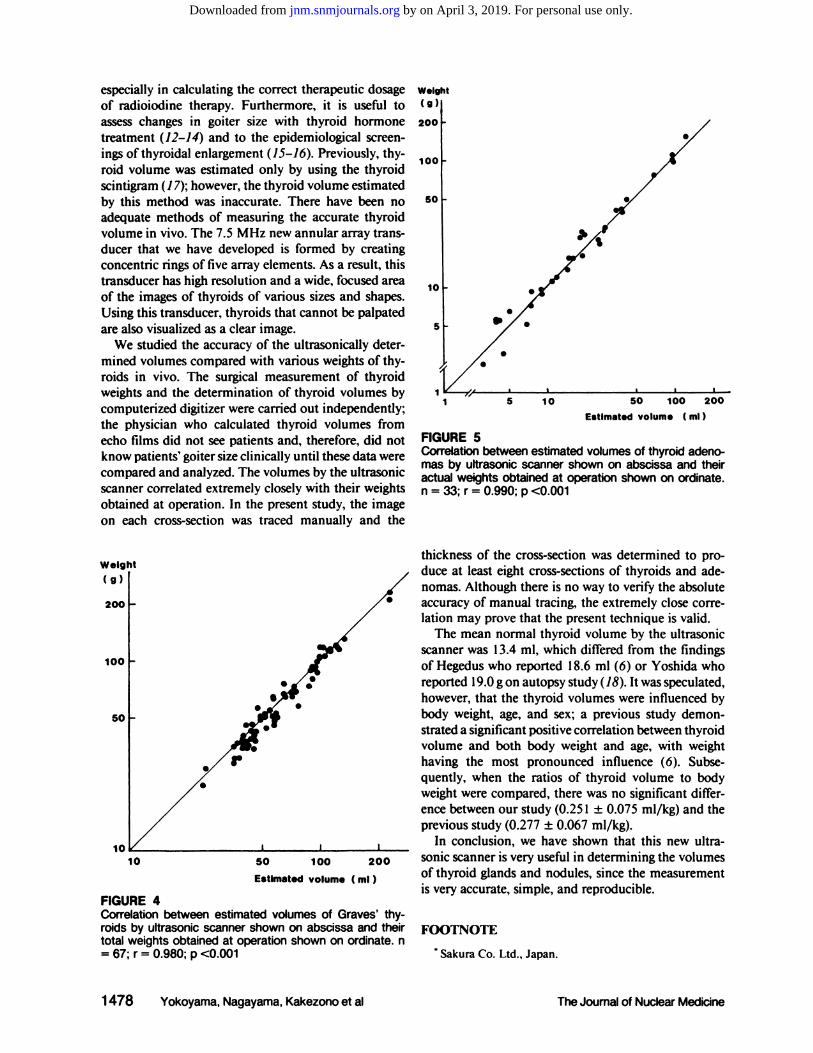
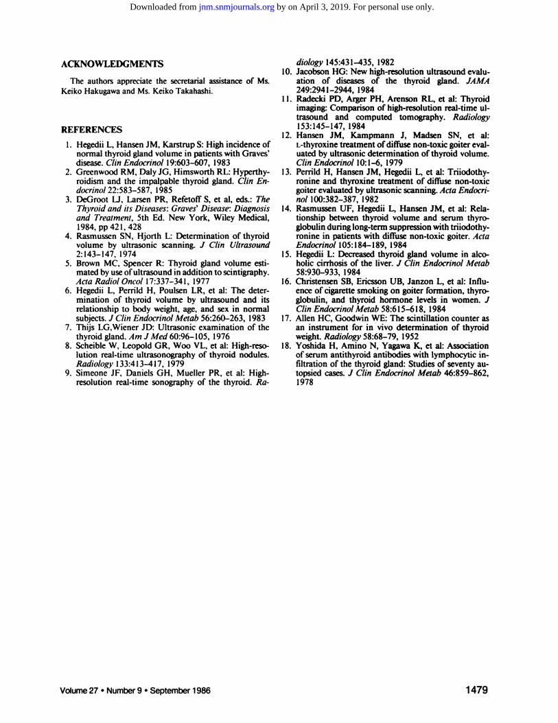
In 100operated patients, the comparison between ultrasonically determined volume and weight measured at operationwas studied. Figure 4 shows the comparison between estimatedvolumes of Graves' thyroids and their total weights by logarithmic scales. In 67 Graves' patients, thyroid weights rangedfrom 27—221g. The estimated volumes by ultrasonic scanningprior to operation correlated very closely with their totalweights obtained at operation. A close correlation was foundfrom thyroids as small as 25 g to those >200 g. The correlationcoefficient was +0.980 (p <0.001, n = 67). In the estimationof the volumes of Graves' thyroids it was important not tomiss the top of each lobe and the possible presence of apyramidal lobe. Figure 5 shows the comparison between theestimated volumes of adenomas and their actual weights. In33 thyroid adenomas, their actual weights ranged from 3—146g. The estimated volumes of adenomas correlated extremely
closely with their actual weights obtained at operation. Fromthe size ofseveral grams to >100 g, the difference between theestimated and actual values were <5% except one patient.The correlationcoefficientwas as high as +0.990 (p <0.001,n= 33). Since these results suggested that the volumes of thethyroids obtained by this technique were very close to theactual weights, the thyroid volumes of the normal subjectswere also measured by this technique. These normal subjectshad a mean (±s.d.) thyroid volume of 13.4 ±4. 1 ml (range,5.9—22.9ml), a mean (±s.d.) body mass of 53.4 ±8.2 kg(range, 40.2—77.4kg), and a mean age of 53.2 yr. The thyroidvolume to body weight ratio was 0.251 ±0.075 mI/kg.
DISCUSSION
In this study of 100 operated thyroid disorders, wehave shown that determination of the volumes of thyroids by a new ultrasonic scanner was extremely accurate.
The accurate estimation ofthyroid volume is considered of value in the assessment of Graves' patients,
FIGURE 3Cross-section images of various thyroid diseases. (A) Graves' disease. (B) Thyroid adenoma
Volume27 •Number9 •September1986 1477
Thyroid
Sterno cleldomastoid M.
Trachea
Longus colIl M.
by on April 3, 2019. For personal use only. jnm.snmjournals.org Downloaded from
especially in calculating the correct therapeutic dosageof radioiodine therapy. Furthermore, it is useful toassess changes in goiter size with thyroid hormone
treatment (12—14) and to the epidemiological screen
ings of thyroidal enlargement (15—16).Previously, thyroid volume was estimated only by using the thyroidscintigram (1 7); however, the thyroid volume estimatedby this method was inaccurate. There have been noadequate methods of measuring the accurate thyroidvolume in vivo. The 7.5 MHz new annular array transducer that we have developed is formed by creatingconcentric rings of five array elements. As a result, thistransducer has high resolution and a wide, focused areaof the images of thyroids of various sizes and shapes.Using this transducer, thyroids that cannot be palpatedare also visualized as a clear image.
We studied the accuracy of the ultrasonically determined volumes compared with various weights of thyroids in vivo. The surgical measurement of thyroidweights and the determination of thyroid volumes bycomputerized digitizer were carried out independently;the physician who calculated thyroid volumes fromecho films did not see patients and, therefore, did notknow patients' goiter size clinically until these data werecompared and analyzed. The volumes by the ultrasonicscanner correlated extremely closely with their weightsobtained at operation. In the present study, the imageon each cross-section was traced manually and the
Weight
10
5
I50 100 200
Estimated volume ( ml)
I 5 10
FIGURE 5Correlation between estimated volumes of thyroid adenomas by ultrasonicscannershown on abscissaand theiractual weights obtained at operation shown on ordinate.n = 33; r = 0.990; p <0.001
thickness of the cross-section was determined to produce at least eight cross-sections of thyroids and adenomas. Although there is no way to verify the absoluteaccuracy of manual tracing, the extremely close correlation may prove that the present technique is valid.
The mean normal thyroid volume by the ultrasonicscanner was 13.4 ml, which differed from the findingsof Hegedus who reported 18.6 ml (6) or Yoshida whoreported 19.0 g on autopsy study (18). It was speculated,however, that the thyroid volumes were influenced bybody weight, age, and sex; a previous study demonstrated a significant positive correlation between thyroidvolume and both body weight and age, with weighthaving the most pronounced influence (6). Subsequently, when the ratios of thyroid volume to bodyweight were compared, there was no significant difference between our study (0.25 1 ±0.075 ml/kg) and theprevious study (0.277 ±0.067 ml/kg).
In conclusion, we have shown that this new ultrasonic scanner is very useful in determining the volumesof thyroid glands and nodules, since the measurementis very accurate, simple, and reproducible.
FOOTNOTE
. Sakura Co. Ltd., Japan.
(9)
200
100
50
10 50 100 200
Estimated volume ( ml)
FIGURE 4Correlation between estimated volumes of Graves' thyroids by ultrasonicscannershownon abscissaand theirtotal weights obtained at operation shown on ordinate. n= 67; r = 0.980; p <0.001
1478 Yokoyama,Nagayama,Kakezonoetal The Journal of Nuclear Medicine
by on April 3, 2019. For personal use only. jnm.snmjournals.org Downloaded from
diology 145:431—435,198210. Jacobson HG: New high-resolution ultrasound evalu
ation of diseases of the thyroid gland. JAMA249:2941—2944, 1984
I I . Radecki PD, Arger PH, Arenson RL, Ct al: Thyroidimaging: Comparison of high-resolution real-time ultrasound and computed tomography. Radiology153:145—147,1984
12. Hansen JM, Kampmann J, Madsen SN, et al:L-thyroxine treatment ofdiffuse non-toxic goiter evaluated by ultrasonic determination of thyroid volume.C/in Endocrinol 10:1—6,1979
13. Perrild H, Hansen JM, Hegedii L, et al: Triiodothyronine and thyroxine treatment of diffuse non-toxicgoiter evaluated by ultrasonic scanning. Ada Endocrinol 100:382—387,1982
14. Rasmussen UF, Hegedii L, Hansen JM, et al: Relationship between thyroid volume and serum thyroglobulin duringlong-term suppression with triiodothyronine in patients with diffuse non-toxic goiter. ActaEndocrinol105:184—189,1984
15. Hegedii L: Decreased thyroid gland volume in alcoholic cirrhosis of the liver. J Cl/n Endocrinol Metab58:930—933,1984
16. Christensen SB, Ericsson UB, Janzon L, Ct al: Influenceofcigarettesmokingongoiterformation,thyroglobulin, and thyroid hormone levels in women. JC/in Endocrinol Metab 58:6 15—618, 1984
17. Allen HC, Goodwin WE: The scintillation counter asan instrument for in vivo determination of thyroidweight. Radiology 58:68—79,1952
18. Yoshida H, Amino N, Yagawa K, et al: Associationof serum antithyroid antibodies with lymphocytic infiltration of the thyroid gland: Studies of seventy autopsied cases. J C/in Endocrinol Mezab 46:859—862,1978
ACKNOWLEDGMENTS
The authors appreciate the secretarial assistance of Ms.Keiko Hakugawa and Ms. Keiko Takahashi.
REFERENCES
1. Hegedii L, Hansen JM, Karstrup 5: High incidence ofnormal thyroid gland volume in patients with Graves'disease. Clin Endocrinol 19:603—607,1983
2. Greenwood RM, Daly JG, Himsworth RL: Hyperthyroidism and the impalpable thyroid gland. Clin Endocrinol22:583—587,1985
3. DeGroot U, Larsen PR, Refetoff S. et al, eds.: TheThyroid and its Diseases: Graves' Disease: Diagnosisand Treatment, 5th Ed. New York, Wiley Medical,1984,pp 421,428
4. Rasmussen SN, Hjorth L: Determination of thyroidvolume by ultrasonic scanning. J Clin Ultrasound2:143—147,1974
5. Brown MC, Spencer R: Thyroid gland volume estimated by use ofultrasound in addition to scintigraphy.ActaRadiolOncol17:337—341,1977
6. Hegedii L, Perrild H, Poulsen LR, et al: The determination of thyroid volume by ultrasound and itsrelationship to body weight, age, and sex in normalsubjects. J Clin Endocrinol Metab 56:260—263,1983
7. Thijs LG,Wiener JD: Ultrasonic examination of thethyroid gland. Am J Med 60:96—105,1976
8. Scheible W, Leopold GR, Woo VL, et al: High-resolution real-time ultrasonography of thyroid nodules.Radiology133:413—417,1979
9. Simeone JF, Daniels GH, Mueller PR, et al: Highresolution real-time sonography of the thyroid. Ra
1479Volume27 •Number9 •September1986
by on April 3, 2019. For personal use only. jnm.snmjournals.org Downloaded from

1986;27:1475-1479.J Nucl Med. Ishikawa, K. Ito and S. NagatakiN. Yokoyama, Y. Nagayama, F. Kakezono, T. Kiriyama, S. Morita, S. Ohtakara, S. Okamoto, I. Morimoto, M. Izumi, N. ScannerDetermination of the Volume of the Thyroid Gland by a High Resolutional Ultrasonic
http://jnm.snmjournals.org/content/27/9/1475This article and updated information are available at:
http://jnm.snmjournals.org/site/subscriptions/online.xhtml
Information about subscriptions to JNM can be found at:
http://jnm.snmjournals.org/site/misc/permission.xhtmlInformation about reproducing figures, tables, or other portions of this article can be found online at:
(Print ISSN: 0161-5505, Online ISSN: 2159-662X)1850 Samuel Morse Drive, Reston, VA 20190.SNMMI | Society of Nuclear Medicine and Molecular Imaging
is published monthly.The Journal of Nuclear Medicine
© Copyright 1986 SNMMI; all rights reserved.
by on April 3, 2019. For personal use only. jnm.snmjournals.org Downloaded from