development: a review of primitive streak ... publications/118.pdf12 spatiotemporal pattern...
TRANSCRIPT

1 2
SPATIOTEMPORAL PATTERN FORMATION IN EARLYDEVELOPMENT: A REVIEW OF PRIMITIVE STREAK FORMATION
AND SOMITOGENESIS
S. SCHNELL1, K.J. PAINTER2, P.K. MAINI1 AND H.G. OTHMER2
Abstract. The basic body plan of a number of vertebrates results from two processes that occurearly in the development of the blastoderm: large scale rearrangements of tissue via a process calledgastrulation, and axial subdivision of tissue in a process called somitogenesis. The £rst step of gas-trulation in avians is formation of the primitive streak, which marks the £rst clear manifestation ofthe anterior-posterior axis. Cell movements that occur through the streak ultimately convert the singlelayered-blastoderm into a trilaminar blastoderm comprising prospective endodermal, mesodermal andectodermal tissue. During streak formation a group of cells moves anteriorly as a coherent columnfrom the posterior end of the blastoderm, and as it proceeds other cells stream over the lateral edgesof the furrow left behind. The anterior end of the streak is a specialized structure called Hensen’snode, which serves as an organizing center for later axis formation and determination of the left-rightasymmetry of the body. Soon after the primitive streak forms, Hensen’s node regresses towards thetail, leaving the notochord and a pair of segmental plates parallel to the primitive streak in its wake.The posterior end of the segmental plate moves down the cranio-caudal axis with the node, as morecells are added to it by cell division within the plate and by cells entering from the primitive streak.A pair of somites forms from the anterior ends of the two plates at regular intervals. Despite the factthat much is known about the basic biological processes, the mechanisms that underlie the formationof the primitive streak and somitogenesis are still unknown, and elucidating them is one of the majorunsolved problems in developmental biology. Mathematical modelling has been a useful tool in thisprocess, as it provides a framework in which to study the outcome of proposed interactions and canmake experimentally testable predictions. In this paper we outline the biological background of theseprocesses and review existing models of them.
Key words. Primitive streak formation, somitogenesis, theoretical models, mathematical models,Hoxgenes,c-hairy-1, Notch-Delta genes
1. Introduction. Early vertebrate development is a complex process thatinvolves cell division, cell-cell signaling, cell movement, and cell differentiation.Many adult vertebrates exhibit common structures, but the developmental pro-cesses that produce them may or may not be similar. For example, formation ofa primitive streak is central to avian, reptilian and mammalian gastrulation, andwhile it is not present in amphibian blastulae, they contain an analogous structure,called the blastopore. On the other hand, somitogenesis is common to all verte-brates. This review focuses on experimental and theoretical aspects of primitivestreak formation and somitogenesis in avian embryogenesis. The chick embryois a widely-used model system for experimental studies and, as a result, there is alarge amount of experimental data. We begin with a brief description of the earlyevents: details of these events can be found in [35], [88], and [50].
The chick embryo develops from a small, disk-shaped blastodisc ¤oatingon top of the yolk. After the egg is fertilized cells divide repeatedly, forming a
1Centre for Mathematical Biology, Mathematical Institute, Oxford University, Oxford, OX1 3LB,UK.
2Department of Mathematics, University of Minnesota,Minneapolis, MN, USA
1

2 S. SCHNELL and P. K. MAINI and K.J. PAINTER and H. G. OTHMER
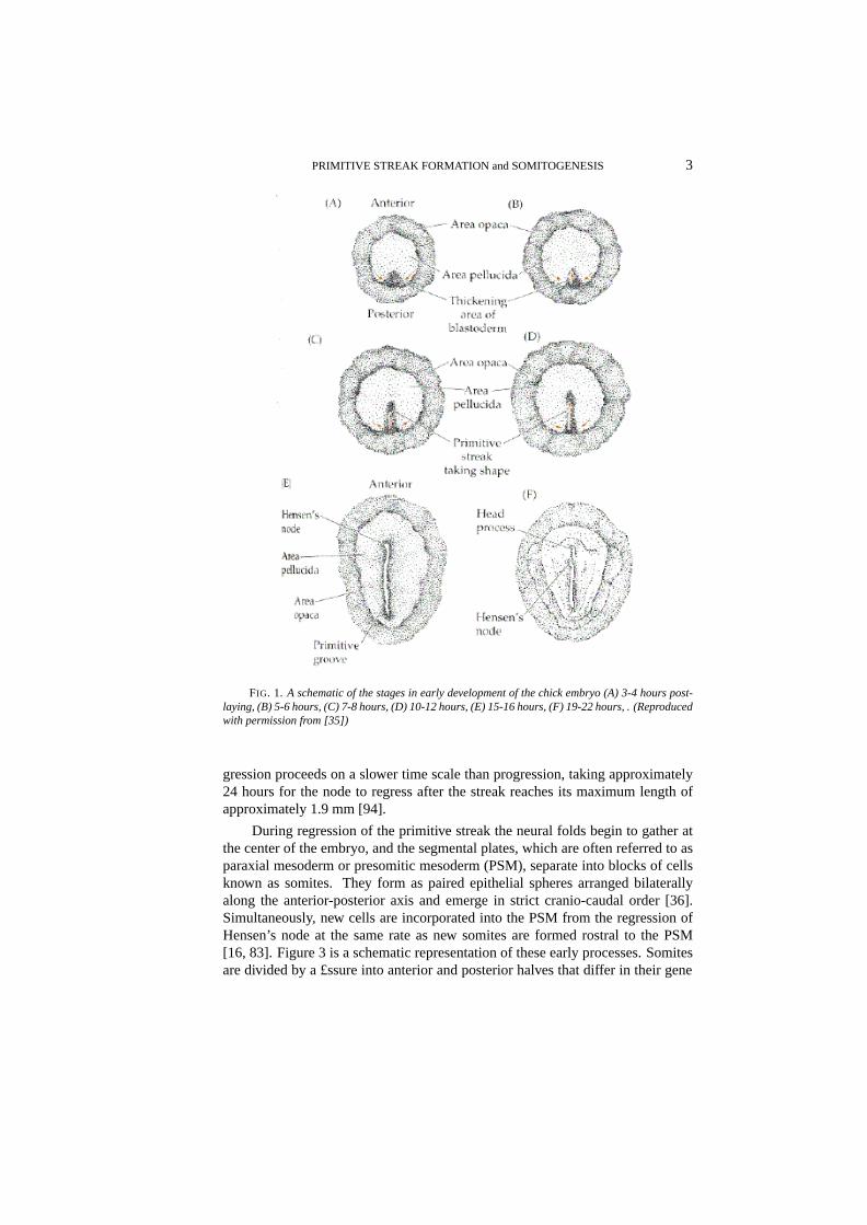
multicellular strati£ed structure called the blastoderm. The period from just priorto laying through several hours afterwards has been subdivided into 14 stages[31, 50]. Cell division is dominant during stages I - VI, and morphogenetic move-ments begin during stages VII-X, when cells of the central blastodisc, called thearea pellucida(cf. Figure 1), separate from the yolk, producing a hollow re-gion beneath the disc called the subgerminal cavity [75, 99]. Subsequently somecells from the central blastodisc move into the subgerminal cavity (either activelyor passively), and simultaneously the disc expands radially over the yolk. Theopaque marginal zone of the blastoderm, known as thearea opaca, remains incontact with the yolk and may play an active role in the radial movement (Figure1 A). The result is that during stages VII-X the central part of the disc changesfrom a layer 4-6 cells deep to a translucent layer one cell thick called the epi-blast. The anterior-posterior axis of the embryo is also determined during thesestages [50]. After stage X some cells within the marginal zone migrate posteriorly,and then leave the marginal zone at the posterior marginal zone (PMZ)(Figure 1B). They spread across the subgerminal cavity beneath the epiblast as a loosely-connected sheet, incorporating islands of cells shed from the blastodisc earlier.By stage XIV this sheet connects with the anterior margin of the disc and formsthe hypoblast, and at this stage the blastoderm is bi-layered with the epiblast andhypoblast separated by the blastocoel cavity. Fate maps for cell movements inthese stages are available [39].
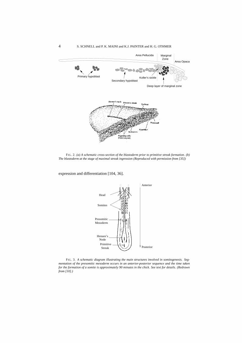
During hypoblast formation the embryonic shield or Koller’s sickle developsat the posterior end of the epiblast (cf Figure 2(a)). This consists of a thickenedepiblast [93] comprising primitive streak precursor cells that have migrated to thisarea by a series of ‘polonnaise movements’ [105]. The £rst visible sign of gastru-lation is formation of the primitive streak, which arises from Koller’s sickle at theposterior midline of the blastodisc [52] (Figure 1 C and D). The sickle narrowsand the primitive streak moves anteriorly between the epiblast and the hypoblast.The tip of the ingressing streak moves∼ 60% of the way across the blastodermbefore it stops, and later, regresses. At full primitive streak stage (Hamburgerand Hamilton stage 4, [38]) the organizer of the avian embryo, Hensen’s node,develops as a bulbous structure at the anterior tip of the streak. The period be-tween the accumulation of cells at the posterior region and full primitive streakis approximately 12 hours. The structure of the blastoderm at this stage is illus-trated in Figure 2(b). During the advance of the node, epiblast cells move throughthe streak and into the interior. Those that migrate through the node form ante-rior structures, those that migrate through the lateral parts of the primitive streakbecome endodermal and mesodermal cells, and the remainder constitute the ecto-derm. Simultaneously, thearea pellucidachanges from circular to pear-shaped,narrowing in the posterior portion. The head structure, notochord and somites arelaid down during regression of the node, and when regression is complete the em-bryo is a ¤at trilaminar blastoderm comprising the ectodermal, mesodermal andendodermal layers. These will form various organs during subsequent morpho-genesis, in addition to the structures formed during regression. The regressingnode and anterior portion of the streak eventually form the tail bud [94]. Re-
PRIMITIVE STREAK FORMATION and SOMITOGENESIS 3
FIG. 1. A schematic of the stages in early development of the chick embryo (A) 3-4 hours post-laying, (B) 5-6 hours, (C) 7-8 hours, (D) 10-12 hours, (E) 15-16 hours, (F) 19-22 hours, . (Reproducedwith permission from [35])
gression proceeds on a slower time scale than progression, taking approximately24 hours for the node to regress after the streak reaches its maximum length ofapproximately 1.9 mm [94].
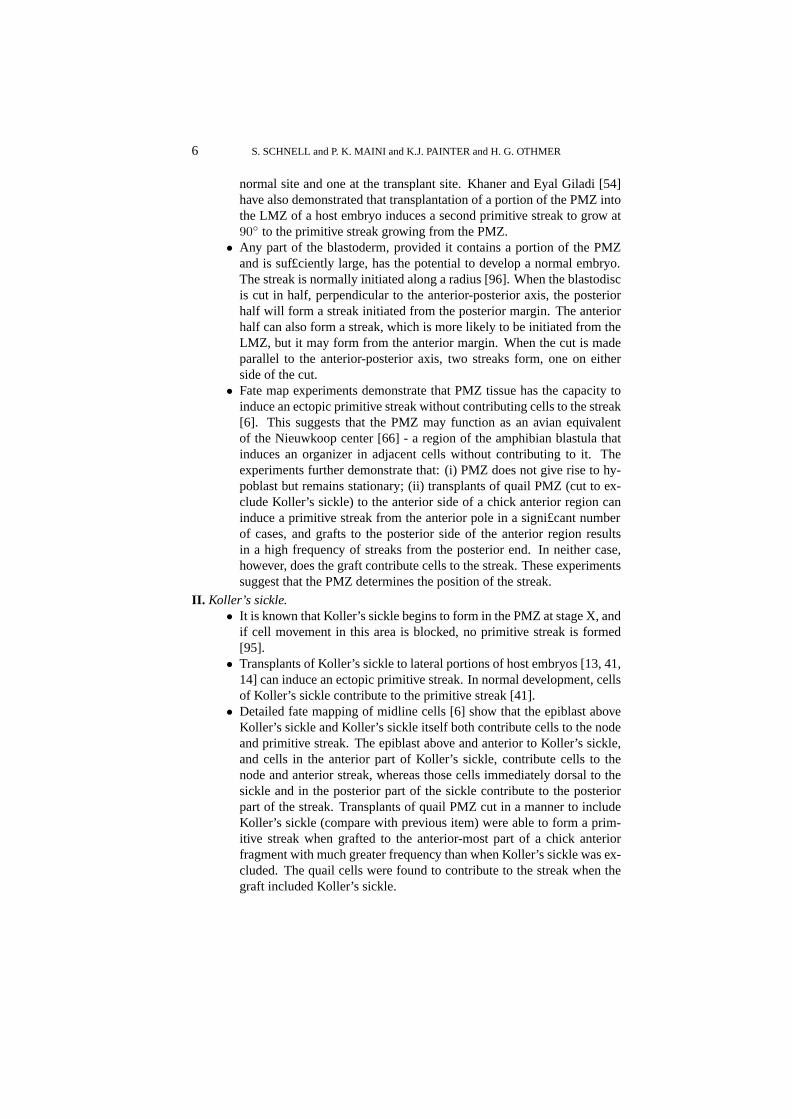
During regression of the primitive streak the neural folds begin to gather atthe center of the embryo, and the segmental plates, which are often referred to asparaxial mesoderm or presomitic mesoderm (PSM), separate into blocks of cellsknown as somites. They form as paired epithelial spheres arranged bilaterallyalong the anterior-posterior axis and emerge in strict cranio-caudal order [36].Simultaneously, new cells are incorporated into the PSM from the regression ofHensen’s node at the same rate as new somites are formed rostral to the PSM[16, 83]. Figure 3 is a schematic representation of these early processes. Somitesare divided by a £ssure into anterior and posterior halves that differ in their gene
4 S. SCHNELL and P. K. MAINI and K.J. PAINTER and H. G. OTHMER
Primary hypoblast
Area Pellucida
Secondary hypoblastKoller’s sickle
Deep layer of marginal zone
Area Opaca
MarginalZone
FIG. 2. (a) A schematic cross-section of the blastoderm prior to primitive streak formation. (b)The blastoderm at the stage of maximal streak ingression (Reproduced with permission from [35])
expression and differentiation [104, 36].
Hensen’sNode
Head
Somites
Anterior
Posterior
PresomiticMesoderm
PrimitiveStreak
FIG. 3. A schematic diagram illustrating the main structures involved in somitogenesis. Seg-mentation of the presomitic mesoderm occurs in an anterior-posterior sequence and the time takenfor the formation of a somite is approximately 90 minutes in the chick. See text for details. (Redrawnfrom [10].)

PRIMITIVE STREAK FORMATION and SOMITOGENESIS 5
The formation and differentiation of somites is the result of three distinctmorphological events progressing in a strict spatio-temporal order: (1) the prepat-terning of the PSM; (2) somite and somitic boundary formation; and (3) the differ-entiation of a somite into anterior and posterior halves [36]. Several experimentalobservations con£rm these events. Scanning electron microscopy observations[42] and transplantation experiments [49] show that the PSM displays a prepatternprior to segmentation. In addition,Hox and Notch-Delta pathway genes are in-volved in all these events [104, 25]. These molecular results suggest the existenceof a conserved mechanism for segmentation in protostomes and deuterostomes[61].
The segmental pattern of somites in turn governs the segmental pattern of theperipheral nervous system and determines the shapes and appendage characteris-tics of the vertebrae. Somites are also the source of cells for muscles, and in¤u-ence the metameric distribution of blood vessels. Genetic or/and environmentalfactors disturbing somitogenesis produce malformations and abnormal develop-ment [117, 27, 36].
Although the sequence of events in early avian development is well docu-mented, less is known about the mechanisms that give rise to primitive streak for-mation and somitogenesis. A number of theoretical models have been proposed toexplain somitogenesis, and while these models are satisfactory in some respects,none can explain the complete set of observations. In the following subsectionswe present a brief exposition of the current experimental facts on primitive streakformation and somitogenesis. We then describe the theoretical models developedto explain some of these observations.
1.1. Formation of the primitive streak and the organizer. The ability ofspeci£c parts of the embryo to induce a primitive streak and node has been iden-ti£ed by a number of experiments. In particular, two regions have been tested, thePMZ and Koller’s sickle. We should stress that references below to the PMZ mayinclude Koller’s sickle, except where stated explicitly.
I. Posterior Marginal Zone (PMZ).• At stage X, transplants or rotation of the PMZ to lateral or anterior po-
sitions can form an ectopic primitive streak; at stage XI the inner regionin contact with the PMZ also has the potential to form primitive streak,and at stage XII the PMZ has lost the ability to induce a primitive streak[53]. At both stages X and XI the size of the transplanted fraction is alsocritical in its capacity to initiate an ectopic axis [30].
• If a fragment of the PMZ is removed and replaced by lateral marginalzone (LMZ) tissue at stage X, a single primitive streak always originatesin the normal position, but if the fragment of PMZ is replaced by beadswhich prevent healing of the wound, then two primitive streaks form[54].
• If donor PMZ tissue is inserted at90◦ to the host PMZ at stage X, asingle primitive streak develops at the site of the host PMZ. However, ifthe host PMZ is removed two small primitive streaks develop, one at the
6 S. SCHNELL and P. K. MAINI and K.J. PAINTER and H. G. OTHMER
normal site and one at the transplant site. Khaner and Eyal Giladi [54]have also demonstrated that transplantation of a portion of the PMZ intothe LMZ of a host embryo induces a second primitive streak to grow at90◦ to the primitive streak growing from the PMZ.
• Any part of the blastoderm, provided it contains a portion of the PMZand is suf£ciently large, has the potential to develop a normal embryo.The streak is normally initiated along a radius [96]. When the blastodiscis cut in half, perpendicular to the anterior-posterior axis, the posteriorhalf will form a streak initiated from the posterior margin. The anteriorhalf can also form a streak, which is more likely to be initiated from theLMZ, but it may form from the anterior margin. When the cut is madeparallel to the anterior-posterior axis, two streaks form, one on eitherside of the cut.
• Fate map experiments demonstrate that PMZ tissue has the capacity toinduce an ectopic primitive streak without contributing cells to the streak[6]. This suggests that the PMZ may function as an avian equivalentof the Nieuwkoop center [66] - a region of the amphibian blastula thatinduces an organizer in adjacent cells without contributing to it. Theexperiments further demonstrate that: (i) PMZ does not give rise to hy-poblast but remains stationary; (ii) transplants of quail PMZ (cut to ex-clude Koller’s sickle) to the anterior side of a chick anterior region caninduce a primitive streak from the anterior pole in a signi£cant numberof cases, and grafts to the posterior side of the anterior region resultsin a high frequency of streaks from the posterior end. In neither case,however, does the graft contribute cells to the streak. These experimentssuggest that the PMZ determines the position of the streak.
II. Koller’s sickle.• It is known that Koller’s sickle begins to form in the PMZ at stage X, and
if cell movement in this area is blocked, no primitive streak is formed[95].
• Transplants of Koller’s sickle to lateral portions of host embryos [13, 41,14] can induce an ectopic primitive streak. In normal development, cellsof Koller’s sickle contribute to the primitive streak [41].
• Detailed fate mapping of midline cells [6] show that the epiblast aboveKoller’s sickle and Koller’s sickle itself both contribute cells to the nodeand primitive streak. The epiblast above and anterior to Koller’s sickle,and cells in the anterior part of Koller’s sickle, contribute cells to thenode and anterior streak, whereas those cells immediately dorsal to thesickle and in the posterior part of the sickle contribute to the posteriorpart of the streak. Transplants of quail PMZ cut in a manner to includeKoller’s sickle (compare with previous item) were able to form a prim-itive streak when grafted to the anterior-most part of a chick anteriorfragment with much greater frequency than when Koller’s sickle was ex-cluded. The quail cells were found to contribute to the streak when thegraft included Koller’s sickle.
PRIMITIVE STREAK FORMATION and SOMITOGENESIS 7
• Grafts of PMZ including the sickle retain the competence to induce aprimitive streak at later stages than grafts excluding the sickle [6]. Theability of Koller’s sickle alone to induce an ectopic axis is lost by stageXIII, but a large fragment of the PMZ together with Koller’s sickle canstill induce an ectopic axis [52].
Stimulated in part by the wealth of data unearthed in other model developmentalsystems, many recent experiments have been directed at discovering the genesregulating development. For example, theHox genegoosecoidis £rst found ina small population of cells corresponding to Koller’s sickle [41]. Later this genecharacterizes cells of the primitive streak, and expression is highest in cells ofHensen’s node and the anterior portion of the streak.Brachyury (Ch-T)genes areexpressed in forming mesoderm in response to inducing factors and at stage XIIin a broad arc in the posterior epiblast. These gene expression patterns suggestthat primitive streak formation can be regulated by gradients of organizer genes[5].
The signals involved in streak formation, particularly the transforming growthfactors, have also been studied recently. A number of members of the transform-ing growth factorbeta family (TGF-β) have been shown to induce primitive streakformation. For example, activin has been shown to induce development of axialstructures [65, 118, 23], but it does not have the spatial and temporal distributionexpected of an inducer.cVg1expressed in the PMZ of pre-primitive streak em-bryos has been shown to induce development of an ectopic primitive streak [91].The activation of the Wnt proto-oncogene pathway potentiates the activity of ac-tivin andcVg1. In contrast, the bone morphogenetic protein-4 (BMP-4) inhibitsprimitive streak formation [102]. Furthermore, BMP antagonists such aschordincan induce both primitive streak formation and organizer genes.
These experiments suggest that areas of the LMZ can form a primitive streakif they are exposed to fragments of PMZ, but they are inhibited from doing so byneighboring PMZ. Thus cells in the PMZ are already differentiated from thosein other parts of the marginal zone and the remainder of the blastoderm wheningression of the primitive streak begins.
Traditionally the blastoderm has been considered homogeneous prior to streakformation, but recent £ndings suggest earlier cell diversity and considerable cellmovement in the early epiblast [98]. Canning and Stern [15] identi£ed a subpop-ulation of cells testing positive for the epitope HNK–1, which is £rst expressed onthe surface of cells of the PMZ and on those which later form primary hypoblast.Later it is found in the area of streak formation, distributed with a distinct anterior-posterior gradient. A primitive streak does not form when these cells are removed.This has led to the suggestion that HNK-1 cells are the source of streak-derivedtissue [98]. The precise role of the epitope itself is not clear, but it may have arole in modulating cell adhesion (see [97] and references therein).
Given the critical role of the organizer in patterning the embryo (for example,formation of the axial structures and left-right asymmetry), it is surprising that inembryos where the node and anterior portion of the streak has been extirpated

8 S. SCHNELL and P. K. MAINI and K.J. PAINTER and H. G. OTHMER
[37, 113, 112, 84], or replaced in reverse orientation [1], a new organizer can beregenerated and development proceeds normally (albeit delayed). In fact, a lateralisolate of the embryo, cut such that both the primitive streak and Hensen’s nodehave been excluded, can reconstitute a primitive streak and organizer [114, 115].
Using labeling techniques, Joubin and Stern [43] have demonstrated that theorganizer is not a static population of cells, as was traditionally believed, but is atransitory population of cells that have moved into the node, acquired organizercharacteristics (i.e. express speci£c organizer genes), and then left the node. Itappears that the central third of the primitive streak (axially), characterized bythe overlapping expression ofcVg-1andWnt-8c, induces the cells anterior to itto acquire organizer characteristics. The organizer prevents neighboring tissuefrom acquiring organizer status by releasing an inhibitory signal. The issue isconfused, however, by the observation of a resident population of cells withinthe epiblast which remain part of the node during its regression [89, 90, 83]. Ithas been suggested that this population constitutes stem cells which divide andproduce notochord/somite progeny.
1.2. Somitogenesis.During somitogenesis, as in other segmentation pro-cesses, the body axis is divided along the anterior-posterior axis into similar repet-itive structures formed from the embryonic layers. In insects, such asDrosophilamelanogaster, segments are generated by the simultaneous division of the synci-tial blastoderm. In other invertebrates such as annelids and crustaceans, andin vertebrates, the mechanism of metamerisation is different; the segments areformed at the cranial end of a multicellular embryo and segmentation propagatescaudally [110].
During somitogenesis, continuous inductive interactions with Hensen’s node,notochord, neural tube and endoderm are not necessary for somite formation[7, 11, 100]. For example, explants of PSM are able to form somites in the ab-sence of all surrounding structures. Further experiments, in which the PSM is cutinto several parts and these parts are rearranged, show that somites do not form.However, if the disrupted PSM is in contact with epithelial structures then somitesdo form, suggesting that some factor derived from the epithelium may in¤uencesomite formation [69].
Scanning electron microscope images show that the PSM is not a homoge-neous tissue. Prior to segmentation, the PSM displays metameric arrangementsof groups of cells, named somitomeres by Meier [62], which are evidently thepredecessors of somites [42, 36]. The existence of this prepattern is con£rmed inmicrosurgical experiments [70, 18], where isolated parts of the PSM form somitesin strict cranio-caudal order some time after their isolation, differentiating into an-terior and posterior halves in each somite. The existence of a prepattern is alsostrongly supported by the periodic pattern ofHox and Notch-Delta gene expres-sion in the PSM [104, 57, 25]. Furthermore, the prepattern of anterior and poste-rior halves is also established before the formation of a somite [49]. Transplanta-tion experiments reversing the anterior-posterior axis of the PSM demonstrate thatthe anterior-posterior polarity of the resulting pattern of somites is also reversed,
PRIMITIVE STREAK FORMATION and SOMITOGENESIS 9
so somite halves develop according to their original orientation [2]. In addition,there is a change in the mechanical properties of the cells in the PSM before theydifferentiate into a somite. There is an increase in cell compaction, and in cell-celland cell-substratum adhesion, followed by epithelialization [49, 104] of the ballof cells as they form a somite. Several studies suggest that adhesion moleculessuch as cadherins play a major role in these processes [26, 85, 59]. It should benoted that cell labeling experiments indicate that cells of the PSM can contributeto more than one somite, suggesting that the prepattern of somitomeres does notpreclude mixing between the prospective somites [101].
The total number of somites is regulated in an embryo. TheAmputatedmouse mutant, which is shorter than the wild-type mouse, has the same numberof somites, but their somites are considerably smaller than those of the wild-typeembryos [32]. However, the number of somites can be altered experimentally[49]. For example, heat shock applied to chick embryos can induce the formationof an extra somite [106, 82], or can result in up to four repeated somite anomalies,con£ned to one or to both rows, separated by relatively constant distances of sixto seven normal somites [82]. The repeated anomalies suggest that heat shockaffects an oscillatory process within the somite precursors [101].
There appears to be some degree of cell cycle synchrony between cells inthe PSM which are destined to segment together to form a somite. The cell cy-cle synchrony is observed in the early somite two cell cycles after segmentation[101, 81]. To some extent, cells of the PSM seem to be arranged in order of devel-opmental age, with cells at a given level having relatively synchronous cell cycles.The rostral end of the PSM has an increased mitotic index, which indicates thatthis region has a high proportion of cells in mitosis [82].
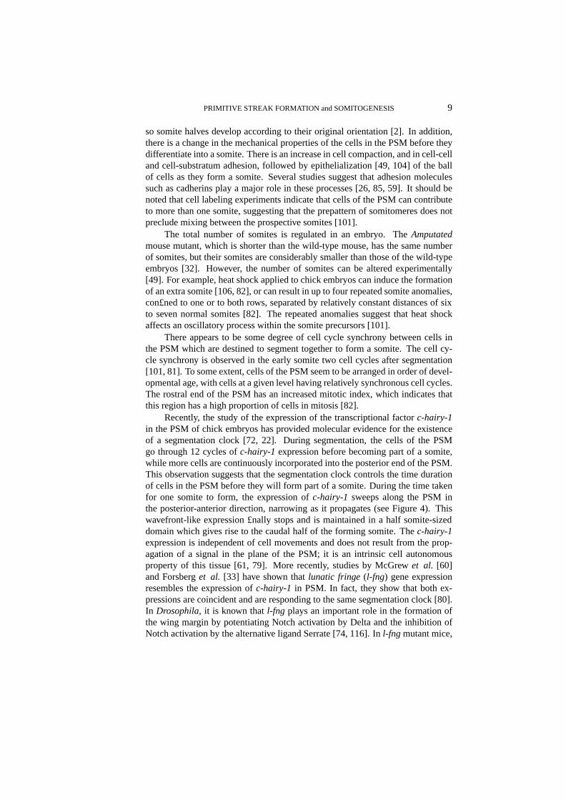
Recently, the study of the expression of the transcriptional factorc-hairy-1in the PSM of chick embryos has provided molecular evidence for the existenceof a segmentation clock [72, 22]. During segmentation, the cells of the PSMgo through 12 cycles ofc-hairy-1 expression before becoming part of a somite,while more cells are continuously incorporated into the posterior end of the PSM.This observation suggests that the segmentation clock controls the time durationof cells in the PSM before they will form part of a somite. During the time takenfor one somite to form, the expression ofc-hairy-1 sweeps along the PSM inthe posterior-anterior direction, narrowing as it propagates (see Figure 4). Thiswavefront-like expression £nally stops and is maintained in a half somite-sizeddomain which gives rise to the caudal half of the forming somite. Thec-hairy-1expression is independent of cell movements and does not result from the prop-agation of a signal in the plane of the PSM; it is an intrinsic cell autonomousproperty of this tissue [61, 79]. More recently, studies by McGrewet al. [60]and Forsberget al. [33] have shown thatlunatic fringe (l-fng) gene expressionresembles the expression ofc-hairy-1 in PSM. In fact, they show that both ex-pressions are coincident and are responding to the same segmentation clock [80].In Drosophila, it is known thatl-fng plays an important role in the formation ofthe wing margin by potentiating Notch activation by Delta and the inhibition ofNotch activation by the alternative ligand Serrate [74, 116]. Inl-fng mutant mice,
10 S. SCHNELL and P. K. MAINI and K.J. PAINTER and H. G. OTHMER
the formation of somites is disrupted and if a somite forms its anterior-posteriorpatterning is disturbed [27, 117].
Time
Anterior
Posterior
FIG. 4. Schematic illustration of the wave ofc-hairy-1sweeping in the posterior-anterior direc-tion (bottom to top) along the PSM with time (left to right). The shading denotes expression of thisfactor. It begins as a broad wave but narrows as it moves anteriorly until it £nally correlates with theposterior half of the new forming somite. Then a new wave begins at the posterior margin of the PSM.Similar behavior is observed forlunatic fringe.
Finally, it is important to mention that the principle differentiation pattern ofall the somites is very similar. However, during morphogenesis subsequent dif-ferentiation forms unique anatomic structures, depending on the position alongthe anterior-posterior axis. Experiments in chick embryos demonstrate that thepositional speci£cation of somites occurs early during somitogenesis [55, 20, 21,19, 107, 17, 12]. When cervical somites are replaced with somites from the trunkregion, rib-like structures develop in the cervical vertebral column of the embryo.When thoracic somites are replaced by cervical somites, embryos do not developribs [55]. There is now a large body of experimental work showing that posi-tional speci£cation of the PSM requires members of theHox gene family [57].Hox gene activation during development correlates with gene position in theHoxcomplex, a property referred to as colinearity. The spatial and temporal colinear-ity in the expression of these genes results in unique combinations ofHox genesin de£ned groups of somites and their derivatives along the anterior-posterior axis[34, 40]. This led to the suggestion that aHox code speci£es the identity ofsomites [48, 47]. The role ofHox genes in positional speci£cation has been an-alyzed by interfering with or altering the expression of singleHox genes or bysimultaneously perturbing the expression with retinoic acid, which is implicatedin the speci£cation of the axes during development [103].
2. Questions. Early organization of the avian blastoderm clearly involvesa carefully controlled sequence of events. At present, very little is known con-

PRIMITIVE STREAK FORMATION and SOMITOGENESIS 11
cerning the mechanisms regulating this development and here we list some of themajor unresolved questions. In the following section we describe some of thetheories postulated to explain these processes.
1. How is the posterior site of the embryo determined? Formation of thearea pellucidainvolves a gravity induced directional shedding of cells(posterior to anterior) to form a one-cell thick layer [56]. How is thistranslated into the structural differences associated with the posterior re-gion (e.g. Koller’s sickle, secondary hypoblast formation)?
2. Development of the primary hypoblast involves an apparent dropping ofcells in thearea pellucidato form isolated islands in the subgerminalcavity [15]. What leads to the early diversi£cation of such cells, andhow do they separate from thearea pellucida? One possibility is to linkthe diversi£cation with the cell cycle, such that at the time of primaryhypoblast formation a randomly scattered population in a speci£c phaseof the cycle experiences a change in its cellular properties, for exampleadhesion. This change in adhesion may result in such cells being forcedfrom the area pellucida. To test such a hypothesis, it is necessary toconstruct a discrete cell model which incorporates cell adhesion [73].
3. What controls formation of the secondary hypoblast, and does the hy-poblast in¤uence streak formation? The role of the hypoblast in streakformation is controversial, and earlier experiments in which the hypoblasthas been shown to induce streak formation [108, 3, 4] have been chal-lenged by recent experiments [51]. However it is still not known whetherthe hypoblast is able to exert some in¤uence over streak formation.
4. What initiates motion and guides the early migration of cells in the lateralregions toward the PMZ? Stern [97] observed migration of a subpopula-tion of thearea pellucidato the posterior marginal zone prior to streakformation and speculated that a chemoattractant is produced at that site.Although collagen-gel assays support this theory, no chemoattractant hasbeen identi£ed.
5. What cues guide elongation and movement of the primitive streak? Asimple anterior-posterior gradient of a diffusible morphogen cannot beused for positional information along that axis [53], for if it were the90◦
transplants of the primitive streak would ingress toward the anterior polerather than along a ray through the center of the disk.
6. What is the role of cell division in streak formation? Recent resultsby Wei and Mikawa [109] suggest that a subpopulation of cells in theposterior region may divide in a directional manner to form the primitivestreak. It remains to be understood whether this division is essential forstreak formation, or if it is simply an associated phenomenon.
7. What mechanisms can account for the fact that the primitive streak main-tains its rod-like structure during ingression? Does the primitive streakingress by convergent extension [46], whereby cells intercalate at theposterior marginal zone and push the primitive streak forward? Are thereadhesive differences between cells in the primitive streak and those in thehypoblast and epiblast, or is the structure maintained by chemotactic at-
12 S. SCHNELL and P. K. MAINI and K.J. PAINTER and H. G. OTHMER
traction between cells in the primitive streak? Alternatively, is the streakmaintained as a rod by the forces occurring throughout the blastoderm atthese stages.
8. There appears to be a gradient within the marginal zone of potential toform a streak, with the posterior being the most capable and the ante-rior the least. At what stage is this potential determined, and by whatmechanisms?
9. The primitive streak seems to inhibit other streaks from forming. Whatis the nature of this inhibition, and is it con£ned to act along the marginalzone?
10. The size and age of a blastodisc segment or donor implant are importantin determining the site of streak formation. How do the key propertiessinvolved change with time?
11. How is the organizer de£ned, and how are the movements of cells throughthe organizer to form notochord, head process, paraxial mesoderm, etc.,regulated? Recent experiments have revealed that the organizer is a tran-sitory population continuously de£ned by cells in the middle part of theprimitive streak [43]. Previous results, however, suggest that there existsa resident population of cells within the organizer that moves back withthe node throughout regression. What is the relevance, if any, of thisresident population?
12. What mechanisms control regression of the streak/organizer? Does re-gression of the streak simply occur through the disappearance of anteriorcells into axial structures. Does the node regress by being pushed backby cells that are ingressing through it? Ablation of the node results inthe regeneration of a new node, yet the new node must regenerate beforeregression proceeds. Does the static population of cells within the nodecontrol the movements of the node during regression?
13. How is the left-right asymmetry established? The earliest indication ofleft-right asymmetry in the avian embryo occurs with the asymmetricexpression ofsonic hedgehog (shh)in the avian node [58]. Studies inthe mouse have revealed the presence of a nodal ¤uid ¤ow from rightto left as a result of unidirectionally rotating cilia on node cells [67, 68]and this has been linked with the establishment of the left-right axis.However, no such cilia have been located in the chick, and the cause ofleft-right asymmetry remains unknown.
14. What regulates the number and size of somites?15. What determines differentiation into anterior and posterior halves within
a somite?16. What are the differentiation and mechanical properties involved in the
epithelialization of somites?17. What determines the regional speci£cation of somites - that is, certain
somites form certain structures. What is the precise role of theHoxfamily in this process and how is it controlled?
18. What drives the segmentation clock? Is there a relation between the cell-cycle and the segmentation clock?

PRIMITIVE STREAK FORMATION and SOMITOGENESIS 13
19. What is the precise role of the segmentation clock during somitogenesis?20. How is the interplay between the segmentation clock and Notch-Delta
and related components established?21. What regulates the re£nement of thec-hairy-1 and l-fng cycles in the
forming somite? How do these cycles interact with the segmentationclock?
22. How can the heat shock experiments be explained?
3. Models of streak formation and somitogenesis.
3.1. Formation of the primitive streak. PI. Model of Induction by Gravity:Eyal-Giladi [29] proposed that substances needed for the initiation of primitivestreak formation become nonuniformly distributed by gravity while the embryois tilted, moving from the vegetal pole toward the region that is incorporated intothe PMZ. Alternatively, Eyal-Giladi also suggests that these factors can be locatedunder the embryo and shifted toward the posterior by the sliding of the yolk, andcould later be found in the PMZ and Koller’s sickle. Classic experiments in chickembryos have established that labile anterior-posterior polarity is determined 20hours after fertilization. During this period there is a critical 2-hour time windowwhere the outer albumen layers are rotated by the uterus while the yolk remainsstationary but slightly tilted within a layer of low friction thin albumen [28]. Inthese experiments, the side of the embryo that is tilted upward during the criticalwindow is de£ned as posterior. This model is unsatisfactory in some aspects. Lit-tle work has been done on this hypothesis due to the dif£culty of obtaining uterineeggs. In addition, this model does not address the ingression and regression of theprimitive streak.
PII. Model of Induction by the PMZ:In this model, proposed by Bachvarova [5],the PMZ is considered analogous to the Nieuwkoop Center of the frog embryo,which is the structure responsible for induction during the £rst stages of amphib-ian development. The PMZ of the chick embryo acts as an extra-embryonic sig-naling center promoting formation of the primitive streak in the adjoining poste-rior central disc epiblast. According to the model, factors such asVg1andWnt8cproduced in the PMZ activate organizer genes such asgoosecoidin Koller’s sickleand chordin in the posterior central-disc epiblast. In turn,chordin suppressesBMP and this decrease promotes activation of organizer genes in the posteriormidline. Lower concentrations ofVg1 or TGF-β factors induceBrachyury-likegenes in a broader crescent of posterior central disc epiblast, leading to meso-derm formation. BMP activity from lateral and anterior marginal zone inducesepidermis in the adjoining central disc. Finally, the activation of the Wnt pathwayin the late uterine and freshly laid egg plays an important role in the asymmetryobserved in cells of Koller’s sickle and the hypoblast.
This model incorporates several aspects of primitive streak formation. How-ever, as in the previous model it does not address the ingression and regressionof the primitive streak. Furthermore, as indicated by Bachvarova [5], many out-standing problems remain with this model. For example, it is not clear if factorssuch asVg1are required in normal development. In addition, other factors such

14 S. SCHNELL and P. K. MAINI and K.J. PAINTER and H. G. OTHMER
asTGF-β cannot be present and active in early embryos; and little is known aboutthe Wnt pathway.
A mathematical formulation of this model could help understand the out-come of the complex of interactions proposed and make experimentally-testablepredictions.
PIII. Chemotaxis Model:Chemotaxis (or haptotaxis) is a plausible mechanismfor the observed oriented cell movements both prior to and during primitive streakformation, and this mechanism has been incorporated into a model designed forformation and subsequent maintenance of the streak (though not the determinationof the initial site of outgrowth) [71].
The model assumes that there is a specialized subpopulation of cells resid-ing at or close to the posterior marginal zone that both respond to and modulatethe level of an attractant. This population serves to mark the site of the primitivestreak and guide the movements of elongation and regression. Several cell popu-lations have been identi£ed [41, 99, 109] as having a role in primitive streak for-mation. The model does not, however, postulate how other cells ingress throughthe streak. In Figure 5 we show the pattern of movements predicted on a two-dimensional domain. To achieve movement of cells as a rod, rather than a generalspreading of cells, it is necessary to choose conditions such that the chemoattrac-tant initially has its highest concentration at the center of the domain (correspond-ing to the center of thearea pellucida) and decreases to zero at the marginal zone.Plausible mechanisms for generating such conditions are given in [71].
The model makes a number of experimentally-testable predictions (Figure6). Firstly, it predicts that any ectopically induced embryonic axis will developalong radial lines. Secondly, it predicts that disruption of the center of theareapellucidawill have a signi£cant effect on the morphology of the streak. It alsopredicts the natural development of anorganizerregion at the anterior portion ofthe streak as a region of higher cell density, and demonstrates a decrease in therate of regression as the streak moves back, in agreement with experimental re-sults [94]. However there is no experimental evidence for chemotactic motion instreak formation, and it is unclear whether the same mechanism that drives prop-agation of the streak is also responsible for regression. Thus this model simplydemonstrates that chemotaxis can produce the observed behavior.
PIV. Cell Division Model:Wei and Mikawa [109] have proposed a model for for-mation of the streak based on directional cell division. In this model, a speci£csubpopulation of cells (localized at stage XII to the epiblast-midline region of thePMZ) undergoes oriented cell division along the anterior-posterior axis to formthe Hamburger and Hamilton stage 3 primitive streak. The model is supportedby cell marking experiments which demonstrate that the Hamburger and Hamil-ton stage 3 streak comprises only cells derived from this region, and not cellswhich have migrated in from lateral regions, as has previously been assumed. Fur-thermore, cells in the streak were shown to have metaphase chromosome plates(which indicate cleavage direction) perpendicular to the anterior-posterior axis.The calculation, based on the number of cells in the pre-streak region and Ham-

PRIMITIVE STREAK FORMATION and SOMITOGENESIS 15
FIG. 5. A time sequence showing the cell density for model PIII on a two-dimensional rectangu-lar domain. White represents high cell density, black represents a zero cell density. The results showcell movement across the domain to form a rod which extends approximately half the way across thedomain (e). Subsequent development shows a period of reverse movement, which occurs on a slowertime scale.
burger and Hamilton stage 3 streak, of a cell cycle time of approximately 4 hoursis consistent with the mitotic index for cells of the chick gastrulae.
This model is consistent with the observation that the epiblast portion of theposterior marginal zone contributes to the primitive streak, and with the idea that aPMZ-derived signal induces primitive streak in the adjoining epiblast (see modelII above). However, it is not yet clear if directional cell division would be able toinduce the streak to form a long straight rod alone, nor is there any suggestion asto how regression of the streak is controlled.
PV. Convergent-Extension Model:Schoenwolf [88] has postulated that primitivestreak formation may occur via a convergent-extension mechanism similar to thatobserved in developing amphibia [44, 45]. In this model, prospective primitivestreak cells from either side of the midline would converge at the midline, in-tercalating with those on the opposite side and thereby producing an elongatingprimitive streak. This also raises the possibility that regression may occur througha reverse process.
This model is speculative, yet some evidence for it can be found in the gen-

16 S. SCHNELL and P. K. MAINI and K.J. PAINTER and H. G. OTHMER
FIG. 6. The time course for the development of an ectopic streak following ‘transplantation’ inmodel PIII. When a second population of “able” cells is placed at another point along the marginalzone (top: lateral, middle: anterior), an ectopic streak develops which moves towards the center ofthe domain. Fairly small changes in model parameters can result in the fusing of these streaks at theanterior ends. In the bottom £gures, this has been effected by increasing the concentration gradientof the chemoattractant.
eral cell movements observed to take place in the epiblast during primitive streakformation [105]. Furthermore, the mechanism could provide an explanation forthe change in morphology of the blastoderm from circular to pear-shaped duringformation of the streak, as intercalation would result in a streak being driven inboth anterior and posterior directions. However the author does not suggest whatdetermines the posterior marginal zone as the site of streak formation, nor whatthe mechanisms are for guiding cell movement during the convergent-extensionprocess so as to maintain the rod-like morphology of the streak.

PRIMITIVE STREAK FORMATION and SOMITOGENESIS 17
3.2. Somitogenesis.During the last three decades, several models have beenproposed to explain the formation of somites [24, 32, 8, 9, 42, 49, 63, 64, 76, 78,77, 82, 81, 87]. Some of these incorporate the different aspects of somitogenesispreviously mentioned, and are satisfactory in many respects. It is important toemphasize that these models cannot explain all the experimental facts presentedin section 1.2, but they do lend insight to many of the observations. The modelscan be divided into four main categories:
SI. Induction Models:In these models, somite formation is explained in termsof inductive interactions with neighboring tissue [8]. These models are unsat-isfactory in many aspects. No single tissue has been shown to induce somiteformation. As we previously indicated, somites can be formed in the absence ofHensen’s node, notochord, neural tube and endoderm [7, 11, 100], but the midlinestructures are necessary after experimental disruption of the PSM [69].
SII. Prepattern Models:These models postulate that there is a spatially-periodicprepattern present in the PSM before formation of the somites. Bellairs and Veini[11] proposed that somitogenic clusters are generated during PSM formation.Meier [62] suggested that prior to segmentation the PSM displays metamericarrangements of groups of cells, named somitomeres. The observation of theprepattern has been con£rmed in microsurgical and transplantation experiments[70, 18, 2]. However, this sort of model does not address the key problems of howthe prepattern is set up and how it is maintained and regulated.
SIII. Positional Information Models:These assume that a spatial pattern in chem-ical morphogen is set up, either via a gradient or a reaction-diffusion mechanism,and this prepattern determines cell differentiation. There are two main models:
1. The wave gradient model was proposed by Wilby and Ede [111] and Flintetal. [32]. This model proposes that regression of Hensen’s node creates two stripsof paraxial mesoderm, and that cells recruited into them start to synthesize a mor-phogen. The morphogen concentration increases in the cells until a threshold isreached, at which point an irreversible change from synthesis to destruction ofmorphogen occurs. The morphogen concentration in these cells falls, establish-ing a sink relative to cells that are still producing the morphogen. Neighboringcells maintain a morphogen concentration below the threshold, as morphogendiffuses from them into the sink, but cells further back in the paraxial mesodermexceed the morphogen concentration threshold, and another trough of concentra-tion is formed. Thus, a pattern of alternating peaks and troughs is created, whichlater gives rise to somites and £ssures respectively. In this model, the size of thesomites is determined by either the rate of incorporation of cells into the PSM orthe speed of node regression. If the rate of node regression depends on the size ofthe embryo, then this model can account for the observation that there is regula-tion of somite number so, for example, in theAmputatedmutant mouse embryo,which is only two thirds the normal size, the number of somites formed is stillthe same as in the normal case. This would be consistent with the assumptionthat the node regresses more slowly in smaller embryos. However, this modelcannot easily explain the observations after the anterior-posterior axis of the PSM

18 S. SCHNELL and P. K. MAINI and K.J. PAINTER and H. G. OTHMER
is reversed. It seems likely that the pattern of morphogen concentration would beseverely disrupted during these experiments and the model would not then predictsomite formation in the normal way.
2. Meinhardt [63, 64] proposed a reaction-diffusion type model, with two cellstates A and P, which locally exclude each other, but stimulate each other overa long range. Cells switch from one state to the other until £nally reaching astable state. These can lead to a pattern of stable. . . APAP . . .stripes formingfrom anterior to posterior. If the transition from, say P to A, allows a change ofsegmental speci£cation then each AP pair (or segment) will have a more posteriorspeci£cation than its predecessor. Thus a segmental pattern can be generated inwhich segments have different regional characteristics.
To set up this pattern, Meinhardt proposed two alternative mechanisms, oneinvolving a morphogen gradient in which threshold concentrations of the mor-phogen are required for successive P to A transitions, the other involving out-growth in which new segments are added as the domain grows. Meinhardt’smodel is in agreement with two observations of Palmeirimet al. [72]: one fullcycle of c-hairy-1 oscillation corresponds to the formation of one somite, andc-hairy-1expression seems to be reminiscent of the spatiotemporal dynamics ofone of the autocatalytic substances, because its wavefront expression stops and ismaintained in the posterior half of the somites. It is also the only model, to ourknowledge, that addresses the regional differences of somites and the anterior-posterior pattern of somites.
This model cannot easily explain the results of the experiment in which anisolated part of the PSM forms normal somites and the experiments that involvereversing the anterior-posterior axis of the PSM. In the former, one would expectany diffusion-based structures to be disrupted by the experiment, while in thelatter, the model would predict that somites would form £rst in the anterior partof the reversed PSM and somitogenesis would proceed as normal, but in realitythey develop according to their original location. One would have to assume thatrostral-caudal determination occurs very early and is £xed before isolation or ro-tation of the PSM. This possible explanation requires more detailed investigation.It is not clear that this scenario is consistent with that envisioned for thec-hairy-1dynamics. Furthermore this model does not explain the cell-autonomous natureof somite formation which is strongly suggested by the experiments of Palmeirimet al. [72], McGrewet al. [60] and Forsberget al. [33]. In these experiments, aportion of one side of the PSM is removed but thec-hairy-1waves propagate insynchrony in both sides of the PSM, including the isolated portion.
As it stands, the model does not appear to explain the heat shock effectswhich seem to require a link between cell fate and cell cycle. Such a link is notapparent in this model.
IV. Clock or Oscillator Models:There are a number of models along these lines:
1. Cooke and Zeeman [24] were the £rst to propose he exisitence of a cellularoscillator, which they assumed interacts with a progressing wave of cell determi-nation travelling along the anterior-posterior axis of the PSM. This model, known
PRIMITIVE STREAK FORMATION and SOMITOGENESIS 19
as the clock and wavefront model, is able to explain the control of somite num-ber [92], but is contradicted by the results of the experiments which reverse theanterior-posterior polarity of the PSM, because, as inPIII , this model would pre-dict that segmentation should continue in the anterior-posterior direction withoutdisturbance. To be consistent with the observation of the repetitive anomaliesobserved after the single heat shock experiments [82, 81, 101] this model wouldhave to additionally assume that the cellular oscillator was closely linked to thecell cycle. The model does not address the formation of the anterior and posteriorhalves of a somite.
2. Sternet al. [101] proposed that the cell cycle plays the role of the oscillator.This cell cycle model relies on an intracellular oscillator that controls cell divi-sion and interacts with a kinematic wave which produces a signal that recruitsother cells in the vicinity shortly before segmentation [49, 101, 81]. It explainsthe periodic anomalies of the heat shock experiments, the cell cycle synchronyobserved in the PSM, as well as the isolation and transplantation experiments.This model addresses pattern formation at the cellular level and therefore doesnot address molecular issues such as the oscillations ofc-hairy-1and its patternin the PSM. A direct link between this model and thec-hairy-1 oscillations isnot obvious, because in chick embryos, the period of the cell cycle in the PSMis 9 hours while the period of the oscillations is only 90 minutes [72]. Further-more, heat shock experiments in zebra£sh embryos show that the periodic unitof somite defect (four normally formed somite + one abnormally formed), whichcorrespond to 2.5 hours, does not match the overall cell cycle length (4 hours).This suggests that the proposed relationship between segmentation clock and cellcycle in vertebrates should be re-evaluated [86].
3. In a similar model to the one above, Polezhaev [76, 78, 77] proposed thata wave of cell determination moves along the PSM causing cell differentiationin a particular phase of the cell cycle, resulting in these cells secreting an in-hibitor which impedes the differentiation of other cells. This model can explainthe results of the heat shock experiments, is consistent with the observations ofthe isolation and transplantation experiments, and the epithelialization observedjust before overt segmentation [49, 104]. However, as in the previous model, thismodel does not address events at the molecular level, nor does it address the for-mation of the anterior and posterior halves. To explain the regulation of somitenumber [49] one would have to assume that the cell determination wave moved atdifferent rates (as inPIII 1).
4. Recently, Schnell and Maini [87] have proposed a clock and induction modelin which, as a group of cells destined to form a somite traverses the PSM, cells un-dergo a series ofl-fng expression pulses, followed by a longer £nal pulse whichwill remain at the posterior half of the newly forming somite.l-fng expressionsynthesizes a protein associated with the cell membrane, which increases its mem-brane levels in a ratchet-like fashion proportional to the segmentation clock oscil-lations experienced. The formation of a somite is then assumed to be triggered at athreshold level of l-fng protein. Elements of the Notch-Delta pathway associated

20 S. SCHNELL and P. K. MAINI and K.J. PAINTER and H. G. OTHMER
with l-fng would allow the formation of a somite boundary and anterior-posteriorpattern, through an induction mechanism. This model is consistent with the rhyth-mical expression ofc-hairy-1 and l-fng and the expression of the Notch-Deltapathway genes in PSM. The model can explain the isolation and transplantationexperiments, and the heat shock defects. However, it cannot explain the cell cyclesynchronization or epithelialization.
4. Discussion. Building the early embryo involves an architectural chal-lenge that higher organisms have addressed through two processes that occur inearly development: large scale rearrangements of tissue via a process called gas-trulation, and the axial subdivision of tissue in a process called somitogenesis.Remarkably, somitogenesis has many elements in common with limb develop-ment. In fact both of these phenomena can be considered as examples of segmen-tation. For example, in limb development the anterior-posterior speci£cation ofdigit elements (see, Dillon, this volume) is determined by theHoxgenes, and dif-ferentiation and boundary formation is determined by the Notch-Delta pathway,as in somitogenesis.
In this paper, we have reviewed the theoretical and mathematical models de-veloped to explain primitive streak formation and somitogenesis. Most of thesemodels have been designed to explain particular aspects of these processes andare successful in doing so. In our critique of previous models, we have comparedthe models only with the experimental results that are widely accepted and whichaddress the gross mechanisms of primitive streak formation and somitogenesis.As the models stand at present, none of them can easily explain all of these exper-imental observations. It should be noted that the majority of these models weredeveloped before the discovery of the molecular evidence for the control of prim-itive streak formation and somitogenesis and are based on cell and tissue levelobservations.
A challenging future problem for theoretical and mathematical modellingwill involve linking the pattern formation mechanisms at the cellular level withthe molecular control of cell properties. In Section 2 we listed 22 key questionsconnected with the problems of primitive streak formation and somitogenesis.The reader will note that the models presented in Section 3 addressed only a smallfraction of these.
Acknowledgments. This research (SS) has been funded by Jose GregorioHernandez and ORS Awards, CONICIT(Venezuela) and a Lord Miles SeniorScholarship in Science. K. Painter and H. G. Othmer are supported in part bygrant GM29123 from the National Institutes of Health.
We thank Daragh Mcinerney for helping with Figure 3 and Paul Kulesa forhelpful comments on the manuscript.
REFERENCES
[1] M. A BERCROMBIE, The effects of antero-posterior reversal of lengths of the primitive streakin the, Phil. Trans. Roy. Soc. Lond. B., 234 (1950), pp. 317–338.
PRIMITIVE STREAK FORMATION and SOMITOGENESIS 21
[2] H. AOYAMA AND K. A SAMOTO, Determination of somite cells: Independence of cell differ-entiation and morphogenesis, Development, 104 (1988), pp. 15–28.
[3] Y. A ZAR AND H. EYAL -GILADI , Marginal zone cells, the primitive streak inducing com-ponent of the primary hypoblast in the chick., J. Embryol. Exp. Morphol., 52 (1979),pp. 79–88.
[4] , Interaction of epiblast and hypoblast in the formation of the primitive streak and theembryonic axis in the chick, as revealed by hypoblast rotation experiments., J. Embryol.Exp. Morphol., 61 (1981), pp. 133–144.
[5] R. BACHVAROVA, Establishment of anterior-posterior polarity in avian embryos, Curr. Opin.Gen. Dev., 9 (1999), pp. 411–416.
[6] R. BACHVAROVA , I. SKROMME, AND C. D. STERN, Induction of primitive streak andhensen’s node by the posterior marginal zone in the early chick embryo, Development,125 (1998), pp. 3521–3534.
[7] R. BELLAIRS, The development of somites in the chick embryo, J. Embryol. Exp. Morph., 11(1963), pp. 697–714.
[8] , The segmentation of somites in the chick embryo, Bull. Zool., 47 (1980), pp. 245–252.[9] R. BELLAIRS, D. A. EDE, AND J. W. LASH, eds.,Somites in Developing Embryos, Plenum
Press, New York, NY, USA; London, UK, 1986.[10] R. BELLAIRS AND M. OSMOND, The Atlas of Chick Development, Academic Press, London,
1998.[11] R. BELLAIRS AND M. V EINI, An experimental analysis of somite segmentation in the chick
embryo., J. Embryol. Exp. Morph., 55 (1980), pp. 93–108.[12] J. BUTLER, E. COSMOS, AND P. CAUWENBERGS, Positional signals: Evidence for a possi-
ble role in muscle £bre-type patterning of the embryonic avian limb, Development, 102(1988), pp. 763–772.
[13] M. CALLEBAUT AND E. V. NUETEN, Rauber’s (koller’s) sickle: the early gastrulation or-ganizer of the avian blastoderm., Eur. J. Morph., 32 (1994), pp. 35–48.
[14] M. CALLEBAUT, E. VAN NUETEN, F. HARRISSON, L. VAN NASSAUW, A. SCHREVENS,AND H. BORTIER, Avian gastrulation and neurulation are not impaired by the removalof the marginal zone at the unincubated blastoderm stage, Eur. J. Morphol., 35 (1997),pp. 69–77.
[15] D. CANNING AND C. STERN, Changes in the expression of the carbohydrate epitope hnk–1 associated with mesoderm induction in the chick embryo., Development, 104 (1988),pp. 643–655.
[16] M. CATALA , M.-A. TEILLET, AND N. M. L. DOUARIN, Organization and developmentof the tail bud analyzed with the quail-chick chimera system, Mech. Dev., 51 (1995),pp. 51–65.
[17] K. CHADA , J. MAGRAM, AND F. CONSTANTINI, An embryonic pattern of expression of ahuman fetal globin gene in transgenic mice, Nature, 319 (1986), pp. 685–689.
[18] E. A. G. CHERNOFF AND S. R. HILFER, Calcium dependence and contraction in somiteformation, Tiss. Cell., 14 (1982), pp. 435–449.
[19] A. CHEVALLIER, Role of the somitic mesoderm in the development of the thorax in bird em-bryos. II. Origin of thoracic and appendicular musculature, J. Embryol. Exp. Morphol.,49 (1979), pp. 73–88.
[20] A. CHEVALLIER , M. KIENY, AND A. M AUGER, Limb-somite relationship: origin of thelimb musculature, Journal of Embryology and Experimental Morphology, 41 (1977),pp. 245–258.
[21] , Limb-somite relationship: Effects of removal of somitic mesoderm on the wing mus-culature, J. Embryol. Exp. Morph., 43 (1978), pp. 263–278.
[22] J. COOKE, A gene that resuscitates a theory — somitogenesis and a molecular oscillator,Trends in Genetics (Personal edition), 14 (1998), pp. 85–88.
[23] J. COOKE, S. TAKADA , AND A. M CMAHON, Experimental control of axial pattern in thechick blastoderm by local expression of wnt and activin: the role of hnk-1 positive cells.,Dev. Biol., 164 (1994), pp. 513–527.
[24] J. COOKE AND E. C. ZEEMAN, A clock and wavefront model for control of the number ofrepeated structures during animal morphogenesis, Journal of Theoretical Biology, 58

22 S. SCHNELL and P. K. MAINI and K.J. PAINTER and H. G. OTHMER
(1976), pp. 455–476.[25] I. DEL BARCO BARRANTES, A. J. ELIA , K. WUNSCH, M. H. DE ANGELIS, T. W. MAK ,
J. ROSSANT, R. A. CONLON, A. GOSSLER, AND J. L. DE LA POMPA, Interactionbetween notch signalling and lunatic fringe during somite boundary formation in themouse, Curr. Biol., 9 (1999), pp. 470–480.
[26] J. DUBAND, S. DUFOUR, K. HATTA , M. TAKEICHI , G. M. EDELMAN , AND J. P. THIERY,Adhesion molecules during somitogenesis in the avian embryo, J. Cell Biol., 104 (1987),pp. 1361–1374.
[27] Y. EVRARD, Y. LUN, A. AULEHLA , L. GAN, AND R. L. JOHNSON, lunatic fringeis anessential mediator of somite segmentation and patterning, Nature, 394 (1998), pp. 377–381.
[28] H. EYAL -GILADI , Gradual establishment of cell commitments during the early states of chickdevelopment, Cell Differ., 14 (1984), pp. 245–255.
[29] , Establishment of the axis in chordates: facts and speculations, Development, 124(1997), pp. 2285–2296.
[30] H. EYAL -GILADI AND O. KHANER, The chick’s marginal zone and primitive streak forma-tion, Dev. Biol., 134 (1989), pp. 215–221.
[31] H. EYAL -GILADI AND S. KOCHAV, From cleavage to primitive streak formation: a comple-mentary normal table and a new look at the £rst stages of the development of the chick.i. general morphology, Dev. Biol., 49 (1976), pp. 321–337.
[32] O. P. FLINT, D. A. EDE, O. K. WILBY, AND J. PROCTOR, Control of somite number in nor-mal andAmputatedmutant mouse embryos: an experimental and a theoretical analysis,Journal of Embryology and Experimental Morphology, 45 (1978), pp. 189–202.
[33] H. FORSBERG, F. CROZET, AND N. A. BROWN, Waves of mouselunatic fringeexpression,in four-hour cycles at two-hour intervals, precede somite boundary formation., Curr.Biol., 8 (1998), pp. 1027–1030.
[34] S. J. GAUNT, Mouse homeobox gene transcripts occupy different but overlapping domainsin embryonic germ layers and organs: A comparison of Hox-3.1 and Hox-1.5, Develop-ment (Cambridge), 103 (1988), pp. 135–144.
[35] S. F. GILBERT, Developmental Biology, Sinauer Associates, £fth ed., 1997.[36] A. GOSSLER AND M. HRABE DE ANGELIS, Somitogenesis, in Curr. Topics in Dev. Biol.,
vol. 38, Academic Press, 1998, pp. 225–287. Jackson Laboratory, Bar Harbor, Maine04609, USA.
[37] C. GRABOWSKI, The effects of the excision of hensen’s node on the early development of thechick embryo, J. Exp. Zool., 133 (1956), pp. 301–344.
[38] V. HAMBURGER AND H. HAMILTON , A series of normal stages in the development of thechick embryo.
[39] Y. HATADA AND C. D. STERN, A fate map of the epiblast of the early chick embryo, Devel-opment, 120 (1994), pp. 2879–2889.
[40] P. W. H. HOLLAND AND B. L. M. HOGAN, Expression of homeo box genes during develop-ment: A review, Genes. Dev., 2 (1988), pp. 773–782.
[41] J. IZPISUA-BELMONTE, E. D. ROBERTIS, K. STOREY, AND C. STERN, The homeoboxgene goosecoid and the origin of organizer cells in the early chick blastoderm., Cell., 74(1993), pp. 645–659.
[42] A. JACOBSON AND S. MEIER, Somites in Developing Embryos, vol. 118 of NATO ASIseries. Series A, Life sciences, New York: Plenum Press., 1986, ch. Somitomeres: Theprimordial body segments, pp. 1–16.
[43] K. JOUBIN AND C. STERN, Molecular interactions continuously de£ne the organizer duringcell movements of gastrulation, Cell, 98 (1999), pp. 559–571.
[44] R. KELLER, Vital dye mapping of the gastrula and neurula ofxenopus laevis. i. prospec-tive areas and morphogenetic movements of the super£cial layer, Dev. Biol., 42 (1975),pp. 222–241.
[45] , The cellular basis of epiboly: An sem study of deep cell rearrangement during gas-trulation in xenopus laevis, J. Embryol. Exp. Morphol., 60 (1980), pp. 201–234.
[46] R. KELLER, J. SHIH, AND P. WILSON, Cell Motility, Control, and Function of Convergenceand Extension During Gastrulation inXenopus, inGastrulation: Movements, Patterns,

PRIMITIVE STREAK FORMATION and SOMITOGENESIS 23
and Molecules, W. C. R. Keller and F. Griffen, eds., Plenum Press, New York, NY, USA;London, UK, 1991.
[47] M. K ESSEL, Respeci£cation of vertebral identities by retinoic acid, Development (Cam-bridge), 115 (1992), pp. 487–501.
[48] M. K ESSEL ANDP. GRUSS, Homeotic transformations of murine prevertebrae and concom-mitant alteration ofHox codes induced by retinoic acid, Cell, 67 (1991), pp. 89–104.
[49] R. J. KEYNES AND C. D. STERN, Mechanisms of vertebrate segmentation, Development(Cambridge), 103 (1988), pp. 413–429.
[50] O. KHANER, Axis determination in the avian embryo, Curr. Topics in Dev. Biol., 28 (1993),pp. 155–180.
[51] , The rotated hypoblast of the chicken embryo does not initiate an ectopic axis in theepiblast, Proc. Nat. Acad. Sci. USA, 92 (1995), pp. 10733–10737.
[52] , The ability to initiate an axis in the avian blastula is concentrated mainly at a poste-rior site., Dev Biol, 194 (1998), pp. 257–266. Department of Cell and Animal Biology,Hebrew University, Jerusalem, Israel.
[53] O. KHANER AND H. EYAL -GILADI , The embryo forming potency of the posterior marginalzone in stage x through xii of the chick., Dev. Biol., 115 (1986), pp. 275–281.
[54] , The chick’s marginal zone and primitive streak formation. I Coordinative effect ofinduction and inhibition, Developmental Biology, 134 (1989), pp. 206–214.
[55] M. K IENY, A. MAUGER, AND P. SENGEL, Early regionalization of somite mesoderm asstudied by the development of axil skeleton of the chick embryo, Dev. Biol., 28 (1972),pp. 142–161.
[56] S. KOCHAV AND H. EYAL -GILADI , Bilateral symmetry in chick embryo, determination bygravity, Science, 171 (1971), pp. 1027–1029.
[57] R. KRUMLAUF, Hox genes in vertebrate development, Cell, 78 (1994), pp. 191–201.[58] M. L EVIN, Left-right asymmetry and the chick embryo, Sem. Cell. Dev. Biol., (1998).[59] K. L INASK, C. LUDWIG, M. D. HANG, X. LIU, AND K. K. G. L. RADICE, N-
Cadherin/Catenin-mediated morphoregulation of somite formation, Dev. Biol., 202(1998), pp. 85–102.
[60] M. M CGREW, J. DALE, S. FRABOULET, AND O. POURQUIE, The lunatic fringegene isa target of the molecular clock linked to somite segmentation in avian embryos, Curr.Biol., 8 (1998), pp. 979–982.
[61] M. J. MCGREW AND O. POURQUIE, Somitogenesis: segmenting a vertebrate, Current opin-ion in genetics and development, 8 (1998), pp. 487–493.
[62] S. MEIER, Development of the chick embryo mesoblast: Formation of the embryonic axis andestablishment of the metameric pattern, Dev. Biol., 73 (1979), pp. 24–45.
[63] H. MEINHARDT, Models of Biological Pattern Formation, Academic Press, New York, USA,1982.
[64] , Somites in Developing Embryos, vol. 118 of NATO ASI series. Series A, Life sci-ences, New York: Plenum Press., 1986, ch. Models of segmentation, pp. 179–189.
[65] E. MITRANI AND Y. SHIMONI , Induction by soluble factors of organized axial structures inchick epiblasts, Science, 247 (1990), pp. 1092–1094.
[66] P. NIEUWKOOP, Origin and establishment of embryonic polar axes in amphibian develop-ment, Curr. Topics Dev. Biol., 11 (1977), pp. 115–132.
[67] S. NONAKA , Y. TANAKA , Y. OKADA , S. TAKEDA , A. HARADA , Y. KANAI , M. KIDO,AND N. HIROKAWA, Randomization of left-right asymmetry due to loss of nodal ciliagenerating leftward ¤ow of extraembryonic ¤uid lacking kif3b motor protein, Cell, 95(1998), pp. 829–837.
[68] Y. OKADA , S. NONAKA , Y. TANAKA , Y. SAIJOH, H. HAMADA , AND N. HIROKAWA,Abnormal nodal ¤ow precedes situs inversus iniv andinv mice, Molecular Cell, 4 (1999),pp. 459–468.
[69] D.-J. PACKARD, R. ZHENG, AND D. C. TURNER, Somite pattern regulation in the aviansegmental plate mesoderm, Development, 117 (1993), pp. 779–791.
[70] D. S.-J. PACKARD AND A. G. JACOBSON, The in¤uence of axial structures on chick somiteformation, Dev. Biol., 53 (1976), pp. 36–48.
[71] K. J. PAINTER, P. K. MAINI , AND H. G. OTHMER, A chemotactic model for the advance

24 S. SCHNELL and P. K. MAINI and K.J. PAINTER and H. G. OTHMER
and retreat of the primitive streak in avian development, Bull. Math. Biol., 62 (2000). Toappear.
[72] I. PALMEIRIM , D. HENRIQUE, D. ISH-HOROWICZ, AND O. POURQUIE, Avianhairy geneexpression identi£es a molecular clock linked to vertbrate segmentation and somitogen-esis, Cell, 91 (1997), pp. 639–648.
[73] E. PALSSON AND H. G. OTHMER, A model for individual and collective cell movement indictyostelium discoideum, Proc. Nat. Acad. Sci, (2000). Submitted.
[74] V. PANIN , V. PAPAYANNOPOULOS, R. WILSON, AND K. D. IRVINE, Fringe modulatesnotch-ligand interactions, Nature, 387 (1997), pp. 908–912.
[75] P. PENNER AND I. BRICK, Acetylcholinesterase and polyingression in the epiblast of theprimitive streak chick embryo., Roux’s Arch. Dev. Biol., 193 (1984), pp. 234–241.
[76] A. A. POLEZHAEV, A mathematical model of the mechanism of vertebrate somitic segmen-tation, Journal of Theoretical Biology, (1992).
[77] , Mathematical model of segmentation in somitogenesis in vertebrates, Biophysics, 40(1995), pp. 583–589.
[78] , Mathematical modelling of the mechanism of vertebrate somitic segmentation, J.Biol. Sys., 3 (1995), pp. 1041–1051.
[79] O. POURQUIE, Clocks regulating developmental processes, Curr. Opin. Neurobiol., 8 (1998),pp. 665–670.
[80] O. POURQUIE, Notch around the clock, Curr. Opin. Gen. Dev., 9 (1999), pp. 559–565.[81] D. R. N. PRIMMETT, W. E. NORRIS, G. J. CARLSON, R. J. KEYNES, AND C. D. STERN,
Periodic segmental anomalies induced by heat shock in the chick embryo are associatedwith the cell cycle, Development (Cambridge), 105 (1989), pp. 119–130.
[82] D. R. N. PRIMMETT, C. D. STERN, AND R. J. KEYNES, Heat shock causes repeated seg-mental anomalies in the chick embryo, Development (Cambridge), 104 (1988), pp. 331–339.
[83] D. PSYCHOYOS AND C. D. STERN, Fates and migratory routes of primitive streak cells inthe chick embryo, Development (Cambridge), 122 (1996), p. 1523.
[84] , Restoration of the organizer after radical ablation of hensen’s node and the anteriorprimitive streak in the chick embryo, Development, 122 (1996), pp. 3263–3273.
[85] G. L. RADICE, H. RAYBURN, H. MATSUNAMI , K. A. K NUDSEN, M. TAKEICHI , AND
R. O. HYNES, Developmental defects in mouse embryos lacking n-Cadherin, Dev. Biol.,181 (1997), pp. 64–78.
[86] M. N. ROY, V. E. PRINCE, AND R. K. HO, Heat shock produces periodic somitic distur-bances in the zebra£sh embryo, Mech. Dev., 85 (1999), pp. 27–34.
[87] S. SCHNELL AND P. K. MAINI , Clock and induction model for somitogenesis, Dev. Dyn.,217 (2000), To appear.
[88] G. C. SCHOENWOLF, Cell movements in the epiblast during gastrulation and neuralationin avian embryoes, in Gastrulation, R. Keller, ed., Plenum Press, New York, NY, USA;London, UK, 1991, pp. 1–28.
[89] M. SELLECK AND C. STERN, Fate mapping and cell lineage analysis of hensen’s node in thechick embryo, Development, 112 (1991), pp. 615–626.
[90] , Formation and Differentiation of Early Embryonic Mesoderm, Plenum Press, NewYork, 1992, ch. Evidence for stem cells in the mesoderm of Hensen’s node and their rolein embryonic pattern formation, pp. 23–31.
[91] S. B. SHAH, I. SKROMNE, C. R. HUME, D. S. KESSLER, K. J. LEE, C. D. STERN, AND
J. DODD, Misexpression of chick Vg1 in the marginal zone induces primitive streak for-mation, Development (Cambridge), 124 (1997), pp. 5127–5138. Department of Physiol-ogy and Cellular Biophysics, College of Physicians and Surgeons of Columbia Univer-sity, New York, NY 10032, USA.
[92] J. M. W. SLACK, From Egg to Embryo. Regional speci£cation in early development, Cam-bridge: Cambridge University Press, 1991.
[93] N. SPRATT, Location of organ-speci£c regions and their relationship to the development ofthe primitive streak in the early chick blastoderm, J. Exp. Zool., 89 (1942), pp. 69–101.
[94] , Regression and shortening of the primitive streak in the explanted chick blastoderm.,J. Exp. Zool., 104 (1947), pp. 69–100.

PRIMITIVE STREAK FORMATION and SOMITOGENESIS 25
[95] , Some problems and principles of development, Am. Zool., 6 (1966), pp. 215–254.[96] N. SPRATT AND H. HAAS, Integrative mechanisms in development of the early chick blasto-
derm. I Regulative potentiality of separate parts, J. Exp. Zool., 145 (1960), pp. 97–137.[97] C. STERN, Gastrulation: Movements, Patterns, and Molecules, Plenum, New York, 1991,
ch. Mesodorm formation in the chick embryo revisited., pp. 29–41.[98] C. STERN AND D. CANNING, Origin of cells giving rise to mesoderm and endoderm in the
chick embryo., Nature, 343 (1990), pp. 273–275.[99] C. D. STERN, The marginal zone and its contribution to the hypoblast and primitive streak
of the chick embryo, Development (Cambridge), 109 (1990), p. 667.[100] C. D. STERN AND R. BELLAIRS, Mitotic activity during somite segmentation in the early
chick embryo, Anat. Embryol. (Berl.), 169 (1984), pp. 97–102.[101] C. D. STERN, S. E. FRASER, R. J. KEYNES, AND D. R. N. PRIMMETT, A cell lineage anal-
ysis of segmentation in the chick embryo, Development (Cambridge), 104 Supplement(1988), pp. 231–244.
[102] A. STREIT, K. LEE, I. WOO, C. ROBERTS, T. JESSELL, AND C. STERN, Chordin regulatesprimitive streak development and the stability of induced neural cells, but is not suf£cientfor neural induction in the chick embryo, Development, 125 (1998), pp. 507–519.
[103] D. SUMMERBELL AND M. M ADEN, Retinoic acid, a developmental signalling molecule,Trends in neurosciences (Regular ed.), 13 (1990), pp. 142–147.
[104] P. P. L. TAM AND P. A. TRAINOR, Speci£cation and segmentation of the paraxial mesoderm,Anat. Embryol. (Berl.), 189 (1994), pp. 275–305.
[105] L. VAKAET, Chimeras in Developmental Biology, Academic Press, London, 1984, ch. Earlydevelopment of birds.
[106] M. VEINI AND R. BELLAIRS, Somites in Developing Embryos, vol. 118 of NATO ASI series.Series A, Life sciences, New York: Plenum Press., 1986, ch. Heat shock effects in chickembryos, pp. 135–145.
[107] F. WACHTLET, B. CHRIST, AND H. J. JACOB, Grafting experiments on determination andmigratory behaviour of presomitic, somitic and somatopleural cells in avian embryos,Anat. Embryol. (Berl.), 164 (1982), pp. 369–378.
[108] C. WADDINGTON, Induction by the endodrem in birds., Roux’s Arch. Dev. Biol., 128 (1933),pp. 502–521.
[109] Y. WEI AND T. MIKAWA , Formation of the avian primitive streak from spatially restrictedblastoderm: evidence for polarized cell division in the elongating streak, Development,127 (2000), pp. 87–96.
[110] R. G. WEISBLAT, C. J. WEDEEN, AND R. G. KOSTRIKEN, Evolution of developmentalmechanisms: Spatial and temporal modes of rostrocaudal patterning, Curr. Top. Dev.Biol., 29 (1994), pp. 101–134.
[111] O. K. WILBY AND D. A. EDE, A model for generating the pattern of cartilage skeletal ele-ments in the embryonic chick limb, Journal of Theoretical Biology, 52 (1975), pp. 199–217.
[112] S. YUAN, D. DARNELL, AND G. SCHOENWOLF, Identi£cation of inducing, responding andsupressing regions in an experimental model of notochord formation in avian embryos,Dev. Biol., 172 (1995), pp. 567–584.
[113] , Mesodermal patterning during avian gastrulation and neurulation: experimentalinduction of notochord from non-notochordal precursor cells, Dev. Genets., 17 (1995),pp. 38–54.
[114] S. YUAN AND G. SCHOENWOLF, De novo induction of the organizer and formation of theprimitive streak in an experimental model of notochord reconstitution in avian embryos,Development, 125 (1998), pp. 201–213.
[115] , Reconstitution of the organizer is both suf£cient and required to re-establish a fullypatterned body plan in avian embryos, Development, 126 (1999), pp. 2461–22473.
[116] Y. P. YUAN, J. SCHULTZ, M. MLODZIK , AND P. BORK, Secreted fringe-like signallingmolecules may be glycosyl-transferases, Cell, 88 (1997), pp. 9–11.
[117] N. ZHANG AND T. GRIDLEY, Defect in somite formation inlunatic fringe-de£cient mice,Nature, 394 (1998), pp. 374–377.
[118] T. ZIV, Y. SHIMONI , AND E. MITRANI , Activin can generate ectopic axial structures in chick

26 S. SCHNELL and P. K. MAINI and K.J. PAINTER and H. G. OTHMER
blastoderm explants, Development, 115 (1992), pp. 689–694.