development and characterization of a hydrogel containing ... · development and characterization...
TRANSCRIPT

RESEARCH ARTICLE – Pharmaceutics, Drug Delivery and Pharmaceutical Technology
Development and Characterization of a Hydrogel Containing SilverSulfadiazine for Antimicrobial Topical Applications
KARIN S. P. JODAR,1 VICTOR M. BALCAO,1,2,3 MARCO V. CHAUD,1 MATTHIEU TUBINO,3 VALQUIRIA M. H. YOSHIDA,1
JOSE M. OLIVEIRA JR,1 MARTA M. D. C. VILA1,3
1LaBNUS – Biomaterials and Nanotechnology Laboratory, i(bs)2 – Intelligent Biosensing and Biomolecule Stabilization Research Group,University of Sorocaba, Sorocaba, Sao Paulo, Brazil2CEB – Centre of Biological Engineering, University of Minho, Braga, Portugal3Institute of Chemistry, University of Campinas, Campinas, Sao Paulo, Brazil
Received 26 January 2015; revised 8 April 2015; accepted 13 April 2015
Published online 7 May 2015 in Wiley Online Library (wileyonlinelibrary.com). DOI 10.1002/jps.24475
ABSTRACT: Development and optimization of a hydrogel with impregnated silver sulfadiazine was pursued, for antimicrobial topicalapplications. The selected hydrogel exhibited a homogeneous appearance, with whitish colloration and devoid of any fractures or cracks.The content in impregnated silver sulfadiazine was within established limits (1%, w/w) with a standard deviation of up to 1.28%. Thehydrogel presented a good characteristic in relation to release of the active antimicrobial principle, verified through swelling tests andantimicrobial activity. The swelling tests indicated a higher increase in weight during the first 6 h of contact with a moist environment,with a maximum value of 266.00 ± 0.81, and with maintenance of the original shape of the hydrogel. The impregnated silver sulfadiazinepresented antimicrobial activity, as expected, indicating a prolonged release of the drug. The infrared spectra of the hydrogel withimpregnated silver sulfadiazine indicated that the drug did not engage in any bonds with the polymeric matrix, which otherwise couldhave reduced its antimicrobial activity. The mechanical resistance tests produced good results, indicating that the hydrogels may be utilizedin different locations of the human body with skin lesions. C© 2015 Wiley Periodicals, Inc. and the American Pharmacists Association JPharm Sci 104:2241–2254, 2015Keywords: hydrogels; PVA; silver sulfadiazine; antimicrobial topical applications; physical characterization; polymeric drug deliverysystems; skin; transdermal drug delivery
INTRODUCTION
Among the various dosage forms developed as modified-releasesystems, hydrogels can be considered controlled-release sys-tems for drugs of topical application, depending on both thetype of formulation and application in which they are used.1
Hydrogels are three-dimensional (3D) networks of hydrophilicpolymers that, in contact with water, swell and may release thedrug by different mechanisms.2 In this sense, hydrogels are ofparticular interest in the treatment of topical wounds becauseof their intrinsic low toxicity, potential for extended release ofdrugs, and the ability to keep the wound hydrated.3–5 Reviewstudies have pointed toward the use of various types of natu-ral and/or synthetic polymers for the preparation of hydrogels,such as poly(acrylic acid), poly(ethylene oxide), poly(ethyleneglycol) (PEG), poly(vinyl pyrrolidone), polyvinyl alcohol (PVA),polyglycerol esters of fatty acids, carbomers, sodium alginate,chondroitin sulfate, pectin, dextran, carboxymethylcellulose,and other cellulose derivatives, chitosan, gelatin, and gums.6–11
PVA is extensively used to produce hydrogels aiming at thetreatment of topical ulcers. PVA is a nontoxic, biocompatiblepolymer that has excellent film-forming properties and ease ofprocessability, as well as mechanical, thermal, and chemicalresistances.5,10,12,13 Dextrans are also widely used for biomed-
Correspondence to: Marta M. D. C. Vila (Telephone: +55-15-2101-7181;Fax: +55-15-2101-7000; E-mail: [email protected])
Journal of Pharmaceutical Sciences, Vol. 104, 2241–2254 (2015)C© 2015 Wiley Periodicals, Inc. and the American Pharmacists Association
ical applications because of their biocompatibility and ease ofmodification and biodegradation.14 For the treatment of skin le-sions, among various substances, there is a consensus in the useof silver sulfadiazine at 1% (w/w) for the treatment of burns andskin infections. Silver sulfadiazine is effective against a widemicrobiota of gram-negative bacteria such as Escherichia coli,Enterobacter, Klebsiella, and Pseudomonas aeruginosa, includ-ing also gram-positive bacteria such as Staphylococcus aureusand yeasts such as Candida albicans.15 Silver sulfadiazine is in-dicated in the treatment and prevention of infections in burns,with classification of anti-infectant agent in the form of creamat 1% (w/w); short-term adjuvant treatment, for infections inboth leg ulcers and pressure ulcers; prophylaxis of infectionin areas of skin grafts prone to abrasion.16 Silver sulfadiazinein the form of cream requires multiple daily applications, in-terfering with the healing process as the wound dressing ex-poses patients to infectious agents, in addition to the pain andtrauma that it causes because the cream is not biodegradableand requires removal before reapplication.17 Considering thegrowing importance of the pharmaceutical form hydrogel, theaim of the present research work was to develop and evaluate ahydrogel using PVA and/or dextran as raw materials, contain-ing incorporated silver sulfadiazine, for antimicrobial topicalapplications. The optimized hydrogel formulation integratingsilver sulfadiazine was subsequently fully characterizedphysicochemically, encompassing determination of pore sizeand porosity via X-ray tomography, surface morphology viascanning electron microscopy (SEM), thermal analyses viathermogravimetric analysis (TGA), and differential scanning
Jodar et al., JOURNAL OF PHARMACEUTICAL SCIENCES 104:2241–2254, 2015 2241
2242 RESEARCH ARTICLE – Pharmaceutics, Drug Delivery and Pharmaceutical Technology
calorimetry (DSC), Fourier transform infrared (FTIR) spec-trophotometry, and X-ray diffraction (XRD) analyses.
MATERIALS AND METHODS
Materials
Chemicals
All reagents used were of analytical grade or better, and wereused without further purification. Tap water was purified ina Milli-Q Elga Purelab system (Molsheim, France) to a finalconductivity of approximately 18.2 M� cm−1. PVA with a hy-drolysis degree of 89% (PVA89), aqueous glutaraldehyde solu-tion (25%, v/v), and PEG 4000 were purchased from DinamicaProdutos Quımicos Ltda (Diadema, Sao Paulo, Brazil). PVAwith a hydrolysis degree of 98.99% (PVA99) was purchasedfrom Sigma–Aldrich (St. Louis, Missouri). Dextran from Am-resco Biochemicals Life Research Products was acquired fromSellex (Sao Paulo, Brazil). Silver sulfadiazine with a purity de-gree of 100.06%, considering 29.3% silver, was purchased fromValdequımica Produtos Quımicos Ltda (Sao Paulo, Brazil). Theointment with silver sulfadiazine at 1% (w/w), with tradenameDermazine R© and manufactured by Silvestre Labs Quımica Far-maceutica Ltda (Rio de Janeiro, Brazil) was purchased fromthe local market at the city of Sorocaba (Sorocaba, Sao Paulo,Brazil).
Biological Materials
The bacterial strain utilized in the antimicrobial assays wasStaphylococcus aureus CCCD S007, and was acquired from Ce-far Diagnostico Ltda (Sao Paulo, Brazil). The microbiologicalgrowth media utilized were BHI broth (Brain Heart Infusion)from Fluka Analytical (St. Louis, Missouri) and Manitol SaltAgar from Prodimol Biotecnologia S.A. (Sao Paulo, Brazil).
Analytical Equipment
Fourier transform infrared spectra were gathered in a FTIRSpectrophotometer from Agilent (model Cary 630; SantaClara, California). X-ray diffractograms were gathered in aX-ray diffractometer from Shimadzu (model XRD7000; Ky-oto, Japan). Thermogravimetric characterization of the hydro-gels was accomplished via thermogravimetric analysis (TGA),whereas thermal analyses were pursued by DSC. TGA anal-yses were carried out using a thermogravimeter from TA In-struments (model 2050; New Castle, Delaware), whereas theDSC analyses were carried out using a differential scanningmicrocalorimeter from TA Instruments (model MDSC 2910).
The surface of the hydrogels containing incorporated silversulfadiazine was duly observed in a scanning electron micro-scope (model JSM-63660 CV; JEOL, Tokyo, Japan). Hydrogelsamples were sputter-coated with colloidal gold under vac-uum, and placed in the microscope chamber. Microphotographswere gathered using electron beams with acceleration speedsof 10–20 keV. The samples were randomly scanned and pho-tomicrographed at magnifications of ×50, ×200, and ×1000. Acomputed tomographer via X-ray transmission from Bruker mi-croCT (model SkyScan 1174; Kontich, Belgium), and an energy-dispersion X-ray fluorescence (EDXRF) spectrometer from Shi-madzu (model EDX-700) were utilized in all nondestructiveanalyses, for gathering tomographic images of the hydrogels.The analysis software utilized for processing the tomographicimages were CTVox
TM (version 2.6.0 r908–64bit, from BrukermicroCT) and CTanTM (version 1.13.5.1–64bit, from BrukermicroCT), and CTvol (version 2.2.3.0–64bit, from Bruker mi-croCT).
Experimental Procedures
Preparation of Hydrogels
To produce the hydrogels, aqueous dispersions of PVA99 (10%,w/v), PVA89 (10%, w/v), dextran (15%, w/v), and silver sulfadi-azine (10%, w/v) were previously prepared. One-hundred gramPVA (99 or 89) was weighed in an analytical scale, added with1000 mL ultrapure water and the resulting mixture kept undermagnetic stirring (�300 rpm) and heating (�80 ± 5°C) dur-ing approximately 2 h, using a magnetic stirring device fromFisaton (model 752 A; Sao Paulo, Brazil). Following this timeperiod, the mixture was stirred during approximately 6 minat 5200 rpm using an UltraTurrax (model T25D; IKA WerkeGmbH and Company KG, Staufen, Germany) homogenizer. Af-terwards, the mixture was left under mechanical stirring at80 ± 5°C for approximately 2 h. The dextran aqueous solu-tion at 15% (w/v) was prepared by adding 150 g dextran with1000 mL ultrapure water, with mechanical stirring in a mag-netic stirring device (�300 rpm) at room temperature (±25°C)during approximately 10 min. The aqueous solution of silversulfadizine at 10% (w/v) was prepared by dispersing 10 g ofsilver sulfadiazine in 100 mL of PEG 4000 at room temper-ature (±25°C). Eleven hydrogel formulations and two cross-linking methodologies were proposed and duly tested, beingone a chemical method (using glutaraldehyde as cross-linkingagent) and the other a physical one (cryogelification). All hy-drogel formulations tested are described in Table 1. PVA dis-persions (either PVA99 or PVA89) and dextran were mixed andheated up to 80 ± 5°C under magnetic stirring (�300 rpm) for
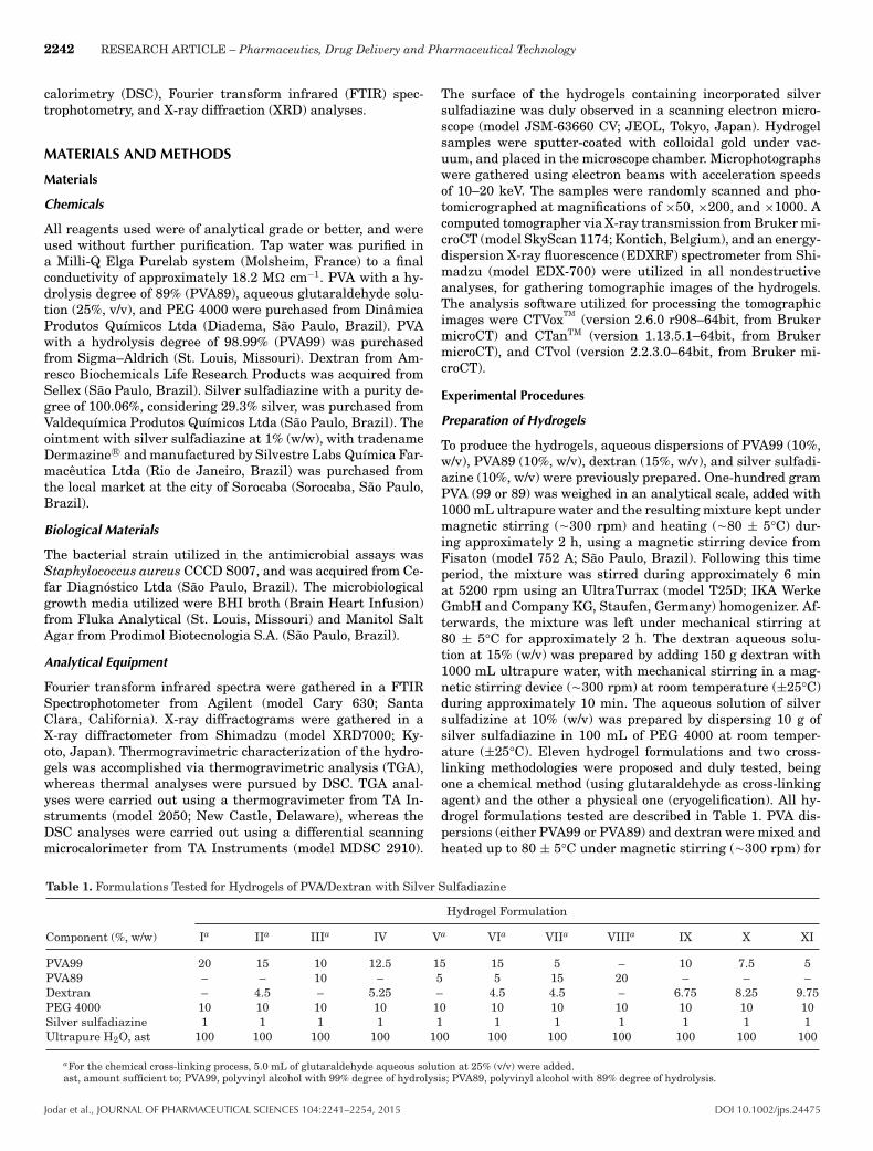
Table 1. Formulations Tested for Hydrogels of PVA/Dextran with Silver Sulfadiazine
Hydrogel Formulation
Component (%, w/w) Ia IIa IIIa IV Va VIa VIIa VIIIa IX X XI
PVA99 20 15 10 12.5 15 15 5 – 10 7.5 5PVA89 – – 10 – 5 5 15 20 – – –Dextran – 4.5 – 5.25 – 4.5 4.5 – 6.75 8.25 9.75PEG 4000 10 10 10 10 10 10 10 10 10 10 10Silver sulfadiazine 1 1 1 1 1 1 1 1 1 1 1Ultrapure H2O, ast 100 100 100 100 100 100 100 100 100 100 100
aFor the chemical cross-linking process, 5.0 mL of glutaraldehyde aqueous solution at 25% (v/v) were added.ast, amount sufficient to; PVA99, polyvinyl alcohol with 99% degree of hydrolysis; PVA89, polyvinyl alcohol with 89% degree of hydrolysis.
Jodar et al., JOURNAL OF PHARMACEUTICAL SCIENCES 104:2241–2254, 2015 DOI 10.1002/jps.24475

RESEARCH ARTICLE – Pharmaceutics, Drug Delivery and Pharmaceutical Technology 2243
approximately 10 min. Subsequently, the silver sulfadiazinesolution at 10% (w/w) was incorporated and the resulting dis-persion was maintained under magnetic stirring for an extra30 min (�300 rpm). After this time period, the dispersions werekept at rest until temperature equilibration with room temper-ature (±25°C). Subsequently, 5.0 mL glutaraldehyde solutionat 25% (v/v) were added, the resulting dispersion duly homoge-nized, poured into plastic molds, and dried at 60°C in a Fanemincubator (model 515 A; Karnataka, India). After completionof the hydrogel’s polymerization process, the hydrogels werethoroughly washed with phosphate buffer pH 7.0, so as to vir-tually eliminating all nonreacted (free) glutaraldehyde. Thehydrogel samples were then cut (2.5 × 3.0 cm2) wrapped inPVC (polyvinyl chloride) film and stored in the dark. For thosehydrogel formulations prepared via criogelification, followinghomogenization and rest, they were poured into plastic moldsand kept at −4°C during 14 h and then at 25°C during 2 h,for five consecutive cycles. The hydrogels were then dried in anincubator set at 60°C.
Morphological Characteristics
The hydrogel films formed were initially analyzed with respectto their macroscopic morphological characteristics, consideringthe absence of air bubbles, the presence of cracks and homo-geneity. This evaluation was performed by both naked eye andmagnifying glass. Thickness of the hydrogel films was deter-mined with the aid of a manual measuring caliper from Mitu-toyo Sul Americana Ltda (Suzano, Brazil). Each hydrogel filmwas measured at 10 different points, and the average thicknessand associated standard deviation were duly calculated.
SEM Analyses
The surface of the hydrogel films containing silver sulfadi-azine was duly observed in a scanning electron microscope(from JEOL; model JSM-63660). Hydrogel samples were coatedwith colloidal gold under vacuum, and placed in the micro-scope chamber. Microphotographs were gathered using electronbeams with 10 keV energy.
FTIR Spectrophotometry Analyses
The infrared spectra of samples of silver sulfadiazine, and ofsamples of selected hydrogels (viz., formulation III) withoutand with incorporated silver sulfadiazine, were gathered usingan FTIR spectrophotometer from Agilent (model Cary 630) inthe wavelength range from 4000 to 400 cm−1, with a resolutionof 4 cm−1 and using Happ-Genzel apodization.
XRD Analyses
X-ray diffractograms of silver sulfadiazine, and of samples ofselected hydrogels (viz., formulation III) without and with in-corporated silver sulfadiazine, were gathered in a X-ray Diffrac-tometer from Shimadzu (model XRD7000), using X-ray radi-ation from a copper lamp with radiation K" (8 = 1.5418 A)filtered through a Cu target. The X-ray scanning was performedat diffraction angles of 2-2 (from 5° to 90°, with increments of0.02° and rate of 2°/min), with a voltage of 40 kV, electric cur-rent intensity of 30 mA, and X-ray power of 3 kW.
Mechanical Resistance Properties
Mechanical properties of the hydrogels were evaluated using atexturometer from Stabile Micro Systems (model TA-TX Plus;
Godalming, United Kingdom), evaluating parameters such asresistance to perforation, relaxation, and resilience. The de-termination parameters were set as distance of 5 mm for theperforation resistance tests, distance of 2 mm for both resilienceand relaxation tests, and a maximum force of 5 kg for all tests.All determinations were performed in triplicate.
Thermal Analyses via TGA and DSC
Thermogravimetric characterization of the hydrogel formula-tions was accomplished via TGA, whereas thermal analyseswere pursued by DSC. The TGA analyses were carried out ina thermogravimeter from TA Instruments (model 2050), usingalumina pans for containing the hydrogel samples under inert(argon) atmosphere. For the thermogravimetric analyses, sam-ples were heated from approximately 20°C up to 500°C, anddata (weight loss) were gathered at a sampling rate of 2.0 s perdata point. For the DSC analyses, one has used a differentialscanning microcalorimeter also from TA Instruments (modelMDSC 2910). For every calorimetric assay, approximately 7 (be-tween 6.48 and 7.93) mg of hydrogel formulation were weigheddirectly into the interior of high-pressure aluminum pans, andduly sealed by pressure. A reference aluminum pan was alsoprepared by simply sealing air inside an empty case. The sam-ples were then subjected to a linear temperature increase fromapproximately 20°C up to 250°C, at a constant heating rate of10°C/min, under an inert atmosphere maintained with a con-stant flow of argon of 50 mL/min, during which the amount ofheat absorbed by the hydrogel samples was recorded. Duringthe analyses, data (heat absorption) were gathered at a sam-pling rate of 0.2 s per data point.
Swelling Degree
The degree of swelling of the hydrogels was determined accord-ing to the methodology described by Pal and Pal,18 using theequation: Swelling degree = [
(Mt − M0)/M0] × 100, where Mt
is the mass of swelled sample at time t, and M0 is the initialmass of dry sample. All hydrogel samples were put in contactwith 50 mL ultrapure water during 1, 2, 3, 6, 12, 24, and 48 h.All swelling assays were carried out in triplicate.
Moisture Content Analyses via Infrared Heating
For determination of the moisture content, a moisture ana-lyzer producing heating via an halogen lamp was used (Shi-madzu; model MOC63U), equipped with a programmable timer,self-calibration, and temperature adjusting between 50°C and200°C. The parameters utilized were temperature of 60°C in“slow” mode.
Silver Content Determination by X-ray Fluorescence Analyses
The silver content of the hydrogels impregnated with silversulfadiazine was determined using a EDXRF spectrophotome-ter from Shimadzu (model EDX-700), which comprised an Rhtube and a Si(Li) semiconductor detector. Voltage applied tothe X-ray tube was of 50 kV, with 25% lag time in the detector.The spectra were gathered sequentially, with a resolution of0.02 keV, from 0 to 40 keV. The irradiation time was set at120 s.
Tomographic Analyses via X-ray Transmission
The tomographic images were gathered using a third gener-ation X-ray transmission tomograph.19 The hydrogel samples
DOI 10.1002/jps.24475 Jodar et al., JOURNAL OF PHARMACEUTICAL SCIENCES 104:2241–2254, 2015

2244 RESEARCH ARTICLE – Pharmaceutics, Drug Delivery and Pharmaceutical Technology
were placed inside the tomograph chamber, and image sliceswere gathered using the following configurations of the tomo-graphic system: operating voltage set at 29 kV and electric cur-rent with 415 :A. The technique employed for obtaining thetomographic image involved acquisition of a large number ofradiographs of the object (image slices), obtained by measuringthe intensity of X-rays transmitted through the hydrogel sam-ple, at different angular positions. The hydrogel samples wererotated 180°, with angular increments of 1°, producing 180 ra-diographs (projections) per image, each containing 652 × 652pixels with a spatial resolution of 15.85 :m. At the outlet of theX-ray source, one utilized an Al filter with 0.25 mm thickness.Appropriate mathematical algorithms were then used to re-construct the 3D tomographic images of the hydrogel samples,through the appropriate composition of bi-dimensional (2D) im-ages. The 3D images possessed 652 × 652 × 652 pixels and thesame spatial resolution of the 2D images, and thus the volumeof data generated for each hydrogel sample is isotropic withrelation to the spatial resolution. Having all the projections(radiographs gathered at each angular position), one utilizedthe software NRecon
TM from Bruker microCT (version 1.6.9.4),which uses the algorithm of Feldkamp et al.20 in the process ofreconstructing the tomographic images.
Evaluation of Antimicrobial Activity of the Hydrogels via the AgarDiffusion Method
The antimicrobial efficacy of the hydrogels integrating sil-ver sulfadiazine was determined by applying the agar diffu-sion technique for susceptibility to antimicrobials describedby Bauer et al.21 and Jorgensen and Ferraro,22 as well as ac-cording to the standards of the Clinical and Laboratory Stan-dards Institute.23 The assays were carried out in duplicateusing a strain of Staphylococcus aureus (CCCD S007 – Ce-far Diagnostico Ltda, Sao Paulo, Brazil). The Staphylococcusaureus strain was, initially, inoculated in BHI nutritive broth(Brain Heart Infusion) and maintained at 37 ± 0.5°C during24 h, to allow growth of the bacteria. Following this time pe-riod, the bacterial culture was inoculated by spreading on twoPetri dishes containing mannitol salt agar as culture medium,and the samples to be tested were then applied on the inocu-lated medium, in duplicate, with the aid of sterile tweezers neara Bunsen burner flame: hydrogel discs with silver sulfadiazine(�7.0 mm in diameter and average weight of 8.00 ± 0.01 mg),the negative standard of inhibition (hydrogel disc without sil-ver sulfadiazine, with �7.0 mm in diameter), and the positivestandard of inhibition [10 mg of reference ointment of silversulfadiazine at 1% (w/w) (with tradename Dermazine R©)]. ThePetri dishes were then inverted and incubated under aerobicconditions at 37 ± 0.5°C during 24 h. After this time period,the plates were visually inspected for observation (or not) ofany growth inhibition halos.
Study of the Kinetics of Drug Release (Bioavailability) from theHydrogel
The drug release experiments were carried out by measuringthe potential difference in an aqueous medium where a por-tion of hydrogel-containing silver sulfadiazine was immersed,using a combined silver electrode (model 9157; ThermoScien-tific Orion, San Jose, California) connected to a millivoltmeter(model pH300; pH analyzer from Analyser InstrumentacaoAnalıtica, Sao Paulo, Brazil). Initially, one tested magnetic
stirring of the aqueous medium with the immersed hydrogelsample, as measures involving specific electrodes require ho-mogenization of the reaction medium. However, the magneticstirring promoted a complete (and accelerated) dissolution ofthe hydrogel. Hence, one opted to integrate a peristaltic pump,for continuous recirculation of the medium without promotingturbulent flow conditions. The peristaltic pump used (modelMP13GJ-4; Ismatec, Glattbrugg, Switzerland) was connectedto Tygon tubing (2.54 mm internal diameter). A weight sam-ple of hydrogel of 0.2030 g [containing �1% (w/w) silver sul-fadiazine] was immersed in 20 mL ultrapure water at roomtemperature (25 ± 0.1°C). Potential difference of the mediumwhere the hydrogel was immersed was gathered at predeter-mined intervals of time, using the aforementioned combined sil-ver electrode connected to a millivoltmeter. All measurementswere gathered in mV by time of immersion of the hydrogel.The experiments were terminated when a silver sulfadiazineconcentration plateau was reached.
RESULTS AND DISCUSSION
In the hydrogel formulations proposed in the research effortdescribed herein, combinations of PVA with several degrees ofhydrolysis, dextran, and silver sulfadiazine at 1% (w/w) wereevaluated. The concentration of 1% (w/w) of silver sulfadiazinewas chosen because of both its consensus in the treatment ofburns and to the fact that commercial pharmaceutical formu-lations present such concentration. PVA and dextran were thepolymers chosen to be utilized in the preparation of suitablehydrogels, as they present good characteristics for producingfilms.24 In a review paper by Baker et al.,25 these researchersclaim that it is possible to produce soft hydrogels with approxi-mately 10% (w/w) PVA and rigid hydrogels with approximately50%–60% (w/w) PVA. The major purpose of the research effortdescribed herein was to produce a hydrogel with malleability,although with a certain resistance so that it could be utilizedfor topical applications. With this in mind, one tested formu-lations encompassing up to 24.5% (w/w) of polymers. PVA sol-ubility increases with decreasing degree of hydrolysis. Thus,by mixing PVA polymers with different degrees of hydrolysis,it is possible to modulate the properties of both solubility andswelling of PVA and, consequently, release of the drug.26 Thecross-linking process via cryogellification produced nonuniformhydrogels with uneven distribution of silver sulfadiazine, andthus one opted by using the chemical cross-linking process withglutaraldehyde. The hydrogels were analyzed with respect tohomogeneity, resistance to manipulation, and color homogene-ity. Formulations I, II, III, V, VI, VII, and VIII (see Table 1),chemically cross-linked with glutaraldehyde, were selected forthe swelling and mechanical resistance tests.
Swelling Degree
Formulations VII and VIII were totally dissolved before com-pleting 1 h of testing, and so they were discarded from furthertesting, by proving to be nonappropriate for using as woundbandages. From all the formulations tested (see Table 1), hy-drogel formulations II, III, and V produced the best results (seeTable 2). From inspection of Table 2, it can be observed that amaximum swelling occurred during the first hours of contactwith ultrapure water (up to 6 h). After this time period, thehydrogels began to lose mass, probably because of dissolution
Jodar et al., JOURNAL OF PHARMACEUTICAL SCIENCES 104:2241–2254, 2015 DOI 10.1002/jps.24475

RESEARCH ARTICLE – Pharmaceutics, Drug Delivery and Pharmaceutical Technology 2245
Table 2. Average Swelling Degrees and Associated StandardDeviations (n = 3) of Selected Hydrogel Formulations FollowingImmersion in Water for 1, 3, 6, 12, 24, and 48 h
Immersion Formulation II Formulation III Formulation VTime (h) (% ± F) (% ± F) (% ± F)
1 200.40 ± 3.88 190.00 ± 1.56 202.50 ± 5.093 210.56 ± 2.42 220.00 ± 0.87 234.20 ± 2.396 232.61 ± 0.73 266.00 ± 0.81 225.99 ± 1.8512 179.42 ± 0.53 197.15 ± 2.20 186.54 ± 2.5624 128.71 ± 1.29 194.32 ± 3.16 155.52 ± 5.0048 104.76 ± 2.34 172.03 ± 3.01 128.85 ± 1.28
of the films. Such loss of mass was smaller for the hydrogel pro-duced according to formulation III, and thus this formulationwas selected for further work.
Mechanical Resistance
To determine the mechanical properties of the selected hydro-gels, these were evaluated with respect to perforation, relax-ation, and resilience. The mechanical properties of the hydrogelfilms are mainly related with the polymer’s ability to form bondsin polymer chains, making their separation difficult when sub-ject to mechanical forces.27 The plasticizer used also has aninfluence on these properties.28 The analysis of the results con-sidered the average thickness (0.50 ± 0.03 mm) and the averageweight (344.18 ± 2.65 mg) of the hydrogels with an area of 25× 30 mm2. The results produced during evaluation of the me-chanical resistance of selected hydrogel formulations with andwithout silver sulfadiazine, with respect to perforation, relax-ation, and resilience are displayed in Table 3.
Hydrogel formulations II and V, containing in their compo-sition a higher amount of PVA99 (15%, w/w, of the formula-tion), exhibited a higher resistance than the hydrogel producedaccording to formulation III, which contained only 10% (w/w)PVA99 in its composition. The hydrogel produced according toformulation II, incorporating silver sulfadiazine, exhibited alower resistance to perforation than its counterpart withoutsilver sulfadiazine; however, it presented higher values rela-tive to resilience and relaxation. Hence, it may be concludedthat the presence of silver sulfadiazine improved its viscoelas-tic characteristics of deformation and molecular relaxation. Hy-drogels produced according to formulations III and V presenteda slight decrease in the mechanical resistance for the parame-ters analyzed, except for the hydrogel produced according to for-mulation V with silver sulfadiazine, which presented a bettermechanical resistance in relation to resilience than its counter-
part without silver sulfadiazine. The major goal of the researchwork undertaken was to develop a hydrogel suitable for appli-cation in skin wounds. In our perspective, a hydrogel bandageapplied directly to a skin wound that produces exudate shouldnot strongly adhere to the wound, because its exchange wouldbecome unnecessary painful to the patient and would not allowre-epithelialization of the wound. Thus, perforation, relaxation,and resilience were the mechanical resistance parameters eval-uated, whereas adhesiveness or bioadhesiveness were not at allconsidered important in the research work developed. The op-timized hydrogel is intended to be applied to a skin wound andmaintained in place by a bandage, and not to stick or adhereto the wounded skin. Our perspective is supported by liter-ature references.29,30 Hydrogels act as moist wound dressingmaterials, and are able to both absorb and retain the woundexudates as well as any contaminating bodies such as bacteria,within their network structure. Additionally, hydrogels havebeen found to both promote fibroblast proliferation by reduc-ing fluid loss from the wound surface and protect the woundfrom external noxae, sine qua non conditions necessary for arapid wound healing.30 Hydrogels also help in maintaining asuitable microclimate on the wound surface, of utmost impor-tance for the biosynthetic reactions necessary for cellular ac-tivities. Proliferation of fibroblasts is necessary for a completere-epithelialization of the wound, which starts from its edge. Ashydrogels help to maintain the wound moist, keratinocytes canthus migrate on its surface.30 Hence, hydrogel-based dressingsare of undeniable value in a wide spectrum of wounds, and havethe potential to be utilized on necrotic, sloughy, granulating,and epithelializing wounds, and can also be applied in infectedwounds, provided that the patient is being treated with sys-temic antibiotics and the dressing is changed on a daily basis.In their work, Rippon et al.29 also describe several wound dress-ing definitions, to include (1) adherent dressings (dressings thatare likely to adhere to any type of drying wound, viz., cottongauze and/or simple dressing pads), (2) low-adherent dressings(dressings with a wound contact surface that is designed toreduce adherence, viz., absorbent wound dressings), (3) nonad-herent dressings (dressings that maintain a moist gel layer overthe wound that is not expected to adhere, provided that it is notallowed to dry out, viz., alginates/hydrocolloids and/or hydro-gels/hydrofibres), and (4) atraumatic dressings (dressings thatdo not cause trauma either to the wound or the peri-woundskin upon removal, viz., soft silicone dressings). Clearly, thehydrogels developed, optimized, and fully characterized in theresearch word described herein, fall in category (3) of the afore-mentioned descriptions.
Table 3. Results from the Mechanical Resistance Tests (kg ± F) Performed to Selected Hydrogels, Relative to Perforation, Relaxation, andResilience (s ± F), as a Function of Applied Force (kg) (AUC)
Perforation Relaxation Resilience
Hydrogel Formulation Force (kg) AUC (kg/s) Force (kg) AUC (kg/s) Force (kg) AUC (kg/s)
II 2.571 ± 0.320 7.445 ± 1.433 0.439 ± 0.072 0.630 ± 0.095 0.352 ± 0.082 0.498 ± 0.135II w/SDZ 0.781 ± 0.165 3.351 ± 0.225 0.690 ± 0.032 2.974 ± 0.790 0.136 ± 0.037 1.880 ± 0.020III 0.791 ± 0.162 0.944 ± 0.269 0.258 ± 0.027 0.355 ± 0.039 0.335 ± 0.020 0.439 ± 0.335III w/SDZ 0.765 ± 0.234 0.915 ± 0.279 0.215 ± 0.043 0.303 ± 0.104 0.193 ± 0.005 0.514 ± 0.032V 1.373 ± 0.063 2.224 ± 0.190 0.224 ± 0.027 0.298 ± 0.056 0.252 ± 0.017 0.535 ± 0.042V w/SDZ 0.862 ± 0.181 3.134 ± 0.077 0.456 ± 0.054 0.580 ± 0.032 0.688 ± 0.076 1.926 ± 0.040
n = 3.AUC, area under the curve; SDZ, silver sulfadiazine.
DOI 10.1002/jps.24475 Jodar et al., JOURNAL OF PHARMACEUTICAL SCIENCES 104:2241–2254, 2015
2246 RESEARCH ARTICLE – Pharmaceutics, Drug Delivery and Pharmaceutical Technology
Wound dressings containing silver may be utilized in acutewounds, such as those produced by burns or surgery and inchronic wounds with infections. The major functions of antimi-crobial dressings, such as those containing silver, involve re-ducing the microbial load in acute or chronic wounds, or act asan antimicrobial barrier for wounds with high risk of infectionor reinfection.31 Additionally, dressings allow the creation ofa moist and warm environment surrounding the wound, thusbringing benefits to the healing process.32 The developed prod-uct presents low adhesion to the injured tissue, which is adesirable feature, as adherent dressings may cause pain andeven more tissue damage during removal.32 However, their fix-ing requires the use of natural or synthetic bandages (cottonwool, lint, and gauzes). Because of the product characteristics,this will virtually not harm the performance of the hydrogel.Hence, hydrogel adhesion to bandage materials will not be aproblem. The developed hydrogels possess PVC in their compo-sition, which is a hydrophobic polymer with a low-surface freeenergy33 that do not favor attraction of the water moleculespresent in the dispersion containing the polymers. As a conse-quence, this promotes the nonadherence of the hydrogel to thebandages and healings that may be utilized.
Structural Microanalysis of Selected Hydrogels IncorporatingSilver Sulfadiazine via SEM
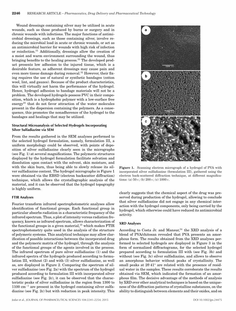
From the results gathered in the SEM analyses performed tothe selected hydrogel formulation, namely, formulation III, auniform morphology could be observed, with points of depo-sition of silver sulfadiazine clearly seen in the micrographs(see Fig. 1) at several magnifications. The polymeric structuresdisplayed by the hydrogel formulation facilitate solvation anddissolution upon contact with the solvent, skin moisture, andwith the skin burn, thus being able to slowly release its sil-ver sulfadiazine content. The hydrogel micrographs in Figure 1were obtained via the EBSD (electron backscatter diffraction)technique, which allows the crystallographic analysis of thematerial, and it can be observed that the hydrogel topographyis highly uniform.
FTIR Analyses
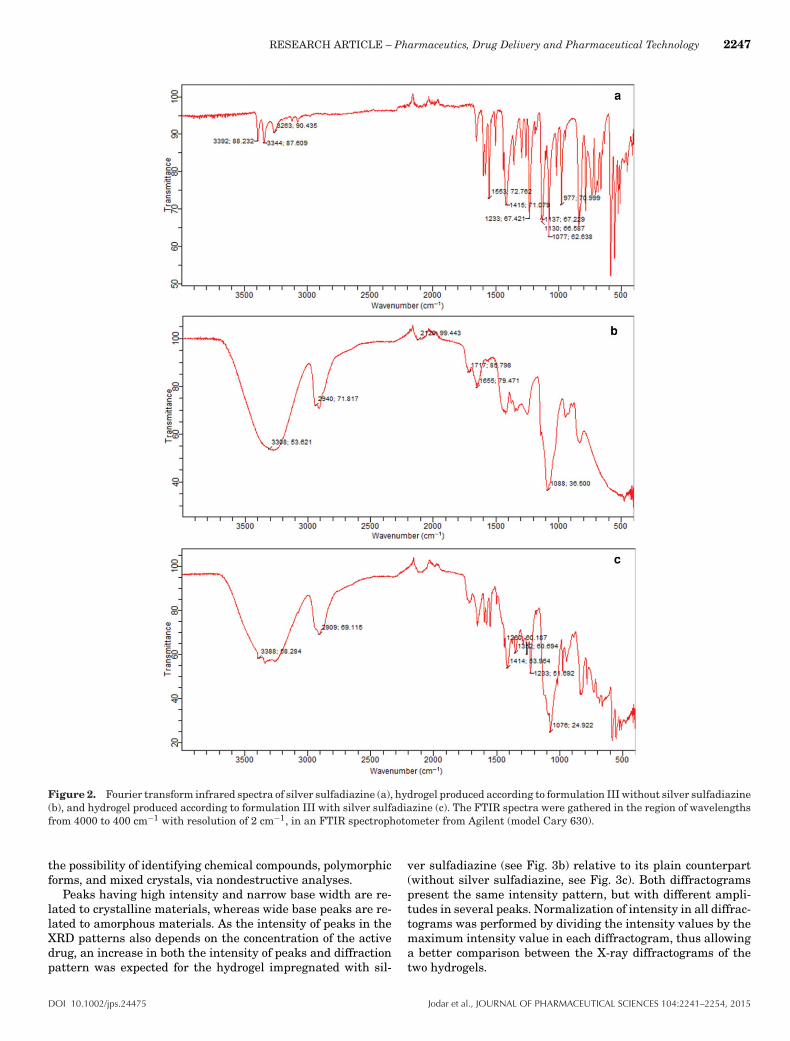
Fourier transform infrared spectrophotometric analyses allowidentification of functional groups. Each functional group inparticular absorbs radiation in a characteristic frequency of theinfrared spectrum. Thus, a plot of intensity versus radiation fre-quency, known as infrared spectrum, allows characterization ofthe functional groups in a given material,35 which makes FTIRspectrophotometry quite used in the analysis of the structureof polymeric systems. This analytical technique may allow clar-ification of possible interactions between the incorporated drugand the polymeric matrix of the hydrogel, through the analysisof the functional groups of the agents involved in the process.The infrared spectrum of pure silver sulfadiazine (1) and theinfrared spectra of the hydrogels produced according to formu-lation III, without (2) and with (3) silver sulfadiazine, as wellas, are displayed in Figure 2. Comparing the spectrum of sil-ver sulfadiazine (see Fig. 2a) with the spectrum of the hydrogelproduced according to formulation III with incorporated silversulfadiazine (see Fig. 2c), it can be observed that the charac-teristic peaks of silver sulfadiazine in the region from 1300 to1100 cm−1 are present in the hydrogel containing silver sulfa-diazine (see Fig. 2c) but with reduction in peak intensity. This
Figure 1. Scanning electron micrograph of a hydrogel of PVA withincorporated silver sulfadiazine (formulation III), gathered using theelectron back-scattered diffraction technique, at different magnifica-tions: (a) 50×; (b) 200×; (c) 1000×.
clearly suggests that the chemical aspect of the drug was pre-served during production of the hydrogel, allowing to concludethat silver sulfadiazine did not engage in any chemical inter-action with the hydrogel components, only being carried by thehydrogel, which otherwise could have reduced its antimicrobialactivity.
XRD Analyses
According to Costa Jr. and Mansur,34 the XRD analysis of ablend of PVA/chitosan revealed that PVA presents an amor-phous form. The results obtained from the XRD analyses per-formed to selected hydrogels are displayed in Figure 3 in theform of normalized diffractograms, for the selected hydrogelprepared according to formulation III with (see Fig. 3b) andwithout (see Fig. 3c) silver sulfadiazine, and allows to observean amorphous behavior without peaks of crystallinity. Thesmall peaks at 22 41° are related with the presence of resid-ual water in the samples. These results corroborate the resultsobtained via SEM, which indicated the formation of an amor-phous film. The decisive advantage of the methods of analysisby XRD over other analytical techniques is based on the unique-ness of the diffraction patterns of crystalline substances, on theability to distinguish between elements and their oxides, and on
Jodar et al., JOURNAL OF PHARMACEUTICAL SCIENCES 104:2241–2254, 2015 DOI 10.1002/jps.24475
RESEARCH ARTICLE – Pharmaceutics, Drug Delivery and Pharmaceutical Technology 2247
Figure 2. Fourier transform infrared spectra of silver sulfadiazine (a), hydrogel produced according to formulation III without silver sulfadiazine(b), and hydrogel produced according to formulation III with silver sulfadiazine (c). The FTIR spectra were gathered in the region of wavelengthsfrom 4000 to 400 cm−1 with resolution of 2 cm−1, in an FTIR spectrophotometer from Agilent (model Cary 630).
the possibility of identifying chemical compounds, polymorphicforms, and mixed crystals, via nondestructive analyses.
Peaks having high intensity and narrow base width are re-lated to crystalline materials, whereas wide base peaks are re-lated to amorphous materials. As the intensity of peaks in theXRD patterns also depends on the concentration of the activedrug, an increase in both the intensity of peaks and diffractionpattern was expected for the hydrogel impregnated with sil-
ver sulfadiazine (see Fig. 3b) relative to its plain counterpart(without silver sulfadiazine, see Fig. 3c). Both diffractogramspresent the same intensity pattern, but with different ampli-tudes in several peaks. Normalization of intensity in all diffrac-tograms was performed by dividing the intensity values by themaximum intensity value in each diffractogram, thus allowinga better comparison between the X-ray diffractograms of thetwo hydrogels.
DOI 10.1002/jps.24475 Jodar et al., JOURNAL OF PHARMACEUTICAL SCIENCES 104:2241–2254, 2015

2248 RESEARCH ARTICLE – Pharmaceutics, Drug Delivery and Pharmaceutical Technology
Figure 3. X-ray diffractograms of samples of silver sulfadiazine (a) and of samples of hydrogel prepared according to formulation III with silversulfadiazine (b), and of hydrogel prepared according to formulation III without silver sulfadiazine (c). The X-ray diffractograms were gatheredusing Cu target-filtered X-rays. The analyses were carried out at diffraction angles of 2-2 (from 5° to 90°, with increments of 0.02° and rateof 2°/min−1) with voltage of 40 kV, current intensity of 30 mA, and X-ray power of 3 kW, in an X-ray diffractometer from Shimadzu (modelXRD7000).
X-ray Transmission Tomography
From the tomographic analyses via X-ray transmission per-formed to the hydrogel prepared according to formulation IIIwith impregnated silver sulfadiazine (see Fig. 4), it can be ob-served that silver sulfadiazine migrate to the surface of thehydrogel (purple-bluish layer; see Figs. 4a and 4b) during thepolymerization process. Because of absorbing more radiationand of its higher atomic density, silver is thus in greater evi-dence as highlighted in the tomographic images displayed asFigure 4. These results are in close agreement with those ob-tained by infrared spectrophotometry that indicated that silversulfadiazine probably did not engage in any chemical bondingwith the polymeric matrix. This is clearly an important and pos-itive data, as by not engaging in any chemical bonding with thepolymeric surface, silver sulfadiazine becomes readily availableand maintains its antimicrobial activity.
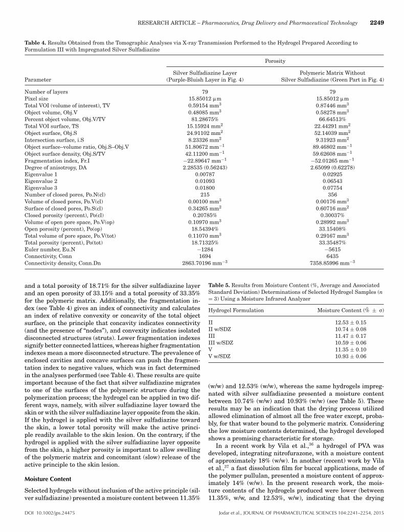
Additionally, as can be seen from inspection of the data inTable 4, the layer of silver sulfadiazine (in purple-bluish inFig. 4) is significantly less porous than the polymeric matrixitself (in green in Fig. 4), and presented a volume 1.5 timessmaller than that of the polymeric film. Anisotropy is the prop-erty of being directionally dependent, as opposed to isotropy,which implies identical properties in all directions. Anisotropycan thus be defined as a difference, when measured alongdifferent axes, in a material’s physical or mechanical prop-erties (e.g., absorbance). The degree of anisotropy, calculatedas Degree of anisotropy =
[1 −
(min Eigenvaluemax Eigenvalue
)], is 0 for total
isotropy and 1 for total anisotropy. Hence, as can be seen frominspection of the data in Table 4, the degree of anisotropy is0.562 and 0.623 for the layer of silver sulfadiazine (in purple-bluish in Fig. 4) and the polymeric matrix (in green in Fig. 4), re-spectively, values that are half-way in the aforementioned scale.When analyzing the structure of these two interconnecting re-gions, one finds that the layer of silver sulfadiazine has a lowernumber of closed pores (viz., 215) than that of the polymericmatrix (viz., 356), corresponding to an open porosity of 18.54%
Figure 4. Images obtained by tomographic analyses via X-ray trans-mission of the hydrogel prepared according to formulation III, withimpregnated silver sulfadiazine, being (a) a cross-cut profile image ofthe hydrogel with silver sulfadiazine, and (b) slant profile image of thehydrogel with impregnated silver sulfadiazine. Three-dimensional im-age slices were gathered using an operating voltage set at 29 kV andelectric current with 415 :A.
Jodar et al., JOURNAL OF PHARMACEUTICAL SCIENCES 104:2241–2254, 2015 DOI 10.1002/jps.24475
RESEARCH ARTICLE – Pharmaceutics, Drug Delivery and Pharmaceutical Technology 2249
Table 4. Results Obtained from the Tomographic Analyses via X-ray Transmission Performed to the Hydrogel Prepared According toFormulation III with Impregnated Silver Sulfadiazine
Porosity
Silver Sulfadiazine Layer Polymeric Matrix WithoutParameter (Purple-Bluish Layer in Fig. 4) Silver Sulfadiazine (Green Part in Fig. 4)
Number of layers 79 79Pixel size 15.85012 :m 15.85012 :mTotal VOI (volume of interest), TV 0.59154 mm3 0.87446 mm3
Object volume, Obj.V 0.48085 mm3 0.58278 mm3
Percent object volume, Obj.V/TV 81.28675% 66.64513%Total VOI surface, TS 15.15924 mm2 22.44291 mm2
Object surface, Obj.S 24.91102 mm2 52.14039 mm2
Intersection surface, i.S 8.23326 mm2 9.31923 mm2
Object surface–volume ratio, Obj.S–Obj.V 51.80672 mm−1 89.46802 mm−1
Object surface density, Obj.S/TV 42.11200 mm−1 59.62608 mm−1
Fragmentation index, Fr.I −22.89647 mm−1 −52.01265 mm−1
Degree of anisotropy, DA 2.28535 (0.56243) 2.65099 (0.62278)Eigenvalue 1 0.00787 0.02925Eigenvalue 2 0.01093 0.06543Eigenvalue 3 0.01800 0.07754Number of closed pores, Po.N(cl) 215 356Volume of closed pores, Po.V(cl) 0.00100 mm3 0.00176 mm3
Surface of closed pores, Po.S(cl) 0.34265 mm2 0.60716 mm2
Closed porosity (percent), Po(cl) 0.20785% 0.30037%Volume of open pore space, Po.V(op) 0.10970 mm3 0.28992 mm3
Open porosity (percent), Po(op) 18.54394% 33.15408%Total volume of pore space, Po.V(tot) 0.11070 mm3 0.29167 mm3
Total porosity (percent), Po(tot) 18.71325% 33.35487%Euler number, Eu.N −1284 −5615Connectivity, Conn 1694 6435Connectivity density, Conn.Dn 2863.70196 mm−3 7358.85996 mm−3
and a total porosity of 18.71% for the silver sulfadiazine layerand an open porosity of 33.15% and a total porosity of 33.35%for the polymeric matrix. Additionally, the fragmentation in-dex (see Table 4) gives an index of connectivity and calculatesan index of relative convexity or concavity of the total objectsurface, on the principle that concavity indicates connectivity(and the presence of “nodes”), and convexity indicates isolateddisconnected structures (struts). Lower fragmentation indexessignify better connected lattices, whereas higher fragmentationindexes mean a more disconnected structure. The prevalence ofenclosed cavities and concave surfaces can push the fragmen-tation index to negative values, which was in fact determinedin the analyses performed (see Table 4). These results are quiteimportant because of the fact that silver sulfadiazine migratesto one of the surfaces of the polymeric structure during thepolymerization process; the hydrogel can be applied in two dif-ferent ways, namely, with silver sulfadiazine layer toward theskin or with the silver sulfadiazine layer opposite from the skin.If the hydrogel is applied with the silver sulfadiazine towardthe skin, a lower total porosity will make the active princi-ple readily available to the skin lesion. On the contrary, if thehydrogel is applied with the silver sulfadiazine layer oppositefrom the skin, a higher porosity is important to allow swellingof the polymeric matrix and concomitant (slow) release of theactive principle to the skin lesion.
Moisture Content
Selected hydrogels without inclusion of the active principle (sil-ver sulfadiazine) presented a moisture content between 11.35%
Table 5. Results from Moisture Content (%, Average and AssociatedStandard Deviation) Determinations of Selected Hydrogel Samples (n= 3) Using a Moisture Infrared Analyzer
Hydrogel Formulation Moisture Content (% ± F)
II 12.53 ± 0.15II w/SDZ 10.74 ± 0.08III 11.47 ± 0.17III w/SDZ 10.59 ± 0.06V 11.35 ± 0.10V w/SDZ 10.93 ± 0.06
(w/w) and 12.53% (w/w), whereas the same hydrogels impreg-nated with silver sulfadiazine presented a moisture contentbetween 10.74% (w/w) and 10.93% (w/w) (see Table 5). Theseresults may be an indication that the drying process utilizedallowed elimination of almost all the free water except, proba-bly, for that water bound to the polymeric matrix. Consideringthe low moisture contents determined, the hydrogel developedshows a promising characteristic for storage.
In a recent work by Vila et al.,36 a hydrogel of PVA wasdeveloped, integrating nitrofurazone, with a moisture contentof approximately 18% (w/w). In another (recent) work by Vilaet al.,37 a fast dissolution film for buccal applications, made ofthe polymer pullulan, presented a moisture content of approx-imately 14% (w/w). In the present research work, the mois-ture contents of the hydrogels produced were lower (between11.35%, w/w, and 12.53%, w/w), indicating that the drying
DOI 10.1002/jps.24475 Jodar et al., JOURNAL OF PHARMACEUTICAL SCIENCES 104:2241–2254, 2015

2250 RESEARCH ARTICLE – Pharmaceutics, Drug Delivery and Pharmaceutical Technology
Table 6. Average Values and Associated Standard Deviations (n = 3)of the Silver Contents (ppm) in Selected Hydrogels Obtained byEnergy-Dispersion X-ray Fluorescence Analyses (n = 3)
Hydrogel Silver Silver Silver SulfadiazineFormulation Content (ppm) Content (%) Content (%) in the Hydrogel
II 301.00 ± 10.00 0.03010 1.030III 325.50 ± 15.00 0.03255 1.085V 384.50 ± 94.00 0.03845 1.280
process utilized was efficient to remove the water, producingdried hydrogels that can be stored for long periods of time be-cause of their relatively low-moisture content.
Silver Content Determination by X-ray Fluorescence Analyses
Table 6 displays the results (average and standard deviation)of silver content in selected hydrogels, obtained by EDXRF.All analyses were carried out in triplicate. The hydrogels wereprepared so as to present 1% (w/w) silver sulfadiazine. Themolecular weight of silver sulfadiazine is 358.4 g/mol, whereasthat of silver is 107.86 g/mol. Thus, in a molecule of silversulfadiazine, 30% of the weight corresponds to silver. Whenpreparing 100 mL of hydrogel formulation, one utilized 0.1 gof silver sulfadiazine (1%, w/w). Hence, in 0.1 g silver sulfa-diazine, there is 0.030% silver. From the results obtained byEDXRF analyses, hydrogels prepared according to formula-tions II and III presented a silver sulfadiazine content withinthe expected value (1.030% and 1.085%, respectively). For thehydrogel prepared according to formulation V, the silver sulfa-diazine content determined was slightly higher than expected(1.280%, w/w), probably because of an uneven distribution ofsilver sulfadiazine in the hydrogel.
Antimicrobial Activity of the Hydrogel
The hydrogel prepared according to formulation III, withoutthe active principle (functioning as negative standard of inhi-bition), did not inhibit bacterial growth (no growth inhibitionhalo produced). The hydrogel film containing silver sulfadiazine(8 mg of hydrogel film, containing �0.08 mg silver sulfadi-azine) promoted a microbial growth inhibition halo (diameter of1.45 cm) slightly higher (�1.54×) than that of the positivestandard of inhibition (silver sulfadiazine ointment at 1%, w/w,namely, 10 mg Dermazine R© containing �0.10 mg silver sulfa-diazine, halo diameter of 0.94 cm), meaning that the hydrogelcontaining impregnated silver sulfadiazine allowed a contin-uous release of the antimicrobial principle, in concentrationsable to inhibit bacterial growth.
Thermal Analyses via TGA and DSC
In general, thermal analyses are useful tools in developing for-mulations as they allow to evaluate the compatibility betweencomponents of a formulation and the stability and thermal de-composition of drugs. The data obtained by thermal analysis aredirectly related to the final quality of a pharmaceutical product,allowing to infer aspects of therapeutic efficacy of the productor the stability of the product throughout the shelf life (or va-lidity) period. TGA accompany the variation in sample massas a function of a linear increase in temperature during a pre-determined time interval, allowing to determine the thermalprofile of the hydrogels prepared. The TGA curves and the firstderivatives of the mass loss curves for the hydrogels prepared
according to formulation III are displayed in Figure 5, respec-tively, for the hydrogel without impregnated silver sulfadiazine(orange curves) and for the hydrogel with impregnated silversulfadiazine (blue curves). As can be observed from inspectionof the curves in Figure 5, the thermal behavior is quite similarin both hydrogels, with a small mass loss around approximately100°C indicating loss of water. Around 250°C, one can notice asignificant loss of mass, indicating degradation of the compo-nents in the hydrogels. The first derivative of the weight losscurve (i.e., the rate of mass change; see Fig. 5) can be used totell the points at which weight loss is most apparent (inflectionpoints), namely, 60°C and 310°C for the hydrogel without silversulfadiazine, and 65°C and 290°C for the hydrogel impregnatedwith silver sulfadiazine.
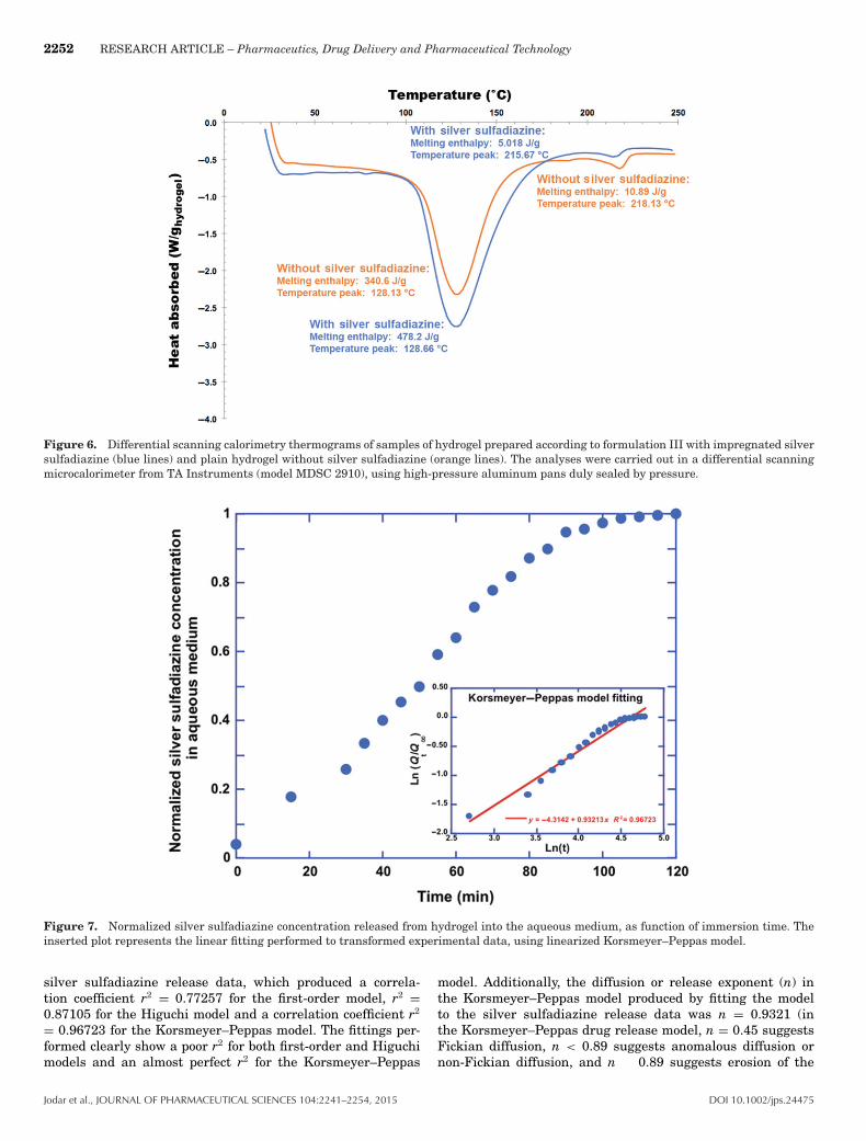
Thermal analysis by DSC is an analysis technique in whichthe temperature of a sample, compared with the tempera-ture of a thermally inert material, is recorded as the sampleis heated or cooled at a constant rate.35 The DSC techniqueof analysis thus measures the enthalpy of the samples, andmay indicate the glass transition temperature and endother-mic and/or exothermic events in the samples under scrutiny.The results of thermal analysis by DSC can be found inFigure 6. The hydrogel produced according to formulation IIIpossesses in its composition 10% (w/w) PVA with a degree ofhydrolysis of 89% and 10% (w/w) PVA with a degree of hydroly-sis of 99%. The thermogram obtained (see Fig. 6) presented twoendothermic events, one at 128.13°C (hydrogel without silversulfadiazine) and one at 128.66°C (hydrogel with silver sulfa-diazine). The other endothermic events occurred at 218.13°C(hydrogel without silver sulfadiazine) and 215.67°C (hydrogelwith silver sulfadiazine). The data obtained indicated that thecomponents utilized, in general, only slightly influenced thethermal behavior of the hydrogels, and also that incorporationof silver sulfadiazine did not compromise the stability of thehydrogel for the intended use. The presence of silver sulfadi-azine promoted a slight shift of the endothermic event closeat 200°C, from 218.13°C to 215.67°C, leading to a better ther-mal stability of the hydrogel produced according to formula-tion III. Differential scanning calorimetric analyses of hydro-gel formulations provided an insight into the state and degreeof crystallinity, and the melting and crystallization behavior ofcrystalline materials. The addition (impregnation) of silver sul-fadiazine promoted a slight increase in the melting temper-ature peak [absorption peak at 128.13°C and heat absorbed(melting enthalpy) of 340.6 J/g for the control hydrogel (de-void of any active drug) formulation, to absorption peak at�128.66°C and heat absorbed (melting enthalpy) of 478.2 J/gfor the optimized hydrogel with impregnated silver sulfadi-azine (see Fig. 6)], and slight decrease in the decompositiontemperature peak [absorption peak at 218.13°C and heat ab-sorbed (melting enthalpy) of 10.89 J/g for the control hydrogel(devoid of any active drug) formulation, to absorption peak at�215.67°C and heat absorbed (melting enthalpy) of 5.018 J/gfor the optimized hydrogel with impregnated silver sulfadiazine(see Fig. 6)], denoting a narrowing of the melting temperaturerange in the case of the optimized hydrogel formulation.
Inclusion of silver sulfadiazine in the hydrogel led to a slightdecrease in the peak of heat absorption (215.67°C), when com-pared with its blank counterpart (218.13°C), because of amor-phization of the system. The results obtained from the DSCthermal analyses carried out with both silver sulfadiazine-loaded and sulfadiazine-unloaded hydrogel (see Fig. 6) are in
Jodar et al., JOURNAL OF PHARMACEUTICAL SCIENCES 104:2241–2254, 2015 DOI 10.1002/jps.24475

RESEARCH ARTICLE – Pharmaceutics, Drug Delivery and Pharmaceutical Technology 2251
Figure 5. Thermogravimetric curves (a) and first derivative of the weight loss curves (b) of samples of hydrogel prepared according to formulationIII with impregnated silver sulfadiazine (blue lines) and plain hydrogel without silver sulfadiazine (orange lines). The analyses were carriedout in a thermogravimeter from TA Instruments (model 2050), using alumina pans for containing the hydrogel samples under inert (argon)atmosphere.
close agreement with those obtained from XRD studies (seeFig. 3); as can be seen from inspection of the DSC thermograms,impregnation of the hydrogel with silver sulfadiazine led to adecreased crystallinity with narrowing of the melting profile.Remarkably, the thermal events depicted in Figure 6 and theX-ray diffractograms depicted in Figure 3 denote a clear tran-sition from a crystalline state of the blank hydrogel (see Fig. 6,orange curve) to an amorphous counterpart (see Fig. 6, hydro-gel with impregnated silver sulfadiazine, blue curve), therebyleading to the increased stability observed for the optimizedantimicrobial hydrogel produced.
Mathematical Modeling of Drug Release from Hydrogel
To shed light on the release profile of silver sulfadiazine (sil-ver sulfadiazine) from the hydrogel, and because after the 2-h
timeframe of assays the amount of silver sulfadiazine releasedreached a plateau, one decided to apply the mathematical mod-els of first-order (ln Qt = ln Q0 + k1 × t, where Qt is the amountof drug released at time t, Q0 is the initial amount of drug indosage form, k1 is a release rate constant, and t is time), Higuchi(Qt = kH
√t, where Qt is the amount of drug released at time
t, kH is the Higuchi constant, and t is time), and Korsmeyer–Peppas (Qt/Q∞ = kKPtn, where Qt is the amount of drug releasedat time t, Q� is the total amount of drug dissolved when thedosage form (hydrogel) is exhausted, kKP is the Korsmeyer–Peppas kinetic constant, n is the diffusion or release exponent,and t is time) to the drug release data, and in fact silver sul-fadiazine release from the hydrogel did not occur by diffusionbut by erosion of the polymeric matrix instead. This is in factsupported by the model fittings performed to the experimental
DOI 10.1002/jps.24475 Jodar et al., JOURNAL OF PHARMACEUTICAL SCIENCES 104:2241–2254, 2015
2252 RESEARCH ARTICLE – Pharmaceutics, Drug Delivery and Pharmaceutical Technology
Figure 6. Differential scanning calorimetry thermograms of samples of hydrogel prepared according to formulation III with impregnated silversulfadiazine (blue lines) and plain hydrogel without silver sulfadiazine (orange lines). The analyses were carried out in a differential scanningmicrocalorimeter from TA Instruments (model MDSC 2910), using high-pressure aluminum pans duly sealed by pressure.
Figure 7. Normalized silver sulfadiazine concentration released from hydrogel into the aqueous medium, as function of immersion time. Theinserted plot represents the linear fitting performed to transformed experimental data, using linearized Korsmeyer–Peppas model.
silver sulfadiazine release data, which produced a correla-tion coefficient r2 = 0.77257 for the first-order model, r2 =0.87105 for the Higuchi model and a correlation coefficient r2
= 0.96723 for the Korsmeyer–Peppas model. The fittings per-formed clearly show a poor r2 for both first-order and Higuchimodels and an almost perfect r2 for the Korsmeyer–Peppas
model. Additionally, the diffusion or release exponent (n) inthe Korsmeyer–Peppas model produced by fitting the modelto the silver sulfadiazine release data was n = 0.9321 (inthe Korsmeyer–Peppas drug release model, n = 0.45 suggestsFickian diffusion, n < 0.89 suggests anomalous diffusion ornon-Fickian diffusion, and n � 0.89 suggests erosion of the
Jodar et al., JOURNAL OF PHARMACEUTICAL SCIENCES 104:2241–2254, 2015 DOI 10.1002/jps.24475

RESEARCH ARTICLE – Pharmaceutics, Drug Delivery and Pharmaceutical Technology 2253
polymeric chain), which clearly suggests that silver sulfadi-azine release occurred by erosion of the hydrogel’s polymericchain. The release results (in the form of normalized silver sul-fadiazine concentration in aqueous medium, see Fig. 7) havethus indicated a continuous liberation within the timeframeassayed, up to 100 min of immersion of the hydrogel in a low-volume aqueous medium, time after which the silver sulfadi-azine concentration maintained a plateau, thus confirming thata complete erosion of the polymeric matrix did occur. Althougha slower liberation is intended for real skin wound applications,one should bear in mind that the experimental setup utilizedinvolved a complete immersion of the hydrogel under staticflow conditions. In a real situation where the hydrogel is in-tended for application in a skin wound, the exudation producedshould not be as abundant, and thus erosion of the hydrogelwith concomitant release of silver sulfadiazine should be muchslower.
CONCLUSIONS
In the research effort just described, development and optimiza-tion of a hydrogel with impregnated SSDZ aiming at antimi-crobial topical applications was pursued. The developed hydro-gel displayed a good characteristic in relation to release of theactive antimicrobial principle, verified through swelling tests,kinetics of release, and antimicrobial activity, and because re-lease of silver sulfadiazine occurred by erosion of the hydrogel’spolymeric chain, it can be utilized in occlusive wound dressingsfor long periods of time. The hydrogel developed can be consid-ered as a nonadherent dressing, able to maintain a moist gellayer over the wound that is not expected to adhere, neither tothe wound or the bandage, provided that it is not allowed to dryout.
ACKNOWLEDGMENTS
Project funding by Fundacao de Amparo a Pesquisa do Es-tado de Sao Paulo (FAPESP, Sao Paulo, Brazil) (FAPESP Ref.Nos. 2012/15651-4, 2013/03181-6, and 2014/21122-0) is herebygratefully acknowledged. This work also received support fromCNPq in the form of a Research Productivity (PQ) fellowshipgranted to Victor M. Balcao. The authors have no conflicts ofinterest whatsoever to declare.
REFERENCES
1. Andrews GP, Laverty TP, Jones DS. 2009. Mucoadhesive poly-meric platforms for controlled drug delivery. Eur J Pharm Biopharm71(3):505–518.2. Ganji F, Vasheghani-Farahani S, Vasheghani-Farahani E. 2010. The-oretical description of hydrogel swelling: A review. Iranian Polym J19(5):375–398.3. Yan C, Pochan DJ. 2010. Rheological properties of peptide-basedhydrogels for biomedical and other applications. Chem Soc Rev39(9):3528–3548.4. Murphy DJ, Sankalia MG, Loughlin RG, Donnelly RF, JenkinsMG, McCarron PA. 2012. Physical characterisation and componentrelease of poly(vinyl alcohol)-tetrahydroxyborate hydrogels and theirapplicability as potential topical drug delivery systems. Int J Pharm423(2):326–334.
5. Song A, Rane AA, Christman KL. 2012. Antibacterial and cell-adhesive polypeptide and poly(ethylene glycol) hydrogel as a potentialscaffold for wound healing. Acta Biomater 8(1):41–50.6. Seabra AB, Oliveira MG. 2004. Poly(vinyl alcohol) and poly(vinylpyrrolidone) blended films for local nitric oxide release. Biomaterials25(17):3773–3782.7. Sudhakar Y, Kuotsu K, Bandyopadhyay AK. 2006. Buccal bioadhe-sive drug delivery: A promising option for orally less efficient drugs. JControl Release 114(1):15–40.8. Kharenko EA, Larionova NI, Demina NB. 2009. Mucoadhesive drugdelivery systems. Pharm Chem J 43(4):200–208.9. Dixit RP, Puthli SP. 2009. Oral strip technology: Overview and fu-ture potential. J Control Release 139(2):94–107.10. Hwang M-R, Kim JO, Lee JH, Kim YI, Kim JH, Chang SW, JinSG, Kim JA, Lyoo WS, Han SS, Ku SK, Yong CS, Choi H-G. 2010.Gentamicin-loaded wound dressing with polyvinyl alcohol/dextran hy-drogel: Gel characterization and in vivo healing evaluation. AAPSPharmSciTech 11(3):1092–1103.11. Ahmed EM. 2015. Hydrogel: Preparation, characterization, and ap-plications. J Adv Res 6(2):105–121.12. Liu Y, Vrana NE, Cahill PA, McGuinness GB. 2009. Physicallycrosslinked composite hydrogels of PVA with natural macromolecules:Structure, mechanical properties, and endothelial cell compatibility. JBiomed Mater Res B Appl Biomater 90(2):492–502.13. Gupta P, Vermani K, Garg S. 2002. Hydrogels: From controlledrelease to pH-responsive drug delivery. Drug Discov Today 7(10):569–579.14. Imren D, Gumusderelioglu M, Guner A. 2006. Synthesis andcharacterization of dextran hydrogels prepared with chlor- andnitrogen-containing crosslinkers. J Appl Polym Sci 102(5):4213–4221.15. Percival SL, Bowler PG, Russell D. 2005. Bacterial resistance tosilver in wound care. J Hosp Infect 60(1):1–7.16. Brasil. 2011. Ministerio da saude [Ministry oh Health]. Secre-taria de Ciencia e Tecnologia e Insumos Estrategicos [Secretary ofScience, Technology, and Strategic Resources]. Formulario TerapeuticoNacional 2010 [National Therapeutic Form 2010]: Rename 2010. 2nded. Brasılia: Ministerio da Saude [Ministry of Health, Brazil].17. Lichtenstein A, Margalit R. 1995. Liposome-encapsulates silver sul-fadiazine for the topical treatment of infected burns: Thermodynamicsof drug encapsulation and kinetics of drug release. J Inorg Biochem60(3):187–198.18. Pal K, Pal S. 2006. Development of porous hydroxyapatite scaffolds.Mater Manuf Process 21(3):325–328.19. Oliveira Junior JMR, Martins ACG. 2009. Construction and test oflow cost X-ray tomography scanner for physical, chemical analysis andnondestructive inspections. AIP Conf Proc 1139:102–105.20. Feldkamp LA, Davis LC, Kress JW. 1984. Practical cone-beam al-gorithm. J Opt Soc Am A 1(6):612–619.21. Bauer AW, Kirby M, Sherris JC, Turck M. 1966. Antibiotic suscep-tibility testing by a standardized single disk method. Am J Clin Pathol45:493–496.22. Jorgensen JH, Ferraro MJ. 2009. Antimicrobial susceptibility test-ing: A review of general principles and contemporary practices. ClinInfect Dis 49(11):1749–1755.23. CLSI. 2011. Performance standards for antimicrobial susceptibil-ity testing: Twenty-first informational supplement. CLSI documentM100-S21. Wayne, Pennsylvania: Clinical and Laboratory StandardsInstitute.24. Clemenson S, Leonard D, Sage D, David L, Espuche E. 2008. Metalnanocomposite films prepared in situ from PVA and silver nitrate.Study of the nanostructuration process and morphology as a functionof the in situ routes. J Polym Sci Part A: Polym Chem 46(6):2062–2071.25. Baker MI, Walsh SP, Schwartz Z, Boyan BD. 2012. A reviewof polyvinyl alcohol and its uses in cartilage and orthopedic ap-plications. J Biomed Mater Res B: Appl Biomater 100(5):1451–1457.
DOI 10.1002/jps.24475 Jodar et al., JOURNAL OF PHARMACEUTICAL SCIENCES 104:2241–2254, 2015
2254 RESEARCH ARTICLE – Pharmaceutics, Drug Delivery and Pharmaceutical Technology
26. Mansur HS, Sadahira CM, Souza AN, Mansur AAP. 2008. FTIRspectroscopy characterization of poly(vinyl alcohol) hydrogel with dif-ferent hydrolysis degree and chemically crosslinked with glutaralde-hyde. Mater Sci Eng C 28(4):539–548.27. Yang T. 2012. Mechanical and swelling properties of hydrogels, KTHChemical Science and Engineering, Royal Institute of Technology, Stock-holm, Sweden. PhD thesis, pp. 77.28. Bourtoom T. 2008. Plasticizer effect on the properties of biodegrad-able blend film from rice starch-chitosan. Songklanakarin J Sci Technol30(Suppl 1): 149–165.29. Rippon M, White R, Davies P. 2007. Skin adhesives and their rolein wound dressings. Wounds UK 3(4):76–86.30. Pal K, Banthia AK, Majumdar DK. 2009. Polymeric hydrogels:Characterization and biomedical applications—A mini review. DesMonomers Polym 12:197–220.31. MacGregor L. (Ed.). 2012. International consensus: Appropri-ate use of silver dressings in wounds. London, United Kingdom:Wounds International Enterprise House. Accessed March 19, 2015:http://www.woundsinternational.com/media/issues/567/files/content10381.pdf.
32. Boateng JS, Matthews KH, Stevens HNE, Eccleston GM. 2008.Wound healing dressings and drug delivery systems: A review. J PharmSci 97(8):2892–2923.33. Asadinezhad A, Lehocky M, Saha P, Mozetic M. 2012. Recentprogress in surface modification of polyvinyl chloride. Materials5:2937–2959.34. Costa Jr ES, Mansur HS. 2008. Preparacao e caracterizacao deblendas de quitosana/poli(alcool vinılico) reticuladas quimicamentecom glutaraldeıdo para aplicacao em engenharia de tecido. QuımicaNova 31(6):1460–1466.35. Skoog DA, Holler FJ, Crouch SR. 2007. Principles of instrumentalanalysis. 6th ed. Canada: Thomson.36. Vila MMDC, Coelho SL, Chaud MV, Tubino M, Oliveira Jr JM,Balcao VM. 2014. Development and characterization of a hydrogelcontaining nitrofurazone for antimicrobial topical applications. CurrPharm Biotechnol 15(2):182–190.37. Vila MMDC, Tardelli ER, Chaud MV, Tubino M, Balcao VM. 2014b.Development of a buccal mucoadhesive film for fast dissolution: Math-ematical rationale, production and physico-chemical characterization.Drug Deliv 21(7):530–539.
Jodar et al., JOURNAL OF PHARMACEUTICAL SCIENCES 104:2241–2254, 2015 DOI 10.1002/jps.24475