development and differentiation of macrophages and related
TRANSCRIPT

Development and Differentiation of
Macrophages and Related Cells:Historical Review and Current Concepts
Kiyoshi Takahashi
Kumamoto University,Kumamoto,Japan (Professor Emeritus)
Two major theories concerning the development and differentiation of macrophages ―― the
reticuloendothelial system proposed by Aschoff(1924)and the mononuclear phagocyte system developed
by van Furth (1972) ―― are critically reviewed. Phylogenetically, mononuclear phagocytic cells
(macrophages)develop in all animals;monocytes are not detected in invertebrates, and both macro-
phages and monocytes appear in vertebrates. The phylogenetic principle that the development and
differentiation of macrophages precede those of monocytes during the evolutionary processes of animals
applies to human and murine ontogeny of macrophages. In early ontogeny,macrophages develop from
hematopoietic stem cells during yolk sac hematopoiesis,and the stage of monocytic cells is bypassed.
Monocytic cells develop during hepatic hematopoiesis,and their development proceeds from the middle
stage of ontogeny.
In postnatal and adult life, macrophages are differentiated from macrophage precursor cells at
different stages or through different pathways of differentiation. In addition to developing via the
differentiation pathway of monocytic cells into macrophages,tissue macrophages develop from macro-
phage precursor cells at or before the stage of granulocyte/macrophage colony-forming cells,and some
macrophage populations are derived from B lymphoid precursor cells. Dendritic cells are also derived
from different precursor cells and are classified into myeloid dendritic cells,monocyte-derived dendritic
cells,and lymphoid dendritic cells according to their precursor cell origin. Thus,macrophages and their
related cells are believed to be differentiated from hematopoietic stem cells through multiple pathways.
Finally, the roles of two major macrophage populations,Kupffer cells and monocyte-derived macro-
phages,in hepatic granuloma formation are analyzed by considering various mouse models.
Key words macrophages,reticuloendothelial system,mononuclear phagocyte system,monocytes,den-dritic cells
INTRODUCTION
Macrophages are a heterogeneous popula-
tion of cells ubiquitously distributed in various
organs and tissues of humans and animals;they
show different cell morphology and have vari-
able functions according to requirements of the
tissues in which they occur. More than a century
has already passed since Metchnikoff in 1892 first
termed large phagocytic cells“macrophages”on
the basis of his phylogenetic studies and de-
scribed their presence in all invertebrates and
vertebrates . During that time, the origin and
differentiation of macrophages and their related
cells have been seriously debated. In 1924, As-
choff proposed the concept of the reticuloendoth-
elial system (RES) and included macrophages
(histiocytes)as a major member of this system,
together with reticulum cells and reticuloendoth-
elia(phagocytic endothelia) . In contrast to this
theory, van Furth and colleagues presented the
concept of the mononuclear phagocyte system
(MPS)and maintained that all macrophages,not
only those appearing in inflammatory foci but
also those residing in tissues under normal
steady-state conditions, are derived from
monocytes,which differentiate via promonocytes Received:November 24,2000
Accepted:December 4,2000
from monoblasts originating in bone marrow .
According to this concept,blood monocytes have
no proliferative capacity and macrophages are
considered to be short-lived, nondividing termi-
nal cells of the MPS. However,data conflicting
with the concept of MPS have been presented.
According to both phylogenetic and ontogenetic
viewpoints, macrophages emerge before the
development of monocytic cells, which contra-
dicts the MPS view that all macrophages are
derived from monocytes. Also in conflict with
the concept of MPS, studies have shown the
differentiation pathways of macrophages from
hematopoietic stem cells without passage
through the developmental stages of monocytic
cells. Some macrophage populations develop
from hematopoietic stem cells via common
lymphoid progenitors.
In this article, the author reviews the con-
cepts of RES and MPS and their related experi-
mental data and discusses the current concept of
the development and differentiation of macro-
phages and their related cells through multiple
pathways of differentiation from their progenitor
cells.
I . Previous theories of development and
differentiation of macrophages and
related Cells
1. Reticuloendothelial system and criticism
of this concept
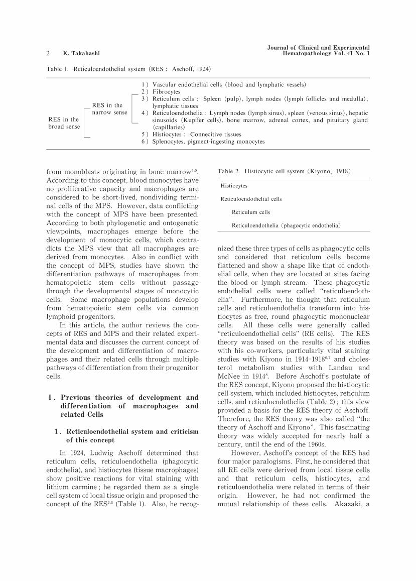
In 1924, Ludwig Aschoff determined that
reticulum cells, reticuloendothelia (phagocytic
endothelia),and histiocytes (tissue macrophages)
show positive reactions for vital staining with
lithium carmine;he regarded them as a single
cell system of local tissue origin and proposed the
concept of the RES (Table 1). Also,he recog-
nized these three types of cells as phagocytic cells
and considered that reticulum cells become
flattened and show a shape like that of endoth-
elial cells,when they are located at sites facing
the blood or lymph stream. These phagocytic
endothelial cells were called “reticuloendoth-
elia”. Furthermore, he thought that reticulum
cells and reticuloendothelia transform into his-
tiocytes as free, round phagocytic mononuclear
cells. All these cells were generally called
“reticuloendothelial cells”(RE cells). The RES
theory was based on the results of his studies
with his co-workers, particularly vital staining
studies with Kiyono in 1914-1918 and choles-
terol metabolism studies with Landau and
McNee in 1914. Before Aschoff’s postulate of
the RES concept,Kiyono proposed the histiocytic
cell system,which included histiocytes,reticulum
cells,and reticuloendothelia (Table 2);this view
provided a basis for the RES theory of Aschoff.
Therefore,the RES theory was also called “the
theory of Aschoff and Kiyono”. This fascinating
theory was widely accepted for nearly half a
century,until the end of the 1960s.
However,Aschoff’s concept of the RES had
four major paralogisms. First,he considered that
all RE cells were derived from local tissue cells
and that reticulum cells, histiocytes, and
reticuloendothelia were related in terms of their
origin. However, he had not confirmed the
mutual relationship of these cells. Akazaki, a
Journal of Clinical and Experimental Hematopathology Vol.41 No.1 K.Takahashi
( )
) ( ))) ( ) ( )
) ( ) ( )( )
( )))

Japanese pupil of Aschoff, extensively studied
the distribution, ontogenesis, and origin of RE
cells, in collaboration with many Japanese co-
workers at both Niigata and Tohoku Univer-
sities,for 30 years . In 1952,Akazaki concluded
that RE cells were composed of two different
groups of cells, 1) reticuloendothelia and 2)
reticulum cells or histiocytes,and that reticuloen-
dothelia had an endothelial cell origin,which was
thus different from that of reticulum cells or
histiocytes . Afterward, Kojima, a pupil of
Akazaki, also actively studied the relationship
between reticulum cells and histiocytes (tissue
macrophages), in collaboration with many co-
workers mainly at Fukushima Medical College,
for 20 years and concluded that histiocytes differ-
ed from reticulum cells in cell morphology,func-
tion,and origin .
Second,with regard to RE cells being posi-
tive for vital staining with lithium carmine,
Aschoff believed that this compound was taken
up by the RE cells through phagocytosis (“cell
eating”). In 1932,Lewis first presented evidence
that all living cells can ingest various materials
by pinocytosis (“cell drinking”) . In distinction
to phagocytosis by macrophages,reticulum cells
and endothelial cells can also take up lithium
carmine by pinocytosis. Third, repeated intra-
venous injections of lithium carmine stimulate
animals. Fourth, this chemical compound accu-
mulates within lysosomes because it is not
degraded therein;the concept of lysosomes was
first established by de Duve and co-workers in
1956 ,about 30 years after proposal of the RES
concept by Aschoff. As for vital staining,current
understanding is that lithium carmine is taken up
by reticulum cells and endothelial cells through
pinocytosis,is not degraded in lysosomes,and is
accumulated in massive amounts in large
lysosomal granules. The large lysosomal gran-
ules of these RE cells were mistaken for evidence
of the phagocytosis usually observed in macro-
phages.
From the 1950s on, various prominent
advances in technology occurred in the field of
histochemistry, and the methodologies of im-
munohistochemistry, electron microscopy, and
immunology developed. Numerous studies using
these advanced technologies provided evidence
that macrophages are distinct from reticulum
cells and endothelial cells,so that by the end of
the 1960s, it was recognized that macrophages,
reticulum cells,and endothelial cells differ from
each other in morphology,function,and origin.
2. Monocyte origin of macrophages
In 1925, Sabin et al. in their studies of su-
pravital staining reported the presence of two
types of macrophages in connective tissues,and
they emphasized that macrophages were derived
from monocytes originating from progenitor
cells in bone marrow . In 1939,during studies of
a chamber made in the ear of rabbits,Ebert and
Florey found that macrophages in inflammatory
foci were derived from monocytes migrating
from peripheral blood in vivo . In 1948,Amano,
a Japanese hematopathologist at Kyoto Univer-
sity,also maintained on the basis of his studies
with supravital staining that macrophages in
inflammatory foci or normal steady-state condi-
tions were derived from monocytes migrating
from peripheral blood and that monocytes devel-
oped via promonocytes from monoblasts
originating from progenitor cells in bone mar-
row . In the 1960s,the development and differen-
tiation of macrophages were studied by various
methods, including radiation-induced chimeras,
skin windows,parabiosis,chromosome markers,
cytochemistry,and autoradiography with [ H]
thymidine. Studies showed that macrophages not
only in inflammatory foci but also in normal
steady-state conditions are differentiated from
blood monocytes originating in bone marrow.
3. Mononuclear phagocyte system and its
experimental basis
In 1969,Langevoort,Cohn,Hirsh,Humphrey,
Spector, van Furth, and many other American
and European researchers had an international
meeting at Leiden, rejected the concept of the
RES,and selected mononuclear phagocytic cells
(mononuclear phagocytes)alone as the basis for
the MPS, excluding reticulum cells, fibroblasts,
endothelial cells,and other nonphagocytic mesen-
chymal cells . These investigators proposed the
concept of the MPS:all macrophages are der-
ived from monocytes,which differentiate through
promonocytes from monoblasts originating in
bone marrow .van Furth promoted the view
that macrophages are nondividing, short-lived,
terminal cells of the MPS and are supplied from
blood monocytes alone . van Furth included,
May 2001 Macrophage Development and Differentiation
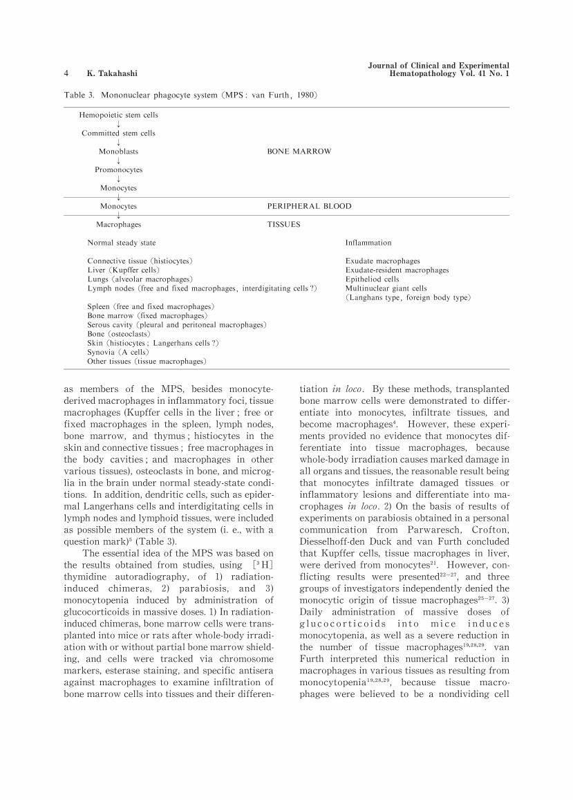
as members of the MPS, besides monocyte-
derived macrophages in inflammatory foci,tissue
macrophages (Kupffer cells in the liver;free or
fixed macrophages in the spleen, lymph nodes,
bone marrow, and thymus;histiocytes in the
skin and connective tissues;free macrophages in
the body cavities;and macrophages in other
various tissues),osteoclasts in bone,and microg-
lia in the brain under normal steady-state condi-
tions. In addition,dendritic cells,such as epider-
mal Langerhans cells and interdigitating cells in
lymph nodes and lymphoid tissues,were included
as possible members of the system (i.e.,with a
question mark) (Table 3).
The essential idea of the MPS was based on
the results obtained from studies, using [ H]
thymidine autoradiography, of 1) radiation-
induced chimeras, 2) parabiosis, and 3)
monocytopenia induced by administration of
glucocorticoids in massive doses.1)In radiation-
induced chimeras,bone marrow cells were trans-
planted into mice or rats after whole-body irradi-
ation with or without partial bone marrow shield-
ing, and cells were tracked via chromosome
markers,esterase staining,and specific antisera
against macrophages to examine infiltration of
bone marrow cells into tissues and their differen-
tiation in loco. By these methods, transplanted
bone marrow cells were demonstrated to differ-
entiate into monocytes, infiltrate tissues, and
become macrophages . However, these experi-
ments provided no evidence that monocytes dif-
ferentiate into tissue macrophages, because
whole-body irradiation causes marked damage in
all organs and tissues,the reasonable result being
that monocytes infiltrate damaged tissues or
inflammatory lesions and differentiate into ma-
crophages in loco. 2)On the basis of results of
experiments on parabiosis obtained in a personal
communication from Parwaresch, Crofton,
Diesselhoff-den Duck and van Furth concluded
that Kupffer cells, tissue macrophages in liver,
were derived from monocytes . However, con-
flicting results were presented , and three
groups of investigators independently denied the
monocytic origin of tissue macrophages . 3)
Daily administration of massive doses of
g l u c o c o r t i c o i d s i n t o m i c e i n d u c e s
monocytopenia,as well as a severe reduction in
the number of tissue macrophages . van
Furth interpreted this numerical reduction in
macrophages in various tissues as resulting from
monocytopenia , because tissue macro-
phages were believed to be a nondividing cell
↓
↓
↓
↓
Journal of Clinical and Experimental Hematopathology Vol.41 No.1 K.Takahashi

population constantly supplied from blood
monocytes . However, the proliferative
capacity of tissue macrophages escaped his
notice. As will be mentioned below,tissue macro-
phages clearly possess a proliferative capacity.
Macrophage proliferation was demonstrated to
be severely impaired by administration of cor-
ticosteroid hormones .
Although the MPS theory has greatly
contributed to definition of the differentiation
pathway of monocytic cells into macrophages in
inflammatory lesions,whether all tissue macro-
phages are derived from monocytes alone in the
normal steady state remains a question .
4. Criticism of the mononuclear phagocyte
system theory
1 )Two major subpopulations of macro-
phages
In 1972, Daems and Brederoo used enzyme
cytochemical studies of peritoneal macrophages
in guinea pigs to demonstrate the presence of two
subpopulations of macrophages that differed in
the localization pattern of endogenous perox-
idase (PO), which allowed discrimination of
exudate macrophages from resident macro-
phages . Like monocytes,exudate macrophages
show PO activity in cytoplasmic granules alone
that is resistant to aminotriazole treatment,and
they appear in stimulated inflammatory foci. In
contrast,resident macrophages show PO activity
in the nuclear envelope and rough endoplasmic
reticulum and usually exist in unstimulated,nor-
mal steady-state conditions . Daems and his
co-workers and Kojima and his colleagues stud-
ied rats,mice,and humans,in addition to guinea
pigs, to demonstrate the existence of resident
macrophages in various tissues under unstimulat-
ed,normal steady-state conditions . In the
following cells,they found PO activity in a loca-
tion similar to its location in peritoneal resident
macrophages:pleural macrophages;alveolar
macrophages in lungs;Kupffer cells in the liver;
histiocytes in dermal, subcutaneous, and inter-
stitial connective tissues;macrophages in the
thymus, lymph nodes, and other peripheral
lymphoid tissues;medullary macrophages in
bone marrow;and macrophages in many other
tissues. Kojima in 1976 believed these resident
macrophages to be a major subpopulation resid-
ing in various tissues and grouped them in a
family of histiocytes or tissue macrophages .
In 1977-1978, Bodel et al. in their in vitro
studies of rabbit blood monocytes found transient
expression of PO activity in the nuclear envelope
and rough endoplasmic reticulum of monocytes
after adherence of the cells to glass slides;they
emphasized that these cells are a transitional or
intermediate form between exudate macrophages
and resident macrophages . This phenomenon
was also demonstrated in mice,rats,guinea pigs,
and humans ,and this type of cell was termed
an “exudate-resident”macrophage, thus adding
evidence to support the concept of the MPS:
monocytes differentiate into exudate macro-
phages,which become resident macrophages by
passing through the stage of exudate-resident
macrophages . Beelen et al., Bainton, and
Watanabe et al.found confirmation of the differ-
entiation pathway of monocytes into tissue ma-
crophages in their in vivo studies . However,
other investigators did not agree with this path-
way for the following reasons:1) the exudate-
resident macrophages have never been found in
tissues under unstimulated normal steady-state
conditions ;2)the expression of PO activity in
the nuclear envelope and rough endoplasmic
reticulum of exudate-resident macrophages is a
transient phenomenon distinct from the stable
and constitutive expression of PO activity usu-
ally found in resident macrophages ;3) bio-
chemical properties of PO differ from exudate
macrophages to resident macrophages:exudate
macrophages contain myeloperoxidase in the
cytoplasmic granules, whereas the PO in the
nuclear envelope and rough endoplasmic
reticulum has cytochemical characteristics of
catalase ;and 4) exudate-resident macro-
phages are indistinguishable from resident ma-
crophages that have taken up PO-positive gran-
ules released from monocytes or exudate macro-
phages .
Using the double staining method of PO
cytochemistry and ultrastructural immunocyto-
chemistry with anti-rat macrophage monoclonal
antibodies ED1, ED2, and ED3, Beelen et al.
demonstrated that ED1-positive monocytes and
exudate macrophages differentiated into ED2-positive resident macrophages via ED3-positive
exudate-resident macrophages . In contrast, in
our previous studies using the same method of PO
cytochemistry and ultrastructural immunocyto-
chemistry with two different anti-rat macro-
May 2001 Macrophage Development and Differentiation

phage monoclonal antibodies, Ki-M2R and
TRPM-3,we clearly demonstrated that TRPM-3
(sialoadhesin,CD169)recognizes exudate macro-
phages and so-called exudate-resident macro-
phages but not resident macrophages, whereas
Ki-M2R is reactive for resident macrophages and
not for exudate macrophages and exudate-
resident macrophages . Also, PO-negative ma-
crophages consist of two different subpopula-
tions:one is positive for TRPM-3 and the other
is positive for Ki-M2R . Results of our studies
therefore indicate that the so-called exudate-
resident macrophages are not a transitional or
intermediate form between exudate macrophages
and resident macrophages. In addition, tissue
macrophage precursors do not show PO activity
in any cytoplasmic organelles and belong to a
PO-negative cell subpopulation . Fig.1 provides
a schematic diagram of the relationship between
monocyte-derived macrophages or tissue macro-
phages and their precursor cells.
2 )Proliferative capacity of macrophages
van Furth reported that the cell cycle of
monoblasts in mice was 11.9 h,a monoblast gave
rise to two promonocytes via cell division,and a
promonocyte divided into two monocytes within
16.2 h . Monocytes lose proliferative capac-
ity;they are released from bone marrow within
24 h after maturation, circulate in peripheral
blood,and enter peripheral tissues . Accord-
ing to the MPS concept,macrophages also have
no proliferative capacity, as evidenced by the
negligible [ H]thymidine uptake of macro-
phages (below 5%)and because a small number
of proliferating cells are promonocytes that have
just arrived at the tissues from the bone mar-
row .
In contrast, it has been repeatedly reported
that macrophages have proliferative capacity.
Parwaresch and Wacker reported in 1984 in their
experimental study of parabiosis that half of the
peritoneal macrophages can survive by cell divi-
sion . Volkman and his co-workers developed a
severely monocytopenic mouse model by use of a
single intravenous injection of strontium-89 (
Sr) . In these mice,within 1 h after injection,
this radioisotope is incorporated into bone tissues
by an exchange with calcium, accumulates in
bone tissues, and irradiates and destroys bone
marrow without any damage to peripheral tis-
sues distant from the bones. From 2 weeks after
injection, monocytes completely disappear in
peripheral blood,and severe monocytopenia con-
tinues for a long time, 10 weeks or more. In
severely monocytopenic mice,however,numbers
of tissue macrophages such as peritoneal,pulmo-
nary alveolar,and splenic macrophages were not
reduced . These results provide evidence that
tissue macrophages can survive for a long time
by self-renewal, without a supply of blood
monocytes. Similar monocytopenic mice were
produced by a technique providing fractional
radiation to bone marrow alone,with shielding of
the other organs and tissues . In these
monocytopenic mice,pulmonary alveolar macro-
phages survived by self-renewal . We also
examined the proliferative capacity of Kupffer
cells in the liver of Sr-treated, severely
monocytopenic mice and confirmed that Kupffer
cells slightly increased in number with time after
injection and that their proliferative capacity
also increased, in parallel with their numerical
increase . All these data clearly show that
macrophages have a proliferative capacity and
can survive for a long time by self-renewal,
without a supply of blood monocytes.
3 )Relationship between monocytes and
tissue macrophages
In response to inflammatory stimuli,
monocytes are prominently produced in and
mobilized from the bone marrow, migrate into
Fig.1. Schematic diagram of the relationship
between the monocyte-derived macro
phages or tissue (resident) macrophages
and their precursor cells (HSC :hematopoietic stem cell, GM-CFC :granulocyte macrophage colony-forming
cell, M-CFC:macrophage colony-forming
cell,Mφ:macrophage).
-
Journal of Clinical and Experimental Hematopathology Vol.41 No.1 K.Takahashi

inflammatory lesions via the blood stream,and
differentiate into exudate macrophages in loco.
Although many factors participate in monocyte
migration,monocyte chemoattractant protein-1
(MCP-1) is the most powerful factor for induc-
tion of monocyte infiltration into inflammatory
foci . However,MCP-1 has no effect on induc-
tion of migration and infiltration of tissue macro-
phages into inflammatory foci. Therefore,
monocytes are the important cells in inflamma-
tory foci and respond to the MCP-1 produced by
many types of cells,such as vascular endothelial
cells, fibroblasts, tissue macrophages, and T
lymphocytes .
To investigate the relationship between
monocytes and tissue macrophages,we produced
prominent monocytosis in mice by daily intra-
venous injections of macrophage colony-
stimulating factor (M-CSF) or in mice with the
use of subcutaneous transplants of M-CSF-and
granulocyte/macrophage colony-stimulating fac-
tor (GM-CSF)-producing mouse fibrosarcoma .
We examined changes in the number of tissue
macrophages in various organs and tissues of
these mice. We found that the organs and tissues
are not stimulated in these monocytic mice:the
numbers of tissue macrophages are not in-
creased . It has recently become clear that
monocytes circulating in peripheral blood
undergo apoptosis under unstimulated conditions
and do not enter peripheral tissues . In trans-
genic mice producing an excess of GM-CSF,
peritoneal macrophages are sustained by self-
renewal, independent of blood monocytes .
These data provide evidence that there is no
relationship between monocytes and tissue ma-
crophages under unstimulated conditions.
4 )Life span of macrophages
According to the MPS concept,macrophages
are terminal cells of the MPS;they lose prolifer-
ative capacity and are supplied from blood mono-
cytes . In studies with [ H]thymidine
autoradiography, van Furth and co-workers
determined that the turnover times for Kupffer
cells, splenic or alveolar macrophages, and per-
itoneal macrophages were 3.8 days,6 days,and
14.9 days, respectively . Thus, monocyte-
derived macrophages are short-lived in periph-
eral tissues and die within 2 weeks.
In contrast,tissue macrophages can survive
by self-renewal for long periods . Previous
studies have presented variable data about their
life span;however, these studies did show that
tissue macrophages have a long life span. In
mice,de Bakker and Daems,using Imferon (iron
May 2001 Macrophage Development and Differentiation

dextran) as a lysosomal marker, reported that
peritoneal resident macrophages can survive for
4 months ,and Melnicoff et al. in their studies
with a fluorescent dye PKH-1 demonstrated that
PKH-1-labeled peritoneal resident macrophages
are detected at 49 days after labeling . For
Kupffer cells,different data were presented. In
contrast with data presented for mice by van
Furth et al.that the life span of Kupffer cells is
about 4 days , other investigators reported a
longer life span,100 days or about 14 months,in
rats . We demonstrated in a recent study of
mouse liver by flash labeling with [ H]
thymidine that the numbers of labeled Kupffer
cells were maintained above the baseline for 5
weeks after labeling,which indicates that the life
span of Kupffer cells is about 5 weeks . In
summary, these data show that tissue macro-
phages are long-lived and can survive for long
periods.
II. Phylogeny of macrophages
Macrophages are present in tissues in all
multicellular invertebrates and vertebrates
(Table 4). Protozoa are unicellular animals that
take up nutrients or pathogenic microorganisms
by phagocytosis, secrete various substances in-
cluding enzymes,and protect themselves against
microorganisms . Because these functions are
common to those of macrophages,Protozoa are
considered to be a prototype of macrophages.
During the evolution from unicellular animals to
multicellular ones,professional phagocytic cells
developed in tissues;these cells have been given
different names. In bidermic animals such as
Porifera, archeocytes are present in a mesoglea
between the epithelial cells of the external body
surfaces and the internal choanocytes,and they
ingest,digest,and degrade various materials and
dead cells . Coelenterata such as sea anemones
or jellyfish have wandering ameboid cells,which
are called amebocytes and possess phagocytic
functions . The archeocytes in Porifera and the
amebocytes in Coelenterata are regarded as prim-
itive forms of macrophages that exist in multicel-
lular animals;these cells are totipotent stem
cells,which can differentiate into various types
of cells. In Hydra, however, there are no cells
corresponding to archeocytes in Porifera,
amebocytes in Coelenterata, or macrophages in
higher multicellular animals;instead, endoder-
mal epithelial cells show phagocytic functions .
Tridermic invertebrates develop,besides the
endoderm and the ectoderm, a mesoderm, from
which ameboid phagocytic cells develop. In
Platyhelminthes, planarian reticular cells are
macrophage-like cells that have phagocytic func-
tions and encapsulate heat-killed human tubercu-
losis bacilli . As shown in Table 4,mononuclear
phagocytes or macrophage-like cells have differ-
ent names according to the species of animal in
which they occur. In invertebrates, monocytes
are not detected in blood. However,monocytes
are observed in the blood of cyclostomes such as
lampreys and hagfish, the most primitive verte-
brates, and macrophages are seen in gills and
spleen . From cartilaginous fish to mammals,
resident macrophages are found in various tis-
sues, and monocytes are detected in peripheral
blood. Although bone marrow is not developed
in cyclostomes,cartilaginous fish,or teleost fish,
macrophages do exist in tissues . In Am-
phibia,bone marrow develops in frogs but not in
salamanders. In frogs, tissue macrophages
involved in phagocytosis of apoptotic muscle
cells during tail involution are considered not to
be derived from monocytes . In Reptiles ,
Aves ,and Mammalia,macrophages have a ubiq-
uitous distribution in various tissues.
In terms of phylogeny,therefore,unicellular
animals are believed to be a primitive form of
macrophages;professional phagocytes (macro-
phages) develop in multicellular invertebrates,
followed by the emergence of monocytes in verte-
brates from cyclostomes to mammals, and the
development of bone marrow in Reptiles,Aves,
and Mammalia (Table 4). This information sug-
gests that during the evolutionary processes in
animals,macrophages develop before monocytes
emerge,which conflicts with the basic concept of
MPS, that all macrophages are derived from
monocytes.
III. Ontogenesis of macrophages and related
cells
1. Ontogeny of Macrophages
The phylogenetic principle that macrophage
development occurs before the emergence of
monocytes is seen in the ontogeny of mammals.
Whereas in postnatal and adult life in normal
animals and humans,hematopoiesis occurs exclu-
Journal of Clinical and Experimental Hematopathology Vol.41 No.1 K.Takahashi

sively in the bone marrow, in human, rat, and
mouse fetuses, primitive fetal hematopoiesis
starts in the yolk sac,and,in line with ontogeny,
progresses to the fetal liver and finally to the
bone marrow. At the stage of progression to the
liver, permanent or definitive hematopoiesis
develops in the gonads,around the aorta and in
the mesonephros (called the GAM area) and
occurs in the fetal liver, gradually replacing
primitive hematopoiesis .
In primitive hematopoiesis in the yolk sac in
human embryos,macrophages make up approxi-
mately 70% of the total nucleated blood cells at 4
weeks . In mouse and rat embryos, macro-
phages predominate in the early stage of primi-
tive hematopoiesis in the yolk sac;monocytic
cells are absent . We observed immature and
mature macrophages in the yolk sac by light and
electron microscopy;we called the immature
macrophages “primitive macrophages”and the
mature macrophages “fetal macrophages” .
The present author maintained that primitive
macrophages are differentiated from
hematopoietic stem cells,bypassing the develop-
mental stage of monocytic cells,and mature into
fetal macrophages (Fig. 2). The hematopoietic
transcription factor PU.1,which determines the
differentiation of hematopoietic stem cells into
macrophages and B lymphocytes,is expressed in
blood cells of the fetal liver and bone marrow .
In the early stage of yolk sac hematopoiesis,
however,PU.1 is not expressed in any type of
hematopoietic cells,although many primitive and
fetal macrophages have already developed
(Table 5). These data provide evidence that the
primitive and fetal macrophages are derived
from early PU.1-negative hematopoietic progen-
itor cells and not from monocytic cells (Table
5). The primitive and fetal macrophages possess
a high proliferative capacity and their [ H]
thymidine labeling rate is approximately 60-70%
in the murine yolk sac .
After the embryo yolk sac connects with the
cardiovascular system via vitelline veins,primi-
tive and fetal macrophages migrate from the
yolk sac into various organs and tissues,particu-
larly the fetal liver, proliferate in these organs
and tissues, and expand their population in
loco . Migration of primitive and fetal macro-
phages from the yolk sac into tissues occurs at 10
fetal days in mice and at 11 fetal days in rats. In
the murine fetal liver, primitive hematopoiesis
occurs at about 12 fetal days, and numerous
primitive and fetal macrophages develop,active-
ly proliferate, and differentiate into Kupffer
cells . In murine fetal lungs,macrophages are
derived from macrophage precursors before the
monocytic cell stage ,proliferate,and differ-
entiate into pulmonary interstitial macrophages
Fig.2. Ontogenetic development, differentiation,and maturation of macrophages and den
dritic cells (HSC:hematopoietic stem cell,GM-CFC : granulocyte macroph a g e
colony-forming cell, M-CFC:macrophage
colony-forming cell,Mφ:macrophage).
-
May 2001 Macrophage Development and Differentiation

and further into alveolar macrophages after
birth . In the other fetal organs and tissues,
primitive and fetal macrophages proliferate and
differentiate into tissue-specific macro-
phages . As gestation progresses, the
proliferative capacity of macrophages declines in
most fetal organs and tissues .
In the liver of human,rat,and mouse fetuses,
primitive hematopoiesis originating in blood
islands of the yolk sac is replaced by permanent
hematopoiesis from the GAM area . In the fetal
mouse liver,monocytic cells start to develop at
14 fetal days and increase their numbers by
proliferation of monoblasts and promonocytes .
At about 18 fetal days,monocytes are detected in
the peripheral circulation ,migrate into tissues,
and differentiate into monocyte-derived macro-
phages . In the early stage of ontogeny,primi-
tive and fetal macrophages circulate in periph-
eral blood;however,their numbers are reduced
in parallel with the progression of gestation,and
they finally disappear at birth. In contrast,
monocytes rather than primitive and fetal macro-
phages are released from fetal hematopoietic
organs in the late stage of ontogeny,circulate in
peripheral blood, and migrate into peripheral
fetal organs and tissues to become monocyte-
derived macrophages .
Primitive and fetal macrophages are thus
different from monocyte/macrophages in terms
of the stage of emergence, the site of develop-
ment,and pathways of differentiation (Fig.2).
2. Ontogeny of dendritic cells
In human yolk sac hematopoiesis, dendritic
cell precursors develop distinctly and indepen-
dently from macrophage precursors . In murine
ontogeny, however, our studies have revealed
that dendritic cell precursors are similar to the
precursors of primitive and fetal macrophages,
express Ia antigens in various fetal tissues at 12
fetal days, and become Ia-positive macro-
phages . At 14 fetal days, Ia-positive macro-
phages migrate into the thymic premordium to
induce Ia expression of thymic epithelial cells,
which occurs at 15 fetal days . These Ia-
positive macrophages show a dendritic cell mor-
phology, differentiate into interdigitating cells,
and develop tubulovesicular structures in the
c y t o p l a s m a t 1 6 f e t a l d a y s .
Tubulovesicular structures are localized around
the Golgi complex area of interdigitating cells,
consisting of many vesicles,tubules and granules.
After birth,Birbeck granules develop in the cyto-
plasm of some interdigitating cells and a minor
population of Langerhans cells appears in the
thymic medulla .
At 12 fetal days in the rat,primitive and fetal
macrophages emerge in the subepidermal mesen-
chyme;some of them express Ia antigens at 14
fetal days,and they migrate into the epidermis at
16 fetal days . From 18 fetal days on,these
cells start to show a dendritic cell morphology
and differentiate into interdigitating cells in the
epidermis . In the perinatal period,the inter-
digitating cells further develop Birbeck granules
in the cytoplasm and differentiate into Langer-
hans cells . The proliferative potential of the
epidermal dendritic cells,which is about 10-20%
in the late fetal stage and 5% after birth, is
important for the expansion and maintenance of
Langerhans cells in the epidermis during per-
inatal,postnatal,and adult life .
In the fetal rat spleen, Ia-positive macro-
phages initially appear around the central
arteries in splenic lymphoid follicles at 16 fetal
days and start extending dendritic processes .
With the advance of gestation, the numbers of
dendritic cells increase, and the cells are local-
ized in the innermost layer of the periarteriolar
lymphoid sheaths . In fetal lymph nodes, Ia-
positive macrophages appear around blood ves-
sels in the paracortical areas from 17 fetal days
on and thereafter differentiate into interdigitat-
ing cells .
In murine ontogeny,Ia-positive macrophages
are derived from hematopoietic stem cells via
primitive macrophages, differentiate into inter-
digitating cells,and mature into Langerhans cells
in the epidermis and thymic medulla after birth
(Fig.2).
IV. Development and differentiation of
macrophages in postnatal and adult life
In normal steady-state conditions, macro-
phages are terminal cells differentiating from
hematopoietic stem cells in bone marrow. The
bone marrow possesses specific mechanisms to
release and mobilize macrophage progenitor
cells into the peripheral circulation,and there are
two pathways for supplying macrophage precur-
sors. When hematopoietic stem cells in the bone
Journal of Clinical and Experimental Hematopathology Vol.41 No.1 K.Takahashi

marrow enter a stage of lineage-specific differen-
tiation, they are not released from the bone
marrow unless they differentiate into monocytes
via monoblasts and promonocytes . Dur-
ing differentiation,monocytes obtain PO-positive
cytoplasmic granules and lose their proliferative
capacity. In addition, a small number of im-
mature macrophage progenitor cells,at the pre-
monocytic cell lineage stage of differentiation,
are released from the bone marrow into periph-
eral blood,migrate into peripheral tissues, and
differentiate into tissue macrophages . Tissue
macrophages have a proliferative capacity, and
their [ H]thymidine labeling rate remains at
approximately 2-5% through their life span .
In the following sections,the differentiation
and maturation of monocyte-derived macro-
phages and tissue macrophages are discussed. In
addition, a differentiation pathway of common
lymphoid progenitors into B cell precursors and
further into B cell-derived macrophages is de-
scribed.
1 . Differentiation and maturation of
monocyte/macrophages
On the basis mainly of the results of studies
by van Furth and co-workers,it has been estab-
lished that hematopoietic stem cells divide sev-
eral times and become monoblasts . One
monoblast divides once into two promonocytes,
and one promonocyte also divides once into two
monocytes. In this differentiation pathway,one
monoblast becomes four monocytes . In
stimulated conditions, monocytes adhere to
activated vascular endothelial cells,migrate into
tissues, and differentiate into exudate macro-
phages . The development of monocytic cells
(monoblasts,promonocytes,and monocytes)and
the differentiation of monocytes into macro-
phages are supported by M-CSF .
Mice with a osteopetrosis (op)mutation,op/
op mice, show a total lack of M-CSF activity
because of a defect in the coding region of the
c-fms gene;show an absence of incisors, a dis-
tinctly domed skull,a short tail,and a small body
at about 10 days after birth;and have a defect in
bone remodeling caused by a complete deficiency
of functional osteoclasts, leading to systemic
osteosclerosis in skeletal bones,prominent stric-
ture of bone marrow cavities,and marked reduc-
tion of medullary hematopoiesis . In addition,
op/op mice show a complete or nearly complete
deficiency of monocytes in peripheral blood,
severe impairment in production of monocytic
cells and their precursors in bone marrow,com-
plete deficiency of monocyte-derived macro-
phages, and defective differentiation of
monocytes into macrophages in tissues . Daily
M-CSF administration to op/op mice induces a
marked increase in the number of blood
monocytes,production of monocytic cells in the
bone marrow,and differentiation and maturation
of monocyte-derived macrophages in tissues and
of osteoclasts in bone . In the bones of op/op
mice, osteoclasts start to develop after daily
M-CSF administration,and the number of these
cells increases up to the level found in normal
littermates at 3 days after administration .
Therefore, osteoclasts and monocyte-derived
macrophages are termed the“M-CSF-dependent
population” (Table 5). This cell population
responds rapidly to M-CSF and are supported by
M-CSF. In op/op mice,marginal metallophilic
macrophages and marginal zone macrophages in
the spleen are absent, and synovial A cells,
Fig.3. Development, differentiation, and matura
tion of macrophages and related cells, in
cluding dendritic cells, in adult life (HSC:hematopoietic stem cell, GM-CFC :granulocyte macrophage colony-forming
cell, M-CFC:macrophage colony-forming
cell,Mφ:macrophage).
--
May 2001 Macrophage Development and Differentiation
macrophages in uterus and ovaries,and microg-
lia in the brain are few,compared with wild-type
mice . Because these cells do not appear or
develop slowly after daily M-CSF adminis-
tration ,they are called the nonresponding or
slowly responding population;their develop-
ment may depend on M-CSF produced in situ
(Table 5). M-CSF supports the proliferation and
differentiation of monocytic cells and their pre-
cursors (macrophage colony-forming cells;M-
CFCs), so all of these macrophage populations
are derived from M-CFCs and/or monocytic
cells (Fig.3).
Although various chemotactic factors such
as MCP-1, MCP-2, MCP-3, MCP-4, M-CSF,
GM-CSF,tumor necrosis factor-α(TNF-α),trans-
forming growth factor-β(TGF-β),and RANTES
(regulated on activation,normal T cell expressed
and secreted)are known to induce chemotaxis of
monocytes and macrophages into tissues,MCP-1
is the most potent CC chemokine responsible for
infiltration of monocytes into tissues . In
pathological conditions, when inflammatory
stimuli occur in tissues, vascular endothelial
cells,fibroblasts,tissue macrophages,infiltrated
monocyte/macrophages, and T cells produce
MCP-1 to induce monocyte infiltration into the
tissues . In unstimulated normal tissues,
MCP-1 is not expressed,and monocytes continue
to circulate in peripheral blood and die by
apoptosis without infiltration into tissues. MCP-1 does not induce migration and infiltration of
any tissue macrophages into inflammatory
lesions .
2. Differentiation and maturation of tissue
macrophages
In contrast to monocyte/macrophages and
their related macrophage populations, tissue
macrophages develop in various organs and tis-
sues of op/op mice . These cells are small,
round, and immature, with an ultrastructure
characterized by poor development of intracel-
lular organelles,particularly lysosomal granules,
and they show no cytochemical localization of
PO activity . These immature tissue macro-
phages are present in various organs and tissues
of op/op mice,particularly in the lungs, spleen,
and brain;their numbers are reduced in other
tissues to a variable degree . Because op/op
mice lack functional activity of M-CSF and
monocytic cells in peripheral blood,the immature
tissue macrophages are called “M-CSF-
independent macrophages”(Table 5) and are
considered to be derived from earlier macro-
phage precursor cells than monocytic cells and
M-CFCs .
As mentioned above,bone marrow releases
macrophage precursor cells at the stage before
lineage-specific differentiation of hematopoietic
cells,and immature macrophage precursor cells
migrate into tissues in response to various
chemotactic factors. Pre-B cell growth-
stimulating factor (PBSF)/stromal-derived fac-
tor (SDF)-1 is one of the CXC chemokines;it
stimulates proliferation of B cell progenitors in
vitro and is constitutively expressed in bone
marrow-derived stromal cells . Mice lacking
PBSF/SDF-1 or CXC receptors die in the per-
inatal stage of development,the number of B cell
progenitors is severely reduced in fetal liver and
bone marrow of mutant embryos, and myeloid
progenitor numbers are reduced only in the bone
marrow,not in the fetal liver . Thus,PBSF/
SDF-1 is responsible for B cell lymphopoiesis
and bone marrow hematopoiesis and can induce
migration of early hematopoietic progenitors
into tissues. In normal mice, PBSF/SDF-1
mRNA is expressed in all organs and tissues
except for the epidermis, which suggests that
these organs and tissues can induce the migration
of early hematopoietic progenitors by the action
of PBSF/SDF-1.
After migration of early hematopoietic pro-
genitors into tissues,these cells can differentiate
into tissue-specific macrophages in response to
interleukin (IL)-6, IL-3, GM-CSF, M-CSF, and
other cytokines produced in situ. In op/op mice,
in spite of the absence of blood monocytes, im-
mature macrophages differentiate from early
hematopoietic progenitors without the action of
M-CSF in various organs and tissues. Immediate-
ly after M-CSF administration, these immature
macrophages proliferate and express PO activity
in the nuclear envelope and rough endoplasmic
reticulum, a finding that is consistent with the
localization pattern of PO activity in resident
macrophages . Thus,resident macrophages in
tissues are considered to be differentiated from
earlier myeloid precursors in tissues;they by-
pass the stage of M-CFCs and monocytic cells.
Although various transcription factors are
known to be involved in the development and
Journal of Clinical and Experimental Hematopathology Vol.41 No.1 K.Takahashi
differentiation of hematopoietic stem cells into
tissue macrophages, PU. 1 is a critical
hematopoietic transcription factor for the differ-
entiation of early hematopoietic progenitors into
macrophages and B cells . In normal mice,PU.
1 mRNA is expressed in all organs and tissues.
PU. 1-deficient mice usually die in the fetal
stages or die of septicemia within 2 days after
birth . In these mutant fetuses or neonatal
mice, all macrophages, including not only
monocytic cells and monocyte-derived macro-
phages but also tissue macrophages, are com-
pletely absent . In the mutant embryos and
fetuses,macrophages are absent in the yolk sac,
feta l l i v er, a n d b o n e ma r r o w .
Hematopoietic progenitor cells from PU. 1
mice do not respond to GM-CSF or M-CSF .
However,when mutant mice are rescued by treat-
ment with antibiotics immediately after birth and
survive for 2 weeks, a small number of large
macrophages develop in various tissues such as
the liver and bone marrow . This result sug-
gests that tissue macrophages can develop from
early hematopoietic progenitor cells in PU.1
mice (Table 5) and that the development and
differentiation of early hematopoietic progeni-
tors into tissue macrophages occur not only in
early ontogeny but also in postnatal life(Fig.3).
3. Development and differentiation of B
lymphoid progenitors into macrophages
As shown in PU. 1- or PBSF/SDF-1-defi-
cient mice,macrophages and B cells are related
to each other at the gene level . B cells
are classified into two major subsets on the basis
of expression of CD5 (Ly-1):CD5-positive B
cells (B-1)and CD5-negative B cells (B-2) .
B-2 cells are conventional B lymphocytes;B-1
cells are derived and differentiated from precur-
sor cells in the omental milky spots,proliferate
by self-renewal in the peritoneal cavity, and
p r o d u c e n a t u r a l a n t i b o d i e s a n d
autoantibodies . One subset of B-1 cells
continues to express CD5 (B-1a cells), whereas
the other subset loses CD5 during maturation (B-1b cells) . In vitro studies of pre-B cell lines or
myeloid cell lines introduced by v-raf and/or
v-myc genes revealed that B cells differentiate
into macrophages . In our previous studies of
pre-B cell lines established in a long-term bone
marrow culture, we confirmed in vitro that a
pre-B cell line,J13,differentiates into CD5-posi-
tive macrophages at 1 month after co-culture
with GM-CSF and a mouse bone marrow stromal
cell line,ST2 . We examined many different
mouse strains including immunodeficient mice
such as nude,severe combined immunodeficiency
(scid), X-linked immunodeficiency (xid), and
viable motheaten (me /me )mice to detect CD5-positive macrophages. In all these mice except
for me /me mice,we did not detect CD5-posi-
tive macrophages as a cell population;however,
we did find many CD5-positive macrophages in
the peritoneal cavity,spleen,and lymph nodes of
me /me mice .
The me /me mice show tyrosine phos-
phatase deficiency in hematopoietic cells because
of mutations in the SHP-1 gene,which results in
abnormal signal transduction in hematopoietic
cells and induces severe impairments of T and B
cell differentiation, a severe reduction in the
numbers of B-2 cells, increased numbers of B-1
cells with abnormal immunoglobulin production,
enhanced production of myeloid cells, and in-
creased production of monocyte/macrophages in
bone marrow and their infiltration into various
organs and tissues . In me /me mice, the
concentration of GM-CSF is elevated in serum
and peritoneal fluid, suggesting that GM-CSF
induces the development and differentiation of
CD5-positive macrophages .
To verify this suggestion, we injected
BALB/c or C3H mice intravenously with 5 ng of
GM-CSF for 5 days . We found the emergence
of numerous CD5-positive macrophages in the
peritoneal cavity of the GM-CSF-treated mice
but not in the untreated mice . Besides the CD5-positive peritoneal macrophages,we found pre-
cursor cells of both B cells and CD5-positive
macrophages or CD5-positive progenitor cells .
The presence of such common precursor cells for
both B-1 cells and macrophages implies that CD5-positive macrophages are derived from common
B lymphoid progenitors. However,daily admin-
istration of GM-CSF to scid, xid, aly/aly mice
failed to produce CD5-positive macrophages in
the peritoneal cavity, because the scid and xid
mice showed severe impairments in B cell devel-
opment and differentiation,and the aly/aly mice
had a severe deficiency of lymphoid tissues .
In addition, daily administration of GM-CSF to
M-CSF-deficient op/op mice did not induce the
development of CD5-positive macrophages in the
May 2001 Macrophage Development and Differentiation
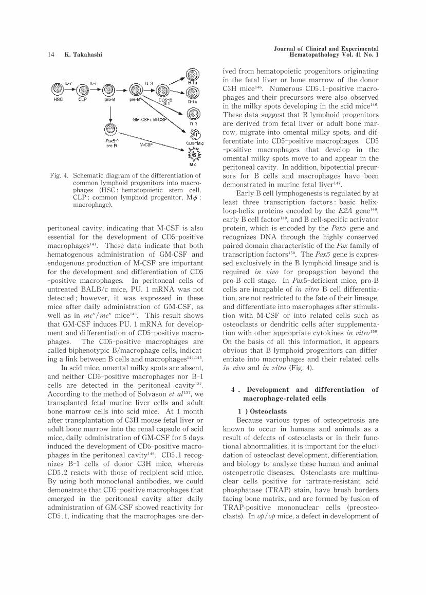
peritoneal cavity,indicating that M-CSF is also
essential for the development of CD5-positive
macrophages . These data indicate that both
hematogenous administration of GM-CSF and
endogenous production of M-CSF are important
for the development and differentiation of CD5-positive macrophages. In peritoneal cells of
untreated BALB/c mice,PU.1 mRNA was not
detected;however, it was expressed in these
mice after daily administration of GM-CSF, as
well as in me /me mice . This result shows
that GM-CSF induces PU.1 mRNA for develop-
ment and differentiation of CD5-positive macro-
phages. The CD5-positive macrophages are
called biphenotypic B/macrophage cells,indicat-
ing a link between B cells and macrophages .
In scid mice,omental milky spots are absent,
and neither CD5-positive macrophages nor B-1
cells are detected in the peritoneal cavity .
According to the method of Solvason et al ,we
transplanted fetal murine liver cells and adult
bone marrow cells into scid mice. At 1 month
after transplantation of C3H mouse fetal liver or
adult bone marrow into the renal capsule of scid
mice,daily administration of GM-CSF for 5 days
induced the development of CD5-positive macro-
phages in the peritoneal cavity . CD5.1 recog-
nizes B-1 cells of donor C3H mice, whereas
CD5.2 reacts with those of recipient scid mice.
By using both monoclonal antibodies,we could
demonstrate that CD5-positive macrophages that
emerged in the peritoneal cavity after daily
administration of GM-CSF showed reactivity for
CD5.1,indicating that the macrophages are der-
ived from hematopoietic progenitors originating
in the fetal liver or bone marrow of the donor
C3H mice . Numerous CD5.1-positive macro-
phages and their precursors were also observed
in the milky spots developing in the scid mice .
These data suggest that B lymphoid progenitors
are derived from fetal liver or adult bone mar-
row,migrate into omental milky spots,and dif-
ferentiate into CD5-positive macrophages. CD5-positive macrophages that develop in the
omental milky spots move to and appear in the
peritoneal cavity. In addition,bipotential precur-
sors for B cells and macrophages have been
demonstrated in murine fetal liver .
Early B cell lymphogenesis is regulated by at
least three transcription factors:basic helix-
loop-helix proteins encoded by the E2A gene ,
early B cell factor ,and B cell-specific activator
protein,which is encoded by the Pax5 gene and
recognizes DNA through the highly conserved
paired domain characteristic of the Pax family of
transcription factors . The Pax5 gene is expres-
sed exclusively in the B lymphoid lineage and is
required in vivo for propagation beyond the
pro-B cell stage. In Pax 5-deficient mice, pro-B
cells are incapable of in vitro B cell differentia-
tion,are not restricted to the fate of their lineage,
and differentiate into macrophages after stimula-
tion with M-CSF or into related cells such as
osteoclasts or dendritic cells after supplementa-
tion with other appropriate cytokines in vitro .
On the basis of all this information, it appears
obvious that B lymphoid progenitors can differ-
entiate into macrophages and their related cells
in vivo and in vitro (Fig.4).
4 . Development and differentiation of
macrophage-related cells
1 )Osteoclasts
Because various types of osteopetrosis are
known to occur in humans and animals as a
result of defects of osteoclasts or in their func-
tional abnormalities,it is important for the eluci-
dation of osteoclast development,differentiation,
and biology to analyze these human and animal
osteopetrotic diseases. Osteoclasts are multinu-
clear cells positive for tartrate-resistant acid
phosphatase (TRAP) stain, have brush borders
facing bone matrix,and are formed by fusion of
TRAP-positive mononuclear cells (preosteo-
clasts). In op/op mice,a defect in development of
Fig.4. Schematic diagram of the differentiation of
common lymphoid progenitors into macro
phages (HSC:hematopoietic stem cell,CLP:common lymphoid progenitor, Mφ:macrophage).
-
Journal of Clinical and Experimental Hematopathology Vol.41 No.1 K.Takahashi

osteoclasts and monocytic cells from
hematopoietic stem cells via granulocyte/macro-
phage colony-forming cells (GM-CFCs) results
from a genetic defect in the production of a
functional M-CSF protein, leading to the occur-
rence of osteopetrosis . Daily administration
of M-CSF induces proliferation of GM-CFCs and
their differentiation into osteoclasts, bypassing
the monocytic cell stage . In aged op/op
mice,GM-CSF and IL-3 levels are increased in
serum,which recover osteopetrosis . In young
op/op mice, osteopetrosis is improved by daily
administration of GM-CSF or IL-3 or both . In
M-CSF-, GM-CSF-or IL-3-treated op/op mice,
the fusion of preosteoclasts into osteoclasts is
evidently observed .
In PU.1-deficient mice,osteopetrosis occurs
as a result of defects in development of osteo-
clasts, monocyte-derived and tissue macro-
phages,dendritic cells,and B cells . c-fos
forms heterodimers of Jun protein and AP-1,
regulates various transcription factors, and is
involved in cell proliferation. In c-fos-deficient
mice, the differentiation of hematopoietic stem
cells into osteoclasts is blocked, leading to the
occurrence of osteopetrosis ;however, differ-
entiation of these cells into macrophages is not
impaired . In c-src-deficient mice, osteoclasts
lack brush borders,which are indispensable for
bone resorption;however,the differentiation of
hematopoietic stem cells into osteoclasts is not
impaired .
OPGL (osteoprotegerin ligand), which
belongs to a TNF family called ODF (osteoclast
differentiation factor), TRANCE (TNF-related
activation-induced cytokine), or RANKL
(receptor-activator of NF-κB ligand), activates
the formation of multinuclear osteoclasts in the
presence of M-CSF,and is a regulating factor for
interactions with T cells and dendritic cells . In
OPGL-deficient mice, severe osteopetrosis
develops because of a complete defect of osteo-
clasts resulting from a functional abnormality of
osteoblasts so that they do not support the forma-
tion of osteoclasts . Mature osteoclasts have a
proton pump and an enzyme,carbonic anhydrase
II,specific for bone resorption . In humans,it
is known that osteopetrosis occurs because of a
deficiency in H -ATPase or carbonic anhy-
drase II .
It is thus concluded that osteoclasts develop
from hematopoietic stem cells and differentiate
via GM-CFCs,bypassing the differentiation stage
of monocytic cells,and become polarized, form-
ing brush borders (Figs.3 and 5).
2 )Microglial cells
Microglia were first described by Del Rio-
Hortega as resting microglia,a population of
cells residing in the central nervous system under
a normal steady state. Since then, many cells
have been proposed for the origin of microglia,
including mesenchymal cells, perivascular cells
(pericytes), monocytes, glial cells, and neural
cells . Fujita and Kitamura in Japan concluded
from electron microscopic studies with [ H]
thymidine autoradiography that microglia
develop in the brain after birth and are derived
from glioblasts, the same progenitor as that for
astrocytes and oligodendrocytes . However,
recent studies using monoclonal antibodies
against macrophages have shown that resting
microglia share a common phenotype with ma-
crophages,providing evidence that the cells are
related to macrophages . On the basis of
differences in cell morphology, ultrastructure,
developmental stage,and precursor cells,microg-
lia are classified into three subtypes:1)ameboid
microglia, 2) resting microglia, and 3) reactive
microglia . Ameboid microglia are found in the
embryonic and fetal stages, emerge in fetal rat
brain at 12 fetal days,and enter,along with the
extension of blood vessels from the meninges,the
fetal brain parenchyma. Ameboid microglia are
differentiated from a subpopulation of primitive
Fig.5. Schematic diagram of osteoclast develop
ment, differentiation, and maturation(HSC:hematopoietic stem cell, GM-CFC:granulocyte macrophage colony-forming
cell, M-CFC:macrophage colony-forming
cell).
-
May 2001 Macrophage Development and Differentiation

and fetal macrophages derived from
hematopoietic stem cells in yolk sac primitive
hematopoiesis . The microglia have a high
proliferative capacity and are distributed in the
fetal rat brain . In the late stage of ontogeny,
they move into the soft meninges, the perivas-
cular areas,and the choroid plexus, around the
brain ventricles , and to the deeper areas of
fetal brain parenchyma . From the late stage
of ontogeny to the early neonatal period,because
ameboid microglia move to the deeper regions of
the brain parenchyma, they disappear in the
brain cortex and subcortical regions. However,
these microglia start to proliferate as progenitors
of resting microglia in the neonatal rat brain and
move to the brain cortex and subcortical
regions ,and resting microglia distribute in the
whole rat brain and appear at 4 weeks after
birth .
In op/op mice,the number of resting microg-
lia in the brain is not reduced in the hippocampal
area, compared with the number of cells in the
brain of wild-type mice . The resting microg-
lia are an M-CSF-independent cell population
and are differentiated from microglial progenitor
cells at the stage before monocytic cell series,
presumably GM-CFCs . However, our studies
showed variable reductions in the numbers of
microglia in areas other than the hippocampus in
op/op mice . This population of microglia did
not recover after daily administration of M-
CSF . They are supported, however, by
growth factors such as GM-CSF and IL-3. Our
study with daily intravenous injections of murine
GM-CSF or IL-3 into op/op mice showed that
F4/80-positive cells increase in number in brain
tissues and that their shape is small,round,and
not ramified (data not published). Therefore,
certain factors other than GM-CSF and IL-3 may
be necessary for the differentiation of microglia
progenitor cells into resting microglia. Studies
with intravenous injections of cells obtained from
primary cultures of microglia or immortalized
microglial cells demonstrated that these cells
enter the brain parenchyma through the walls of
blood vessels, move into brain tissues, and
change into ramified resting microglia .
Reactive microglia are usually found in
inflammatory or damaged brain tissues and are
derived from monocytes invading in loco. Exper-
imental studies with intravenous injections of
isolated microglia, blood monocytes, or macro-
phages revealed that these cells enter inflamma-
tory or damaged brain lesions, particularly
around blood vessels . However, monocytes
and macrophages do not change into ramified
resting microglia . A bone marrow transplanta-
tion study with green fluorescent protein (GFP)-
expressing cells from GFP-transgenic mice
demonstrated the migration and cluster forma-
tion of GFP-positive cells and their differentia-
tion and maturation into ramified resting microg-
lia in brain parenchyma . These GFP-positive
cells clustered in brain parenchyma showed a
positive reaction for ER-MP12,a marker of early
myeloid cells (GM-CFCs) . These data suggest
that resting microglia are derived from
hematopoietic stem cells via early myeloid pro-
genitors,presumably GM-CFCs.
Results of the above-mentioned studies indi-
cate that resting microglia are derived from
proliferating ameboid microglia in the fetal stage
and that after birth they are differentiated from
microglial progenitors with a proliferative capac-
ity, presumably GM-CFCs, and not from
monocytes under normal steady-state conditions
(Fig. 3). In the brain under inflammatory or
stimulated conditions, activated microglia are
found,which are transformed from both ramified
resting microglia and reactive (monocyte-
derived)microglia.
3 )Synovial A cells
Synovial A cells are a macrophage popula-
tion existing in the synovial membrane of joints.
Ontogenetically, they emerge in the late fetal
stage . Because synovial A cells are absent in
op/op mice, these cells are classified as an M-
CSF-dependent macrophage population;how-
ever, their population does not recover after
daily administration of M-CSF . Therefore,
locally produced M-CSF seems to be essential for
the development and differentiation of synovial
A cells.
5. Development and differentiation of den
dritic cells
-
1 )Heterogeneity and migration of den-
dritic cells
Dendritic cells are a heterogeneous popula-
tion and are classified into two major popula-
tions:T cell-associated and B cell-associated
dendritic cells . T cell-associated dendritic cells
Journal of Clinical and Experimental Hematopathology Vol.41 No.1 K.Takahashi

include epidermal Langerhans cells, veiled cells
in the dermis(dermal dendritic cells)and afferent
lymphatics, interdigitating cells in the paracor-
tical area of lymph nodes or in the T cell-
dependent area of peripheral lymphoid tissues,
and lymphoid dendritic cells . B cell-associated
dendritic cells are follicular dendritic cells
(FDCs) located in the germinal center of
lymphoid follicles . Two theories have been
presented for the origin of FDCs:one proposes
their derivation from reticulum cells in a fol-
licular germinal center , and the other pro-
Fig.6. Development, differentiation, and trafficking of T cell-associated den
dritic cells. A:lymphogenous route of dendritic cells from skin to
regional lymph node(2),and hematogenous route of dendritic cell progen
itors from bone marrow to skin or lymph node (1), B :Blood-lymph
translocation of dendritic cells in the liver(3)(LC:Langerhans cell,IDC:interdigitating cell,VC:veiled cell,FDC:follicular dendritic cell).
-
-
Fig.7. Development, differentiation, and maturation of dendritic
cells (HSC:hematopoietic stem cell, MPC:myeloid pro
genitor cell,CLP:common lymphoid progenitor,GM-CFC:granulocyte macrophage colony-forming cell,M-CFC:ma
crophage colony-forming cell, TCP:T cell progenitor,BCP:B cell progenitor,TTCP:thymic T cell progenitor,PTC:plasmacytoid T cell,IDC:interdigitating cell).
-
-
May 2001 Macrophage Development and Differentiation

poses a bone marrow origin . However,the
former theory has been widely recognized up to
the present.
Hence, from the viewpoint of macrophage-
related cells, the present author should explain
here the maturation and migration of T cell-
associated dendritic cells. It is known that
epidermal Langerhans cells are differentiated
from indeterminate (interdigitating)cells,which
are derived via dendritic cell progenitors from
hematopoietic stem cells in bone marrow .
During this differentiation process,the epidermal
Langerhans cells develop Birbeck granules in
their cytoplasm and express E-cadherin. In
humans, the monoclonal antibody Lag-1 recog-
n i z e s m a t u r e B i r b e c k g r a n u l e s .
Tubulovesicular structures are characteristic of
interdigitating cells. The epidermal Langerhans
cells migrate into the dermis and become veiled
cells(dermal dendritic cells),which then enter the
afferent lymphatics and move to the paracortical
area of regional lymph nodes (Fig.6A). Veiled
cells extend fan-like processes and are highly
mobile. They mature into interdigitating cells in
loco and lose Birbeck granules from the cyto-
plasm. Against this view,epidermal Langerhans
cells and dermal dendritic cells are currently
believed to be different populations . In the
lymph nodes, compared with bone marrow-
derived dendritic cells, epidermal Langerhans
cells and dermal dendritic cells accumulate 3 to 4
times slower, turnover that is dramatically in-
creased by cutaneous inflammation . In normal
steady-state conditions, dendritic cells die by
apoptosis in the lymph nodes and there are few
dendritic cells in the efferent lymphatics and
thoracic duct under normal steady-state condi-
tions . Besides this lymphogenous route from
the skin to the regional lymph nodes, there is a
hematogenous route for the migration of den-
dritic precursor cells from bone marrow into
lymph nodes and for their differentiation into
interdigitating cells in the paracortical area, as
demonstrated by the finding of numerous den-
dritic cells in the lymph nodes at 4 weeks after
ligation of the afferent lymphatics draining into
the regional lymph nodes in mice (Fig. 6A).
Besides both the lymphogenous a n d
hematogenous routes,blood-lymph translocation
of dendritic cells occurs in liver . In normal
steady-state conditions,dendritic cell precursors
derived from bone marrow adhere to Kupffer
cells in the hepatic sinusoids,enter the space of
Disse, and differentiate into dendritic cells in
situ . These dendritic cells enter the lymphatic
vessels of the periportal area of the liver and
move into the paracortical area of the hepatic
hilar lymph nodes (Fig.6B).
In adults,although the life cycle of epidermal
Langerhans cells is about 16.3 days ,they pos-
sess a proliferative capacity and include a long-
lived,slowly replicating, self-renewing subpopu-
lation . In contrast,interdigitating dendritic
cells in lymph nodes and/or peripheral lymphoid
tissues are generally short-lived, existing about
10-14 days in mice and 2-4 weeks in rats,and are
constantly supplied by and differentiated from
dendritic cell precursors . Recent studies have
revealed two different types of dendritic cell
precursors:1)myeloid dendritic cell precursors
and 2) lymphoid dendritic cell precursors.
According to the type of precursor cell,the den-
dritic cells are termed “myeloid dendritic cells”
or“lymphoid dendritic cells”(Fig.7).
2 )Multiple pathways of dendritic cell
differentiation
Myeloid dendritic cells include Langerhans
cells in the epidermis and lymphoid tissues,inter-
digitating cells in the lymphoid tissues,and veiled
cells in the dermis and afferent lymphatics.
These myeloid cells are closely related to the
dendritic cells developed during ontogeny under
unstimulated conditions in the fetus and are
derived from myeloid dendritic precursor cells
earlier than the stage of monocytes. Monocytes
are also differentiated into dendritic cells,which
are called “monocyte-derived dendritic cells”,in
response to inflammatory stimuli.
(1)Dendritic cells derived from myeloid precursor
Cells earlier than the stage of monocytic cells
This dendritic cell population includes
epidermal Langerhans cells that express CD1a,
CD11b,CD11c,CD68,Lag-1,and E-cadherin and
are characterized by Birbeck granules in the
cytoplasm. In PU.1-deficient mice,all dendritic
cells including epidermal Langerhans cells and
interdigitating cells are completely absent,as are
macrophages and their related cells and their
progenitor cells . Differentiation of all these
cells is blocked because of the PU.1 deficiency.
In contrast, in Sr-induced, severely
Journal of Clinical and Experimental Hematopathology Vol.41 No.1 K.Takahashi

monocytopenic, splenectomized mice, the num-
bers of epidermal Langerhans cells and dendritic
cells in thymic medulla and lymph nodes are
similar to those of splenectomized control
mice . Compared with normal littermates,op/
op mice show no statistically significant differ-
ence in the number of dendritic cells in the skin,
thymic medulla, lymph nodes, and spleen .
These results indicate that neither monocytes nor
M-CSF contributes to the development and differ-
entiation of dendritic cells in these tissues and
suggest that dendritic cells are an M-CSF-
independent cell population,presumably derived
from GM-CFCs or earlier myeloid precursor cells
without passing through the monocytic cell
stage . In cultures supplemented with GM-
CSF,the presence of proliferating dendritic pre-
cursors was demonstrated in peripheral blood
and bone marrow,and dendritic cells were gener-
ated in in vitro studies of murine bone marrow
cells with GM-CSF . In cultures of mononu-
clear cells from human cord blood supplied with
GM-CSF and TNF-α or TGF-β1, Langerhans
cells with Birbeck granules were generated and
were demonstrated to be differentiated from
CD1a-positive cells originating from CD34-posi-
tive hematopoietic stem cells,but not from CD14-positive cells (monocytes) . In TGF-α1-
deficient mice, epidermal Langerhans cells are
completely absent . In in vitro studies of CD34-
positive hematopoietic stem cells and GM-CSF
and TNF-a,Langerhans cells developed and their
population grew,particularly when supplement-
ed with TGF-β1 . This type of dendritic cell
is called a “myeloid dendritic cell”,because, as
shown in these in vitro studies, these cells are
derived from myeloid progenitor cells earlier
than the monocytic cell stage, do not pass
through the differentiation stage of monocytic
cells,and are thus distinct from the cells belong-
ing to the MPS (Fig.7).
(2)Monocyte-derived dendritic cells
In addition to being derived from myeloid
progenitors, dendritic cells are derived from
monocytes or macrophages . In vitro
studies have shown that dendritic cells are gener-
ated from human peripheral blood monocytes in
cultures with GM-CSF and IL-4 and express DR,
DQ,CD1a,CD1b,CD1c,CD11,and costimulatory
molecules such as intracellular adhesion mole-
cule 1 (ICAM-1), leukocyte function-associated
antigen 1 (LFA-1), B7, or B70;these cells also
have T cell-stimulatory activity in autologous
and heterologous mixed leukocyte reactions .
These dendritic cells are called “dendritic cell-
like cells” or “monocyte-derived dendritic
cells” . In our studies, these dendritic cells
showed an ultrastructure of secretory cells
resembling epithelioid cells and had neither Bir-
beck granules nor a tubulovesicular structure .
However, studies showed that TGF-β1 induced
the development of Birbeck granules in the cyto-
plasm of monocyte-derived dendritic cells in
vitro and that these cells expressed E-cadherin
and Lag-1,suggesting that monocytes differenti-
ate into Langerhans cells in vitro . It remains
unknown whether this differentiation pathway of
monocytes into Langerhans cells occurs as a
major route in vivo under unstimulated normal
steady-state conditions.
In op/op mice,the development and differen-
tiation of myeloid dendritic cells are not impair-
ed;however, the development of monocyte-
derived dendritic cells is severely impaired in
lymph nodes under inflammatory conditions .
In addition, we found that monocyte-derived
dendritic cells do not develop in hepatic
granulomas induced by Zymosan administration
(data not published). In me /me mice,the num-
bers of epidermal Langerhans cells and inter-
digitating cells in lymph nodes are reduced with
aging,whereas monocytes and monocyte-derived
macrophages infiltrate various organs and tis-
sues,accompanied by increased numbers of ER-
MP20-positive monocyte-derived dendritic cells
in inflammatory foci of lungs, liver, and other
tissues . Monocyte-derived dendritic cells thus
show behaviors distinct from those of myeloid
dendritic cells. They develop in inflammatory
foci, M-CSF is essential for their development,
and their number increases in response to inflam-
matory stimuli,as shown in studies of op/op mice
or me /me mice in inflammatory reactions(Fig.
7).
Lymphoid dendritic cells are a heterogenous
population and at least two subsets have been
reported in lymphoid tissues. First,Shortman et
al. reported in mice that thymic dendritic cells
are derived from progenitor cells similar to T
cells in the thymus and express major his-
tocompatibility complex class II antigens (Ia
May 2001 Macrophage Development and Differentiation

antigens),CD11c,NLDC-145(DEC205),BP-1,and
CD8 . The CD8α-positive,DEC205-positive,
CD11b-negative dendritic cells belong to a
subtype of T cells and are differentiated from
precursor cells weakly positive for CD4,positive
for CD44 and c-kit,and negative for CD25. IL-3
is important,but GM-CSF is unnecessary for the
differentiation of thymic T cell progenitor cells
into CD8α-positive, DEC205-positive, CD11b-
negative dendritic cells. The thymic progenitor
cells differentiate into T, B, and natural killer
cells besides thymic dendritic cells,but not into
macrophages. The CD8α-positive,DEC205-posi-
tive,CD11b-negative dendritic cells are distribut-
ed from the thymus into lymphoid tissues,partic-
ularly the spleen . CD8α-negative, DEC205-
negative,CD11b-positive dendritic cells are found
in the spleen and lymph nodes,which also contain
a third subpopulation of dendritic cells:CD8α-negative, DEC205-positive, CD11b-negative .
In the lymph nodes,compared with the myeloid
dendritic cells,the lymphoid dendritic cells have
a 30% faster turnover .
In humans, cells previously called plas-
macytoid T cells in humans have been thought to
be a type of dendritic cell . These cells resem-
ble plasma cells,express CD4,and are also ter-
med “plasmacytoid monocytes”. The plas-
macytoid monocytes are IL-3-dependent, differ-
entiate and mature into dendritic cells,and show
an intense T cell-stimulatory activity in response
to stimuli induced by IL-3 and CD40 ligand.
These dendritic cells lack phagocytic activity,do
not express any myeloid cell-related antigens
such as CD11b,CD11c,CD13,and CD33,and are
GM-CSF-independent. Electron microscopically,
these dendritic cells resemble plasma cells and
extend dendritic cell processes and develop
lamellarly arranged rough endoplasmic reticula.
The dendritic cells are distributed in the T cell-
dependent area of peripheral lymphoid tissues,
particularly around high endothelial venules,as
well as in the thymus . On the basis of these
results,it is speculated that dendritic cell progen-
itors develop in the thymus,move via the blood
stream into peripheral lymphoid tissues, and
differentiate into dendritic cells. Some
invesigators have pointed out the relationship
between plasmacytoid T cells and interdigitating
cells in lymph nodes and other lymphoid tissues.
However, the heterogeneity and lymphogenous
route of migration of interdigitating cells must be
considered when discussing the origin of these
cells,as mentioned above .
In addition, in Pax 5-deficient mice, pro-B
cells are incapable of in vitro B cell differentia-
tion,are not restricted to the fate of their lineage,
and differentiate into dendritic cells after stimu-
lation with GM-CSF in vitro . However, this
dendritic cell subpopulation has not yet been
verified in vivo. All these data suggest the
presence of differentiation pathways of T or B
lymphoid progenitors into dendritic cells, with
their differentiation and maturation supported by
GM-CSF or IL-3 in vitro (Fig. 7). This last-
mentioned result should challenge the finding
that the development of dendritic cells is not
impaired in GM-CSF-deficient mice or in
mice deficient in GM-CSF/IL-3/IL-5βc chain
receptor orβIL-3 receptor .
V. Multiple roles of macrophages in inflam
mation-
As mentioned above, there are two major
macrophage populations ―― tissue macro-
phages and monocyte-derived macrophages――
and both populations participate in different
stages of inflammation. The liver has a simple
structure composed of hepatic parenchymal cells
and sinusoidal cells;Kupffer cells are present in
the hepatic sinusoidal lumen, and monocytes
circulate in peripheral blood. To examine the
roles of Kupffer cells and monocyte-derived
ma cr o p h a g e s p o p u l a t i o n i n h e p a t i c
granulomatous inflammation,the following vari-
ous mouse models were have been used. In mice
depleted of Kupffer cells by administration of
liposome-encapsulated dichloromethylene di-
phosphonate(lipo-MDP),hepatic granuloma for-
mation is delayed from 3 to 28 days after
zymosan injection, accompanied by suppressed
expression of M-CSF,IL-1,MCP-1,TNF-αand
IFN-γmRNA, compared with lipo-MDP-
untreated control mice . These data imply that
Kupffer cells are indispensable for hepatic
granuloma formation,play a part act as an initia-
tor, and produce various cytokines including
MCP-1 and M-CSF. In mice lacking class A type
I and II macrophage scavenger receptors(MSR-A
I & and II),the development and distribution of
Kupffer cells in the liver are similar to those of
the wild-type mice . In MSR-A mice, how-
ever,hepatic granuloma formation was delayed
Journal of Clinical and Experimental Hematopathology Vol.41 No.1 K.Takahashi

markedly after intravenous injection of Cor-
ynebacterium parvum (C.parvum),because MSR-
A macrophages can not ingest and digest C.
parvum due to the lack of MSR-A, and their
signal transduction through MRS-A for the
expression of MCP-1, TNF-αand IFN-γmRNA
is impaired. In the early stage of hepatic
granuloma formation, monocyte migration into
liver granulomas,phagocytosis of C. parvum by
and activation of granuloma macrophages are
markedly impaired in MSR-A mice.
In Sr-treated,severely monocytopenic mice,
hepatic granuloma formation is severely impair-
ed until a week after Zymosan injection,marked
proliferation of Kupffer cells occurs at 5 days,
and the Kupffer cells start to collect and aggre-
gate to form granulomas in the hepatic sinusoids
and change into epithelioid cells and multinu-
clear giant cells . Monocytes do not partici-
pate in such hepatic granuloma formation;Kupf-
fer cells alone can form hepatic granulomas by
their proliferation and differentiation into epith-
elioid cells and/or multinuclear giant cells. In
M-CSF-deficient op/op mice,hepatic granuloma
formation is delayed early after Zymosan injec-
tion, and hepatic granulomas are formed by
proliferation of immature Kupffer cells alone,
because monocytes are completely or nearly
completely absent in peripheral blood and
because differentiation of monocytes into macro-
phages is severely impaired . Although prolifer-
ation of Kupffer cells is most important for
hepatic granuloma formation in both
monocytopenic mouse models in response to
Zymosan stimulation, proliferation of Kupffer
cells is more marked in the 89Sr-induced,severe-
ly monocytopenic mice than in the op/op mice.
CC chemokine receptor 2 (CCR2) is a rece-
ptor for MCP-1,the most potent chemokine for
monocyte chemotaxis. In mice lacking CCR2,
monocyte infiltration into Zymosan-induced he-
patic granulomas is severely impaired, and he-
patic granuloma formation is markedly delayed
compared with granuloma formation in control
mice . All these data imply that,in addition to
Kupffer cells, monocytes are important for he-
patic granuloma formation, particularly in its
early stage.
GM-CSF is an important growth factor for
the proliferation,differentiation,and survival of
macrophages,as well as for monocyte chemotax-
is. In mice lacking GM-CSF, Zymosan-induced
hepatic granuloma formation is delayed in the
early stage,and the granulomas disappear more
rapidly,compared with wild-type mice. In GM-
CSF mice, monocyte influx into liver
granulomas and[ H]thymidine uptake rates of
granuloma macrophages are impaired in the
early stage of hepatic granuloma formation,and
the number of apoptotic cells is increased in the
granulomas at 8 days after Zymosan injection .
GM-CSF mice showed expression of MCP-1,
TNF-α,IFN-γ,M-CSF,and IL-1 mRNA in liver
granulomas even though there was a total lack of
GM-CSF. Thus,GM-CSF plays important roles
in monocyte migration into hepatic granulomas
and proliferation and survival of granuloma
macrophages in vivo.
In vitro,T lymphocytes are known to partici-
pate in activation of macrophages during the
formation of various types of granulomas. To
elucidate the roles of T cells in hepatic
granuloma formation, we injected Zymosan
intravenously into three different types of im-
munodeficient mice―― nude (Hfhllnu/Hfhllnu)
mice, C3H/HeN and CB-17/clr-scid mice, and
CBA/N (xid/xid)mice―― and compared results
with those from control mice . Nude mice
are congenitally athymic,have a profound defi-
ciency of T cells, and lack T cell-dependent
immune responses in the early stage of adult life.
Compared with control mice, in nude mice the
number and size of hepatic granulomas and the
influx of monocytes into the liver are slightly
Fig.8. Schematic representation of macrophage
involvement in hepatic granuloma forma
tion in mice.1 Kupffer cell-depleted mice,by administration of lipo-MDP 2 MSR-A mice;3 Sr-induced monocytopenic
mice;4 CCR2 mice;5 op op mice;6 GM-CSF mice; 7 nude mice;8 scid mice;9
xid mice.
-
May 2001 Macrophage Development and Differentiation

reduced and the differentiation,maturation,and
activation of granuloma macrophages are
delayed. Also,the percentages of Thy 1.2-posi-
tive T cells in the granulomas and peripheral
blood are lower in the nude mice than in the
control mice .
The scid mouse is a murine model of T and
B cell dysfunction. In these mice, the develop-
ment and formation of hepatic granulomas are
delayed for more than a week after Zymosan
injection, the influx of monocytes into
granulomas during the early stage of granuloma
formation is reduced,and the percentages of Thy
1.2-positive T cells in the granulomas and
peripheral blood are lower than in the control
mice . However,[ H]thymidine uptake rates
of granuloma macrophages in nude and scid mice
do not differ from those of control mice. In both
immunodeficient mice,T cell deficiency seems to
result in a delay of hepatic granuloma formation
because of a failure in the differentiation,matu-
ration,and activation of macrophages in loco.
The xid mouse is a murine model of X-linked
immunodeficiency, with impaired proliferation
and differentiation of B cells. After Zymosan
injection, the number of hepatic granulomas
increases slightly in xid mice, peaks at 5 days,
and rapidly declines from 8 days on . Also,the
influx of monocytic cells and earlier macrophage
precursors into granulomas, the proliferative
capacity of granuloma macrophages, and the
phagocytic and digestive capacity of the macro-
phages for glucan particles are increased in xid
mice compared with control mice . In xid mice,
although the number of T cells in peripheral
blood is normal,the percentages of T cells in the
granulomas are higher than in the control mice.
Such increased percentages of T cells in hepatic
granulomas in xid mice seem to result in activa-
tion of granuloma macrophages, leading to a
rapid disappearance of granulomas via enhanced
phagocytosis and digestion of glucan particles by
granuloma macrophages. These data for three
different types of immunodeficient mice indicate
that T lymphocytes are involved in the activation
of macrophages in hepatic granuloma formation
in vivo.
The present author has thus explained the
multiple roles of two major macrophage popula-
tions,Kupffer cells and monocyte-derived macro-
phages, in the multistep processes of hepatic
granuloma formation, their participation in dif-
ferent stages of granuloma formation, and
involvement of various cytokines in vivo by the
use of various mouse models (Fig.8).
CONCLUSION
This article has critically reviewed two
major theories concerning the development and
differentiation of macrophages,the RES and the
MPS;pointed out that the development and
differentiation of macrophages occurs not in a
single cell lineage as proposed by both theories;
and explained that macrophages are differentiat-
ed through multiple pathways from their progeni-
tor cells via different routes of differentiation.
With regard to animal phylogeny, ameboid
phagocytic mononuclear cells (macrophages)do
develop but monocytes are not detected in inver-
tebrates, whereas both macrophages and
monocytes develop in vertebrates. This fact
indicates that the development and differentia-
tion of macrophages precede those of monocytes
during the evolutionary processes of animals.
This phylogenetic principle is applied to human
and murine ontogeny of macrophages. In the
yolk sac, primitive and fetal macrophages first
develop and monocytes are not detected in primi-
tive hematopoiesis, indicating that in early
ontogeny these macrophages are differentiated
from hematopoietic stem cells,and the develop-
mental stage of monocytic cells is bypassed. In
the fetal liver,monocytes develop in permanent,
or definitive,hematopoiesis,are released into the
peripheral circulation,migrate into fetal tissues,
and differentiate into monocyte-derived macro-
phages in late ontogeny. A subpopulation of
primitive and fetal macrophages expresses Ia
antigens and differentiates into interdigitating
cells in late ontogeny and further into Langer-
hans cells in perinatal and neonatal life.
In postnatal and adult life,macrophages are
differentiated from macrophage precursor cells
at different stages or lineages of differentiation
originating in bone marrow. In myeloid cell
lineages, monocytes or myeloid precursor cells
from an earlier stage than the monocytic cells
differentiate into macrophages. As advocated by
the MPS concept, it has been established that
monocytes derived from monoblasts via
promonocytes in bone marrow are released into
peripheral circulation, migrate into tissues in
response to inflammatory stimuli,and differenti-
Journal of Clinical and Experimental Hematopathology Vol.41 No.1 K.Takahashi
ate into macrophages in loco. This differentia-
tion pathway is supported by M-CSF,is complete-
ly blocked in op/op mice,and is evident in inflam-
mation. Tissue macrophages are differentiated
from macrophage precursor cells earlier than the
monocytic cell stage (M-CFCs, monoblasts,
promonocytes,monocytes)under normal steady-
state conditions. The differentiation pathway of
macrophage precursor cells into immature tissue
macrophages is supported by growth factors
other than M-CSF and is not blocked in op/op
mice. M-CSF induces proliferation of immature
tissue macrophages and their maturation into
mature tissue macrophages. Although the differ-
entiation of most tissue macrophages and
m o n o c y t e -d e r i v e d m a c r o
phages is blocked in PU. 1 mice, PU. 1
macrophages develop in PU.1 mice that sur-
vive when administered antibiotics after birth.
Tissue macrophages possess a proliferative
capacity, can survive by self-renewal, and are
long-lived. Although osteoclasts are an M-CSF-
dependent cell population,they are differentiated
from preosteoclasts, which are derived from
osteoclast progenitors at or before the stage of
GM-CFCs, bypassing the stage of monocytic
cells. Resting microglia in the hippocampal area
of the brain are a M-CSF-independent cell popu-
lation and are differentiated from microglial
progenitors at or before the stage of GM-CFCs,
bypassing the stage of monocytic cells. In addi-
tion, some macrophage populations are derived
from hematopoietic stem cells via B lymphoid
progenitors.
Dendritic cells are a heterogeneous popula-
tion and are differentiated through multiple path-
ways of differentiation from dendritic cell pro-
genitors:1)myeloid cells of a stage earlier than
monocytic cells, 2)monocytes, and 3) lymphoid
progenitors. In normal steady-state conditions,
epidermal Langerhans cells and dendritic cells in
lymph nodes and lymphoid tissues are differ-
entiated from myeloid precursor cells earlier
than monocytes,whereas monocyte-derived den-
dritic cells appear in inflammatory lesions. Some
lymphoid dendritic cells are derived from T
lymphoid progenitors or pro-B cells.
Therefore,in addition to the differentiation
pathway of monocyte/macrophages as advocat-
ed by the MPS concept,there are multiple path-
ways of differentiation of macrophages and their
related cells,some of which bypass the stage of
monocytic cells.
ACKNOWLEDGMENT
The author thanks Drs. Aye Aye Wynn and
Katsunori Jinnouchi,postgraduate students,Sec-
ond Department of Pathology, Kumamoto Uni-
versity School of Medicine,for typing and prepa-
ration of this article.
REFERENCES
1 Metchnikoff E:Leçns sur la pathologie
comparee de l’inflammation. Paris, Masson,
1892
2 Aschoff L:Das reticuloendotheliale System.
Ergeb Inn Med Kinderheilkd 26:1-118,1924
3 Akazaki K,Watanuki T, Teshima T :A fur-
ther consideration on the reticuloendothelial
system. In Organizing Committee of the 6th
International RES Symposium (eds):The
Reticuloendothelial System, Morphology, Im-
munology,and Regulation.Proceedings of the
6th International Symposium on RES,May 29-June 1,1964, in Otsu and Kyoto,Japan.1964,
pp3-16.
4 van Furth R,Cohn ZA,Hirsch JG,Humphrey
JH, Spector WG, Langevoort HL : The
mononuclear phagocyte system:A new classi-
fication of macrophages,monocytes,and their
precursor cells. Bull WHO 46:845-852,1972
5 van Furth R :Cells of the mononuclear
phagocyte system. Nomenclature in terms of
sites and conditions. In van Furth R (ed):
Mononuclear Phagocytes:Functional Aspects.
Part 1. The Hague, Martinus Nijhoff Pub-
lishers,1980,pp1-30
6 Kiyono K:Zur Frage der histiocytaen Blutzel-
len.Folia Haematol 18:149-170,1914
7 Kiyono K:Die vitale Karminspeicherung.
Jena,Fischer,1914
8 Landau M, NcNee JW :Zur Physiologie des
Cholesterinstoffwechsels.Beitr Pathol Anat 58:
667-693,1914
9 Akazaki K:Reticulo-endothelial system.Acta
Pathol Jpn 2:113-142,1952
10 Kojima M :Macrophages, reticuloendothelial
system and mononuclear phagocyte system.
Rec Adv RES Res 16:1-25,1976
11 Lewis WH :Pinocytosis. Bull Johns Hopkins
Hosp 49 :17-26,1931
12 de Duve C:The lysosome in retrospect. In
Dingle JT, Fell HB (eds):Lysosomes in Biol-
ogy and Pathology.Vol 1. Amsterdam,North
May 2001 Macrophage Development and Differentiation

Holland/American Elsevier Publishing Co.,
1969,pp3-40
13 Sabin FR, Doan CA, Cunningham RS:The
discrimination of two types of phagocytic cells
in the connective tissues by the supravital tech-
nique. Contrib Embryol(Am)16:125-162,1925
14 Ebert RH,Florey EJ:The extravascular devel-
opment of monocytes observed in vivo. Br J
Exp Pathol 20:342-356,1939
15 Amano S:Fundamentals of Hematology.Vol
1. Tokyo,Maruzen,1948,pp332-364.(Japanese)
16 Langevoort HL, Cohn ZA, Hirsch JG, Hum-
phrey J, Spector WG, van Furth R :The
nomenclature of mononuclear phagocytic cells:
Proposal for a new classification. In van Furth
R (ed):Mononuclear Phagocytes. Oxford,
Blackwell Scientific Publications,1970,pp1-6
17 van Furth R, Langevoort HL, Schaberg A:
Mononuclear phagocytes in human pathology.
Proposal for an approach to improved classifi-
cation. In van Furth R (ed):Mononuclear
Phagocytes in Immunity,Infection and Pathol-
ogy.Oxford,Blackwell Scientific Publications,
1975,pp1-156
18 van Furth R :Development of mononuclear
phagocytes. In Foster O, Landy M (eds):
Heterogeneity of Mononuclear Phagocytes.
London,Academic Press,1981,pp3-10
19 van Furth R :Origin and turnover of
monocytes and macrophages. In Iverson O
(ed):Cell Kinetics of the Inflammatory Reac-
tion. Berlin,Springer-Verlag,1989,pp125-150
20 van Furth R :Production and migration of
monocytes and kinetics of macrophages. In van
Furth R (ed):Mononuclear Phagocytes. Biol-
ogy of Monocytes and Macrophages. Dordre-
cht,Kluwer Academic Publishers,1992,pp3-13
21 Crofton RW, Diesselhoff-den Dulk MMC, van
Furth R :The origin,kinetics and characteris-
tics of the Kupffer cells in the normal steady
state. J Exp Med 143:1-17,1978
22 Parwarsch MR, Wacker HH :Origin and
kinetics of resident macrophages. Parabiosis
studies with radiolabelled leukocytes. Cell
Tissue Kinet 17:25-39,1984
23 Wacker HH, Radzun HJ, Parwaresch MR:
Kinetics of Kupffer cells as shown by par-
abiosis and combined autoradiographic/im-
munohistochemical analysis. Virchows Arch[B]51:71-78,1986
24 Volkman A:Disparity in origin of mononu-
clear phagocyte population. J Reticuloendothel
Soc 19 :249-268,1976
25 Sawyer RT :The ontogeny of pulmonary alve-
olar macrophages in parabiotic mice. J Leukoc
Biol 40:347-354,1986
26 Collins FM, Auclair LK : Mononuclear
phagocytes within the lungs of unstimulated
parabiotic rats. J Reticuloendothel Soc 27:429-441,1980
27 Thompson J, van Furth R :The effects of
glucocorticoids on the kinetics of mononuclear
phagocytes. J Exp Med 131:429-442,1970
28 Blusse van Oud Alblas A, van der Linden-
Schrever R,Mattie H,van Furth R :The effect
of glucocorticosteroids on the kinetics of pul-
monary macrophages. J Reticuloendothel Soc
30:1-14,1981
29 van Furth R :Cellular biology of pulmonary
macrophages. Int Arch Allergy Appl Immunol
76:21-27,1985
30 North RJ:The action of cortisone on cell-
mediated immunity to infection. J Exp Med
134:1485-1500,1971
31 Baybutt HN,Holsboer F:Inhibition of macro-
phage differentiation and function by cortisol.
Endocrinology 127:476-480,1990
32 Carr I, Daems WT :The macrophage. A
bird’s-eye view. In Carr I, Daems WT (eds):
The Reticuloendothelial System:A Compre-
hensive Treatise. New York, Plenum Press,
1980,pp1-17
33 Volkman A, Chang NCA, Strausbauch PM,
Morahan PS:Maintenance of resident macro-
phage populations in monocyte depleted mice.
In Volkman A (ed):Mononuclear Phagocyte
Biology. New York, Marcel Dekker, 1984,
pp419-437
34 Morahan PS,Volkman A,Melnicoff M,Demp-
sey WL:Macrophage heterogeneity. In Hepp-
ner GH, Fulton AM (eds):Macrophages and
Cancer.Boca Raton,Florida,CRC Press,1988,
pp1-25
35 Leenen PJM,Campbell PA:Heterogeneity of
mononuclear phagocytes. An interpretive
review. Blood Cell Biochem 5:29-85,1993
36 Takahashi K, Naito M, Takeya M :Develop-
ment and heterogeneity of macrophages and
their related cells through their differentiation
pathways. Pathol Int 46:473-485,1996
37 Daems WT, Brederoo P:The fine structure
and peroxidase activity of resident and exudate
macrophages in the guinea-pigs. In DiLuzio
NR, Flemming K (eds):The Reticuloendoth-
elial System and Immune Phenomena. New
York,Plenum Press,1972,pp19-31
38 Daems WT,Brederoo P:Electron microscopi-
cal studies on the structure, phagocytic prop-
erties,and peroxidatic activity of resident and
exudate peritoneal macrophages in the guinea
Journal of Clinical and Experimental Hematopathology Vol.41 No.1 K.Takahashi
pig. Z Zellforsch Mikrosk Anat 144:247-297,
1973
39 Daems WT,Koerten HK,Soranzo MR:Differ-
ences between monocyte-derived and tissue
macrophages. In Reichard SM, Escobar MR,
Friedman HH (eds):The Reticuloendothelial
System in Health and Disease:Functions and
Characteristics.Adv Exp Med Biol 73A:27-40,
1976
40 Soranzo MR,Koerten HK,Daems WT :Perox-
idase activity and morphometric analysis of
alveolar macrophages in guinea pigs. J
Reticuloendothel Soc 23:343-359,1978
41 Daems WT,Roos D,van Berkel ThJC,van der
Rhee HJ:The subcellular distribution and bio-
chemical properties of peroxidase in monocytes
and macrophages. In Dingle JT, Jacques PJ
(eds):Lysosomes in Biology and Pathology.
Vol 6. Amsterdam,North-Holland Publishing
Co.,1979,pp463-516
42 Kojima M,Sato T :Cytological characteristics
of bone marrow reticular cells, tissue macro-
phages and monocytic cell line. In Escobar MR,
Friedman H (eds): Macrophages and
Lymphocytes, Nature, Functions and Interac-
tion. Part A. Adv Exp Med Biol 121B:195-
210,1980
43 Bodel PT, Nichols BA, Bainton DF:Appear-
ance of peroxidase reactivity within rough
endoplasmic reticulum of blood monocytes
after surface adherence. J Exp Med 145:264-274,1977
44 Bodel PT, Nichols BA, Bainton DF:Differ-
ences in peroxidase localization of rabbit per-
itoneal macrophages after surface adherence.
Am J Pathol 91:107-117,1978
45 Beelen RHJ, van’t Veer M, Fluitsma DM,
Hoefsmit ECM :Identification of different per-
oxidatic activity patterns in human macro-
phages in vitro and in vivo. J Reticuloendothel
Soc 24:351-362,1978
46 Beelen RHJ,Fluitsma DM,van der Meer JWM,
Hoefsmit ECM :Development of different per-
oxidatic activity patterns in peritoneal macro-
phages in vitro and in vivo. J Reticuloendothel
Soc 25:513-523,1979
47 Beelen RHJ,Fluitsma DM,van der Meer JWM,
Hoefsmit ECM :Development of exudate-
resident macrophages,on the basis of the pat-
tern of peroxidatic activity in vitro and in vivo.
In van Furth R (ed):Mononuclear Phagocytes.
Functional Aspects.Part 1. The Hague,Mar-
tinus Nijhoff Publishers,1980,pp87-112
48 Bainton DF:Changes in peroxidase distribu-
tion within organelles of blood monocytes and
peritoneal macrophages after surface adher-
ence in vitro and in vivo. In van Furth R (ed):
Mononuclear Phagocytes. Functional Aspects.
Part 1. The Hague, Martinus Nijhoff Pub-
lishers,1980,pp66-86
49 Watanabe K, Masubuchi S, Kageyama K:
Ultrastructural localization of peroxidase in
mononuclear phagocytes from the peritoneal
cavity,blood,bone marrow,and omentum,with
a special reference to the origin of peritoneal
macrophages. Rec Adv RES Res 11:120-147,
1972
50 Daems WT, van der Rhee:Peroxidase and
catalase in monocytes, macrophages, epith-
elioid cells and giant cells of the rat. In van
Furth R (ed):Mononuclear Phagocytes.Func-
tional Aspects. Part 1. The Hague,Martinus
Nijhoff Publishers,1980,pp43-60
51 Naito M,Oka K,Miyazaki M,Sato T,Kojima
M :Ultrastructural and cytochemical changes
of monocytes and macrophages in vivo and in
vitro. Rec Adv RES Res 18:66-82,1979
52 Fahami HD, Gray BA, Herzog VK:Cyto-
chemical localization of catalase and perox-
idase in sinusoidal cells of rat liver. Lab Invest
34:192-201,1976
53 Deimann W :Endogenous peroxidase activity
in mononuclear phagocytes. Prog Histochem
Cytochem 15:1-56,1984
54 Shellito J, Sniezek M, Warnock M :Acquisi-
tion of peroxidase activity by rat alveolar
macrophages during pulmonary inflammation.
Am J Pathol 129 :567-577,1987
55 Schmekel B,Hornblad Y,Linden M,Sundstrom
C, Venge P:Myeloperoxidase in human lung
lavage. II. Internalization of myeloperoxidase
by alveolar macrophages. Inflammation 14:
455-461,1990
56 Beelen RHJ, Estermans IL, Dop EA, Dijkstra
CD:Monoclonal antibodies ED1,ED2,and ED3
against rat macrophages:Expressions of rec-
ognized antigens in different stages of differen-
tiation. Transplant Proc 19 :3166-3170,1987
57 Miyamura S,Naito M,Takeya M,Takahashi
K:Analysis of rat peritoneal macrophages
with combined ultra structural cytochemistry
and immunoelectron microscopy using anti-rat
macrophage monoclonal antibodies. J Clin
Electron Miscrosc 21:5-6,1988
58 Ginsel LA, de Water R, van der Meer JWM,
Daems TW :Heterogeneity of 5’-nucleotidase
activity and wheat-germ agglutinine binding in
mononuclear phagocytes. In van Furth R (ed):
Mononuclear Phagocytes, Characteristics,
Physiology,and Function. Dordrecht,Martinus
May 2001 Macrophage Development and Differentiation
Nijhoff Publishers,1985,pp99-113
59 Sawyer RT, Strausbauch PH, Volkman A:
Resident macrophage proliferation in mice de-
pleted of blood monocytes by strontium-89.Lab
Invest 46:165-170,1982
60 Volkman A, Chang NC, Strausbauch PH,
Morahan PS:Differential effects of chronic
monocyte depletion on macrophage popula-
tions. Lab Invest 49 :291-298,1983
61 Sawyer RT :The significance of local resident
pulmonary alveolar macrophage proliferation
to population renewal. J Leukoc Biol 39 :77-87,1986
62 Oghiso Y, Kubota Y, Takahashi S, Sato H :
Effect of 89Sr-induced monocytopenia on
splenic and pulmonary alveolar macrophage
population in a normal steady state. J Radiat
Res (Tokyo)29 :189-202,1988
63 Tarling JD, Lin H, Hsu S:Self-renewal of
pulmonary alveolar macrophages:Evidence
from radiation chimera studies. J Leukoc Biol
42:443-446,1987
64 Yamada M, Naito M, Takahashi K:Kupffer
cell proliferation and glucan-induced
granuloma formation in mice depleted of blood
monocytes by strontium-89. J Leukoc Biol 47:
195-205,1990
65 Yoshimura T, Takeya M, Takahashi K:
Molecular cloning of rat monocyte
chemoattractant protein-1 (MCP-1) and its
expression in rat spleen cells and tumor cell
lines. Biochem Biophys Res Commun 174:504-509,1991
66 Yamashiro S,Takeya M,Kuratsu JI,Ushio Y,
Takahashi K,Yoshimura T :Intradermal injec-
tion of monocyte chemoattractant protein-1
induces emigration and differentiation of blood
monocytes in rat skin. Int Arch Allergy Im-
munol 115:15-23,1998
67 Oghiso Y, Yamada Y, Ando K, Ishihara H,
Shibata Y:Differential induction of prostag-
landin E2-dependent and independent immune
suppressor cells by tumor-derived GM-CSF and
M-CSF.J Leukoc Biol 53:86-92,1993
68 Honda Y,Takahashi K,Naito M,Sato T :The
role of macrophage colony-stimulating factor in
the differentiation and proliferation of Kupffer
cells in the liver of protein-deprived mice. Lab
Invest 72:696-706,1995
69 Heidenreich S:Monocyte CD14:A multifun-
ctional receptor engaged in apoptosis from both
sides. J Leukoc Biol 63:737-743,1999
70 Metcalf C:Granulocyte-macrophage colony-
stimulating factor transgenic mice. In Jacob
CO (ed):Overexpression and Knockout of
Cytokines in Transgenic Mice. London, Aca-
demic Press,1994,pp161-186
71 van Furth R,Diesselhoff-den Dulk MMC,Suiter
W, van Dissel JT :New perceptives on the
kinetics of mononuclear phagocytes. In van
Furth R (ed):Mononuclear Phagocytes, Char-
acteristics, Physiology, and Function. Dordre-
cht, Martinus Nijhoff Publishers, 1985, pp201-210
72 de Bakker JM,Daems WT :The heterogeneity
of mouse peritoneal macrophages. In Foster O,
Landy M (eds):Heterogeneity of Mononuclear
Phagocytes. London, Academic Press, 1981,
pp11-18
73 Melnicoff MJ, Horan PK, Breslin EW,Mora-
han PS:Maintenance of peritoneal macro-
phages in the steady state. J Leukoc Biol 44:
367-375,1988
74 Bouwens L,Baekeland M,De Zanger R,Wisse
E:Quantitation, tissue distribution and prolif-
eration kinetics of Kupffer cells in normal rat
liver. Hepatology 6:718-722,1986
75 Steinhoff G, Behrend M, Sorg C, Wonight K,
Pichlmayer R :Sequential analysis of macro-
phage tissue differentiation and Kupffer cell
exchange after human liver transplantation. In
Wisse E,Knook DL,Decker K (eds):Cells of
the Hepatic Sinusoid. Vol 2. Rijwijk, The
Netherlands, Kupffer Cell Foundation, 1989,
pp406-409
76 Miyakawa K, Myint YY, Takahashi K:
Effects of recombinant human macrophage
colony-stimulating factor on proliferation, dif-
ferentiation and survival of Kupffer cells in the
liver of adult mice. Anal Quant Cytol Histol
21:329-335,1999
77 Korn ED,Weisman RA:Phagocytosis of latex
beads by Acanthameba. Cell Biol 34:219-227,
1967
78 Galatiuc C,Ciobanu M,Panaitescu D,Sulica A:
Identification of IgG-binding site on E. his-
tolytica cell membrane. Dev Comp Immunol 5:
205-215,1981
79 Cheng TC,Rifkin E,Yee HHWF:Studies on
the internal defense mechanisms of sponges.
Phagocytosis and elimination of India ink and
carmine particles by certain parenchymal cells
of Terpois zeteki. J Invertebr Pathol 11:302-309,1968
80 Bosch TCG,David CN :Growth regulation in
Hydra:Relationship between endothelial cell
cycle length and growth rate. Dev Biol 104:
161-71,1984
81 Hideman WH,Jokiel PL,Bigger CH,Johnston
IS :Allogenic polymorphism and alloimmune
Journal of Clinical and Experimental Hematopathology Vol.41 No.1 K.Takahashi

memory in the coral, Montipora verrucosa.
Transplantation 30:297-301,1980
82 Morita M :Phagocytic response of planarian
reticular cells to heated-killed bacteria.
Hydrobiologia 227:193-199,1991
83 Tomonaga S, Yamaguchi K, Ihara K, Awaya
K:Mononuclear phagocytic cells (Kupffer
cells)in hagfish liver sinusoids. Zool Sci 3:613-620,1986
84 Mainwaring G, Rowley AF:Studies on
granulocyte heterogeneity in elasmobranchs. In
Manning MJ, Tatner MF (eds):Fish Im-
munology. London, Academic Press, 1985,
pp57-69
85 Watanabe T,Kamijo A, Narita H, Kitayama
K, Ohta H, Kubo N, Moritomo T, Kono K,
Furukawa K:Resident peritoneal cells in red
sea bream Pargrus major. Fish Sci 61:937-
941,1995
86 Watanabe K,Horiguchi T,Sasaki F:Scanning
electron microscopy of macrophages in the tail
musculature of the metamorphosing anuran
tadpole,Rana japonica. Cell Tissue Res 241:
545-550,1985
87 Terebey N :A light microscopic study of the
mononuclear cells infiltrating skin homografts
in garter snake,Thamnophis sirtalis (Reptilia:
Colubridae). J Morphol 137:149-159,1972
88 Powell PC:Macrophages and other nonlym-
phoid cells contributing to immunity. In
Toivanen A, Toivanen P (eds):Avian Im-
munology:Basis and Practice. Boca Raton,
Florida,CRC Press,1986,pp195-212
89 Dzierzak E,Medvinsky A:A mouse embryonic
hematopoiesis. Trends Genet 11:359-367,1997
90 Kelemen E, Calvo W, Fiedner TM :Atlas of
Human Hematopoietic Development. Berlin,
Springer-Verlag,1979,pp1-212
91 Takahashi K,Yamamura F,Naito M :Differ-
entiation,maturation,and proliferation of ma-
crophages in the mouse yolk sac:A light-
microscopic,enzyme-histochemical,and ultras-
tructural study. J Leukoc Biol 45:87-96,1989
92 Takahashi K,Naito M :Development,differen-
tiation,and proliferation of macrophages in the
rat yolk sac. Tissue Cell 25:534-539,1993
93 Naito M,Yamamura F,Nishikawa SI,Takaha-
shi K:Development,differentiation,and matu-
ration of fetal mouse yolk sac macrophages in
cultures. J Leukoc Biol 46:1-10,1989
94 Lichanka AM,Browne CM,Henkel GW,Mur-
phy KM,Ostrowski MC,McKercher SR,Maki
RA,Hume DA:Differentiation of the mononu-
clear phagocyte system during mouse em-
bryogenesis:The role of transcription factor
PU.1.Blood 94:127-138,1999
95 Shepard JL,Zon LI:Developmental derivation
of embryonic and adult macrophages. Curr
Opin Hematol 7:3-8,2000
96 Naito M, Takahshi K, Takahashi H, Kojima
M :Ontogenetic development of Kupffer cells.
In Knook DL,Wisse E (eds):Sinusoidal Liver
Cells.Amsterdam,Elsevier Biochemical Press,
1982,pp155-164
97 Naito M,Yamamura F,Takeya M,Takahashi
K:Ultrastructural analysis of Kupffer cell
progenitors. In Kirn A, Knook DL, Wisse E
(eds):Cells of the Hepatic Sinusoid. Vol 1.
Rijwijk,The Netherlands,Kupffer Cell Founda-
tion,1986,pp143-20
98 Naito M, Takahashi K, Nishikawa SI:Yolk
sac macrophages:A possible Kupffer cell pre-
cursor in the fetal mouse liver. In Wisse E,
Knook DL, Decker K (eds):Cells of the He-
patic Sinusoid. Vol 2. Rijwijk, The Nether-
lands,Kupffer Cell Foundation,1989,pp419-420
99 Naito M,Hasegawa G,Takahashi K:Develop-
ment,differentiation,and maturation of Kupf-
fer cells. Microsc Res Tech 39 :350-364,1997
100 Sorokin SP,Hoyt RF Jr,Grant MM :Develop-
ment of macrophages in the lungs of fetal rab-
bits, rats, and hamsters. Anat Rec 208:103-121,1964
101 Sorokin SP, Hoyt RF Jr:Pure population of
non-monocyte-derived macrophages arising in
organ cultures of macrophages of embryonic
rat lungs. Anat Rec 217:35-52,1987
102 Sorokin SP, Kobzik L, Hoyt RFJr, Goldleski
JJ:Development of surface membrane charac-
teristics of “premedullary” macrophages in
organ cultures of embryonic rat and hamster
lungs. J Histochem Cytochem 37:365-376,
1989
103 Sorokin SP, McNelly NA, Hoyt RFJr:CFU-
rAM, the origin of lung macrophages and the
macrophage lineage. Am J Physiol 263:299-307,1992
104 Higashi K, Naito M, Takeya M, Ando M,
Araki S, Takahashi K:Ontogenetic develop-
ment, differentiation, and phenotypic expres-
sion of macrophages in fetal rat lungs. J
Leukoc Biol 51:444-454,1992
105 Takeya M, Takahashi K:Ontogenetic devel-
opment of macrophage populations and Ia-
positive dendritic cells in fetal and neonatal rat
spleen. J Leukoc Biol 52:516-523,1992
106 Robinson JH :The ontogeny of thymic macro-
phages express Ia antigens from 15 days of
gestation onward in the mouse. Cell Immunol
84:422-426,1984
May 2001 Macrophage Development and Differentiation

107 Takahashi K,Takahashi H,Naito M,Sato T,
Kojima M :Ultrastructural and functional
development of macrophages in the dermal
tissues of rat fetuses. Cell Tissue Res 232:539-
552,1983
108 Izumi S,Takeya M,Takagi K,Takahashi K:
Ontogenetic development of synovial A cells in
fetal and neonatal rat knee joints. Cell Tissue
Res 262:1-8,1990
109 Morioka Y, Naito M, Sato T, Takahashi K:
Immunophenotypic and ultrastructural heter-
ogeneity of macrophage differentiation in bone
marrow and fetal hematopoiesis of mouse in
vitro and in vivo. J Leukoc Biol 55:642-651,
1994
110 Wake K,Decker K,Kirn A,Knook DL,McCur-
skey RL, Bouwens C, Wisse E:Cell biology
and kinetics of Kupffer cells in the liver. Int
Rev Cytol 118:173-229,1989
111 Janossy G,Bofill M,Poulter LW,Rowlings E,
Burford GD,Navarrete C,Ziegler A,Kelemen
E:Separate ontogeny of two macrophage-like
accessory cell populations in the human fetus. J
Immunol 136:4354-4361,1986
112 Takahashi K, Takeya M, Hsiao L, Arao T :
Ontogenetic development of Langerhans cell
lineage in the thymus and skin of rat fetuses
and newborn infants. In Thivolet J,Schmitt D
(eds):The Langerhans Cell.London,John Lib-
bey Eurotext,1988,pp21-31
113 Hsiao L, Takahashi K, Takeya M, Arao T :
Differentiation and maturation of macrophages
into interdigitating cells and their multicellular
complex formation in the fetal and postnatal
rat thymus. Thymus 17:219-235,1991
114 Takahashi K, Naito M, Takeya M, Hsiao L,
Arao T :Primitive/fetal macrophage popula-
tion is a precursor of interdigitating cells in the
fetal rat thymus during early ontogeny. In Imai
Y, Tew JG, Hoefsmit ECM (eds):Dendritic
Cells in Lymphoid Tissues.Amsterdam,Excer-
pta Medica Series 191,1991,pp253-254
115 Hsiao L,Takeya M,Arao T,Takahashi K:An
immunohistochemical and immunoelectron
microscopic study of the ontogeny of rat Lan-
gerhans cell lineage with anti-macrophage and
anti-Ia monoclonal antibodies. J Invest Der-
matol 93:780-786,1989
116 Mizoguchi S,Takahashi K,Takeya M,Morio-
ka T :Development,differentiation,and prolif-
eration of epidermal Langerhans cells in rat
ontogeny studied by a novel monoclonal anti-
body against epidermal Langerhans cells RED-1.J Leukoc Biol 52:52-61,1992
117 Travassoli M, Yoffey JM :Bone Marrow,
Structure and Function. New York, Alan R
Liss,1983,pp325-256
118 Novak JP, Skamene E, Gervais F:Quantita-
tive model of mononuclear phagocyte lineage
proliferation in murine bone marrow. J Leukoc
Biol:25-33,1989
119 Naito M, Hayashi SI, Yoshida H, Nishikawa
SI,Schultz LD,Takahashi K:Abnormal differ-
entiation of tissue macrophage populations in
osteopetrosis (op)mice defective in the produc-
tion of macrophage colony-stimulating factor.
Am J Pathol 139 :657-667,1991
120 Umeda S,Takahashi K, Shultz LD,Naito M,
Takagi K:Effects of macrophage colony-
stimulating factor on macrophages and their
related cell populations in the osteopetrosis
mouse defective in production of functional
macrophage colony-stimulating factor. Am J
Pathol 149 :559-574,1996
121 Takahashi K,Uemeda S,Shultz DL,Hayashi S,
Nishikawa SI:Effects of macrophage colony-
stimulating factor(M-CSF)on the development,
differentiation, and maturation of marginal
metallophilic macrophages and marginal zone
macrophages in the spleen of osteopetrosis(op)
mutant mice lacking functional M-CSF activ-
ity. J Leukoc Biol 55:581-588,1994
122 Sakanashi H,Takeya M,Yoshimura T, Feng
L,Morioka T,Takahashi K:Kinetics of ma-
crophage subpopulations and expression of
monocyte chemoattractant protein-1 (MCP-1)
in bleomycin-induced lung injury of rats studied
by a novel monoclonal antibody against rat
MCP-1.J Leukoc Biol 56:741-750,1994
123 Yamashiro S,Takeya M,Nishi T,Kuratsu J,
Yoshimura T,Ushio Y,Takahashi K:Tumor-
derived monocyte chemoattractant protein-1
induces intratumoral infiltration of monocyte-
derived macrophage subpopulation in trans-
planted rat tumors. Am J Pathol 145:856-867,
1994
124 Aiuti A, Webb IJ, Bleul C, Springer T,
Gutierrez-Ramos JC:The chemokine SDF-1 is
a chemoattractant for human CD34
hematopoietic progenitor cells and provides a
new mechanism to explain the mobilization of
CD34 progenitors to peripheral blood. J Exp
Med 185:111-120,1997
125 Moehle R,Bautz F,Rafi S,Moore AS,Brugger
W,Kantz L:The chemokine receptor CXCR-4
expressed on CD34 hematopoietic progenitors
and leukemic cells and mediates transendoth-
elial migration induced by stromal cell-derived
factor-1.Blood 91:4525-4530,1998
126 Nagasawa T,Hirota S,Tachibana K,Takakur-
Journal of Clinical and Experimental Hematopathology Vol.41 No.1 K.Takahashi

a N, Nishikawa SI, Kitamura Y, Yoshida N,
Kikutani H, Kishimoto T :Defects of B-cell
lymphopoiesis and bone-marrow myelopoiesis
in mice lacking the CXC chemokine PBSF/SDF-1.Nature 382:635-638,1996
127 Ma Q, Jones D, Borghesani PR, Segal RA,
Nagasawa T,Kishimoto T,Bronson RT,Prin-
ger TA : Impaired B-lymphopoiesis,
myelopoiesis, and detailed cerebellar neuron
migration in CXCR4- and SDF-1-deficient
mice. Proc Natl Acad Sci USA 95:9448-5453,
1998
128 Kawabata K,Ujikawa M,Egawa T,Kawamoto
H, Tachibana K, Iizasa H, Katsura Y, Ki-
shimoto T, Nagasawa T :A cell-autonomous
requirement for CXCR4 in long-term lymphoid
and myeloid reconstitution. Proc Natl Acad
Sci USA 96:5663-5667,1999
129 Takahashi K,Naito M,Umeda S,Shultz LD:
Effects of macrophage colony-stimulating fac-
tor in hepatic glucan-induced granuloma forma-
tion in the osteopetrosis mutant mouse defec-
tive in the production of macrophage colony-
stimulating factor. Am J Pathol 144:1381-
1392,1994
130 Vallender AF, Borras FE, Cullell-Young M,
Celada A:Transcription factors that regulate
monocyte/macrophage differentiation. J Leu-
koc Biol 63:405-417,1998
131 McKrecher SR, Torbett BE, Anderson KL,
Henkel GW,Vestal DJ, Baribailt H, Klemesz
M, Feeney AJ, Wu GE, Paige J, Maki RA:
Targeted disruption of the PU.1 gene results in
multiple hematopoietic abnormalities. EMBO J
15:5647-5658,1996
132 Scott EW, Simon MC,Anastasia J, Singh H :
Requirement of transcription factor PU. 1 in
the development of multiple hematopoietic line-
ages. Science 265:1573-1577,1994
133 Anderson KL, Smith KA, Cooners K, McKer-
cher SR, Maki RA, Torbett BE:Myeloid
development is selectively disrupted in PU. 1
null mice. Blood 91:3702-3710,1998
134 Su I,Tarakhovsky A:B-1 cells:orthodox or
conformist? Curr Opin Immunol 12:191-194,
2000
135 Herzenberg LA,Kantor AB,Herzenberg LA:
Layered evolution in the immune system:A
model for the ontogeny and development of
multiple lymphocyte lineages. Ann NY Acad
Sci 651:1-9,1992
136 Stall AM, Adams S, Herzenberg LA, Kantor
AB:Characteristics and development of the
murine B-1b (Ly-1 B sister) cell population.
Ann NY Acad Sci 651:33-43,1992
137 Solvason N,Chen X,Shu F,Kearney JF:The
fetal omentum in mice and humans. A site
enriched for precursors of CD5 B cells early in
development. Ann NY Acad Sci 651:10-20,
1992
138 Klinken SP, Alexander WS, Adams JM :
Hemopoietic lineage switch:v-raf oncogene
converts Emu-myc-transgenic B cells into ma-
crophages. Cell 53:857-867,1993
139 Principato M,Cleveland JL,Rapp UR,Holmes
KL,Pierce JH,Morse HC,Klinken SP:Trans-
formation of murine bone marrow cells with
combined v-raf-v-myc oncogenes yields clonally
related mature B cells and macrophages. Mol
Cell Biol 10:3562-3568,1991
140 Katoh S, Tomonaga A, Migita M, Kudo A,
Takatsu K:Conversion of normal Ly-1-posi-
tive B-lineage cells into Ly-1-positive macro-
phages in long-term bone marrow cultures. Dev
Immunol 1:113-125,1990
141 Takahashi K, Miyakawa K, Wynn AA, Na-
kayama KI, Myint YY, Naito M, Shultz LD,
Tominga S, Takatsu K : Effects of
granulocyte/macrophage colony-stimulating
factor on the development and differentiation
of CD5-positive macrophages and their poten-
tial derivation from a CD5-positive B-cell line-
age in mice. Am J Pathol 152:445-456,1998
142 Nakayama SI,Takahashi K,Shultz LD,Miyak-
awa K,Tomita K:Abnormal development and
differentiation of macrophages and dendritic
cells in viable motheaten mutant mice deficient
in haematopoietic cell phosphatase. Int J Exp
Pathol 98:245-257,1998
143 Miyakawa K,Takahashi K,Myint YY,Wynn
AA,Nakagawa T :Development of CD5-posi-
tive macrophages in the mouse. Dendritic Cells
9 :57-59,1999
144 Cumano A, Paige CJ, Iscove NN, Brandy G :
Bipotential precursors of B cells and macro-
phages in murine fetal liver. Nature 356:612-614,1992
145 Borrello MA,Phipps RP:The B/macrophage
cell:An elusive link between CD5 B
lymphocytes and macrophages. Immunol
Today 17:471-475,1996
146 Takahashi K,Miyakawa K,Wynn AA:Devel-
opment and differentiation of CD5 macro-
phages in peritoneal cavity of mice:a trans-
plantation study with scid mice. Pathol Int 50
(Suppl):A136,2000
147 Graf BA,Nazarenko DA,Borrello MA,Rober-
ts LJ, Morrow JD, Palla J, Phipps RP:Bi-
phenotypic B/macrophage cells express COX-1
and up-regulate COX-2 expression and prostag-
May 2001 Macrophage Development and Differentiation

landin E2 production in response to proinflam-
matory signals. Eur J Immunol 29 :3793-3803,
1999
148 Zhuang Y,Soriano P,Weintraub H :The helix-
loop-helix gene EA2 is required for B cell
formation. Cell 79 :875-884,1994
149 Lin H,Grosschedl R :Failure of B-cell differen-
tiation in mice lacking the transcription factor
EBF.Nature 376:263-267,1995
150 Nutt SL,Heavey B,Rolink AG,Busslinger M :
Commitment to the B-lymphoid lineage
depends on the transcription factor Pax5.
Nature 401:556-562,1999
151 Umeda S,Takahashi K,Naito M, Shultz LD,
Takagi K:Neonatal changes of osteoclasts in
osteopetrosis (op/op)mice defective in produc-
tion of functional macrophage colony-
stimulating factor (M-CSF)and the effects of
M-CSF on osteoclast development and differen-
tiation. J Submicrosc Cytol Pathol 28:13-26,
1996
151 Myint YY,Miyakawa K,Naito M,Shultz LD,
Oike Y, Yamamura K-I, Takahashi K:
Granulocyte/macrophage colony-stimulating
factor and interleukin-3 correct osteopetrosis
in mice with osteopetrosis mutation. Am J
Pathol 154:553-566,1999
152 Tondravi MM, McKrecher SR, Anderson K,
Erdmann JM,Quiroz M,Maki R, Teitelbaum
SL : Osteopetrosis in mice lacki n g
hematopoietic transcription factor PU. 1.
Nature 386:81-83,1997
153 Teitelbaum SL, Tondravi MM, Ross FP:
Osteoclasts, macrophages, and the molecular
mechanisms of bone resorption. J Leukoc Biol
61:381-388,1997
154 Grigoniadis AE,Wang ZQ,Cecchini MG,Hof-
stetter W, Felix R, Fleish HA, Wagner EF:
c-fos : A key regulator of osteoclast-
macrophage lineage determination and bone
remodeling. Science 266:443-448,1994
155 Sariano P,Montogomery C,Gesek R,Bradley
A:Targeted disruption of the c-src proto-
oncogene leads to osteopetrosis in mice. Cell
64:639-702,1991
156 Kong YY, Yoshida H, Sarosi I, Tan H-L,
Timms E,Caparelli C,Morony S,Olivera-dos-
Santos AJ,van G,Itie A,Khoo W,Wakaham A,
Dunstan CR,Lancy DL,Mak TW,Boyle WJ,
Penninger JM :OPGL is a key regulator of
osteoclastogenesis, lymphocyte development,
and lymph node organogenesis. Nature 367:
315-323,1999
157 Yamamoto,Kurihara N,Yamaoka K,Ozono K,
Okada M, Yamamoto K, Matsumoto S, Mi-
chigami T, Ono J, Okada S:Bone marrow-
derived osteoclast-like cells from a patient with
craniometaphyseal dysplasia lack expression of
osteoclast-reactive vacuolar proton pump. J
Clin Invest 91:362-367,1993
158 Sly WS,Hewett-Emett D,Whyte MP,Yu YS,
Tashian RE:Carbonic anhydrase II deficiency
identified as the primary defect in the
autosomal recessive syndrome of osteoclasts
with renal tubular acidosis and cerebral cal-
cification. Proc Natl Acad Sci USA 80:2752-2756,1983
159 Del Rio-Hortega P:Microglia. In Penfield W
(ed):Cytology and Cellular Pathology of the
Central Nervous System. Vol 3. New York,
Hafner,1932,pp438-534
160 Baldwin F:Microglia and brain macrophages.
In Carr I, Daems TW (eds):The Reticuloen-
dothelial System:A Comprehensive Treatise.
Vol 1.New York,Plenum Press, 1980, pp635-
669
161 Fujita S,Kitamura T :Origin of brain macro-
phages and the nature of the microglia. In
Zimmerman HM (ed):Progress in Neuropath-
ology. New York,Grune& Stratton,1976,pp1-
50
162 Perry VH,Hume DA,Gordon S:Immunohisto-
chemical location of macrophages and microg-
lia in the adult and developing mouse brain.
Neuroscience 15:313-326,1985
163 Perry VH, Gordon S:Macrophages and mi-
croglia in the nervous system. Trends Neurosci
11:273-277,1988
164 Lig EA,Wong WC:The origin and nature of
ramified ameboid microglia:A historical
review and current concepts. Glia 7:9-18,1993
165 Suyoshi N :Cytological study on macrophages
in fetal rat brain. Jpn J RES Soc 25:305-47,
1985(Japanese)
166 Imamura K,Ito M,Suzumura A,Asai J,Taka-
hashi A:Generation and characterization of
monoclonal antibodies against microglia and
ontogenetic distribution of positive cells. Lab
Invest 63:853-861,1990
167 Giulian D, Baker TJ:Characterization of
ameboid microglia isolated from developing
mammalian brain. J Neurosci 6:2163-2178,
1988
168 Alliot F,Lecain E,Grima B,Pessac B :Microg-
lial progenitors with a high proliferative poten-
tial in the embryonic and adult brain. Proc
Natl Acad Sci USA 88:1541-1545,1991
169 Blevins G, Fedoroff S :Microglia in colony-
stimulating factor 1-deficient op/op mice. J
Neurosci Res 40:535-544,1995
Journal of Clinical and Experimental Hematopathology Vol.41 No.1 K.Takahashi

170 Pollard JW :Role of colony-stimulating factor-1 in reproduction and development. Mol
Reprod Dev 46:54-60,1997
171 Imai F,Sawada M,Suzuki H,Kiya N,Nadatsu
T,Marunouchi T,Kanno T :Migration activ-
ity of microglia and macrophages into rat
brain. Neurosci Lett 237:49-52,1997
172 Sawada M, Imai F, Suzuki H, Hayakawa M,
Kanno T, Nagatsu T :Brain-specific gene
expression by immortalized microglial cell-
mediated gene transfer in the mammalian
brain. FEBS Lett 433:37-40,1998
173 Imai F, Sawada M, Suzuki H, Zlokovic BV,
Kojima J, Kuno S, Nagatsu T, Nitatori T,
Uchiyama Y,Kanno T :Exogenous microglia
enter the brain and migrate into ischemic
hippocampal lesions. Neurosci Lett 272:127-
130,1999
174 Ono K,Takii T,Onozaki K, Ikawa M,Okabe
M, Sawada M :Migration of exogenous im-
mature hematopoietic cells into adult mouse
brain parenchyma under GFP-expressing bone
marrow chimera. Biochem Biophys Res Com-
mun 262:610-614,1999
175 Tew JG :Dendritic cells and dendritic cell
nomenclature. Adv Exp Med Biol 329 :467-468,1993
176 Imai Y,Yamakawa M :Morphology, function
and pathology of follicular dendritic cells.
Pathol Int 46:807-833,1996
177 Parwaresch MR, Radzun HJ, Hansmann ML,
Peters KP:Monoclonal antibody Ki-M4 spe-
cifically recognizes human dendritic reticulum
cells (follicular dendritic cells) and their pos-
sible precursors in blood. Blood 62:585-590,
1983
178 Wood GS, Turner RR, Shiura RA, Eng L,
Warnke RA:Human dendritic cells and ma-
crophages. In situ immunophenotypic defini-
tion of subsets that exhibit specific mor-
phologic and microenvironmental characteris-
tics. Am J Pathol 119 :73-82,1985
180 Carbone A, Manconi R, Poletti A, Volpe R :
Heterogeneous immunostaining patterns of fol-
licular dendritic reticulum cells in human
lymphoid tissue with selected antibodies
reactive with different cell lineages. Hum
Pathol 19 :51-56,1988
181 Fossum S:The life history of dendritic leuko-
cytes(DL).In Iverson OH (ed):Cell Kinetics of
the Inflammatory Reaction. Berlin, Springer-
Verlag,1989,pp101-124
182 Fujita M,Horiguchi Y,Kashihara-Sawami M,
Inamura S:Langerhans cells in the late phase
of allergic contact dermatitis. In Thivolet J,
Schmitt D (eds):The Langerhans Cells,
London-Paris, John Libbey Eurotext, 1988,
pp405-411
183 Ruedl C, Koebel P, Bachmann M, Hess M,
Karjalainen K:Anatomical origin of dendritic
cells determines their life span in peripheral
lymph nodes. J Immunol 165:1910-1916,2000
184 Takahashi K, Sakashita N :Experimental
analysis on the origin of interdigitating cells in
popliteal lymph nodes of mice by ligation of
afferent lymphatics. Dendritic Cells 4:139-143,1994
185 Matsuno K,Ezaki T :Dendritic cell dynamics
in the liver and hepatic lymph. Int Rev Cytol
197:83-136,2000
186 Czernielewski JM, Valgot P, Pruniers M :
Epidermal Langerhans cells -a cycling cell
population. J Invest Dermatol 84:424-426,
1985
187 Czernielewski JM,Demarchez M :Further evi-
dence for the self-reproducing capacity of Lan-
gerhans cells in human skin. J Invest Dermatol
88:17-20,1987
188 Takahashi K,Araki M,Miyakawa K:Devel-
opment and distribution of T cell-associated
dendritic cells in organs and tissues of mice
depleted of blood monocytes by administration
of strontium-89. In Banchereau J,Schmitt D,
Valette L (eds):Dendritic Cells in Fundamen-
tal and Clinical Immunology. New York,
Plenum Press,1995,pp155-157
189 Takahashi K, Naito M, Schultz LD, Hayashi
S-I,Nishikawa S-I:Differentiation of dendritic
cell populations in macrophage colony-
stimulating factor-deficient mice homozygous
for osteopetrosis (op)mutation. J Leukoc Biol
53:19-28,1993
190 Inaba K,Steinman RM,Witmer-Pack M,Aya
H,Inaba M,Sudo T,Wolf W,Schuler G :Iden-
tification of proliferating dendritic cell precur-
sors in mouse blood. J Exp Med 175:1157-
1167,1992
191 Inaba K,Inaba M,Romani N,Aya H,Deguchi
M, Ikehara S, Muramatsu S, Steinman RM :
Generation of dendritic cells from mouse bone
marrow culture supplemented wi t h
granulocyte/macrophage colony-stimulating
factor. J Exp Med 176:1693-1702,1992
192 Takahashi K,Naito M,Inaba K,Akagawa K:
Development and differentiation of dendritic
cells:Two functionally related but ultras-
tructurally distinct dendritic cell subpopula-
tions. Dendritic Cells 5:1-5,1995
193 Caux C,Denzutter -Dambuyand C,Schmitt D,
Banchereau J:GM-CSF and TNF-αcooperate
May 2001 Macrophage Development and Differentiation

in the generation of dendritic Langerhans cells.
Nature 360:258-261,1992
194 Caux C,Vanbervliet B,Massacrier C,Dubois B,
Denzutter-Dambuyant C, Schmitt D :Charac-
terization of human CD34 -derived dendritic
Langerhans cells. In Banchereau J,Schmitt D,
Valette L (eds):Dendritic Cells in Fundamen-
tal and Clinical Immunology. New York,
Plenum Press,1995,pp155-157
195 Borkowski TA, Letterio JJ, Farr AG, Udey
MC:A role for endogenous transforming
growth factor-β1 in Langerhans cell biology. J
Exp Med 184:2417-2422,1996
196 Strobl H, Riell E, Scheinecker C, Bello-
Fernandez C, Pickl WF, Rappersberger K,
Majdic O, Knapp W :TGF-β1 promotes in
vitro development of Langerhans type dendritic
cells from CD34 hematopoietic progenitors. J
Immunol 157:1499-1507,1996
197 Strobl H, Bello-Fernandenz C, Riedl E, Pickl
WF, Majdic O, Lyman SD, Knapp W :Flt3
ligand in cooperation with TGF-beta1
potentiates in vitro development of Langerhans
type dendritic cells and allows single-cell den-
dritic cell cluster formation under serum-free
conditions. Blood 90:1425-1434,1997
198 Akagawa K,Takasuka N,Nozaki Y,Komuro
I,Azuma M,Ueda M,Naito M,Takahashi K:
Generation of CDI RelB dendritic cells and
tartrate-resistant acid phosphatase-positive
osteoclast-like giant cells from human
monocytes. Blood 88:4029-4039,1996
199 Jaksits S, Kreihuber E, Stingl G, Maurer D :
Langerhans cell development from CD14
hematopoietic precursors. J Leukoc Biol
(Suppl 2):27,1998 (Abstract)
200 Rossi G,Heveker N,Thiele H,Gelderblom H,
Steinbach F:Development of a Langerhans
cell phenotype from peripheral blood
monocytes. Immunol Lett 31:189-197,1992
201 Kasirerk W, Baumruker T, Majdic O, Knapp
W,Stockinger H :CD1 molecule expression on
human monocytes induced by granulocyte-
macrophage colony-stimulating factor. J Im-
munol 150:579-594,1993
202 Sallusto F, Lanzavecchia A:Efficient presen-
tation of soluble antigen by cultured human
dendritic cells is maintained by granulocyte/
macrophage colony-stimulating factor plus
interleukin 4 and downregulated by tumor ne-
crosis factor alpha. J Exp Med 179 :1109-1118,
1994
203 Geissman F,Prost C,Monn JP,Dy M,Brousse
N, Hermine O:Transforming growth factor
b1,in the presence of granulocyte/macrophage
colony-stimulating factor and interleukin-4,
induces differentiation of human peripheral
blood monocytes into dendritic Langerhans
cells. J Exp Med 187:961-966,1998
204 Randolph GJ,Inaba K,Robbiani DF,Steinmann
RM,Muller WA:Differentiation of phagocytic
monocytes into lymph node dendritic cells in
vivo. Immunity 11:753-761,1999
205 Shortman K, Wuu L, Ardavin C, Vremec D,
Sotzik F,Winkel K,Suess C:Thymic dendritic
cells:Surface phenotype,developmental origin
and function. In Banchereau J, Schmitt D,
Valette L (eds):Dendritic Cells in Fundamen-
tal and Clinical Immunology. New York,
Plenum Press,1995,pp21-29
206 Wu L,Li CL,Shortman K:Thymic dendritic
cell precursors: Relationship to the T
lymphocyte lineage and phenotype of the den-
dritic cell progeny. J Exp Med 184:903-911,
1996
207 Vremec D, Shortman K:Dendritic cell
subtypes in mouse lymphoid organs:cross-
correlation of surface markers changes with
incubation, and differences among thymus
spleen,and lymph nodes. J Immunol 159 :565-573,1997
208 Grouard G,Rissoan M-C,Filgureira L,Durand
I,Banchereau J,Liu Y-J:The enigmatic plas-
macytoid T cells develop into dendritic cells
with interleukin (IL)-3 and CD40-ligand.J Exp
Med 189 :1101-1111,1997
209 Olweus J,BitMansour A,Warnke R,Thompson
PA,Chaballidos J:Dendritic cell ontogeny:A
human dendritic cell lineage of myeloid origin.
Proc Natl Acad Sci USA 94:12551-12556,1997
210 Dranoff G,Crawford AD,Sadelain M,Ream B,
Rashi A,Bronson RT,Dickesin GR,Bachurski
CJ, Mark EL, Whitsett JA, Mulligan RC:
Involvement of granulocyte-macrophage
colony-stimulating factor in pulmonary
homeostasis. Science,264:7713-7716,1994
211 Stanley E, Lieshake GJ, Grail D, Metcalf D,
Hodgson G, Gall JAM, Paher DW, Cebon J,
Sinickas V, Dunn AR:Granulocyte/macro-
phage colony-stimulating factor-deficient mice
show no major perturbation of hematopoiesis
but develop a characteristic pulmonary pathol-
ogy. Proc Natl Acad Sci USA 91:5592-5596,
1994
212 Nishinakamura R,Nakayama N,Hirabayashi
Y, Inoue T, Aud D, McNeil T, Azuma S,
Yoshida S, Toyoda Y, Arai K, Miyajima A,
Murray R :Mice deficient for the IL-3/GM-
CSF/IL-5 bc receptor exhibit lung pathology
and impaired immune response, while bIL-3
Journal of Clinical and Experimental Hematopathology Vol.41 No.1 K.Takahashi
receptor-deficient mice are normal. Immunity
2:211-222,1995
213 Nishinakamura R、Miyajima A, Mee PJ,
Tubulewicz VLJ,Murray R :Hematopoiesis in
mice lacking entire granulocyte-macrophage
colony-stimulating factor/Interleukin-3/Inter-
leukin-5 functions. Blood 88:2458-2464,1996
214 Moriyama H, Yamamoto T, Takatsuka H,
Umezu H,Tokunaga K,Nagano T,Arakawa
M, Naito M :Expression of macrophage
colony-stimulating factor and its receptor in
hepatic granulomas of Kupffer cell-depleted
mice. Am J Pathol 150:2047-2060,1997
215 Hagiwara S,Takeya M,Suzuki H,Kodama T,
van der Laan LJW, Kraal G, Kitamura N,
Takahashi K:Role of macrophage scavenger
receptors in hepatic granuloma formation in
mice. Am J Pathol 154:705-720,1999
216 Naito M, Takahashi K:The role of Kupffer
cells in glucan-induced granuloma formation in
mice depleted of blood monocytes by adminis-
tration of strontium-89. Lab Invest 64:664-
674,1991
217 Takahashi K,Naito M,Umeda S,Schultz LD:
The role of macrophage colony-stimulating
factor in hepatic glucan-induced granuloma
formation in the osteopetrosis mutant mouse
defective in the production of macrophage
colony-stimulating factor. Am J Pathol 144:
1381-1392,1994
218 Kunziel W, Morgan S, Dawson T, Griffin S,
Smith O,Ley K,Maeda N :Severe reduction in
leukocyte adhesion and monocyte extravasa-
tion in mice deficient in CC chemokine receptor
2. Proc Natl Acad Sci USA 94:12053-12058,
1997
219 Wynn AA,Miyakawa K,Miyata E,Dranoff G,
Takeya M,Takahashi K:Role of granulocyte/
macrophage colony-stimulating factor in
Zymocel-induced hepatic granuloma formation.
Am J Pathol 2000(in press).
220 Naito M, Honda Y, Umeda S, Takahashi K:
Glucan-induced granuloma formation of
osteopetrosis (op) and nude (nu) mice. In
Knook DL,Wisse E (eds):Cells of the Hepatic
Sinusoid. Vol 4. Leiden Kupffer Cell Founda-
tion,1993,pp95-100
221 Takahashi K,Miyakawa K,Myint YY, Ikebe
M :Zymocel-induced granuloma formation in
the liver of immunodeficient mice. In Wisse E,
Knook DL,Wake K,Balabaud C(eds):Cells of
the Hepatic Sinusoid. Vol 5. Leiden,Kupffer
Cell Foundation,1996,pp290-292
222 Takahashi K,Takeya M,Miyakawa K, Hag-
iwara S,Wynn AA,Naito M,and Yamada M :
The multiple roles of macrophages in hepatic
granuloma formation in mice. In Tanikawa K,
Ueno T (eds):Liver Diseases and Hepatic
Sinusoidal Cells,Tokyo,Springer-Verlag,1999,
pp128-140
May 2001 Macrophage Development and Differentiation