development, differentiation and derivation of the ... · development, differentiation and...
TRANSCRIPT

Diabetologia 9, 120 129 (1973) �9 by Springer-Verlag 1973
Development, Differentiation and Derivation of the Endocrine Polypeptide {Jells of the Mouse Pancreas
Immunofluorescenee, {Jytochemical and lJltrastructural Studies*
A. G. E. Pearse, J. 3/[. Polak and C.M. Heath
Department of Histochemistry, I~PMS, Hammersmith Hospital, W12 OIlS England
Received: August 3, 1972, accepted: December 1t, 1972
Summary. Studies on the developing mouse pancreas indicate that neuroectodermal cells from the neural crest, identifiable by their APUD-FIF characteristics, colonize the foregut at sround the 10th day. Carried into the pancreatic anlagen, their primitive p]eomorphic granules are progressively replaced by spherical granules which are ultimately (around 16 days) identifiable as of A, B or D type. -- Insulin and glucagon are first demonstrable, by immunofiuorescence, at the 14th day, at which time zymogen granules are detectable by electron microscopy.
-- I t is postulated that the neuroectodermal cell of the neural crest may be the precursor of some or all of the three known endocrine cells of the pancreatic islets. In the case of the A and D cells present evidence is considered sufficiently strong to make this a tenable hypothesis.
Key words: Pancreatic islets, endocrine (APUD) cells, embryology, neural crest, immunofluorescence, ultra- structure.
Introduction
Numerous and extensive studies on different aspects of the development of the mammalian endocrine pancreas have been made since the pioneer work of Laguesse (1869) [1]. These studies (e. g. Hard, 1944 [2]; MeAlpine, 1951 [3]; Ferreira, 1957 [4]; Frye, 1957 [5]; Munger, 1958 [6]; Esterhuizen, 1959 [7]; Golosow and Grobstein, 1962 [8]; Grille, 1964 [9]; Hellman, 1965-- 66 [10]; Pictet, Clark, Renold, Williams and l~utter, 1968 [11]; Wessel]s and t~utter, 1969 [12]; Perrier, 1970 [13]; Spooner, Walther and Rutter, 1970 [14]; Pictet, Levine, Phelps and Rutter, 1971 [15]) have produced a mass of controversial findings. There is, however, universal acceptance of the view that, what- ever their subsequent behaviour or degree of autonomy, the majority of endocrine cells of the islets are derived from the duct epithelium. As the sole exception it is conceded by some that acinar-insular transitions may also occur.
On the other hand it has been shown in the mouse embryo that cells possessing the main characteristics of the APUD series (Pearse, 1969) [16] which develop, ill the various tissues of the adult, into fully functional endocrine polypeptide cells, arise from the neural crest. Cells derived from this structure have been shown to invade the pharyngeal tube and its pouches (to give rise to the caleitonin-secreting C cells (Le Donarin et al., 1970 [17], Pearse and Polak, 1971a [18]) and the carotid body type i cells (Le I)ouarin et al., 1972 [19]) inter alia. Neural crest cells also have been shown to colonize the foregut and its derivatives including the pancreas (Pearse and Polak, 1971b [20]).
* Supported by Grants from the Wellcome Trust and the Cancer Campaign (CMH).
In the adult state three distinct cell types, which can be distinguished by staining, cytochemical and ultrastructural techniques, have been identified as the source of the three known polypeptide hormones of the pancreas. These are the B cell, first shown conclusively to contain insulin by Lacy and Davies (1959) [211, the A cell shown by Bantu, Simons, Unger and Madison (1962) [22] to contain glucagon and the D cell, identi- fied as the source of gastrin by Lomsky, Langr and Vortel (1969) [23].
All these cell types possess the cytochemical and ultrastructural characteristics of APUD cells. There is, however, a gap in continuity between the APUD cells from the neural crest which invade the foregut and are carried into its derivatives, including the developing pancreatic diverticula, and the fully differentiated hormone-secreting cells of the adult islets.
Our present investigations involve the application of immunofluorescence, eytoehemical, ultrastructural and electron eytochemical techniques to the earliest stages of pancreatic development. They were carried out primarily in order to provide information on the transitional period between the early embryo and, effectively, the adult state. Our objectives were thus to detect the first appearance of the three hormone-con- taining cells, and to determine their relationships to each other and to the primitive endocrine cell of the gut wall, already shown to be derived from the neural crest. Not possessing an anti-gastrin serum capable of reacting with murine gastrin, we were obliged to confine our immunofluorescence studies to the two best established hormones, insulin and glucagon. Ultra- structurally, however, we could follow the three cell types individually.

A.G.E. Pearse et al. : Development of the Endocrine Ceils of the Mouse Pancreas 121
Material and Methods
Seventy-two embryos from 11 pregnant CSI albino mice were studied. Gestation was terminated at periods from the 10th to the 19th day and the age of each embryo verified from the external features present using criteria established by Griineberg (1943) [24]. Where possible the pancreas was dissected out (from 14 days onward) other- wise the pancreatic region was fixed as a whole.
Optical Microscopy. The pancreas (from 39 embryos between 10 and 19 days old) was fixed in methanol-free formaldehyde (MFF), as described by Polak, Bussolati and Pearse (1971) [25], for histology, eytoehemistry and immunofluorescenee. In addition to staining with hae- matoxylin and eosin, the Masson-Fontana method (Pearse, 1972) [26] was used to distinguish enteroehromaffin gra- nules. For A cell granules we used the silver method of Grimelius (1968) [27], for B cell granules the aldehyde- fuehsin technique (Gomori, 1952) [28], and for the D cell granules the Davenport method (Hellerstr6m and Hell- man, 1960) [29].
Immunofluorescence. Blocks from the pancreatic re- gion (up to the 14th day) and from the whole pancreas (14th day onwards) were fixed in MFF, dehydrated in gradient aleohols, cleared in xylene and embedded in 56 ~ paraffin wax. Sections (5 Bin) were cut and mounted on glycerine-albumin coated slides and allowed to dry at 37 ~ overnight. They were then dewaxed in light petroleum and indirect immunofluorescenee techniques (Coons, Ledue and Connolly, 1955) [30] were carried out.
The first layer was either rabbit anti-porcine glueagon antiserum (Polak, Bloom, Coulling and Pearse, 1971) [31] or guinea-pig anti-porcine insulin antiserum (Welleome). The second layer was either goat anti-rabbit IgG labelled with fluorescein isothioeyanate (ttyland) or fluoreseein- labelled rabbi t anti-guinea pig IgG (I-Iyland). Controls included, 1. antiserum absorbed with the pancreatic antigen, followed by the second layer, 2. normal rabbit or guinea-pig serum followed by fluoreseein-labelled anti- globulin and, 3. second layer alone.
Sections were observed with a Laborlux microscope fitted with a quartz-iodine lamp or with a Zeiss (Oberko- chert) microscope and HB2O0 mercury vapour lamp. Pho- tographs were taken on Ilford FP4 film.
Electron Microscopy. Blocks from younger embryos, containing the pancreas, and small pieces of the pancreas from older animals, were fixed in 3~o glutarMdehyde in 0 . i N phosphate buffer (ptI 7.2) for at least 3 h. After fixation the tissue was washed out for 2 h in several changes of 0.1M phosphate buffer (pH 7.2) containing O. 1 M sucrose. They were then post-fixed in 1 o/o osmium tetroxide in Millonig buffer (pH 7.0) for one hour. After dehydration in ascending grades of ethyl alcohol, and two ehanges in propylene oxide, the blocks were left overnight at 45 ~ in a mixture of AI"aldite resin, hardener and dibutyl- phthalate. They were then embedded in Araldite mixture with BMDA acceleration and polymerized at 60 ~ The blocks from the younger embryos were embedded flat in polythene embedding trays and the others in capsules in the usual manner.
Blocks of whole pancreatic region were tr immed on an LKB Pyramitome unti l the area of the pancreas was reached, this being found by staining 2 Bm sections with Toluidine blue in 3 ~o borax. The blocks were then trimm- ed to a pyramid containing the pancreatic region and ul t ra thin sections were cut on an LKB Ultratome I I I microtome. Sections from blocks of pancreas from older animals were also cut on this instrument. All sections were stained with 15~ uranyl acetate in methanol, followed by lead citrate. They were viewed in an AEI EM 6B microscope.
Electron Cytochemistry. In order to provide positive correlation between the DOPA-uptaking, dopamine-
loaded APUD cells of neural crest origin, demonstrated by the APUD-FIF method (Pearse and Polak, 1971 b) [20], and the potymorphic granulated primitive endocrine cells observed in electron micrographs of foregut epithelium and pancreatic rudiment, a Masson-Fontana silver reduc- tion method was applied in the following manner: The pancreatic region from young embryos or the pan- ereatic glands from older embryos, removed from mothers who had received intraperitoneal doses of 3,4-dihydroxy- phenylalanine (1O0 mg/kg) 2 h previously, were fixed ~or 3 h in 3O/o glutarMdehyde in 0.1M phosphate buffer at pI-I 7.2. Avoiding post-fixation in osmium tetroxide, blocks were then processed and embedded in Araldite as described previously.
Ultrathin sections were cut and placed on nickel grids which were then floated on an ammoniaeal silver solution for 1 to 4 h (optimum time 2 h) at 60 ~
The solution was prepared as follows: Strong ammonia was added dropwise to a 10 ~o solution
of silver nitrate unt i l the precipitate first formed was redissolved. Fresh silver nitrate solution (10~o) was then added unti l an opalescence appeared and persisted. To each volume of this 9 volumes of distilled water were added. Grids were then rinsed in distilled water, fixed for 30 see in 1 ~o sodium thiosulphate, rinsed again and air- dried before viewing in the AEI EM 6B microscope.
Control sections from embryos of the same age whose mothers had not received 3,4-dihydroxyphenylalanine were exposed to the silver solution simultaneously. In addition, parallel sections of test and control blocks were stained in the conventional manner.
Results
The development of the endocrine pancreas could be divided into three stages, the first from day 9 to day 14, the second from day 14 to day 16, and the th i rd from day 16 onwards. Our results were ma in ly confined to the first two stages.
Day 10 to Day 14. Sections of the pr imit ive gut at the level of the presumptive pancreat ic divert icula (9-- 10 days) showed amongst the epithelial cells of the wall of the intest ine, numerous clear cells (Fig. 1). Electron micrographs at this stage revealed endocrine ceils of a single type (Fig. 2), conta ining pleomorphic, moderate ly electron dense granules. The cell form was often elongated with an apical process reaching or almost reaching the lumen.
Forma t ion of tile dorsal pancreas was observed to commence at about the 1 l t h day by the bulging out- wards of a group of cells in the upper wall of the intest- ine. This bu lge gave rise to a p lump diver t iculum closely apposed to and surrounded by condensed mesenchyme (Fig. 3). At this stage (11 days) all specific granule stains were negative, as were the immune- fluorescence reactions for both insul in and glucagon. As shown in Fig. 3, however, the A P U D (Amine Precursor Uptake and Decarboxylat ion) characteris- tics of the pr imit ive endocrine cells were readi ly demon- strable by their formaldehyde- induced fluorescence.
At the electron microscope level the undifferen- t ia ted cells of the pancreat ic r ud imen t were observed to be round or oval in shape, with dense cytoplasm containing numerous free ribosomes (Fig. 4). Some of the

122 A. G.E. Pearse et al. : Development of the Endocrine Cells of the Mouse Pancreas Diabetologia
cells contained secretion granules of endocrine type, moderately electron dense, irregular and predominantly nonspherical in shape, and surrounded by a closely attached membrane. Not infrequently mitoses could be observed in these primitive endocrine polypeptide cells.
Between day 12 and day 14 the pancreatic rudiment was observed to grow continuously. Optical microscopy revealed numerous branching cords of primitive cells with occasional glandular lumina (Fig. 5). Specific granule stains and both immunofluorescence reactions remained negative. Ultrastructural studies showed a
cilium. According to Mnnger (1958) [6] cells with these characteristics were to be regarded as B cells. A further feature of interest was the large number of coated vesicles, clearly shown in Fig. 6. Day 14 to Day 16. The second stage in the development of the endocrine pancreas begins on the ]4th day with the onset of islet formation. From day 14 to day 16 insulin-producing and glucagon-producing cells were found either singly or in small clusters (islets). Their granules now had the characteristic appearance found in the adult pancreas and specific staining methods could be carried out successfully. Immunofluorescence for both insulin
Fig. 1. 2Yiouse Embryo 10.5 days. Resin-embedded 1 y.m section, stained with Toluidine blue. Shows foregut surrounded by mesenchyme. Many clear cells are visible, some of which extend from the basement membrane to
the lumen. X 340
higher proportion of cells containing endocrine-type secretion granules and the latter were more electron dense, with a visible limiting membrane and sometimes a halo in addition (Fig. 6). Intermediate forms could be seen between the primitive undifferentiated endocrine granules and more mature forms, recognizable as A or B granules. Very often, but not invariably, the granulated endocrine cells were less dense than their undifferentiated companions, some of which presum- ably were destined to form duct epithelium.
In some of the primitive endocrine cells there were very numerous mitochondria, a prominent Golgi region, much fibrillar material and, sometimes, a
Fig. 2. Mouse Embryo 10.5 days. Shows part of an endo- crine (clear)cell in the foregut entoderm, with poly- morphic granules adjacent to the basement membrane.
Lead citrate-Uranyl acetate, x 20000
(Fig. 7) and glucagon (Fig. 8) became positive on the 14th day. Despite repeated efforts we could never show either hormone at an earlier stage than this.
Between the 14th and 15th days zymogen granules became numerous in the epithelial cells of the ducts. Endocrine cells with mixed pleomorphic and adult type granules were still visible. By the 16th day the pres-

Vol. 9, No. 2, 1973 A.G.E. Pearse et al. : Development of the Endocrine Cells of the Mouse Pancreas 123
enee of three types of endocrine cell (A,B,D) could clearly be demonstrated. The predominant endocrine cell, the B cell, situated especially in the centre of the islets, was eharaeterised by the presence of secretory granules (average diameter 500 nm), with a highly electron dense eccentric core (280 nm) and a prominent halo (Figs. 9 and 10). I t s cytoplasm was normally darker than tha t of the other endocrine cells, containing numerous mitochondria, free ribosomes, and a promi- nent Golgi region. The second type of cell, the A cell, possessed equally electron dense but smaller granules (up to 250 nm, average diameter 190 nm) with a con-
reducing enterochromaffin cells. Both the pleomorphic endocrine granules in the earliest embryos from DOPA- injected mothers, and the round granules from later stages, were shown to be positive with the Masson- Fontana method. This indicates tha t they contain a silver-reducing aldehyde condensation product, derived from the uptake of DOPA and its subsequent decarb- oxylation and storage as the amine. Hence it follows tha t they can be correlated absolutely with the cells showing formaldehyde-induced fluorescence after I )OPA uptake, which are illustrated in Fig. 3. The evidence of the two methods, taken together, is con-
Fig. 3. Mouse Embryo 10-11 days. Processed by the APUD-FIF technique. Shows the presumptive dorsal pancreatic rudiment, surrounded by condensed mesenchyme, in which several fluorescent endocrine (APUD) cells can be seen.
• 340
eentrie core and a slender halo (Fig. 10). This halo was sometimes lacking altogether. The third cell type, the D cell, was eharaeterised by small granules (average diameter 150 nm) of variable electron density (Fig. 10), mainly round but often still pleomorphie (Fig. 9).
Specific staining techniques for A, B and D cells gave much clearer results in the third stage of devel- opment, after the 16 th day. At the beginning of the third stage (16--17 days) immunofluoreseence reac- tions showed the A cells, and the B cells in their normal adult distribution in the islets.
When the Masson-Fontana method was applied to tissues from embryos from uninjeeted mothers at no time, in any stage of the development, was a positive result obtained. This indicates, within the limits of the method, tha t the primitive endocrine cells with pleo- morphic granules are not to be regarded as silver-
sidered to indicate that, the primitive endocrine cells identifiable by electron microscopy are to be regarded as coming front the neural crest.
Discussion
We have followed the development and differentia- tion of the endocrine cells of the pancreas from their earliest stages until the stage of cellular matur i ty at, around 16 days. Our findings can be compared with those of other workers by reference to Table 1.
With regard to the development of the islets it can be seen t h a t we are in general agreement with the majori ty opinion while our observations place the origin of the zymogen granules much earlier than previously recorded. From the table it can also be seen

124 A.G.E. Pearse et al. : Development of the Endocrine Cells ef the Mouse Pancreas Diabetologia
t ha t there are variable opinions on the A cells. There is one recording of their appearance earlier t h a n noted by us. This paper (Pietet et al., 1971) [15] claims the detection of A cells conta ining secretory granules "a t the onset of organogenesis".
The earliest observat ion of B cell granules is an- other area of wide disagreement bu t for the D cell no record other t h a n our own has appeared up to the present time.
Broadly speaking we conclude tha t in relat ion to the origin of the (dorsal) pancreas at a round the l l t h day there is a period of 3 - - 4 days when the pr imit ive
component is responsible for the adopt ion and main- tenance of the more usual spherical form of endocrine granule. I t would seem most l ikely to be connected with the type or amoun t of membrane and/or mat r ix l ipoprotein synthesized by the cell. An a l ternat ive hypothesis would connect i t with the na tu re of the granule contents or with the proport ions therein of hormone, hormone precursor, and matr ix . If either hypothesis is correct it follows tha t adul t endocrine cells with non-spherical grannies mus t be regarded as less t han fully differentiated, perhaps even as poten- t ial ly t ransi t ional .
Table 1. Ontogenesis of Islets, Endocrine and Zymogen Granules
Author(s) and Species Islets Granules first observed (Day) Noted Zymogen A (Day)
Techniques B D Employed
Hard (1944) [2] 13 -- Rat Manger (1958) [6] 15 -- Meuse Pictet et al. (1968) [11] -- 16 Rat Pictet et al. (1971) [15] -- 16.5 Rat (culture) Ferreira (1957) [4] -- -- Rat I-Iellman (1965-- 66) [10] -- -- Rat Orei et al. (1969) [57] -- -- Rat Perrier et al. (1969) [58] 19 18 Rat Spooner et al. (1970) [14] -- 17 Rat (culture) Frye (1957) [5] -- -- Rat Grillo (1964) [9] 13 -- Rat and Mouse Esterhuisen (1959) [7] -- -- Rat Wessels and Evans (1968) [59] -- Not seen Mouse Von Denffer and Merts (1972) [60] -- Mouse This paper 14 14-- 15 Mouse
After 18 -- Histology bir th After 13 -- Histology birth 15 (EM) E.M. - - 1 3 - - E . M .
11.5 16.5 -- E.M.
After 18 -- E.M. bir th After 2nd half -- E.M. birth pregnancy 18 18 -- E.M.
17 17 -- E.M.
17 17 -- E.M.
- - 1 8 - - E . M .
-- 17 -- 18 -- Cytochemistry Immunofluoreseence
- - 1 7 - - E . M .
Not seen 10 -- E.M. (24 somites) ( 18 -- 32 sernites)
-- 14 Cytochemistry
13 -- 14 13-- 14 15-- 16 }Iistology Immunofluoreseenee E.M.
endocrine cells, which are dividing very rapidly, most ly conta in only pleomorphie granules. Transi t ions be- tween pleomorphie and round granules are readi ly observed bu t i t remains difficult to identify the la t ter confidently as A or B unt i l a round the 14th day. Trans i t ion from the cell with pleomorphic granules to a fully differentiated endocrine cell is deares t in the case of the D cell since in termediate forms are still present at 16 days.
Our identif icat ion of the pleomorphie granule as the pr imit ive form requires further elucidation. I n the adul t m a m m a l or bird granules of this type are found in the enteroehromaffin cell of the gastrointest inal t ract and, in some eases, in the noradrenal in cells of the adrenal medulla. We do not know precisely what
We failed to observe t rans i t ional forms between acini and islets. Many workers have found no evidence of this t ransformat ion (Bensley, 1911 [32]; Liegner, 1932 [33] ; Ra thery et al., 1938 [34] ; Gomori, 1941 [35]). Others have ma in ta ined tha t the relat ionship between islets and aeini is dynamic rather t h a n static and tha t conversion of aeini into islets and vice versa does occur under funct ional stimuli, even in the adul t (Laguesse, 1905 [36] ; Collin et al., 1931 [37] ; F loren t in et al., 1934 [38], 1936 [39]; Pieard, 1935 [40]; Tusques, 1938 [41]; Woerner, 1938 [42]; Auber t in et al., 1938 [43]; Serge- yeva, 1940 [44]; Simard, 1942 [45]). I n m a n y of these papers morphological evidence for aeinar- insular con- version is lacking while the lack of complete delimita- t ion of the islets by a connective tissue capsule and the
Vol. 9, No. 2, 1973 A.G.E. Pearse et al. : Development of the Endocrine Cells of the Mouse Pancreas 125
apparent continuity of the two components is taken by many observers as evidence of transition. Pieard (1935) [40] claimed to have observed all stages of the formation of islet cells from aeinar cells by a process which he called endoeytogenesis. This consisted of the formation of a daughter cell within the aeinar cell and of its extrusion as a functioning islet cell.
Evidence derived from electron microscopy pro- vides undoubted examples of cells containing zymogen and endocrine-type granules. Stoeekenius and Kraeht (1958) [46] found exoerine and endocrine granules in the same cell in normal rat pancreas and Gusek and
rat pancreas regenerating after subtotal removal Marx, Sehmidt and Goberna (1970) [52] observed "light cells" migrating from the duets to form both A and B cells. They concluded tha t "obviously the new forma- tion of islets takes origin from certain cells of the due- tular system". In a further paper Marx, Sehmidt, Herrmann and Goberna (1970) [53] reported that they could find no "proof of formation of new functioning beta cells by a transformation of acinar cells". In explanted rat pancreas grown in vitro, on the other hand, Brown et al. (1971) [54] found islet cells in the wails of small duets and numerous examples of aeinar-
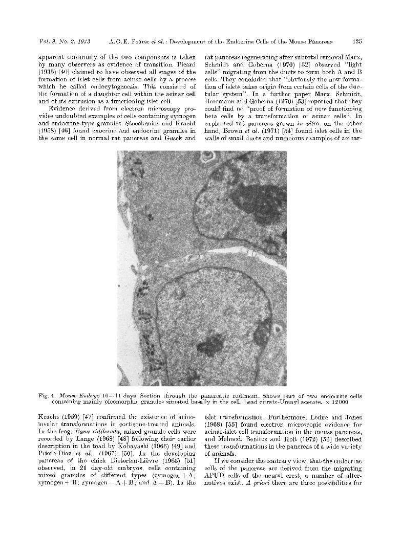
Fig. 4. Mouse Embryo 10--11 days. Section through the pancreatic rudiment. Shows part of two endocrine cells containing mainly pleomorphie granules situated basally in the cell. Lead eitrate-Uranyl acetate. • 12000
Kraeht (1959) [47] confirmed the existence of aeino- insular transformations in cortisone-treated animals. In the frog, Rana ridibunda, mixed granule cells were recorded by Lange (1968) [48] following their earlier description in the toad by Kobayashi (1966) [49] and Prieto-Diaz et al., (1967) [50]. In the developing pancreas of the chick Dieterlen-Litvre (1965) [51] observed, in 21 day-old embryos, cells containing mixed granules of different types (zymogenq-A; zymogen -~ B ; zymogen + A q- B ; and A q- B). In the
islet transformation. Furthermore, Leduc and Jones (t968) [55] found electron microscopic evidence for aeinar-islet cell transformation in the mouse pancreas, and Melmed, Benitez and Holt (1972) [56] described these transformations in the pancreas of a wide variety of animals.
I f we consider the contrary view, tha t the endocrine cells of the pancreas are derived from the migrating APUD cells of the neural crest, a number of alter- natives exist. A priori there are three possibilities for

126 A . G . E . Pea r se et al. : D e v e l o p m e n t of t h e E n d o c r i n e Cells of t h e Mouse P a n c r e a s Diabetologia
Fig. 5. Mouse Embryo 13 days. Resin- e m b e d d e d t ~m sect ion, s t a ined w i t h To lu id ine blue. The deve lop ing panc rea s consis ts of due t - l ike ep i the l ia l cords w i t h n u m e r -
ous lumina . Mitoses are f requen t . • 340
Fig. 6. Mouse Embryo 13 days. Shows p a r t of a n endocr ine cell in t h e deve lop ing pancreas . The granu les are m a i n l y spher ical , some resemble A granu les b u t o the r s are pleo- morph ic . A p r o m i n e n t Golgi region a n d m a n y coa ted vesi- cles can be seen. Lead e i t r a t e - U r a n y l ace ta te . • 12000
Fig. 7. 2VJouse Embryo 14 days. I n d i r e c t t e c h n i q u e for immunof luorescence . A n t i - h u m a n a n d porc ine insu l in sera r eac t w i t h B cells in t he n o w recognizable islets. • 300
Fig. 8. As Fig. 7. An t i -po rc ine g lucagon sera r eac t w i t h A cells in t h e def in i t ive islets a n d also in t h e ducts . Single
cells are found ou ts ide t he se s i tua t ions . • 300

Vol. 9, No. 2, 1973 A.G.E. Pearse et al. : Development of the Endocrine Cells of the Mouse Pancreas 127
the clear cells illustrated in Figs. 1 and 2, and for the dopamine-fluorescent cells in Fig. 3. Either they are arising from the gut epithelium and remaining in situ, or they are arising from the gut epithelium and going out, or they are arising outside the epithelium and coming in. We consider that there is sufficient evidence available to support the third deduction. If this is accepted then clearly there remain three more possi- bilities :
necessary to assume that the colonizing neural crest cells die out, having perhaps first induced endocrine differentiation in the duet cells. The finding of dop- amine labelled endocrine cell granules in the developing pancreas and of cells containing both the primitive pleomorphic granules characteristic of the neural crest- derived APUD cells and spherical granules of specific endocrine type, is sufficient to make this supposition unlikely.
Fig. 9. Mouse Embryo 14 days. Pancreatic islet. Shows parts of five endocrine cells. One cell contains both round and pleomorphic granules. Two identifiable B cells, with prominent Golgi regions and coated vesicles are present and two cells with mixed granules identified as D cells.
Lead citrate-Uranyl acetate. • 5000
1. All the endocrine cells of the pancreatic islets are derived from duet epithelium or aeinar cells.
2. All the endocrine cells of the pancreatic islets are derived from a neuroeetodermal precursor which colonizes the gut wall and primordial pancreas at an early stage of development.
3. Some of the endocrine cells are derived from the duet epithelium and others from the neural crest.
To accept the first of these suppositions as true it is
Fig. 10. Mouse Embryo 16 days. Pancreatic islet shows parts of several endocrine cells. An easily identifiable B cell now contains much rough endop]asmie reticulum and a single plcomorphic (biconcave) granule. A small portion of A cell cytoplasm contains the characteristic granules and a recognizable D cell is present. Lead citrate-Uranyl
acetate. • 10000
Our views on the second assumption are expressed in the diagram (Fig. 11) where the undifferentiated endocrine cells from the neural crest are shown (as clear cells) already in situ in the gut epithelium at the stage when the pancreatic anlagen are forming. Is there then sufficient evidence to warrant the assump- tion that all pancreatic endocrine cells are neuro- ectodermal, and thus lodgers in the duct epithelium. The answer at present is probably no, since the next

128 A.G.E. Pearse et al. : Development of the Endocrine Cells of the Mouse Pancreas Diabetologia
step from the proven neural crest origin of the primi- t ive endocrine cells has had to be made by ul t ras t ruc- rural criteria alone except for the eytoehemical evidence of cont inui ty between the A P U D cell and a t least one of the emdoerine cells wi th round granules. We cannot ye t dismiss, therefore, the th i rd assump- tion, and the possible der iva t ion of some endocrine cells f rom true duet epithelial (entodermal) cells.
........... - -??f!i:i:i??iii:i~:i?!.:-.:..::---.~-.-i!i-~: :.i~:- :-ii.!.~ii i.::J~iii~:-.:
::";'i'~;~ ..... : :""'.:" f-"[:??;"::.":-:'
"":~':::".':'!"-':" :': ' '" "" I ::::::::::::::::::::: ....... !
Duodenum
Fig. 11. Schematic representation of the distribution of endocrine (APUD) cells of neural crest origin in the primitive foregut (9 days) and its derivatives (9-- i 1 days)
References
1. Laguesse, E. : gecherches sur l 'histogenie du pancreas chez le Mouton. J. Anat. Physiol. 32,209--350 (1869).
2. I~ard, W.L. : The origin and differentiation of the alpha and beta cells in the pancreatic islets of the rat. Amer. J. Anat. 75, 369--403 (1944).
3. MeAlpine, 1%. J. : Alkaline phosphatase in the develop- ing endocrine pancreas of the albino rat. Anat. Rec. 109, 189--216 (1951).
4. Ferreira, D.: L 'ul trastrueture des cellules du pan- er6as endocrine ehez l 'embryon et le rat nouveau-n@. J. Ultrastructure l~es. 1, 14--25 (1957).
5. Frye, B.E. : The differentiation of the endocrine pan- ereas in foetuses of Alloxan diabetes and insulin- treated rats. J. Morph. 101, 325--350 (1957).
6. Munger, B.L. : A light and electron microscopic study in eellMar differentiation in the pancreatic islets of the mouse. Amer. J. Anat. 103, 275--312 (1958).
7. Esterhuizen, A. C. : The relationship between the alpha and beta cells in the islets of Langerhans of the albino rat. Sth. Aft. reed. J. 33, 197--201 (1959).
8. Golosow, N., Grobstein, C.: Epithelie-mesenehymal interaction in pancreatic morphogenesis. Develop. Biol. 4, 242--255 (1962).
9. Grille, T. A. I. : The occurrence of insulin in pancreases of foetuses of some rodents. J. Endocr. 31, 67--73 (1964).
10. Ilellman, B.: The development of the mammalian endocrine pancreas Biol. Neonat. 9, 263--278 (1965/ 66).
11. Pietet, 1~., Clark, W.R. , Renold, A.E., Williams, 1~., Rutter , W . J . : An electron microscopic study of the i n vitro development of the embryonic rat panereas. J. cell. Biol. 39, 105a (Abstract) (1968).
12. Wessells, N.K. , l%utter, W.J . : Phases in cell differen- tiation. Sei. Amer. 220, 36--44 (1969).
t 3. Perrier, H. : Evolut ion de l 'ultrastructure du pancr6as chez le foetus de rat. Diabetologia 6, 605--615 (1970).
14. Spooner, B.S., Walther, B.T., 1Rutter, W . J . : The development of the dorsal and ventral mammalian pancreas i n vivo and i n vitro. J. cell. Biol. 47, 235-- 246 (1970).
15. Pictet, R., Levine, S., Phelps, P., l~utter, W.J.: In vitro cytodifferentiation of rat embryonic pancreatic epithelium. Diabetes 29, Suppl. i, 326 (1971).
16. Pearse, A.G.E.: The cytochemistry and ultrastruc- ture of polypeptide hormone-producing cells of the APUD series, and the embryologic, physiologic and pathologic implications of the concept. J. I-listochem. Cytochem. 17, 303--313 (1969).
17. Le Douarin, N., Le Li~vre, C.: D6monstration de l'origine neural des cellules ~ ealcitonine du dorps ultimobranchial ehez l'embryon de Poulet. C. R. Acad. Sei. S6r. D. 270, 2857--2860 (1970).
18. Pearse, A.G.E., Polak, Julia M.: Cytochemical evi- dence for the neural crest origin of mammalian ultimo- branchial C cells. Histochemie 27, 96--102 (19Via).
19. Le Douarin, N., Le Li6vre, C., Fontaine, J . : Recher- ches experimentales sur l 'origine embryologique du corps caroditien chez les oiseaux. C. R. Acad. Sci. S6r. D. (1972 in press).
20. Pearse, A.G.E. , Polak, Jul ia M. : Neural crest origin of the endocrine polypeptide cells of the gastroin- testinal t ract artd pancreas. Gut 12, 783--788 1971b).
21. Lacy, P .E. , Davies, J . : Demonstration of insulin in mammalian pancreas by the fluorescent antibody method. Stain Teehnol. 34, 85 (1959).
22. Baum, J., Simons, B.E. , Unger, R.H. , Madison, L.L. : Localization of glucagon in the alpha cells in the pancreatic islet by immunofluorescent technics. Dia- betes 11, 371--374 (1962).
23. Lomsky, R., Langr, F., Vortel, V. : Site of glueagon in the islets of Langerhans of man as studied by the immunofiuoreseent technic. Sborn. ved. Praei l@d. Fak. Hradci Kralov6 I t , 585--590 (1968).
24. Griineberg, H. : The development of some external features in mouse embryo. J. Heredi ty 84, 89--92 (1943).
25. Polak, J .M., Bussolati, G., Pearse, A .G.E . : Cyto- chemical, immunofluorescence and ultrastructural in- vestigations on the antral G cells in hyperparathy- roidism. Virehows Arch. Abt. B. Zellpath. 9, 187-- 197 (1971).
26. Pearse, A .G.E . : Histoehemistry, Theoretical and Applied. Vol. 2, Ed. 3. London: Churchill-Livingstone 1972.
27. Grimelius, L. : A silver nitrate stain for ~2 cells in human pancreatic islets. Aeta See. Med. upsalien 73, 243--270 (1968).
28. Gomori, G. : Microscopic histochemistry. University Press, Chicago (1952).
29. I-IeIlerstr6m, C., Itellman, B. : Some aspects of silver impregnation of the islets of Langerhans in the rat. Aeta endoer. (Kbh.) 35, 518--532 (1960).
30. Coons, A. I-I., Leduc, E.H. , Connolly, J .M. : Studies on anibody production. I. A method for the histoehemical demonstration of specific antibody and its application to a study of the hyperimmune rabbit. J. exp. Med. 102, 49--60 (1955).
31. Polak, J.M., Bloom, S., Coulling, I., Pearse, A .G.E . : Immunofluorescent localization of enteroglueagon ceils in the gastrointestinal tract of the dog. Gut 12, 311--318 (1971).

Vol. 9, No. 2, 1973 A.G.E. Pearse et aI. : Development of
32. Bensley, 1R.R. : Studies on the pancreas of the guinea pig. Amer. J. Anat. 12, 297--388 (1911).
33. Liegner, B.: Studien zur Entwicklung des Pankreas, besonders der Langerha~sschen Inseln. Z. Zellforsch. 30, 494--529 (1932).
34. Rathery, F., Turiaf, J.: Influence de la castration et des injections de testosterone sur les ilots de Langer- hans du pancr6as du cobaye. C. R. Soc. Biol. 128, 155--156 (1938).
35. Gomori, G. : Observations with differential stains on human islets of Langcrhans. Amer. J. Path. 17, 395-- 406 (1941).
36. Laguesse, M. E. : Ilots endocrines et formes de transi- tion dans le lobule pancr6tique (heroine). C. r. S6anc. Soc. Biol. 58, 542--544 (1905).
37. Collin, R., Drouet, P.L., Watrin, J., Florentin, P.: Action histophysiologique de l'hypoglye6mie sur la glande pituitaire la neurohypophyse et le tuber einereum. C. 1R. See. Biol. 108, 61--64 (1931).
38. Florentin, P., Picard, D., Weis, M. : Modifications du pancr6as endocrine au tours de la gestation. C. R. Soc. Biol. 117, 188--189 (1934).
39. Florentin, P., Picard, D. : Reeherches sur le pancr6as endocrine. Rev. franq, d'endocrinol. 14, 1--27 (1936).
40. Picard, D.: S u r u n mode de formation de cellules langerhansiennes dans le pancr6as. C. R. See. Biol. 120, 153--154 (1935).
41. Tusques, J. : La formation d'ilots de Langerhans aux d6pens des acini duns le puncr6as des castrats. C. R. Soc. Biol. 129, 1103--1106 (1938).
42. Woerner, C. A. : Studies of islands of Langerhans after continuous intravenous injection of dextrose. Anat. Rec. 71, 33--57 (1938).
43. Aubertin, E., Lacoste, A., Saric, R.: Action des in- jeetions r6p6t6es d'insuline sur l '6tat structural et fonetionnel du tissue langerhansien (6rude exp6rimen- tal et clinique). Ann. de m6d. 43, 253--284 (1938).
44. Sergeyeva, M.A.: Microscopic changes in islands of Langerhans produced by sympathetic and parasym- pathetic stimulation in cat. Anat. Rec. 77, 297--317 (1940).
45. Simard, L.C.: Le complexe neuro-insulaire du pan- crdas chez les mammif6res adultes. Rev. Canad. de Biol. 1, 2--49 (1942).
46. Stoeckenius, W., I~2racht, J . : Elektronenmikroskopi- sche Untersuchungen an den Langerhansschen Inseln der Ratte. Endokrinologie 36, 135--142 (1958).
47. Gusek, W., Kracht, J.: Elektronenmikroskopische Untersuchungen fiber Inselwuehstum und acino-insu- l~re Transformation. Frankfurt Z. ffir Path. 7{), 98-- 106 (1959).
the Endocrine Cells of the Mouse Pancreas 129
48. Lunge, R.: Uber die Variabilitgt und experimentelle Beeinflussung der Morphologie der Zelltypen im In- selappurat des Forsches Rana ridibunda. Z. Zell- forsch. 88, 353--364 (1968).
49. Kobayashi, K.: Electron microscope studies of the Langerhans islets in the toad pancreas. Arch. histol. jap. 26, 439--482 (1966).
50. Prieto-Diaz, I-I.E., Iturriza, F.C., Rodriguez, i%.1%.: Acino-insular relationship in the pancreas of the toad investigated with the electron microscope. Aeta anat. (Basel) 67, 291--303 (1967).
51. Dieterlen-Li6vre, F. : Etude rnorphologique et expgri- mentale de la diff6r6neiation du pancr6as chez l'em- bryon de poulet. Bull. Biol. 99, 1--116 (1965).
52. Marx, M., Schmidt, W., Goberna, i%.: Elektronen- mikroskopische Untersuehungen zur Inselregenera- tion im Rattenpankreas nach subtotaler pankreatek- tomie. Z. Zellforsch. 110, 569--587 (1970).
53. Marx, M., Sehmidt, YV., I-Iernaann, M., Goberna, I~. : Electron microscopic studies on the existence of the so called "acinar-islet cells" in the regenerating Pan- creas of the rat. florin. & Metab. Rcs. 2, 204--212 (1970).
54. Brown, R.E. , Still, W.J , S.: Acinar-islct cells in the exocrine pancreas of the adult eat. Amer. J. dig. Dis. 15, 327--335 (1970).
55. Leduc, E. J., Jones, E. E. : Acinar-islet cell transforma- tion in mouse pancreas. J. Ultrastructure Res. 24, 165--169 (1968).
56. Melmed, R.N., Benitez, C.J., Holt, S. J. : Intermediate cells of the pancreas. I. Ultrastructural characteriza- tion. J. Cell Sei. 2, 449--475 (1972).
57. Orci, L., Lambert, A.E., Rouiller, Ch., Renold, A.E., Samols, E. : Evidence for the presence of A-cells in the endocrine fetal pancreas of the rat. Horm. & Metab. Res. I, 108--110 (1969).
58. Perrier, If., Porte, A., Jacquot, R.: Pr6sence de cellnles A duns le pancr6as foetal de rat. C. I%. Acad. Sci. 269, 841--843 (1969).
59. Wessells, N.K., Evans, J. : Ultrastructural studies of early morphogenesis and eytodifferentiation in the embryonic mammalian pancreas. Dev. Biol. 17, 413-- 446 (1968).
60. yon Denffer, l-I., Mertz, M. : Empfindliehkeit einiger Farbstoffe zum Naehweis yon/~-Granula in den Insel- zellen des Pankreas w/~hrend der Ontogenese. Hi- stochemie 29, 54--64 (1972).
Prof. A.G.E. Pearse Dept. of Histochemistry Royal Postgraduate Medical School Hammersmith Hospital London W. 12 England
Diabetologia, Vol. 9 9