development of a chemically defined fermentation medium for … · 2019-03-15 · chemically...
TRANSCRIPT
Development of a Chemically Defined
Fermentation Medium for the Production of a
New Recombinant Fructosyltransferase
1Institute of Bioprocess Engineering and Pharmaceutical Technology, University of Applied Sciences Mittelhessen,
Giessen, Germany 2Faculty of Biology and Chemistry, Justus Liebig University Giessen, Giessen, Germany
3Fraunhofer Institute for Molecular Biology and Applied Ecology (IME), Aachen, Germany 4Kansas State University, Manhattan (Kansas), United States of America
Email: {jan.burghardt, peter.czermak }@lse.thm.de
Abstract—The industrial-scale production of fructo-
oligosaccharides from sucrose requires large
quantities of the enzyme fructosyltransferase. An
Aspergillus terreus fructosyltransferase was therefore
expressed in Kluyveromyces lactis GG799 and secreted
into the medium. K. lactis was cultivated in shaking
flasks at 30°C and 250 rpm using either rich or
chemically defined fermentation media. In order to
limit the accumulation of unwanted side products that
tend to form in rich media such as yeast extract
peptone dextrose, a chemically defined FM22 medium
was optimized in a two stages, focusing on biomass
accumulation and enzyme production, respectively. A
design-of-experiments strategy was used to screen for
essential vitamins. A two-level fractional factorial
design revealed that only biotin, nicotinic acid,
pyridoxine and D-pantothenic acid were necessary for
biomass accumulation, and an additional D-optimal
design was used to optimize the concentration of
inorganic salts (MgSO4, (NH4)2SO4, CaSO4, FeSO4
and KH2PO4) and the fermentation temperature. For
enzyme production, the integrated LAC4 promoter
was induced with galactose, which was provided in
addition to glucose as the carbon source in the
adapted FM22 medium.
Index Terms—fructo-oligosaccharide, FOS,
fructosyltransferase, Kluyveromyces lactis, FM22, 1-kestose,
prebiotic
I. INTRODUCTION
Fructo-oligosaccharides (FOS) are low-calorie
sweeteners that occur naturally as long-chain
carbohydrates in plants and as exopolysaccharides in
microorganisms [1]. FOS cannot be digested by humans,
but they can stimulate the growth of beneficial gut
bacteria and thus have a prebiotic effect [2], [3]. FOS can
be produced industrially by the enzymatic elongation of
Manuscript received June 21, 2018; revised September 10, 2018.
sucrose or the hydrolysis of polysaccharides such as
inulin. Hydrolysis is catalyzed by endo-inulinases (EC
3.2.1.7), whereas the elongation reaction is catalyzed by
fructosyltransferases (EC 2.4.1.9), such as those found in
commercial enzyme mixes like Pectinex Ultra SP-L, with
sucrose acting as both donor and acceptor [4]-[7].
Fructose released from the donor sucrose is bound via a
β(2-1) linkage to the fructose moiety of an acceptor
sucrose, yielding 1-kestose (GF2). Further
transfructosylation extends the linear chain of β(2-1)-
linked fructose moieties yielding nystose (GF3) and 1F-
fructofranosylnystose (GF4). In addition to the β(2-1)-
linked FOS type, two related trisaccharides (GF2) can be
produced by fructosylation: 6-kestose via a β(2-6) linkage
to the fructose moiety and neokestose via a β(2-6) linkage
to the glucose moiety of the acceptor sucrose [8], [4], [9].
Kluyveromyces lactis has long been used for the
industrial-scale production of native β-galactosidase,
which is used to manufacture dairy products for people
with lactose intolerance. For β-galactosidase production,
K. lactis is classified “generally recognized as safe”
(GRAS) by the US Food and Drug Administration (FDA)
[10]. In a dried and inactivated form, K. lactis is used as a
protein additive for food and feed [11]. K. lactis was one
of the first fully sequenced yeasts, and one of the first
yeasts successfully used for heterologous protein
expression [12]. K. lactis is therefore used for the
commercial production of bovine chymosin, which
coagulates milk in the calf stomach. In the food industry,
chymosin and prochymosin are used during cheese
production [13]. An emerging use of K. lactis, due to its
capacity for post-translational modification [10], is the
production of complex therapeutic proteins such as
interleukin 1β, interferon α, lysozyme, serum albumin,
and preproinsulin [14]-[18].
In terms of process development, rich complex media
generally promote better growth and higher productivity
than chemically defined media due to the presence of
precursors that are used by the microorganisms in their
core metabolic processes [19]. However, rich
fermentation media are more expensive than chemically
International Journal of Pharma Medicine and Biological Sciences Vol. 7, No. 4, October 2018
©2018 Int. J. Pharm. Med. Biol. Sci. 71doi: 10.18178/ijpmbs.7.4.71-77
Jan P. Burghardt , Arne M. Oestreich , Tobias Weidner , Doreen Gerlach , and Peter Czermak1,2 1,2 1 3 1,2,3,4

defined media and the concentrations of various indigents
are inconsistent [20]. For the manufacture of low-margin
products such as enzymes in the food industry, low-cost
substrates are necessary for economic feasibility and
chemically defined media also increase process
robustness. Here, we adapted the chemically defined
medium FM22 to achieve K. lactis cell growth and
fructosyltransferase yields comparable to those realized
in a complex medium, namely yeast extract peptone
dextrose (YPD).
II. MATERIALS AND METHODS
A. Strain and Materials
We used a recently described Kluyveromyces lactis
GG799 strain from New England Biolabs GmbH
(Germany) expressing an integrated fructosyltransferase
gene from Aspergillus terreus NIH2624 and secreting the
enzyme to the medium [21]. All chemical reagents, if not
otherwise stated, were purchased from Merck KGaA
(Germany). Ammonium sulfate, trisodium citrate and
LC-MS grade triethylamine were purchased from VWR
International GmbH (Germany). Biotin, galactose,
tryptone and sucrose were purchased from Carl Roth
GmbH & Co KG (Germany). We obtained D-pantothenic
acid, inositol, nicotinic acid, pyridoxine and analytical
grade glucose, fructose and sucrose for HPLC analysis
from Sigma-Aldrich (Germany). Iron(II) sulfate and
4-amino-benzoic acid were purchased from AppliChem
GmbH (Germany) and cobalt(II)-chloride was purchased
from Alfa Aesar Haverhill (USA). We purchased a FOS
standard set comprising 1-kestose, nystose and
1F-fructofranosylnystose from Wako Pure Chemical
Industries Ltd (Japan). LC-MS grade acetonitrile was
purchased from Altmann Analytik GmbH & Co. KG
(Germany).
B. Small-Scale Shake Flask Conditions and Media
Cultures were grown in 500-mL shake flasks with a
working volume of 50 mL, and were incubated at 250
rpm in an INFORS HT Multitron (Infors AG,
Switzerland). FM22 medium [22], [23] was used at 30°C
and pH 5. If not otherwise stated, the medium comprised
5 g L-1 (NH4)2SO4, 1 g L-1 CaSO4 H2O, 1.75 g L-1
K2HPO4, 8.6 g L-1 K2SO4, 20 g L-1 glucose, 10 g L-1
inositol, 25.7 g L-1 KH2PO4, 16.4 g L-1 MgSO4 H2O, 15.6
g L-1 Na3C6H5O7, 160 mg L-1 biotin, 410 mg L-1 thiamin,
410 mg L-1 D-pantothenic acid, 410 mg L-1 nicotinic acid,
410 mg L-1 pyridoxine, 80 mg L-1 4-aminobenzoic acid,
410 mg L-1 riboflavin, 2.4 mg L-1 CuSO4, 0.01 mg L-1
NaI, 3.6 mg L-1 MnSO4 H2O, 0.24 mg L-1
Na2MoO4 2H2O, 0.02 mg L-1 H3BO3, 1 mg L-1 CoCl2, 14
mg L-1 ZnCl2 and 26.4 mg L-1 FeSO4. The vitamins and a
trace elements were prepared as individual filter sterilized
stock solutions before mixing.
Stock solutions containing the carbon source, the
potassium phosphate buffer, and the remaining salts were
autoclaved at 121°C for 15 min to avoid phosphorus salt
precipitation and caramelization reactions. Cultures were
inoculated from frozen glycerol stocks stored at –80°C to
an optical density of ΔOD600 = 0.2 and were cultivated
until they reached the stationary phase.
C. Reactor-Scale Expression Conditions
Small cultures were transferred to MiniBio 500
reactors (Applikon Biotechnology, Netherlands)
containing 300 mL medium, including 30 g L-1 glucose
and 7.5 g L-1 galactose as carbon sources. For production
of the recombinant fructosyltransferase, the integrated
LAC4 promoter was induced with galactose. The K. lactis
dry cell weight and protein yields were compared in the
adapted FM22 medium and YPD medium at pH 5 and 6
in duplicate fermentations. The pH was regulated using a
PID controller by adding 25% concentrated ammonia
hydroxide as a base when necessary. The reactor vessel
was aerated at 1 vvm and with 0–0.27 vvm oxygen to
maintain the dissolved oxygen (DO) saturation above
50%. We used 42 µL J673A solution as an anti-foaming
agent (Schill + Seilacher “Struktol” GmbH, Germany).
The YPD medium contained 20 g L-1 peptone ex casein
and 10 g L-1 yeast extract in addition to the carbon source.
D. Cell Dry Weight
The empty weight of a 2-mL tube was measured on a
precision balance after drying the tube for at least 24 h at
70°C. We then added 2 mL fermentation broth,
centrifuged for 3 min (4°C, 16,100 x g), and the
supernatant was removed. In addition, to remove
disruptive medium components, the pellet was dissolved
in physiological sodium chloride solution and treated as
described above. This washing step was repeated. The
open tube containing the biomass was then incubated in
the drying oven for 24h at 70°C, cooled to room
temperature in a desiccator and then re-weighed on the
precision balance. The difference between the final
weight and the initial weight of the empty tube was
reported as the cell dry weight.
E. HPLC Analytics and Enzyme Assay
Samples were analyzed on a Dionex UltiMate 3000
HPLC system coupled with a Corona Veo Charged
Aerosol Detector (Thermo Fisher Scientific, USA).
Samples were fractionated in an XBridge Amide, 3.5 μm,
4.6 x 150 mm column (Waters GmbH, Germany). The
mobile phase was 70:30 (v/v) acetonitrile/water. The
water fraction was supplemented with 0.2% (v/v)
trimethylamine before analysis.
To determine the enzyme activity, 100 µL cell-free
fermentation broth was incubated with 900 µL substrate
solution at 60°C for 20 min, agitated at 1000 rpm in a
Thermomixer Comfort (Eppendorf AG, Germany). The
substrate solution consisted of 600 g L-1 sucrose in
50 mM potassium phosphate (pH 6). After incubation, the
reaction was interrupted by heating the sample to 95°C
for 20 min. The samples were diluted 1:50 with 50:50
(v/v) acetonitrile/water and clarified by passing through a
0.45-µm nylon membrane filter. Enzyme activity was
determined by measuring the amount of released glucose
minus the amount of free fructose [24].
International Journal of Pharma Medicine and Biological Sciences Vol. 7, No. 4, October 2018
©2018 Int. J. Pharm. Med. Biol. Sci. 72

F. Design of Experiments
Design Expert v10 (Stat-Ease, USA) was used to
identify the essential vitamins and to adjust the salt
concentrations and temperature. The concentration of the
vitamins was varied as follows, defining the –1/1 values:
biotin (A), 0.0-0.41 g L-1; thiamin (B), 0.0-0.41 g L-1;
inositol (C), 0.0–25.0 g L-1; D-pantothenic acid (D), 0.0-
0.41 g L-1; nicotinic acid (E), 0.0-0.41 g L-1; pyridoxine
(F), 0.0-0.41 g L-1; 4-aminobenzoic acid (G), 0.0-0.08 g
L-1; riboflavin (H), 0.0-0.41 g L-1. Optimized salt
concentrations and the fermentation temperature were
determined using a D-optimal design with the following–
1/1 values: KH2PO4 (A), 1.0-18.9 g L-1; MgSO4∙H2O (B),
0.85-16.4 g L-1; (NH4)2SO4 (C), 0.5-10.0 g L-1;
CaSO4∙H2O (D), 0.004-1.007 g L-1; FeSO4 (E). 0.002-
0.19 g L-1; and the temperature (F) 20-30°C.
G. Polyacrylamide Gel Electrophoresis (SDS PAGE)
Protein samples from aliquots of cell-free fermentation
broth from the shake flask and reactor scale cultures were
analyzed by SDS-PAGE using Criterion TGX Stain-Free
Protein 4-20% Gels from Bio-Rad (Germany). For each
sample, 31 µL of the protein solution was incubated with
11.7 µL of 4x Laemmli Sample Buffer (Bio-Rad) and 1.3
µL 2-mercaptoethanol (Bio-Rad) at 95°C for 5 min. We
used 5 µL Precision Plus Protein Unstained Protein
Standards (Bio-Rad) as size markers. The gel were run
for 25 min at 250 V, then activated with the Chemidoc
MP Imaging System (Bio-Rad).
III. RESULTS AND DISCUSSION
A. Vitamin Screening
A two-level fractional factorial design was used to
determine which vitamins are essential for the growth of
K. lactis. The responses in terms of cell dry weight are
summarized in Table I.
TABLE I. VITAMIN SCREENING
Factors Response
Run A B C D E F G H CDW
(g L-1)
1 1 –1 1 1 –1 –1 1 –1 0.30±0.04
2 –1 –1 1 1 1 –1 –1 1 0.08±0.01
3 1 –1 1 –1 –1 1 –1 1 0.07±0.01
4 –1 –1 –1 –1 –1 –1 –1 –1 0.07±0.01
5 0 0 0 0 0 0 0 0 4.52±0.08
6 1 1 1 –1 1 –1 –1 –1 0.34±0.01
7 –1 1 1 1 –1 1 –1 –1 0.06±0.00
8 –1 1 1 –1 –1 –1 1 1 0.04±0.00
9 1 1 1 1 1 1 1 1 4.20±0.18
10 –1 –1 1 –1 1 1 1 –1 0.23±0.01
11 –1 1 –1 –1 1 1 –1 1 0.25±0.04
12 1 –1 –1 –1 1 –1 1 1 0.24±0.05
13 1 1 –1 –1 –1 1 1 –1 0.04±0.00
14 –1 –1 –1 1 –1 1 1 1 0.07±0.01
15 0 0 0 0 0 0 0 0 4.93±0.13
16 0 0 0 0 0 0 0 0 5.30±0.01
17 –1 1 –1 1 1 –1 1 –1 0.75±0.07
18 1 1 –1 1 –1 –1 –1 1 0.07±0.01
19 1 –1 –1 1 1 1 –1 –1 5.08±0.07
Concentration in coded format for the factors: A = biotin; B = thiamin;
C = inositol; D = D-pantothenic acid; E = nicotinic acid; F = pyridoxine;
G = 4-aminobenzoic acid, and H = riboflavin. CDW = cell dry weight.
These results show which vitamins must be added to
the medium because they cannot be synthesized by the
cells. Table I shows that despite the three centerpoints
(Runs 5, 15 and 16) and the high concentrations of all
vitamins (Run 9), only Run 19 achieved a cell dry weight
(5.08 g L-1) comparable to the nonadapted FM22 medium
(4.40 g L-1).
Figure 1. Pareto chart of the two-level fractional factorial vitamin
screening. The chart shows effects above the Bonferroni limit indicating
factors with a significant impact on K. lactis growth in the model [25].
Orange bars define positive effects and blue bars define negative effects.
DE, AE, AD and AF indicate two-factor interactions.
The vitamins necessary for the growth of K. lactis are
biotin, D-pantothenic acid, nicotinic acid and pyridoxine
(Fig. 1). To confirm the model, Run 19 was repeated and
subsequent experiments were carried out lacking
individual vitamins, resuling in the absence of
measurable cell dry weights. These results confirmed that
all four vitamins must be present in the medium. The
entire model, as well as the factors biotin, D-pantothenic
acid and nicotinic acid, have p-values < 0.0001.
Pyridoxine and the interactions of the other factors have
p-values < 0.001 and are thus also clearly in the
significant range. This means the probability that the
effect is caused by noise in the process model is < 0.1%.
In further experiments, only the essential vitamins
nicotinic acid, biotin, D-pantothenic acid and pyridoxine
were included in the vitamin adapted FM22 medium.
B. D-opitmal Optimization of Salt Cocnentrations
Next we investigated the impact on cell dry weight of
different salt concentrations in the vitamin adapted FM22
medium and different fermentation temperatures. Each
factor was examined in individual ranges based on data
available in the literature. A two-factor interaction model
was used, as suggested by Design Expert, and square root
data transformation, derived from the Box Cox diagram,
was therefore applied to evaluate the model. The
evaluation of the process model confirmed significance
with a p-value < 0.0001. Furthermore, significant model
terms included potassium phosphate, ammonium sulfate,
International Journal of Pharma Medicine and Biological Sciences Vol. 7, No. 4, October 2018
©2018 Int. J. Pharm. Med. Biol. Sci. 73
the temperature and the two-level interactions between
potassium phosphate and magnesium sulfate, potassium
phosphate and iron(II) sulfate, magnesium sulfate and
iron(II) sulfate, and ammonium sulfate and iron(II)
sulfate.
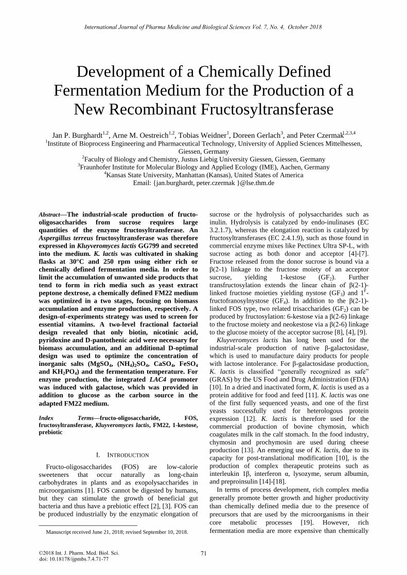
TABLE II. D-OPTIMAL DESIGN
Factors Response
Run A B C D E F CDW
(g L-1) 1 –0.3 –1.0 1.0 –0.3 –1.0 –1.0 5.87±0.13
2 1.0 –0.8 –1.0 –1.0 1.0 1.0 0.83±0.03
3 1.0 1.0 0.5 –1.0 0.9 1.0 6.83±0.03
4 –1.0 –0.2 1.0 –0.5 1.0 1.0 5.38±0.28
5 1.0 –1.0 –1.0 –1.0 –1.0 –1.0 0.47±0.08
6 –1.0 –1.0 –1.0 –1.0 1.0 –1.0 0.47±0.03
7 0.3 0.3 –1.0 0.5 0.5 0.0 0.83±0.03
8 0.0 0.0 0.0 –0.2 0.0 0.0 3.00±0.08
9 –1.0 1.0 –1.0 0.5 1.0 –1.0 1.05±0.13
10 –1.0 1.0 –1.0 –1.0 1.0 1.0 1.35±0.12
11 0.0 0.0 0.0 –0.2 0.0 0.0 2.98±0.03
12 1.0 1.0 –1.0 –1.0 1.0 –1.0 1.00±0.05
13 0.3 1.0 1.0 0.5 1.0 0.0 5.12±0.06
14 1.0 –1.0 1.0 –1.0 1.0 –1.0 5.15±0.05
15 1.0 1.0 1.0 –1.0 –1.0 1.0 6.73±0.13
16 –1.0 1.0 1.0 0.5 –0.8 1.0 6.86±0.16
17 0.0 0.0 0.0 –0.2 0.0 0.0 3.55±0.22
18 1.0 –1.0 –1.0 –1.0 –1.0 –1.0 0.52±0.03
19 1.0 –1.0 0.9 0.4 0.8 1.0 6.13±0.13
20 –1.0 –1.0 –1.0 0.5 1.0 1.0 0.77±0.18
21 1.0 –1.0 –1.0 0.5 1.0 –1.0 0.57±0.06
22 –1.0 1.0 1.0 0.5 –0.8 1.0 4.62±0.19
23 –1.0 –1.0 –1.0 –1.0 1.0 –1.0 0.45±0.01
24 –1.0 –1.0 –1.0 0.5 –1.0 –1.0 5.03±0.08
25 1.0 1.0 0.9 –1.0 –0.4 –1.0 0.97±0.08
26 –1.0 1.0 –1.0 –1.0 –1.0 –1.0 0.92±0.13
27 0.9 1.0 –1.0 –1.0 –1.0 1.0 0.70±0.09
28 0.2 1.0 0.2 0.5 –1.0 –1.0 3.12±0.08
29 0.1 0.1 –1.0 0.5 –1.0 1.0 0.58±0.06
30 1.0 1.0 –0.2 0.5 1.0 1.0 6.23±0.08
31 –1.0 1.0 1.0 –1.0 1.0 –1.0 4.42±0.08
32 1.0 –1.0 –1.0 –0.2 –0.1 1.0 0.58±0.06
33 –1.0 –1.0 –0.6 –0.9 –1.0 1.0 7.45±0.13
34 1.0 0.4 1.0 0.5 –0.1 –1.0 2.83±0.03
35 –1.0 –1.0 1.0 0.5 1.0 –1.0 5.43±0.06
36 –1.0 –0.2 1.0 –1.0 –1.0 0.0 6.60±0.05
37 1.0 1.0 –1.0 0.5 –1.0 0.0 0.67±0.06
38 1.0 –1.0 1.0 0.5 –1.0 1.0 5.77±0.23
39 –0.3 –1.0 1.0 –1.0 0.1 1.0 7.10±0.13
Concentration in coded format for the factors: A = potassium phosphate;
B = masgnesium sulfate; C = ammonium sulfate; D =calcium sulfate;
E = Iron(II) sulfate, and F = temperature. CDW = cell dry weight.
The difference between the predicted R-squared
(0.7659) and the adjusted R-squared (0.8415) was < 0.2.
In a confirmation run, a cell dry weight of 8.18±0.58 g L-
1 was achieved with the vitamin and salt adjusted medium
at 30°C, based on the following salt concentrations:
KH2PO4, 9.95 g L-1; MgSO4∙H2O, 1 g L-1; CaSO4∙H2O, 1
g L-1; FeSO4, 0.19 g L-1; (NH4)2SO4, 10 g L-1. In shake
flasks, the cell dry weight increased by 186%. Similar
increases in production yield (210%) have been achieved
[22] by reducing the concentration of salts and trace
elements compared to the original FM22 medium [26].
Figure 2. Contour plot of the D-optimal model with cell dry weight as
the response. Actual concentration of the remaining factors: KH2PO4,
9.95 g L-1; MgSO4∙H2O, 1 g L-1; CaSO4∙H2O, 1 g L-1; FeSO4, 0.19 g L-1.
C. Enzyme Production
Four duplicate fermentation runs were conducted with
the vitamin and salt adapted FM22 medium at pH 5 and 6.
The cell dry weights increased to 17.48±0.20 g L-1 at pH
5 and to 16.53±0.55 g L-1 at pH 6 (Fig. 3). The higher cell
dry weights were achieved due to the adjusted carbon
source concentrations at the larger reactor scale. The
transfer activities of the cell-free fermentation broth in
each case reflect the expressed fructosyltransferase.
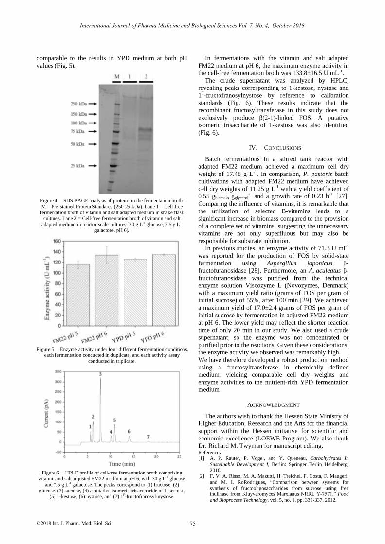
Compared to shake flask cultures, a new 80-kDa protein
band was identified by SDS-PAGE (Fig. 4),
corresponding to the glycosylated form of the expressed
fructosyltransferase in K. lactis, whereas this band is not
expressed by wild-type K. lactis GG799 [21].
Figure 3. Cell dry weight under four different fermentation conditions,
each fermentation conducted in duplicate, and each cell dry weight
measured in triplicate.
Fermentation broths without galactose showed no
measurable transfer activity (data not shown). By
measuring the expression level of the enzymes, the
activities in adapted FM22 medium were found to be
International Journal of Pharma Medicine and Biological Sciences Vol. 7, No. 4, October 2018
©2018 Int. J. Pharm. Med. Biol. Sci. 74
comparable to the results in YPD medium at both pH
values (Fig. 5).
Figure 4. SDS-PAGE analysis of proteins in the fermentation broth.
M = Pre-stained Protein Standards (250-25 kDa). Lane 1 = Cell-free
fermentation broth of vitamin and salt adapted medium in shake flask
cultures. Lane 2 = Cell-free fermentation broth of vitamin and salt
adapted medium in reactor scale cultures (30 g L-1 glucose, 7.5 g L-1
galactose, pH 6).
Figure 5. Enzyme activity under four different fermentation conditions,
each fermentation conducted in duplicate, and each activity assay
conducted in triplicate.
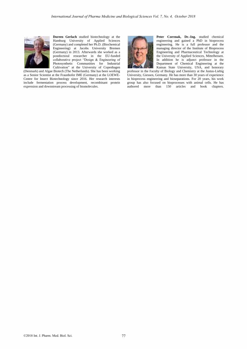
Figure 6. HPLC profile of cell-free fermentation broth comprising
vitamin and salt adjusted FM22 medium at pH 6, with 30 g L-1 glucose
and 7.5 g L-1 galactose. The peaks correspond to (1) fructose, (2)
glucose, (3) sucrose, (4) a putative isomeric trisaccharide of 1-kestose,
(5) 1-kestose, (6) nystose, and (7) 1F-fructofranosyl-nystose.
In fermentations with the vitamin and salt adapted
FM22 medium at pH 6, the maximum enzyme activity in
the cell-free fermentation broth was 133.8±16.5 U mL-1.
The crude supernatant was analyzed by HPLC,
revealing peaks corresponding to 1-kestose, nystose and
1F-fructofranosylnystose by reference to calibration
standards (Fig. 6). These results indicate that the
recombinant fructosyltransferase in this study does not
exclusively produce β(2-1)-linked FOS. A putative
isomeric trisaccharide of 1-kestose was also identified
(Fig. 6).
IV. CONCLUSIONS
Batch fermentations in a stirred tank reactor with
adapted FM22 medium achieved a maximum cell dry
weight of 17.48 g L-1. In comparison, P. pastoris batch
cultivations with adapted FM22 medium have achieved
cell dry weights of 11.25 g L-1 with a yield coefficient of
0.55 gbiomass gglycerol-1 and a growth rate of 0.23 h-1 [27].
Comparing the influence of vitamins, it is remarkable that
the utilization of selected B-vitamins leads to a
significant increase in biomass compared to the provision
of a complete set of vitamins, suggesting the unnecessary
vitamins are not only superfluous but may also be
responsible for substrate inhibition.
In previous studies, an enzyme activity of 71.3 U ml-1
was reported for the production of FOS by solid-state
fermentation using Aspergillus japonicus β-
fructofuranosidase [28]. Furthermore, an A. aculeatus β-
fructofuranosidase was purified from the technical
enzyme solution Viscozyme L (Novozymes, Denmark)
with a maximum yield ratio (grams of FOS per gram of
initial sucrose) of 55%, after 100 min [29]. We achieved
a maximum yield of 17.0±2.4 grams of FOS per gram of
initial sucrose by fermentation in adjusted FM22 medium
at pH 6. The lower yield may reflect the shorter reaction
time of only 20 min in our study. We also used a crude
supernatant, so the enzyme was not concentrated or
purified prior to the reactions. Given these considerations,
the enzyme activity we observed was remarkably high.
We have therefore developed a robust production method
using a fructosyltransferase in chemically defined
medium, yielding comparable cell dry weights and
enzyme activities to the nutrient-rich YPD fermentation
medium.
ACKNOWLEDGMENT
The authors wish to thank the Hessen State Ministry of
Higher Education, Research and the Arts for the financial
support within the Hessen initiative for scientific and
economic excellence (LOEWE-Program). We also thank
Dr. Richard M. Twyman for manuscript editing. References
[1] A. P. Rauter, P. Vogel, and Y. Queneau, Carbohydrates In
Sustainable Development I, Berlin: Springer Berlin Heidelberg,
2010.
[2] F. V. A. Risso, M. A. Mazutti, H. Treichel, F. Costa, F. Maugeri,
and M. I. RoRodrigues, “Comparison between systems for
synthesis of fructooligosaccharides from sucrose using free
inulinase from Kluyveromyces Marxianus NRRL Y-7571,” Food
and Bioprocess Technology, vol. 5, no. 1, pp. 331-337, 2012.
International Journal of Pharma Medicine and Biological Sciences Vol. 7, No. 4, October 2018
©2018 Int. J. Pharm. Med. Biol. Sci. 75

[3] Z. KovÁCs, E. Benjamins, K. Grau, A. Ur Rehman, M. Ebrahimi,
and P. Czermak, “Recent developments in manufacturing
oligosaccharides with prebiotic functions,” Advances in
Biochemical Engineering/Biotechnology, vol. 143, pp. 257-295,
2014.
[4] S. P. Singh, J. S. Jadaun, L. K. Narnoliya, and A. Pandey,
“Prebiotic oligosaccharides: Special focus on
fructooligosaccharides, its biosynthesis and bioactivity,” Applied
Biochemistry and Biotechnology, vol. 183, no. 2, pp. 613-635,
2017.
[5] J. J. Virgen-OrtÍZ, V. Ibarra-Junquera, P. Escalante-Minakata, S.
Centeno-Leija, H. Serrano-Posada, J. de Jesús Ornelas-Paz, et al.,
“Identification and functional characterization of a
fructooligosaccharides-forming enzyme from aspergillus
aculeatus,” Applied Biochemistry and Biotechnology, vol. 179, no.
3, pp. 497-513, 2016.
[6] A. Ur Rehman, Z. Kovacs, H. Quitmann, M. Ebrahimi, and P.
Czermak, “Enzymatic production of fructo-oligosaccharides from
inexpensive and abundant substrates using a membrane reactor
system,” Separation Science and Technology, vol. 58, no. 4, P.
548, 2016.
[7] B. Erdős, M. Grachten, P. Czermak, and Z. Kovács, “Artificial
neural network-assisted spectrophotometric method for monitoring
fructo-oligosaccharides production,” Food and Bioprocess
Technology, vol. 8398, no. 2014, P. 37, 2017.
[8] N. Duchateau, K. Bortlik, U. Simmen, A. Wiemken, and P. Bancal,
“Sucrose: Fructan 6-fructosyltransferase, a key enzyme for
diverting carbon from sucrose to fructan in barley leaves,” Plant
Physiology, vol. 107, no. 4, pp. 1249-1255, 1995.
[9] S. Kilian, S. Kritzinger, C. Rycroft, G. Gibson, and J. du Preez,
“The effects of the novel bifidogenic trisaccharide, neokestose, on
the human colonic microbiota,” World Journal of Microbiology
and Biotechnology, vol. 18, no. 7, pp. 637-644, 2002.
[10] A. J. J. Van Ooyen, P. Dekker, M. Huang, M. M. A. Olsthoorn, D.
I. Jacobs, and P. A. Colussi, et al., “Heterologous protein
production in the yeast kluyveromyces lactis,” FEMS Yeast
Research, vol. 6, no. 3, pp. 381-392, 2006.
[11] F. J. Bonekamp and J. Oosterom, “On the safety of kluyveromyces
LACTIS: A review,” Applied Microbiology and Biotechnology, vol.
41, no. 1, pp. 1-3, 1994.
[12] S. Das And C. P. Hollenberg, “A high-frequency transformation
system for the yeast kluyveromyces lactis,” Current Genetics, vol.
6, no. 2, pp. 123-128, 1982.
[13] J. A. Van Den Berg, K. J. Van Der Laken, A. J. J. Van Ooyen, T.
C. H. M. Renniers, K. Rietveld, A. Schaap, et al.,
“Kluyveromyces as a host for heterologous gene expression:
Expression and secretion of prochymosin,” Nature Biotechnology,
vol. 8, no. 2, pp. 135-139, 1990. [14] R. Fleer, X. J. Chen, N. Amellal, P. Yeh, A. Fournier, F. Guinet,
et al., “High-level secretion of correctly processed recombinant
human interleukin-1 beta in kluyveromyces lactis,” Gene, vol.
107, no. 2, pp. 285-295, 1991.
[15] X. Chen, B. Gao, W. Shi, and Y. Li, “Expression and secretion of
human interferon Alpha a in yeast kluyveromyces lactis,” Yi
Chuan Xue Bao=Acta Genetica Sinica, vol. 19, no. 3, pp. 284-288,
1992.
[16] T. Iwata, R. Tanaka, M. Suetsugu, M. Ishibashi, H. Tokunaga, and
M. Kikuchi, “Efficient secretion of human lysozyme from the
yeast, kluyveromyces lactis,” Biotechnology Letters, vol. 26, no.
23, pp. 1803-1808, 2004.
[17] M. Saliola, C. Mazzoni, N. Solimando, A. Crisà, C. Falcone, G.
Jung, et al., “Use of the kladh4 promoter for ethanol-dependent
production of recombinant human serum albumin in
kluyveromyces lactis,” Applied and Environmental Microbiology,
vol. 65, no. 1, pp. 53-60, 1999.
[18] Y. M. Feng, B. Y. Zhang, Y. S. Zhang, and H. Fukuhara,
“Secretory expression of porcine insulin precursor in
kluyveromyces lactis and its conversion into human insulin,” Acta
Biochimica et Biophysica Sinica, vol. 29, no. 2, pp. 129-134, 1997.
[19] B. Hahn-Hägerdal, K. Karhumaa, C. U. Larsson, M. Gorwa-
Grauslund, J. Görgens, and W. H. van Zyl, “Role of cultivation
media in the development of yeast strains for large scale industrial
use,” Microbial. Cell Factories, vol. 4, no. 31, 2005.
[20] G. G. Moulton, Fed-Batch Fermentation: A Practical Guide to
Scalable Recombinant Protein Production in Escherichia Coli,
2014.
[21] S. C. Spohner And P. Czermak, “Heterologous expression of
Aspergillus Terreus fructosyltransferase in Kluyveromyces Lactis,”
New Biotechnology, vol. 33, no. 4, pp. 473-479, 2016.
[22] B. A. Plantz, J. Sinha, L. Villarete, K. W. Nickerson, and V.
L.Schlegel, “Pichia pastoris fermentation optimization: Energy
state and testing a growth-associated model,” Applied
Microbiology and Biotechnology, vol. 72, no. 2, pp. 297-305,
2006.
[23] P. P. Jacobs, M. Inan, N. Festjens, J. Haustraete, A. Van Hecke, R.
Contreras, et al., “Fed-batch fermentation of GM-CSF-producing
glycoengineered pichia pastoris under controlled specific growth
rate,” Microbial Cell Factories, vol. 9, no. 93, 2010. [24] S. Van Hijum, M. Van Der Maarel, and L. Dijkhuizen, “Kinetic
properties of an inulosucrase from lactobacillus reuteri 121,”
FEBS Letters, vol. 534, no. 1-3, pp. 207-210, 2003.
[25] M. G. Borines, R. L. De Leon, and J. L. Cuello, “Bioethanol
production from the macroalgae sargassum spp,” Bioresource
Technology, vol. 138, pp. 22-29, 2013.
[26] J. Stratton, V. Chiruvolu, and M. Meagher, “High cell-density
fermentation,” Methods in Molecular Biology (Clifton, N. J.), vol.
103, pp.107-120, 1998.
[27] A. Ghosalkar, V. Sahai, and A. Srivastava, “Optimization of
chemically defined medium for recombinant pichia pastoris for
biomass production,” Bioresource Technology, vol. 99, no. 16, pp.
7906-7910, 2008.
[28] S. I. Mussatto, C. N. Aguilar, L. R. Rodrigues, and J. A. Teixeira,
“Fructooligosaccharides and β-fructofuranosidase production by
aspergillus japonicus immobilized on lignocellulosic materials,”
Journal Of Molecular Catalysis B: Enzymatic, vol. 59, no. 1-3, pp.
76-81, 2009.
[29] A. S. G. Lorenzoni, L. F. Aydos, M. P. Klein, R. C. Rodrigues,
and P. F. Hertz, “Fructooligosaccharides synthesis by highly
stable immobilized β-fructofuranosidase from aspergillus
aculeatus,” Carbohydrate Polymers, vol. 103, pp. 193-197, 2014.
Jan Philipp Burghardt was born in 1988. He
studied bioprocess engineering at the Hamburg
University of Technology. He studied for his
Master’s thesis at BASF Personal Care and
Nutrition GmbH (Germany) based on mass
transfer studies in different systems. He is
currently a PhD candidate under Professor
Czermak at the University of Applied Sciences
Mittelhessen. His research topic focuses on
recombinant enzymes in food biotechnology.
Arne Michael Oestreich was born in Gießen, in
1986. He studied biotechnology and biopharma-
ceutical technology at the University of Applied
Sciences Mittelhessen. He studied for his Master’s
thesis at the Institute of Bioprocess Engineering
and Pharmaceutical Technology (IBPT) focusing
on process and medium development. Currently
he is a Ph.D. student at the IBPT and his research
focuses on recombinant protein expression.
Tobias Weidnet, born in 1982, studied chemical
and bioprocess engineering at Friedrich-
Alexander University Erlangen-Nuremberg. After
earning his PhD in the field of cell culture
technology at the Institute of Bioprocess
Engineering, University Erlangen-Nuremberg, he
is now a postdoctoral fellow at the Institute of
Bioprocess Engineering and Pharmaceutical
Technology, University of Applied Sciences,
Mittelhessen.
International Journal of Pharma Medicine and Biological Sciences Vol. 7, No. 4, October 2018
©2018 Int. J. Pharm. Med. Biol. Sci. 76
Doreen Gerlach studied biotechnology at the
Hamburg University of Applied Sciences
(Germany) and completed her Ph.D. (Biochemical
Engineering) at Jacobs University Bremen
(Germany) in 2013. Afterwards she worked as a
postdoctoral researcher in the EU-funded
collaborative project “Design & Engineering of
Photosynthetic Communities for Industrial
Cultivation” at the University of Copenhagen
(Denmark) and Algae Biotech (The Netherlands). She has been working
as a Senior Scientist at the Fraunhofer IME (Germany) at the LOEWE-
Centre for Insect Biotechnology since 2016. Her research interests
include fermentation process development, recombinant protein
expression and downstream processing of biomolecules.
Peter Czermak, Dr.-Ing. studied chemical
engineering and gained a PhD in bioprocess
engineering. He is a full professor and the
managing director of the Institute of Bioprocess
Engineering and Pharmaceutical Technology at
the University of Applied Sciences, Mittelhessen.
In addition he is adjunct professor in the
Department of Chemical Engineering at the
Kansas State University, USA, and honorary
professor in the Faculty of Biology and Chemistry at the Justus-Liebig
University, Giessen, Germany. He has more than 30 years of experience
in bioprocess engineering and bioseparations. For 20 years, his work
group has also focused on bioprocesses with animal cells. He has
authored more than 150 articles and book chapters.
International Journal of Pharma Medicine and Biological Sciences Vol. 7, No. 4, October 2018
©2018 Int. J. Pharm. Med. Biol. Sci. 77