development of a functional testing platform for the
TRANSCRIPT

University of Central Florida University of Central Florida
STARS STARS
Electronic Theses and Dissertations
2019
Development of a Functional Testing Platform for the Sensory Development of a Functional Testing Platform for the Sensory
Segment of the Neuromuscular Reflex Arc Segment of the Neuromuscular Reflex Arc
Alisha Colon University of Central Florida
Part of the Animal Experimentation and Research Commons, and the Medical Neurobiology
Commons
Find similar works at: https://stars.library.ucf.edu/etd
University of Central Florida Libraries http://library.ucf.edu
This Doctoral Dissertation (Open Access) is brought to you for free and open access by STARS. It has been accepted
for inclusion in Electronic Theses and Dissertations by an authorized administrator of STARS. For more information,
please contact [email protected].
STARS Citation STARS Citation Colon, Alisha, "Development of a Functional Testing Platform for the Sensory Segment of the Neuromuscular Reflex Arc" (2019). Electronic Theses and Dissertations. 6821. https://stars.library.ucf.edu/etd/6821

DEVELOPMENT OF A FUNCTIONAL TESTING PLATFORM FOR THE SENSORY
SEGMENT OF THE NEUROMUSCULAR RELFEX ARC
by
ALISHA MARIE COLÓN
B.S. University of Central Florida, 2012
M.S. University of Central Florida, 2015
A dissertation submitted in partial fulfillment of the requirements
for the degree of Doctor of Philosophy
in the Burnett School of Biomedical Sciences
in the College of Medicine
at the University of Central Florida
Orlando, Florida
Summer Term
2019
Major Professor: James J. Hickman

ii
© 2019 Alisha Marie Colón

iii
ABSTRACT
Investigations of human biology and disease have been hindered by the use of animal
models. The information obtained from such studies often results in clinically irrelevant results
and drug trial failures. Additionally, several governing bodies have been formulating legislation
to move away from animal models and toward more ethical and efficient testing platforms for
drug discovery and cosmetic research. As an answer to these issues, “body-on-a-chip” systems
have been a rapidly developing field which easily recapitulates in vivo functionality, providing a
more relevant, repeatable, and ethical testing platform to better predict biology. These systems
can be used as human-based testing platforms to evaluate human physiology, disease
progression, and drug responsiveness for specific cell types and multi-organ systems. Diseases
such as amyotrophic lateral sclerosis (ALS) and spinal muscular atrophy (SMA) have significant
research challenges, specifically with translating research findings into treatment plans. The
complexity of the neuromuscular reflex arc, the biological system affected by these diseases, is
difficult to study with traditional molecular techniques, namely because the many components of
this disease system interact with each other using complex pathways. This work pushes the
existing platform to a more complete human model of neuromuscular disease with the
incorporation of gamma motoneurons, development of the first human induced pluripotent cell
(iPSC) derived intrafusal fibers, and proposals to incorporate nociceptive neurons all on a
functionally interrogative platform. The incorporation of these components will allow for a more
complete, clinically relevant model to study neuromuscular disorders and for preclinical dug
discovery.

iv
I dedicate this work to my parents, my husband, and my siblings. This work is not forged by my
hands, but by your love and encouragement.

v
ACKNOWLEDGEMENTS
I would like to thank my committee chair, James Hickman and committee members, Stephen
Lambert, Annette Khaled, and William Self, for their dedication to my success. I would like to
thank Xiufang “Nadine” Guo, Christopher McAleer, Christopher Long, John Rumsey, and Frank
Sommerhage for their detailed guidance, training, and astonishing patience during my
development into a proficient scientist. I would also like to thank all of my past and present lab
mates for making work seem a lot less like “work.”

vi
TABLE OF CONTENTS
LIST OF FIGURES ....................................................................................................................... ix
LIST OF TABLES ........................................................................................................................ xv
CHAPTER ONE: INTRODUCTION ............................................................................................. 1
CHAPTER TWO: TISSUE ENGINEERING THE MECHANOSENSORY CURCUIT OF THE
STRETCH REFLEX ARC WITH HUMAN STEM CELLS: SENSORY NEURON
INNERVATION OF INTRAFUSAL FIBERS .............................................................................. 3
Introduction ................................................................................................................................. 3
Results ......................................................................................................................................... 6
Development of a defined system for the induction of intrafusal fibers, utilizing a serum-free
medium .................................................................................................................................... 6
Phase contrast evaluation of the induction of intrafusal fibers ................................................ 6
Immunocytochemical evaluation of intrafusal fiber induction ................................................ 9
Immunocytochemical evaluation of sensory nerve endings on intrafusal fibers ................... 11
Electrophysiological properties of intrafusal fibers in the co-culture ................................... 14
Immunocytochemical analysis indicates innervation of intrafusal fibers by human sensory
neurons................................................................................................................................... 16
Discussion ................................................................................................................................. 18
Materials and Methods .............................................................................................................. 21
Induction of intrafusal fibers from human satellite cells ....................................................... 21
Co-culture of human sensory neurons with human intrafusal fibers ..................................... 24
Immunocytochemistry and microscopy................................................................................. 25
Electrophysiological recording .............................................................................................. 26
Quantification ........................................................................................................................ 27
CHAPTER THREE: FUNCTIONAL ANALYSIS OF HUMAN INTRAFUSAL FIBER
INNERVATION BY HUMAN 𝜞-MOTONEURONS ................................................................. 28
Introduction ............................................................................................................................... 28
Results ....................................................................................................................................... 32
Morphological analysis.......................................................................................................... 32
Immunocytochemical analysis .............................................................................................. 35
RNA expression analysis ....................................................................................................... 41
Electrophysiological analysis ................................................................................................ 42
Discussion ................................................................................................................................. 44

vii
Materials and Methods .............................................................................................................. 47
Surface modification .............................................................................................................. 47
Cell culture ............................................................................................................................ 47
Immunocytochemistry ........................................................................................................... 49
Quantitative polymerase chain reaction ................................................................................ 50
Electrophysiology .................................................................................................................. 51
Statistical analysis.................................................................................................................. 52
CHAPTER FOUR: DIFFERENTIAITON OF HUMAN INTRAFUSAL FIBERS FROM
INDUCRED PLURIPOTENT STEM CELLS ............................................................................. 53
Introduction ............................................................................................................................... 53
Results ....................................................................................................................................... 57
Morphological analysis and quantification of bag fibers ...................................................... 57
Immunocytochemical analysis .............................................................................................. 63
Discussion ................................................................................................................................. 66
Materials and Methods .............................................................................................................. 68
Surface modification .............................................................................................................. 68
Satellite cell culture ............................................................................................................... 69
iPSC extrafusal cell culture ................................................................................................... 69
iPSC intrafusal cell culture .................................................................................................... 70
Immunocytochemistry-pErbB2 and phalloidin ..................................................................... 71
Immunocytochemistry-S46 and phalloidin ........................................................................... 71
Image analysis ....................................................................................................................... 72
CHAPTER FIVE: INVESTIGATION OF NOCICIEPTIVE MUSCLE SPINDLE ACTIVITY 73
Introduction ............................................................................................................................... 73
Results ....................................................................................................................................... 75
Immunocytochemical analysis of hNP1-derived sensory neurons ........................................ 75
Functional evaluation of hNP1-derived neurons ................................................................... 76
Immunocytochemical evaluation of intrafusal fibers ............................................................ 77
Discussion ................................................................................................................................. 78
Materials and Methods .............................................................................................................. 79
Surface preparation ................................................................................................................ 79
Human neural progenitor cell expansion and differentiation ................................................ 80
Electrophysiological Evaluation of hNP1 cells ..................................................................... 81

viii
Human Intrafusal fiber differentiation ................................................................................... 82
Immunocytochemical analysis .............................................................................................. 82
CHAPTER SIX: PIEZOELECTRIC BIOMEMS CANTILEVER FOR MEASUREMENT OF
MUSCLE CONTRACTION AND FOR ACTUATION OF MECHANOSENSITIVE CELLS . 84
Introduction ............................................................................................................................... 84
Experimental Details ................................................................................................................. 86
Design and fabrication of piezoelectric cantilevers ............................................................... 86
Surface modification of piezoelectric cantilevers ................................................................. 89
Cell culture on the piezoelectric cantilevers and housing system assembly ......................... 89
Contractile force measurement with piezoelectric cantilevers .............................................. 90
Immunocytochemical imaging of cardiomyocytes on piezoelectric cantilevers ................... 91
Detection system for actuation of piezoelectric cantilevers .................................................. 92
Results and Discussion .............................................................................................................. 95
Piezoelectric sensing for cardiomyocyte force measurements .............................................. 95
Actuation of piezoelectric cantilevers ................................................................................... 96
Conclusion ................................................................................................................................. 98
CHAPTER SEVEN: GENERAL DISCUSSION ......................................................................... 99
LIST OF REFERENCES ............................................................................................................ 103

ix
LIST OF FIGURES
Figure 1: Phase contrast micrographs. A) A bag fiber in an induced culture after 16 days of
differentiation. Note the equatorial distribution of multiple nuclei in the bag-shaped myofiber.
Scale bar: 20 μm. B) A chain fiber in an induced culture after 16 days of differentiation. Note the
linear assembly of the nuclei inside the myotube. Scale bar: 20 μm. C) A low magnification
image of an induced culture after 17 days of differentiation. Scale bar: 80 μm. D) Graph
comparison of the percentage of bag fibers when NRG/LMN/FN was added to the culture at
different times during differentiation (* means P<0.05). ............................................................... 8
Figure 2: A&B) Immunocytochemistry of induced intrafusal muscle cultures stained with the bag
fiber-specific antibody S46 (green), and co-stained with Phalloidin (red) which is a general
marker for all myofibers. A) A representative bag fiber is highlighted with a yellow arrow and a
blue arrow indicates a myofiber that was partially stained by S46 but did not present apparent
bag morphology, suggests the possibility of chain fiber or an emerging bag fiber formation. B)
An image of a bag-fiber at higher magnification. C) A bag fiber immunostained with the BA-G5
antibody......................................................................................................................................... 10
Figure 3: Activation of Neuregulin signaling pathway demonstrated by immunocytochemistry.
A) Co-immunostaining of Phalloidin and erbB2-℗. To visualize the erbB2-℗ clusters on the cell
membrane, two regions of the low magnification image were enlarged. Image a’ and b’ are the
higher magnifications of regions a and b respectively. Abundant erbB2-℗ signals (indicated by
arrows) were observed only on multi-nuclei bag fibers (b and b’), and rarely observed on the
others (a and a’). B) Immunostaining of Egr3 co-stained with S46. Egr3-positivity was only
observed in S46-positive myofibers, confirming its specificity. C) A bag fiber under higher
magnification. ............................................................................................................................... 11

x
Figure 4: Immunocytochemical analysis of the co-culture of human sensory neurons and
intrafusal fibers indicates connectivity. A) Phase image of a Day 10 co-culture. Note the
pseudounipolar sensory neuron (orange arrow) and bipolar sensory neuron (red arrow) in the
vicinity of a bag-shaped myofiber. B&C) Co-immunostaining of Peripherin and BA-G5 revealed
two typical sensory terminal structures around intrafusal fibers: annulospiral wrappings (B) and
flower spray endings (C), as indicated by the arrows in both images .......................................... 13
Figure 5: Optical section of a sensory terminal structure indicating annulospiral wrappings on
intrafusal fibers immunostained with Peripherin and BA-G5. A) Projected image of an intrafusal
fiber with sensory axons. B) Series of optical sections of the intrafusal fiber demonstrated in A).
....................................................................................................................................................... 14
Figure 6: Patch clamp recording from intrafusal fibers in the co-culture system. A) Phase
micrograph of the recorded cell. B) Current clamp recording indicating repetitive firing of APs.
C) An example trace of active Na+ and K+ currents from a voltage clamp recording. D) An
example trace of an Action Potential (AP) elicited when the cell received a saturated stimulus (2
msec 200pA inward current). ........................................................................................................ 15
Figure 7: Immunocytochemical analysis of the connection of human sensory neurons and
intrafusal fibers from a day 5 co-culture by co-immunostaining with PICK1 and Neurofilament
antibodies. Co-localization of PICK1 expression on the myofiber at the neural terminal ending
site is indicated by the arrow ........................................................................................................ 17
Figure 8: Schematic diagram of the culture protocol and timeline. .............................................. 25
Figure 9: The cell culture scheme for the differentiation of the human intrafusal fibers derived
from satellite cells. MNs added on day 10 of the culture were subjected to 2–10 DIV
differentiation. .............................................................................................................................. 33

xi
Figure 10: Phase contrast analysis of the human skeletal muscle and motoneurons. (A) Human
muscle at 4 DIV. (B) Human MNs at 4 DIV. (C and D) Human muscle cultured
without (C) and with (D) MNs at 17 DIV. (E and F) Morphological confirmation of intrafusal
fibers in both muscle only (E) and muscle and MNs co-cultures (F). (G) An enlarged view of
image F to demonstrate the contacts of axon terminals with intrafusal fibers. Intrafusal fibers
are indicated with arrows and MNs are indicated with black arrowheads. Neuromuscular
contacts are indicated with white arrowheads. Black scale bars are 500 µM. White scale bars
are 50 µM. .................................................................................................................................... 34
Figure 11: Immunocytochemical analysis of intrafusal fibers and γ-MNs. (A and B) Intrafusal
fibers were identified with the antibodies against EGR3 (A) and pErbB2 (B). Myotubes were
visualized with the antibody against All Myosin Heavy Chains, and nuclei were identified by
DAPI (A and B). A sample image of a chain fiber is shown in panel A and bag fiber in panel
B. (C)Motoneuron cultures were stained with Neurofilament and γ-MNs were additionally
stained with an antibody against ERRγ. Scale bars are 50 µM. ................................................. 37
Figure 12: Immunocytochemical analysis of motoneuron only cultures. Markers for Wnt7a
(Abcam ab100792) (A) NeuN (Millipore MAB377) (B) and ERRGamma (C)
immunocytochemistry in motoneuron only controls. Scale bars are 50 µM. ............................... 39
Figure 13: Immunocytochemical analysis of intrafusal fibers and motoneurons co-cultures.
(A) Co-cultures were stained with Bungarotoxin (BTX), Neurofilament, and ERRγ. (B) High
definition image for the boxed area in (B) indicates a close apposition of an axonal terminal
from a γ-MN with BTX-488 patches (white arrow). Scale bars are 50 µM. ............................. 40
Figure 14: Quantitative PCR: Quantification of ERRγ in the motoneuron culture compared to
that in their undifferentiated precursors, human SCSCs ............................................................ 41

xii
Figure 15: Patch-clamp analysis of intrafusal fibers. (A,B) Representative bright field
microscopy of patched intrafusal cells in motoneuron-muscle co-cultures (A) and muscle only
controls (B). (C,D) Gap-free recordings from patched intrafusal fibers in motoneuron–muscle
co-cultures (C) and muscle only controls (D). Addition of glutamate (marked with green
arrows) in the co-culture elicited increased activity and addition of curare (marked with red
arrow) terminated activity (C) but no activity change was induced by either of them in
intrafusal fibers in the muscle culture alone (D). ....................................................................... 43
Figure 16: Statistical analysis. Percentages of glutamate responding and glutamate
nonresponding intrafusal fibers in muscle-motoneuron co-culture conditions and muscle only
conditions. P value = 2.17e-9. ..................................................................................................... 44
Figure 17: Cell culture protocol. Cells were differentiated into myoblasts from a previous
protocol and frozen into stocks. Cells were then expanded and treated with either intrafusal fiber
differentiation or extrafusal fiber differentiation. ......................................................................... 61
Figure 18: Phase Contrast Analysis. Morphological comparison between Intrafusal fibers derived
from satellite cells and iPSC myoblasts treated with different differentiation protocols. A) and D)
Cultures of satellite cells treated with an intrafusal induction protocol. B) and E) Cultures of
iPSC myoblasts treated with an extrafusal-centered differentiation protocol C) and F) Cultures of
iPSC myoblasts treated with an intrafusal induction protocol. White arrowheads show cells with
intrafusal morphology. G) Quantification of morphologically assessed bag fibers. (Error
bars=SEM, p=0.002). All scale bars are 100 microns. ................................................................. 62
Figure 19: Immunocytochemical Analysis of the myotubes induced under different conditions.
Molecular comparison between ErbB2 phosphorylation in Intrafusal fibers derived from satellite
cells and iPS cells treated with different differentiation protocols. A-E) Cultures of satellite cells

xiii
treated with an intrafusal induction protocol. F-J) Cultures of iPS cells treated with an extrafusal-
centered differentiation protocol K-O) Cultures of iPS cells treated with an intrafusal induction
protocol. P) Quantification of pERBb2 clustering within iPSC cultures. (Error bars are SEM,
***p=0.00054). All scale bars are 100 microns. ........................................................................... 64
Figure 20: Immunocytochemical Analysis of the myotubes induced under different conditions.
Molecular comparison between s46 expression in Intrafusal fibers derived from iPS cells treated
with different differentiation protocols. (Error bars are SEM, *p=0.03) All scale bars are 100
microns. ......................................................................................................................................... 65
Figure 21: Immunocytochemical analysis of hNP1-derived neurons. Evaluation of TRPV1 and
TRKA expression in hNP1-derived neurons. All scale bars are 100 microns. ............................. 76
Figure 22: Raster plot demonstrating the electrophysiological response of hNP1-derived neurons
to capsaicin.................................................................................................................................... 77
Figure 23: Immunocytochemical analysis of human skeletal muscle. Cultures were evaluated for
TRPV1 and S46. All scale bars are 100 microns. ......................................................................... 78
Figure 24: Fabrication of piezoelectric cantilevers. (a) Ti/Pt bottom electrodes and contact pads
were deposited on an SOI wafer with insulation layers on the front and backside and patterned
via liftoff. (b) Piezoelectric AlN was reactively sputtered and patterned via liftoff. (c) Top metal
electrodes and contact pads were deposited and patterned via liftoff. (d) Top insulation layer
(ONO) was deposited via PECVD. (e) The insulation layer was patterned by RIE. (f) The front
and backside were etched via DRIE to define and release the cantilevers, followed by an HF dip
to remove the buried oxide layer. .................................................................................................. 88
Figure 25: Piezoelectric cantilever chip layout (a) Piezoelectric cantilever chip design showing
location of cantilevers, wires, and contact pads. (b) SEM image of piezoelectric cantilevers

xiv
showing top surface of platinum and SiO2 insulation. Because the cantilever chip was not coated
with a separate conductive layer, the piezoelectric element and platinum wires appear darker, as
the adjacent insulation layers charge slightly under the electron beam. ....................................... 89
Figure 26: Sensing of muscle contraction using piezoelectric cantilevers. (a) Confocal image of
skeletal muscle on a piezoelectric cantilever, with myosin heavy chain shown in red and DAPI in
blue. (b) Schematic of recording setup for cardiomyocyte force measurements using piezoelectric
cantilevers. (c) Measurement of cardiomyocytes on piezoelectric cantilevers showing control
(top) and after treatment with addition of 3 µm epinephrine (bottom). ........................................ 91
Figure 27: Actuation of piezoelectric cantilevers. (a) Schematic of optical deflection system. (b)
Piezoelectric cantilever actuation under a 5V monophasic 150 ms stimulation pulse. (c)
Piezoelectric cantilever actuation under a ±5V biphasic (total duration 300 ms) stimulation pulse.
Slight offset observed at end of actuation in (a) and (b) due to baseline drift in raw data (Figure
28). ................................................................................................................................................ 94
Figure 28: Raw data from actuation of piezoelectric cantilevers. (a) Piezoelectric cantilever
actuation under a 5V monophasic 150 ms stimulation pulse. (b) Piezoelectric cantilever actuation
under a ±5V biphasic (total duration 300 ms) stimulation pulse. ................................................. 94

xv
LIST OF TABLES
Table 1: Electrophysiological properties of induced intrafusal bag fibers in the co-culture with
human sensory neurons ................................................................................................................. 16
Table 2: Composition of Enriched Co-culture Media (Medium 2) .............................................. 23
Table 3: Listing of primary antibodies used to characterize γ-motoneurons, intrafusal fibers
and the co-cultures. ...................................................................................................................... 36
Table 4: Medium formulations used to differentiate intrafusal fibers from induced pluripotent
stem cells ....................................................................................................................................... 58
Table 5: Medium formulations used to differentiate extrafusal fibers from induced pluripotent
stem cell derived myoblasts .......................................................................................................... 60

1
CHAPTER ONE: INTRODUCTION
Investigations of human disease is an enormous motivator for the progression of scientific
inquiry. The utilization of animal models has provided a way to investigate drugs and other
products used by humans for efficacy and potential hazards. Unfortunately, animal modeling has
several pitfalls which are influencing the outcome of research pertaining to clinical trials and
disease modeling(1). Also, several governing bodies have chosen to move toward more ethical
and high-throughput methods of human-based biological inquiry. As an answer to these concerns
more in vitro testing platforms have become a large area of interest in the scientific community.
More specifically, “body-on-a-chip” technology can integrate modern micro-scale engineering
along with human-based investigational platforms to provide an ethical way of predicting clinical
trials and testing the effects of drugs and cosmetics (1). This technology has already been used to
model several single and multi-organ systems (1-10), providing a platform of in vitro
investigation for human pathology and drug discovery. Further development of these systems
will allow for a more comprehensive array of high-throughput platforms for human biology
investigation
While several platforms have been developed for interrogating human organ systems, the
neuromuscular reflex arc is a complex system that needs further development to be more
representative of in vitro biology. This system is very complex and has multiple interacting cell
types, each of which can mediate disease states and drug efficacy (11, 12). The neuromuscular
reflex arc has two basic cell types (neurons and muscle) each of which has their own further
subcategories of cell types. Motoneurons are an efferent neurons that sends signals from the
central nervous system (CNS) to the periphery (13). Motoneurons typically innervate extrafusal
fibers, the contractile component of muscle tissue. Upon stimulation from the motoneurons,

2
these fibers can contract and relax, controlling limb position, dexterity, and strength. Conversely,
there is an afferent component to this system. Embedded within the extrafusal fibers are muscle
spindles. These are encapsulations of specialized muscle fibers called intrafusal fibers. These act
as the sensory organ of muscle tissue (14). They are innervated by sensory neurons and relay
information regarding muscle tension, speed of contraction or relaxation, and proprioception up
through afferent sensory neurons and up to the CNS (15). Additionally, there are specialized
motoneurons (γ-motoneurons) which innervate these intrafusal fibers of the muscle spindle (16).
They send the intrafusal fibers signals to contract or relax in response to muscle length in order
to maintain an efficient feedback loop. There has also been evidence of nociceptive nerve
endings within the muscle spindle (17). These neurons are responsible for conveying sensations
of pain up to the CNS. Collectively, these cell types interact with may intrinsic feedback systems
which do not accurately depict human physiology or disease states. Therefore, it is integral to the
progression of neuromuscular research to find an alternative model for investigating
neuromuscular diseases and potential drug treatments with more clinically relevant, human-
based results.
To further the clinical relevance of this technology, investigating the integration of induced
pluripotent stem cells (iPSCs) have become a relevant inquiry (11). These cells are obtained
from simple procedures, namely biopsies, and are treated to reverse the differentiation process.
These cells can then be differentiated into a wide variety of cell types, allowing for the
investigation of disease with complex genetic and posttranslational onsets as well as a more
personalized approach for specific diseases or biological variants within a range. Integration of
this technology with “body-on-a-chip” platforms advance the potential for having personalized
medicine and a testing platform that can relate functional in vitro data to clinical outcomes.

3
CHAPTER TWO: TISSUE ENGINEERING THE MECHANOSENSORY
CURCUIT OF THE STRETCH REFLEX ARC WITH HUMAN STEM
CELLS: SENSORY NEURON INNERVATION OF INTRAFUSAL FIBERS
The work in this chapter was published as listed below and, as per the publication agreement,
does not require permission for use in this thesis.
Guo, X., Colon, A., Akanda, N., Spradling, S., Stancescu, M., Martin, C., & Hickman, J. J.
(2017). Tissue engineering the mechanosensory circuit of the stretch reflex arc with human stem
cells: Sensory neuron innervation of intrafusal muscle fibers. Biomaterials, 122, 179–187.
doi:10.1016/j.biomaterials.2017.01.005
Introduction
Proprioception is the sensation of axial body position and the awareness of limb and body
movement through space. Muscle spindle fibers, or muscle mechanoreceptors, are small
encapsulated sensory organs that lie in parallel with skeletal (extrafusal or contractile) muscle
fibers. While extrafusal muscle fibers generate force via muscle contraction to initiate skeletal
movement, intrafusal fibers serve as musculoskeletal sensory organs to detect the amount and
rate of change of muscle length and monitor muscle position (proprioceptors). This mechanical
information is converted to electrical action potentials which are then sent to the central nervous
system (CNS) through its connection with proprioceptive type Ia and type II sensory neurons
(18, 19). Reciprocally, after integrating the inputs from sensory neurons and those from the
motor cortex through corticospinal tract, the CNS can regulate motor activity through
motoneurons (MNs): α MNs for extrafusal fibers to induce muscle contraction and γ MNs for
intrafusal fibers to modulate the sensitivity of the proprioceptors. Despite their lower number in
human muscle compared to extrafusal fibers(20), intrafusal fibers are indispensable for

4
proprioception and coordination of movement. Impairment of this sensory circuit can cause
motor deficits, especially in fine or coordinated motor activity (21, 22). This proprioceptive
system has been extensively investigated with animal models (23, 24), but research to address
function in human systems has been gaining increased attention. A representative clinical
problem is human deafferentation, in which a patient loses their afferent sensory input due to a
number of causes, including neuropathy (25). In the absence of proprioceptive feedback, these
patients suffer difficulties in motor control. They cannot control the magnitude and speed of the
motion of their limbs leading to problems in balance and gait (26). Basic life skills such as
mastication, swallowing, even walking can become difficult unless the patient exerts increased
levels of mental concentration and visual monitoring (27, 28). In addition, impairment of
proprioceptive sensory feedback has been found in a wide range of diseases such as autism,
ADHD (29), Parkinson’s disease (30), Huntington’s disease, dystonia (31), neuropathy (27) and
multiple sclerosis (32). However, deficits in proprioception and motor control may only be a side
effect of the disease and therefore not a primary focus in the investigation of these diseases.
However, these side effects do cause significant impact to their clinical symptoms and quality of
a patient’s life(28, 30). Pathology of the proprioceptive function could be caused by deficits in
central integration of sensory and motor systems such as in neurodegenerative/development
diseases like Parkinson’s and autism (30, 33), or by direct damage to the intrafusal sensory
system such as in deafferentation due to neuropathy which can be induced by immune system
attack, viral infection, drug toxicity or other pathological conditions (25, 28, 32). Developing an
in vitro model of this circuit would not only provide a valuable platform for the investigation of
developmental, physiology as well as function of the somatosensory-motor system, but also for
the construction of relevant disease models. Due to difficulties in translating the findings from

5
animal models to clinical applications, human-based in vitro systems are becoming increasingly
utilized for etiological studies and for drug development. The rapid expansion of the availability
of stem cells in recent years has provided an avenue for the unlimited supply of human cells for
tissues, and a straightforward way to generate desired cell types. In addition, induced pluripotent
stem cells (iPSC) technology provides the flexibility to investigate patient genetic diversity in
these in vitro models. Our goal was to develop a human-based, in vitro muscle-sensory neuron
circuit utilizing human stem cells as the source of the intrafusal fibers and sensory neurons, and
to establish and characterize the resultant intrafusal innervations. Intrafusal muscle fibers have
been induced in vitro from rat embryonic muscle cells (34), and the establishment of connections
with rat sensory neurons from primary DRG neurons in a defined in vitro system has previously
been demonstrated (14). In vitro induction of intrafusal fibers from human myoblasts had also
been reported but by utilizing a serum-containing system and with a focus on molecular
mechanism of signal transduction (35). We have successfully differentiated functional myotubes
(extrafusal fibers) from human satellite cells (36), then co- cultured them in a serum-free defined
medium with motoneurons (MNs) to form NMJs (37). We have also differentiated functional
proprioceptive sensory neurons from human neural progenitors (38). The identity of these
sensory neurons has been confirmed by the expression of sensory neuron markers Brn3a and
peripherin, and their proprioceptive identity has been further confirmed by the markers of
parvalbumin and vGluT1 (38). In this study, we generated intrafusal fibers from human satellite
cells and established innervation by the stem cell-derived human proprioceptive sensory neurons
and established functional connections. Surprisingly, the intrafusal fibers were capable of
repetitively firing upon stimulation, which hadn’t previously been observed in any extrafusal
myofibers (36, 39) or rat intrafusal myofibers (34). This human-based, in vitro mechanosensory

6
model could find important applications in the etiological study of relevant diseases such as
deafferentiation, neuropathies and possibly neuropathic pain. It could also provide a plausible
phenotypic model for the drug screening and potential therapy development for these diseases
Results
Development of a defined system for the induction of intrafusal fibers, utilizing a serum-free
medium
Considering the common origin, environment and multiple needs shared by both intrafusal and
extrafusal fibers during development, the protocol for generating functional extrafusal fibers (14,
34) developed in our laboratory was used as a starting point for designing protocols for intrafusal
fiber induction. As previously reported, neuregulin 1-β-1 (NRG) is necessary for the
specification of nuclear bag fibers and for spindle development (35, 40). Additionally, the basal
laminar molecules, laminin and agrin, are especially important for NRG signaling for intrafusal
induction by activating a dystroglycan receptor on the myotube membrane which then
potentiates the NRG-Egr3 signaling pathway (41). Therefore, these three factors, NRG, laminin
and agrin, were included in the differentiation process. The composition of medium 2 is as
described in Table 2.
Phase contrast evaluation of the induction of intrafusal fibers
The human satellite cells were plated, expanded and differentiated as described in the Methods.
Myocyte fusion was initiated after switching to medium 1, which defines the start of the
differentiation process, and multinuclear myocytes were evident after 3~4 days in vitro. The
cultures are then switched to medium 2 and multinuclear myotubes continued to mature as

7
indicated by more individualized myotubes with better defined morphology under phase
microscopy. Starting approximately at day 7, myofibers with morphology consistent with bag
fibers could be identified and were increasingly evident and numerous as the culture matured.
Some demonstrated equatorial nucleation which is typical for bag fibers (18, 42) (Figure1 A, C).
Myofibers with a chain of nuclei were also observed frequently and identified as chain fibers
(Figure 1 B). During development, nuclear bag morphology is thought to be the earliest reliable
feature for identifying young intrafusal fiber bundles in human muscle (42), however, the
identification of chain fibers under phase microscopy in not as well defined. Thus, the number of
induced muscle bag fibers was used to optimize the induction protocol. To determine whether
the generation of bag fibers was NRG-dependent as postulated from in vivo studies(43), the
percentage of bag fibers over the total number of myofibers was compared with control cultures
in which the three factors were omitted (NRG, laminin and agrin). Also, to determine the optimal
timing of factor addition, the percentage of bag fibers was analyzed from factors inclusive from
day 0, 4 & 7 of differentiation in medium 1. As displayed in Figure 1 D, inclusion of the three
factors caused a significant increase of the percentage of bag fibers from 3.02 +/- 3.95% to at
least 15.80 +/- 6.62% (P<0.05) But there is no significant difference when added at different
time points during differentiation in medium 1. Later analysis indicated that switching to
medium 2 on day 4 generated more consistent results in intrafusal induction than on day 7.
Therefore, cells were induced for 4 days in medium 1 before switching to medium 2 in all
subsequent experiments.

8
Figure 1: Phase contrast micrographs. A) A bag fiber in an induced culture after 16 days of
differentiation. Note the equatorial distribution of multiple nuclei in the bag-shaped myofiber.
Scale bar: 20 μm. B) A chain fiber in an induced culture after 16 days of differentiation. Note the
linear assembly of the nuclei inside the myotube. Scale bar: 20 μm. C) A low magnification
image of an induced culture after 17 days of differentiation. Scale bar: 80 μm. D) Graph
comparison of the percentage of bag fibers when NRG/LMN/FN was added to the culture at
different times during differentiation (* means P<0.05).

9
Immunocytochemical evaluation of intrafusal fiber induction
Immunostaining was used to confirm the identity of the induced bag fibers. The hSKM culture
treated with NRG/LMN/Agrin as described above, was evaluated for the expression of the bag
fiber-specific myosin heavy chain (MHC) with the S46 antibody (44, 45). As indicated in Figure
2 A, a sub-population of myofibers expressed the slow tonic MHC and were identified as S46-
positive myotubes. In addition, these bag fibers expressed α cardiac-like MHC which was
indicated by immunocytochemical staining with the BA-G5 antibody (Figure 2 C) (44, 46). In
vivo, NRG induction functions through the ErbB2 receptor (47). Its activation through
phosphorylation can then increase the expression of the transcription factor Egr3 (43), which
triggers downstream genes that then delineate intrafusal fiber differentiation. Therefore, the
induced cultures were analyzed for the expression and distribution of phosphorylated ErbB2
(ErbB2-℗) and Egr3 (Figure 3). ErbB2-℗ was specific for a subset of myofibers and was found
in all S46- positive myotubes (Figure 3 A). The induced cultures were then analyzed for the
expression of Egr3, the intrafusal fiber-specific transcription factor. As in Figure 3 B&C, Egr3
was found to be expressed in bag fibers which were also identified by staining for the S46-
antibody. Both the expression of ErbB2-℗ and Egr3 in the induced intrafusal fibers suggest the
activation of the ErbB2 receptor-mediated signaling pathway by NRG treatment, similar to what
occurs during in vivo development.

10
Figure 2: A&B) Immunocytochemistry of induced intrafusal muscle cultures stained with the
bag fiber-specific antibody S46 (green), and co-stained with Phalloidin (red) which is a general
marker for all myofibers. A) A representative bag fiber is highlighted with a yellow arrow and a
blue arrow indicates a myofiber that was partially stained by S46 but did not present apparent
bag morphology, suggests the possibility of chain fiber or an emerging bag fiber formation. B)
An image of a bag-fiber at higher magnification. C) A bag fiber immunostained with the BA-G5
antibody.

11
Figure 3: Activation of Neuregulin signaling pathway demonstrated by immunocytochemistry.
A) Co-immunostaining of Phalloidin and erbB2-℗. To visualize the erbB2-℗ clusters on the cell
membrane, two regions of the low magnification image were enlarged. Image a’ and b’ are the
higher magnifications of regions a and b respectively. Abundant erbB2-℗ signals (indicated by
arrows) were observed only on multi-nuclei bag fibers (b and b’), and rarely observed on the
others (a and a’). B) Immunostaining of Egr3 co-stained with S46. Egr3-positivity was only
observed in S46-positive myofibers, confirming its specificity. C) A bag fiber under higher
magnification.
Immunocytochemical evaluation of sensory nerve endings on intrafusal fibers
The induced intrafusal fibers were co-cultured with human stem cell-derived sensory neurons
(38) and evaluated for innervation characteristic of this mechanosensory system. The
differentiated sensory neurons were added to the partially differentiated muscle culture (after

12
step 1 differentiation) and co-cultured for approximately 10 days (Figure 8). Both sensory
neurons and intrafusal fibers survived well in the co-culture system as indicated by the phase
images (Figure 4 A). In order to evaluate the synaptic connections the co-cultures were
immunostained with the sensory neuron marker peripherin and the intrafusal marker BA-G5. In
vivo sensory nerve endings on intrafusal fibers normally demonstrate two distinct morphological
structures: annulospiral wrappings (ASWs) and flower spray endings (FSEs) (18). As shown in
Figure 5 B-C, both terminal structures were observed in the developed co-culture system. An
example of the annulospiral association between sensory terminals and the intrafusal fibers is
demonstrated in the detailed optical series depicted in Figure 5.

13
Figure 4: Immunocytochemical analysis of the co-culture of human sensory neurons and
intrafusal fibers indicates connectivity. A) Phase image of a Day 10 co-culture. Note the
pseudounipolar sensory neuron (orange arrow) and bipolar sensory neuron (red arrow) in the
vicinity of a bag-shaped myofiber. B&C) Co-immunostaining of Peripherin and BA-G5 revealed
two typical sensory terminal structures around intrafusal fibers: annulospiral wrappings (B) and
flower spray endings (C), as indicated by the arrows in both images

14
Figure 5: Optical section of a sensory terminal structure indicating annulospiral wrappings on
intrafusal fibers immunostained with Peripherin and BA-G5. A) Projected image of an intrafusal
fiber with sensory axons. B) Series of optical sections of the intrafusal fiber demonstrated in A).
Electrophysiological properties of intrafusal fibers in the co-culture
The electrophysiological properties of the intrafusal fibers in the co-culture were evaluated using
voltage and current clamp recordings. Representative voltage-clamp and current-clamp
recordings for intrafusal fibers are shown in Figure 6. Similar to extrafusal fibers (7, 36),
intrafusal fibers demonstrated prominent sodium and potassium currents and generated action
potentials. Most interestingly repetitive firing was observed in 10 out or 19 bag fibers recorded in
the system (Figure 6 B). The electrophysiological properties of intrafusal fibers are listed in
Table 1.

15
Figure 6: Patch clamp recording from intrafusal fibers in the co-culture system. A) Phase
micrograph of the recorded cell. B) Current clamp recording indicating repetitive firing of APs.
C) An example trace of active Na+ and K+ currents from a voltage clamp recording. D) An
example trace of an Action Potential (AP) elicited when the cell received a saturated stimulus (2
msec 200pA inward current).

16
Table 1: Electrophysiological properties of induced intrafusal bag fibers in the co-culture with
human sensory neurons
Resting Membrane Potential (mV)
Membrane Resistance (MΩ)
Membrane Capacitance (pF)
No. of Aps (s)
Inward Current (pA)
Outward Current (pA)
AP Amplitude (mv)
Average +/- STDEV
-53.0 +/- 9.6
365.2 +/- 235.9
207.9 +/- 65.0
3.1 +/- 2.5
5043.6 +/- 1888.8
1932.3 +/- 1311.9
95.9 +/- 11.5
Immunocytochemical analysis indicates innervation of intrafusal fibers by human sensory
neurons
To examine in detail the connections between the sensory neuron terminals and intrafusal fibers,
the co-culture was analyzed by immunostaining with PICK1 (PRKCA-binding protein) and
peripherin. PICK1 contains a PDZ domain and functions as an adaptor protein that organizes a
variety of membrane proteins including the stretch sensitive sodium channel BNaC1 on the
membrane of intrafusal fibers. Co-localization of PICK1 with nerve terminals was observed.
Figure 7 demonstrates an example of a flower spray endings associated with a myofiber and its
co-localization with PICK1, suggesting a functional connection between intrafusal fibers with
the axonal terminals of sensory neurons, which is a key connection for mechanosensory function.

17
Figure 7: Immunocytochemical analysis of the connection of human sensory neurons and
intrafusal fibers from a day 5 co-culture by co-immunostaining with PICK1 and Neurofilament
antibodies. Co-localization of PICK1 expression on the myofiber at the neural terminal ending
site is indicated by the arrow

18
Discussion
This study reports the de novo induction of human intrafusal skeletal muscle fibers from satellite
cells and their synaptic connection with human stem cell-derived sensory neurons in a defined in
vitro system. The induction of intrafusal fibers was characterized by morphology and fiber-type
specific transcription factor expression using phase microscopy and immunocytochemistry. The
connections between sensory neurons and the intrafusal fibers were observed morphologically
type-specific innervation patterns using phase microscopy and immunocytochemistry. The
functional maturity of the induced intrafusal fibers was confirmed by patch-clamp
electrophysiological analysis. During development, induction of muscle spindles depends on
trophic factors released by sensory neurons. Neuregulin (NRG), specifically the Ig-Nrg1 isoform,
is a proteoglycan released by type Ia proprioceptive sensory neurons and is sufficient to induce
muscle spindle differentiation in vivo (43). The NRG-ErbB2-Egr3 signaling pathway has been
proposed to be the central pathway for this effect (19). Mice with a conditional knockout of
ErbB2 have progressive defects in proprioception due to the loss of muscle spindles (21). Egr3
has an essential role for the phenotypic differentiation of spindles (48) and mice deficient in Egr3
have gait ataxia and lack muscle spindles (49). Therefore, it is hypothesized that NRG released
by sensory axons, through the activation of ErbB2 receptors on the myotube membrane, can turn
on the transcription factors Egr3/Pea3/Erm, which then initiates downstream genes and
delineates the differentiation of intrafusal fibers and muscle spindles (19, 21, 35, 40, 48, 49). In
addition to this major pathway, the basal laminar molecules, laminin and agrin, are especially
important for NRG signaling and intrafusal induction by activating a dystroglycan receptor on
the myotube membrane which then potentiates the NRG-Egr3 signaling pathway (41). In this
study, both pathways were employed for the in vitro induction of intrafusal myofibers as

19
evidenced by the increased expression of ErbB2-℗ and Egr3 in BA-G5-positive myotubes. The
electrical function of the induced intrafusal fibers was confirmed by patch-clamp
electrophysiological analysis. However, these results differed from previous experiments with
human extrafusal fibers, which only fire a single action potential during depolarization (36), in
that the majority (53%) of the intrafusal fibers examined exhibited repetitive firing. The rest fired
single action potentials probably due to maturation issues. This repetitive firing phenomenon has
not been observed in the electrophysiology of rat intrafusal fibers (34). This is the first
electrophysiological analysis of human intrafusal fibers and the first report concerning the unique
electrophysiological property of these myotubes. This unique electrophysiological result for a
human muscle fiber may actually be intrinsic to its function as a mechano-electrical transducer.
Sensory receptors of multiple modalities utilize firing frequency to encode stimulus intensity
such as the sensing of temperature, pain and touch (50-54). Thus, repetitive firing may be
important for the normal function of these sensory transducers, endowing them the potential to
code stimulus intensity with firing frequency. This result is in agreement with numerous previous
studies from human microneurography, an in vivo approach to monitor the impulses in sensory
nerves while applying the stimulus, such as touch or movement (54, 55). All the dynamic
responses of human muscle intrafusal sensory afferents from these studies displayed multiple
discharges at various frequencies depending on the stimulation protocols (54, 56-58). Overall,
these results establish that there are important electrophysiological differences between intrafusal
and extrafusal fibers, and also between human intrafusal and rat intrafusal fibers, which could
have new implications for understanding muscle physiology. In vitro systems composed of the
main component of the specialized stretch receptors could allow studies of perception and
coordination of limb movement in a defined, controlled system. Mice lacking spindle fibers

20
within skeletal muscle present with profound gait ataxia and other motor deficits (49).
Impairment of proprioception has been well represented in deafferentation cases in which
proprioceptive sensory nerves are damaged due to neuropathy (25, 59), and can also be
associated with many diseases including multiple sclerosis (60), spinal cord injury (61), diabetes
mellitus (62) and other degenerative diseases (30). Humans with proprioception dysfunction
typically have deficits in motor planning, motor control, grading movement and postural
stability. While previous publications covering motor control/learning as well as rehabilitation
have been obtained from deafferentated patients (63-65), a human-based in vitro system would
constitute a valuable phenotypic model for this sensorimotor circuit. The in vitro system reported
in this study using a well-defined serum-free medium provides a platform for the dissection of
the physiological/molecular/developmental mechanism and regulation of this mechanosensory
system. The human nature of this biological system makes it more relevant and amenable to the
study of neurological and/or muscular disease modeling, drug discovery and regenerative
medicine.

21
Materials and Methods
DETA Surface Modification Glass coverslips (6661F52, 22x22 mm No. 1; Thomas Scientific,
Swedesboro, NJ, USA) were cleaned using HCl/methanol (1:1) for a minimum of 2 hours, rinsed
with water, soaked in concentrated H2SO4 for at least 2 hours and rinsed with water. The
coverslips were then boiled in nanopure water and oven dried. The
trimethoxysilylpropyldiethylenetri-amine (DETA, T2910KG; United Chemical Technologies
Inc., Bristol, PA, USA) film was formed by the reaction of the cleaned surfaces with a 0.1%
(v/v) mixture of the organosilane in freshly distilled toluene (T2904; Fisher, Suwanne, GA,
USA). The DETA coated coverslips were heated to ~80oC, cooled to room temperature (RT),
rinsed with toluene, reheated to approximately 80oC, and then cured for at least 2 hours at
110oC. Surfaces were characterized by contact angle and X-ray photoelectron spectroscopy as
previously described (39, 66, 67).
Induction of intrafusal fibers from human satellite cells
Human skeletal muscle stem cells (hSKM SCs)/progenitors were isolated, proliferated and
differentiated as described in Thorrez et al. (68). Briefly, the primary human skeletal muscle
cells (hSKMs) were isolated by needle biopsy (69) and expanded in the myoblast growth
medium (MGM; SkGM (Cambrex Bio Science, Walkersville, MD) plus 15% (v/v) fetal bovine
serum. Biopsies were performed on adult volunteers according to procedures approved by the
Institutional Clinical Review Board of the Miriam Hospital. Cell preparations averaged 70%
myogenic content based on desmin-positive staining (70).
To induce intrafusal fibers from these hSKM SCs, a unique protocol was developed in this study.
For each culture, hSKM SCs/progenitors with about 20 doubling times were plated on DETA

22
coverslips at a density of 100 cells/mm2 in hSKM Growth Medium (Lonza, CC-3160) and fed
every 2 days by changing the entire medium until confluency. Myoblast fusion was then induced
by switching to the differentiation medium 1(36) (high-glucose DMEM (Invitrogen, Carlsbad,
CA) supplemented with Insulin (10 μg/ml), bovine serum albumin (BSA) (50 μg/ml), Epidermal
Growth Factor (10 ng/ml) and Gentamicin (50 μg/ml)). For intrafusal fiber induction, Neuregulin
(10 ng/ml), Laminin (10 ng/ml) and Agrin (10 ng/ml) were added to the differentiation medium
on day 0, day 4 or day 7 after the initiation of differentiation. The cells were fed every 2 days by
changing half of the medium. Four days after differentiation initiation, the medium was switched
to the co-culture medium or medium 2 (Table 2). The cells were fed once using the same
medium 2 days later by changing half of the medium. Thereafter, the cultures were fed every 2
days using NBactive4 (Brain Bits, Nb4-500) by replacing half of the medium.

23
Table 2: Composition of Enriched Co-culture Media (Medium 2)
Component Full Name Concentration Company Catalog Number
Neurobasal
/Neurobasal
A
Invitrogen 10888/21103
B27 (50X) 1X Invitrogen 17504-044
Glutamax
(100X)
1X Invitrogen 35050
rhNRG Neregulin 1-β1
EGF domain
100 ng/ml R&D 396-GF/CF
RHβ-NGF Nerve Growth
Factor
100 ng/ml R&D 256-GF/CF
GDNF Glial-derived
growth factor
10 ng/ml Cell Sciences CRG400B
BDNF Brain-derived
growth factor
20 ng/ml Cell Sciences CRB600B
Shh Sonic
Hedgehog, N-
terminal
peptide
50 ng/ml R&D 1845-SH-025
RA Retinoic Acid 0.1 µM Sigma R2625
IGF-1 Insulin-like
Growth Factor
10 ng/ml PeproTech 100-11
cAMP Adenosine
3’,5’-cyclic
Monophosphat
e
1 µM Sigma A9501
CNTF Ciliary
Neurotrophic
Factor
5 ng/ml Invitrogen CRC400A
NT-3 Neurotrophin-3 20 ng/ml Invitrogen CRN500B
NT-4 Neurotrophin-4 20 ng/ml Cell Sciences CRN501B
Vitronectin 100 ng/ml Sigma V8379
Laminin Mouse Laminin 4 µg/ml Invitrogen 23017-015
G5 (100X) 1X Invitrogen 17503-012
Agrin 100 ng/ml R&D 550-AG-100

24
Co-culture of human sensory neurons with human intrafusal fibers
Co-cultures were established according to the procedures depicted in Figure 8. Human sensory
neurons (hSNs) were differentiated from human neural progenitor cells, STEMEZTMhNP1
(Neuromics, Edina, Minnesota) as described in Guo et al. (37). Neuromics’ product information
states that their cells can be expanded through 10 passages before any genotypic monitoring is
necessary (http://www.neuromics.com/). In this study, passage 9 or 10 cells were used. Briefly,
hNP1 cells in the growth phase were manually dissociated and replated onto glass coverslips pre-
coated with DETA, followed by Polyornathine/Laminin/Fibronectin (71), at a density of 400
cells/mm2 . The cells were expanded in the proliferation medium for 2 to 3 days to achieve
~90% confluence before induction. To initiate sensory neuron differentiation, the medium was
replaced with KSR medium which contained 10 µM SB43152 (Tocris, Cat 1614) and 500 ng/ml
Noggin (R&D, Cat 6057-NG-025). To feed the cells during differentiation, the medium was
replaced and gradually switched from KSR medium (ThermoFisher, Cat 10828028) to N2B
medium (NeuralStem Inc) according to the following schedule: day 0 (100%KSR, 0% NB), day
2 (75% KSR, 25% N2B), day 4 (50% KSR, 50% N2B), day 6 (25% KSR, 75% N2B), days 8 &
10 (0% KSR, 100% N2B). However, the content of SB43152and Noggin (10 µM and 500 ng/ml,
respectively) remained constant throughout the procedure. Starting with day 12, the cells were
fed with a differentiation medium by changing 1/3 of the medium every 2 days. After 14 days of
differentiation, SNs were harvested by trypsinization and added to the muscle culture at a density
of 75 cells/mm2 , timed so the muscle culture was on day 4 of differentiation and right after it
had been switched to co-culture medium.

25
Figure 8: Schematic diagram of the culture protocol and timeline.
Immunocytochemistry and microscopy
Cultures were analyzed with immunocytochemistry and microscopy as described before (37, 38).
Cells on the DETA coverslips were fixed utilizing freshly prepared 4% paraformaldehyde in
PBS for 15 min. Cells were then washed twice in Phosphate Buffered Saline (PBS) (pH 7.2, w/o
Mg2+ , Ca2+) for 10 min at room temperature and then permeabilized with 0.1% triton X-
100/PBS for 15 min. Non-specific binding sites were blocked using Blocking Buffer (5%
Donkey serum plus 0.5% BSA in PBS) for 45 min at room temperature. Cells were then
incubated with primary antibodies overnight at 4 °C. After being washed with PBS 3x10 min, the

26
cells were then incubated with secondary antibodies for 2.5 hours at room temperature. The cells
were again washed with PBS 3x for 10 min and mounted utilizing Vectashield with 4'-6-
Diamidino-2-Phenylindole (DAPI) (Vector laboratories, Inc.). Primary antibodies used in this
study included: Goat-anti-Peripherin (Santa Cruz Biotech, 1:25), Rabbit-anti-MHC H-300 (Santa
Cruz, 1:200), Goat-anti-PICK1 (Santa Cruz, 1:200), Mouse-anti-Neurofilament (Sigma, 1:1000),
Rabbit-anti-Egr3 (Santa Cruz, 1:100), erbB2-℗-Alexa488 (Millipore, 1:250). The monoclonal
antibody against slow tonic muscle heavy chain (S46, 1:10) was obtained from the
Developmental Studies Hybridoma Bank which is under the auspices of the NICHD and
maintained by the University of Iowa. Phalloidin-568 (Life Technologies, 1:40) was included
during the secondary antibody incubation. The monoclonal antibody against cardiac muscle
heavy chain BA-G5 was produced as described in Rumsey et al.(34). Secondary antibodies
include: Donkey-anti-Mouse-568 (Invitrogen, 1:250), Donkey-anti-Rabbit-488 (Invitrogen,
1:250), and Donkey-anti-Rabbit-568 (Invitrogen, 1:250) Donkey-anti-Gt-488 (Invitrogen,
1:250). All antibodies were diluted in Blocking Buffer.
Electrophysiological recording
Electrophysiological properties of human intrafusal fibers were investigated after ~10 days of
co-culture with human sensory neurons utilizing whole-cell patch-clamp recording techniques
(67). The recordings were performed in a recording chamber located on the stage of a Zeiss
Axioscope 2FS Plus upright microscope (72). Intrafusal fibers were identified visually under an
infrared DIC-video microscope by their typical bag fiber morphology. Patch pipettes, with a
resistance of 6-10 MΩ, were made from borosilicate glass (BF 150-86-10; Sutter, Novato, CA)
with a Sutter P97 pipette puller (Sutter Instrument Company). Current-clamp and voltage-clamp
recordings were made utilizing a Multiclamp 700A amplifier (Axon, Union City, CA).

27
According to the standard protocol that has been used routinely (67, 73, 74), the pipette
(intracellular) solution contained (in mM) Kgluconate 140, MgCl2 2, Na2ATP 2,
Phosphocreatine 5, Phosphocreatine kinase 2.4 mg and Hepes 10; pH 7.2. After the formation of
a giga ohm seal and membrane puncture, the cell capacitance was compensated. The series
resistance was typically <23 MΩ, and it was compensated 60% using the amplifier circuitry.
Signals were filtered at 3 kHz and sampled at 20 k Hz using a Digidata 1322A interface (Axon
instrument). Data recording and analysis were performed with pClamp8 software (Axon
instrument). Membrane potentials were corrected by the subtraction of a 15 mV tip potential,
which is the liquid junction potential between intracellular solution and extracellular solution,
was calculated using Axon’s pClamp8 program, and it is within the normal range of junction
potential for patch clamp (10~20 mV) (75). Membrane resistance and capacitance were
calculated using 50 ms voltage steps from –85 to – 95 mV without any whole-cell or series
resistance compensation. The resting membrane potential and depolarization-evoked action
potentials were recorded in current-clamp mode. Depolarization-evoked inward and outward
currents were examined in voltage-clamp mode. Single APs were elicited by a brief saturated
depolarization current.
Quantification
For the quantification of bag fiber induction under phase microscopy, a minimum of 10 random
fields were imaged under phase microscopy. The number of bag fibers out of the total number of
myotubes was quantified for each experimental condition and compared to the control. At least 3
independent cultures were analyzed for each condition. Statistical analysis by T-Test (one tail,
two samples with unequal variance) was performed to compare differences between samples and
P<0.05 was considered significantly different.

28
CHAPTER THREE: FUNCTIONAL ANALYSIS OF HUMAN INTRAFUSAL FIBER
INNERVATION BY HUMAN 𝜞-MOTONEURONS
The work in this chapter was published as listed below under a Creative Commons license and
therefore does not require copyright permission.
Colón A, Guo X, Akanda N, Cai Y, Hickman JJ. Functional analysis of human intrafusal fiber
innervation by human γ-motoneurons. Sci Rep. 2017 Dec 8;7(1):17202.
A.C. performed preparations of all muscle and some motoneuron cultures, all
immunocytochemistry, some experimental design and most data analysis. X.G. performed
experimental design, data analysis, set data interpretation guidelines and helped with
manuscript revision. N.A. performed all electrophysiology recordings. Y.C. performed most
of the motoneuron culture. J.J.H. designed the experiments and directed the work as well as
edited the manuscript until it was in its final form.
Introduction
There has been a recent emphasis on the development of in vitro “human-on-a-chip” systems for
use in drug discovery studies as well as basic cell biology investigations. While animal models
have been the standard for disease and drug evaluation, many of the effects seen in these
models are incongruent with the effects seen in humans. Furthermore, various organizations
have restricted the use of animal models for ethical considerations. To avoid these
difficulties, in vitro systems that integrate human cell culture with bio-
microelectromechanical systems (BioMEMS) devices are being investigated for their ability
to recapitulate functional human organ systems(1, 8, 9, 76-80). The high content analysis

29
capabilities, reproducibility, ethical considerations, and biological flexibility with relevant
cell types have increased the demand for realistic functional in vitro systems. The
neuromuscular reflex arc is a highly complex biological circuit, where the actuation segment
has been successfully evaluated using in vitro systems(13, 14, 37, 81-83). We have previously
shown functional neuromuscular junction formation in vitro for both animal and human cells
on a non-biological substrate(13, 37). We have also demonstrated the fundamental sensory
portion of the arc by animal and human sensory neuron innervation of intrafusal fibers (14,
15). Further elaboration of this system, by incorporating additional sensory components,
allows for a more accurate recapitulation of in vivo functionality and serves as a better
representative platform for investigating prosthetic design, neuromuscular diseases and
understanding the mechanism of action for relevant drugs and their targets(84, 85).
The stretch reflex arc is the physiological system that regulates skeletal muscle movement and
tension. This arc can be broken down into two primary components: the efferent and afferent
domains. The efferent domain regulates mechanical actuation, muscle tension and relaxation
through extrafusal fibers(86). The regulatory control of tension and contraction is mediated by
innervating α-motoneurons. Embedded within the extrafusal fiber tissue are muscle spindles,
the sensory organ for afferent (sensory) signals to regulate muscle function(86). The spindles
are composed of intrafusal fibers encapsulated in collagen and innervated by both sensory and
gamma-motoneurons (γ-MNs)(86-88). The primary role of intrafusal fibers is the detection of
the magnitude and speed of stretch or flexion of the muscle and the position of the limbs, or
proprioception(86, 87). Upon a change in muscle tension, the muscle spindles send signals
through afferent sensory neurons which are relayed to neurons within the spinal cord.
Motoneurons receiving afferent information can then signal to intrafusal or extrafusal fibers to

30
relax or contract in response to sensory input(86). With the afferent sensory feedback, the
reflex arc acts as an automated closed loop so that voluntary movement can be achieved
accurately and properly.
Several types of neurons innervate both the extrafusal and intrafusal fibers of the muscle
tissue. The extrafusal fibers are primarily innervated by α motoneurons while intrafusal fibers
are innervated by both sensory neurons and γ-MNs(86). The Type Ia sensory neurons
innervate the intrafusal fibers with annulospiral wrappings around the equatorial region and
Type II sensory neurons employ flower spray endings towards the peripheral ends of the fiber.
γ-MNs innervate the peripheral ends of the intrafusal fibers via flower-spray endings as well.
The peripheral ends of the intrafusal fibers slowly relax or contract under the control of γ -
MNs. These γ-MNs modulate the tension, sensitivity, and length of the intrafusal fibers so
muscle spindles can maintain constant sensitivity during dynamic muscle action and prevent
overextension, which can lead to undue stress on the muscle as well as tendon and joint
damage(87). Unlike α motoneurons, γ-MNs do not directly induce muscle tissue contraction
or relaxation but modulate the sensitivity of the muscle spindle instead(86), which then
modifies the activity of α-motoneurons (α-MN) and subsequent muscle contraction. Human-
based in vitro models for the innervation of extrafusal fibers by α-MNs and intrafusal fibers
by proprioceptive sensory neurons have been established, but an in vitro model for the
innervation of intrafusal fibers by γ-MNs has yet to be developed, despite being crucial for
proper function of the neuromuscular system. Development and incorporation of this γ -
motoneuron - intrafusal fiber system into the reflex arc would provide a better platform for the
study of neuromuscular development, prosthetic design, relevant diseases and the evaluation
of potential drug candidates.

31
Investigations surrounding γ-MN interactions have been sparse due to the nature of the cell
types involved, and the lack of in vitro studies demonstrating this particular cellular
interaction, especially with human cells. Although γ-MNs have a specific function in the
neuromuscular system, their developmental similarity to α-MNs has made it difficult to
identify them for study. γ-MNs originate in the ventral spinal cord along with three subtypes
of α-MNs(86, 87). Recently, molecular markers have become available for identifying γ-MNs
in co-culture with α-MNs from murine tissue(89-94). However, functional data of postnatal in
vitro γ-MNs has yet to be observed and no human systems have been studied.
Utilization of human reflex arc test platforms with the inclusion of γ-MNs could allow for
more appropriate analysis of neuromuscular diseases, particularly in relation to proprioceptive
function. This is specifically applicable for Spinal Muscular Atrophy (SMA) research. The
mechanism for SMA progression in murine models indicated a difference in motoneuron
subtype (α versus γ) survival rates compared to analysis of human cells(95). These data imply
that animal models may be insufficient to recapitulate the human form of the disease and
more accurate, reproducible, and modular models are needed to fully understand the
mechanisms of disease onset and progression. There is also a distinct possibility that this
circuit can be involved in neuropathic pain in some cases(96) and a model system that could
investigate this possibility should significantly advance our understandings in this field. In
this study, we aimed to develop a de novo defined human-based functional in vitro fusimotor
system in which the innervation of intrafusal fibers by γ-MNs was evaluated by both
immunocytochemical and electrophysiological approaches.

32
Results
Morphological analysis
Human intrafusal fibers were co-cultured with human motoneurons (MNs) on
trimethoxysilylpropyldiethylenetriamine (DETA) coated glass coverslips in serum-free
medium. This surface modification has been used as a means of providing a surface amenable
for the proliferation and maturation of an array of cell types for long term experimentation(7,
13-15, 34, 36, 37, 79, 81, 82, 97-102) Figure 9 indicates the protocol for cell expansion and
differentiation for developing the co-culture and more detail can be found in Guo et al.(15)
and Guo et al.(36). Cellular proliferation and differentiation of intrafusal fibers and neurons
were temporally monitored via phase contrast microscopy and assessed via morphological
analysis as shown in Figure 10. Intrafusal fibers and neurons were identified in the culture by
their unique morphological features. Chain fibers have long segments of aligned nuclei in the
equatorial region and bag fibers have a large cluster of nuclei in the center of the fiber, which
tapers down to thin myotube endings used to anchor both ends of the elongated cell within the
spindle in vivo(44). Phase contrast images demonstrated both chain and bag morphologies of
intrafusal fibers. Direct physical contacts observed morphologically between intrafusal fibers
and neurons indicated possible intrafusal fiber innervation by MNs (Figure 10 F-G).

33
Figure 9: The cell culture scheme for the differentiation of the human intrafusal fibers derived
from satellite cells. MNs added on day 10 of the culture were subjected to 2–10 DIV
differentiation.

34
Figure 10: Phase contrast analysis of the human skeletal muscle and motoneurons. (A) Human
muscle at 4 DIV. (B) Human MNs at 4 DIV. (C and D) Human muscle cultured
without (C) and with (D) MNs at 17 DIV. (E and F) Morphological confirmation of intrafusal
fibers in both muscle only (E) and muscle and MNs co-cultures (F). (G) An enlarged view of
image F to demonstrate the contacts of axon terminals with intrafusal fibers. Intrafusal fibers
are indicated with arrows and MNs are indicated with black arrowheads. Neuromuscular
contacts are indicated with white arrowheads. Black scale bars are 500 µM. White scale bars
are 50 µM.

35
Immunocytochemical analysis
After maturation, at approximately day 15 of the human muscle and MN co-culture, cells
were immunostained utilizing the molecular markers for both intrafusal fibers and γ-MNs
(Table 3). The general marker used to identify muscle fibers in the cultures was All myosin
heavy chain (AllMyHC). Neurofilament, a neuronal cytoskeleton marker, was used to non-
specifically identify neurons. Intrafusal fibers were identified using pERB2 and EGR3. ErbB2
is a receptor tyrosine kinase for neuregulin, a molecule released by neurons as part of the
intrafusal induction mechanism during differentiation(103). Upon the binding of neuregulin to
ERB2, the receptor becomes phosphorylated (pErbB2)(104), leading to an increase in the
transcription factor EGR3, which initiates the expression of genes for intrafusal
differentiation(105). Immunocytochemistry indicated positive identification of both bag and
chain intrafusal fibers (Figure 11).

36
Table 3: Listing of primary antibodies used to characterize γ-motoneurons, intrafusal fibers
and the co-cultures.
Antibody Target Host
Species
Dilution/
Concentration
Source Catalog
Number
All
MyHC
All muscle Mouse 1:10 Developmental
studies hybridoma
bank
A4. 1025
Phospho-
erbB2
Intrafusal
fibers,
activated
NRG
receptors
Rabbit 2 µg/ml Millipore 06-229
EGR3 Intrafusal
fibers,
transcription
factor
Rabbit 1:100 Santa Cruz
Biotechnology
SC-191
Errγ γ-MNs Mouse 1:100 Perseus
Proteomics
PD-
H6812-00
α-
Bungaro-
toxin
Acetylcholine
receptors
N/A 1:100 Thermo Fisher B13422
Neurofila
ment
Neuronal
cytoskeleton
Chicken 1:1000 Millipore AB5539

37
Figure 11: Immunocytochemical analysis of intrafusal fibers and γ-MNs. (A and B) Intrafusal
fibers were identified with the antibodies against EGR3 (A) and pErbB2 (B). Myotubes were
visualized with the antibody against All Myosin Heavy Chains, and nuclei were identified by
DAPI (A and B). A sample image of a chain fiber is shown in panel A and bag fiber in panel
B. (C)Motoneuron cultures were stained with Neurofilament and γ-MNs were additionally
stained with an antibody against ERRγ. Scale bars are 50 µM .
Very few markers have been reported for γ-MNs, but ERRγ (95) was found to be the most
robust. Other markers were evaluated, including Wnt7a (marker for γ -MNs) and NeuN
(marker for α-MNs), but did not have as robust and repeatable of a signal as ERRγ (Figure
11). γ-MNs were recognized by positive immunostaining for ERRγ and the axonal terminals
were visualized with neurofilament (Figure 12 A). Since the cultures used have a mixed
population, γ-MNs were quantified in a motoneuron preparation and it was found to have

38
3.63% γ-MNs (±0.86%). Previous work has shown these cultures contain about 20% of
motoneurons(101) so the MN culture consisted of about 18% γ-MNs in the culture. Recent
literature from in vivo animal studies has also shown that γ-MN innervation of intrafusal
fibers is an acetylcholine (ACh)-based neuromuscular interaction(106). In order to detect the
possible innervation of intrafusal fibers by γ-MNs in the co-culture, intrafusal fibers were
visualized by fluorescently-labeled bungarotoxin (BTX), a toxin that specifically binds to
ACh receptors (Fig. 4). The close apposition of γ-MN axonal terminals or soma with the ACh
receptor -positive intrafusal fibers were frequently observed under high definition confocal
microscopy, demonstrating immunocytochemical evidence for the potential innervation of
intrafusal fibers by γ-MNs. These interactions were quantified as 53.4% (±7.59%) of all
neuromuscular appositions. Considering the low percentage of γ-MNs in the motoneuron only
culture, interactions between intrafusal fibers and γ-MNs appear to be enhanced over
interactions between intrafusal fibers and other neuron types in the culture. This may imply a
selective mechanism to promote NMJs between γ-MNs and intrafusal fibers.

39
Figure 12: Immunocytochemical analysis of motoneuron only cultures. Markers for Wnt7a
(Abcam ab100792) (A) NeuN (Millipore MAB377) (B) and ERRGamma (C)
immunocytochemistry in motoneuron only controls. Scale bars are 50 µM.

40
Figure 13: Immunocytochemical analysis of intrafusal fibers and motoneurons co-cultures.
(A) Co-cultures were stained with Bungarotoxin (BTX), Neurofilament, and ERRγ. (B) High
definition image for the boxed area in (B) indicates a close apposition of an axonal terminal
from a γ-MN with BTX-488 patches (white arrow). Scale bars are 50 µM.

41
RNA expression analysis
Immunocytochemistry results for γ-MN percentage in the MN cultures were confirmed by
quantitative PCR analysis. RNA expression of ERRγ was examined in human motoneuron
cultures, utilizing the undifferentiated neural progenitors (human spinal cord stem cells, or
SCSCs) as the control. There was found to be a 5.3 fold increase in ERRγ expression in
differentiated human motoneurons compared to the control (Figure 14). This confirmed the
increase in expression levels of the γ-MN specific marker ERRγ in the immunocytochemical
data, demonstrating the presence of γ-MNs in the heterogeneous culture.
Figure 14: Quantitative PCR: Quantification of ERRγ in the motoneuron culture compared to
that in their undifferentiated precursors, human SCSCs

42
Electrophysiological analysis
The immunocytochemical evidence strongly suggested that the stimulation of γ-MNs would
lead to electrophysiological activity in the intrafusal fibers. In order to determine if the
innervations identified immunocytochemically were functional, patch-clamp
electrophysiological recordings were performed on intrafusal fibers (15–30 days in
vitro (DIV)). Glutamate is an excitatory neurotransmitter that has been previously used to
stimulate neurons in co-cultures with muscle without directly initiating myotube
contraction(13). The electrophysiological response to the addition of glutamate, both with and
without MNs in the culture, was recorded from intrafusal fibers, identified by their
morphological characteristics, in the co-culture (Figure 15 A,B). Theoretically, the intrafusal
fiber, if innervated, should be excited upon glutamate addition, while those not innervated
should not be affected. As indicated, bursts of myotube action potentials (APs) were induced
in multiple intrafusal fibers upon glutamate addition, suggesting innervation by γ-MNs
(Figure 15 C). To confirm that the increased AP firing was initiated by ACh-mediated
innervation, a blocking agent for ACh mediated synaptic transmission, curare, was applied
after increased firing was initiated by glutamate treatment(13). Immediate cessation of
electrophysiological activity was observed (Figure 15 D). This demonstrated the functional
innervation of the intrafusal fibers by γ-MNs. As a control, the same experiment was
performed on intrafusal fibers in the absence of MNs where no significant change of activity
was detected. Quantification of the co-cultures indicated that 28 out of 34 intrafusal fibers
were excited upon glutamate addition, while only 1 out of 23 muscle-only controls were
excited (p value = 0.2.17e-9) (Figure 16). The few intrafusal fibers not excited in the co-
culture may not have been innervated, resulting in a lack of response to MN activity. This

43
data confirms not only the formation of functional synapses but also demonstrates a high
percentage of γ-MN innervation of the intrafusal fibers under these co-culture conditions.
Figure 15: Patch-clamp analysis of intrafusal fibers. (A,B) Representative bright field
microscopy of patched intrafusal cells in motoneuron-muscle co-cultures (A) and muscle only
controls (B). (C,D) Gap-free recordings from patched intrafusal fibers in motoneuron–muscle
co-cultures (C) and muscle only controls (D). Addition of glutamate (marked with green
arrows) in the co-culture elicited increased activity and addition of curare (marked with red
arrow) terminated activity (C) but no activity change was induced by either of them in
intrafusal fibers in the muscle culture alone (D).

44
Figure 16: Statistical analysis. Percentages of glutamate responding and glutamate
nonresponding intrafusal fibers in muscle-motoneuron co-culture conditions and muscle only
conditions. P value = 2.17e-9.
Discussion
We have developed a human-based in vitro fusimotor system in which functional innervation
of intrafusal fibers by γ-MNs was demonstrated by morphological, immunocytochemical and
electrophysiological analysis. Neither the culture of human γ-MNs nor their innervation by
intrafusal fibers has previously been shown in vitro. The recapitulation of the spindle
fusimotor circuit in vitro with human cells provides a defined model to investigate the
physiology of this circuit, which is otherwise extremely difficult to observe for human
systems. The use of MNs and muscle derived from iPSCs also offers the possibility for their
incorporation into human-on-a-chip systems designed for the investigation of patient specific
neuromuscular diseases and deficits.
The functional sensory circuits, especially human based systems, have previously not been
extensively investigated in vitro and γ-MN identification and functional innervation had not

45
been demonstrated with human cells at all. Here, a co-culture of human intrafusal fibers and
human MNs was established to evaluate the interactions between these two important cell
types from the sensory portion of the reflex arc. Initial morphological analysis via phase
contrast microscopy indicated the two cell types were compatible in a serum-free defined
system and formed close physical contacts suggesting synaptic interactions.
Immunocytochemical analysis confirmed the identity of the cell types in these intercellular
interactions. Intrafusal fibers stained positive for pERB2 and EGR3, while the γ-MNs were
identified by immunostaining with a γ-MN specific marker ERRγ(87, 89, 93).
Electrophysiological analysis confirmed functional interactions between the γ-MNs and
intrafusal fibers. γ-MNs were stimulated using the neurotransmitter glutamate. The majority
of intrafusal fibers in co-culture demonstrated increased excitation upon the application of
glutamate (28 out of 34), while only one intrafusal fiber in the monoculture responded.
Furthermore, the glutamate-induced responses were terminated by the application of curare, a
competitive antagonist of acetylcholine receptors. These data indicate functional innervation
of human intrafusal fibers by human γ-MNs in our defined in vitro system.
It should be pointed out that a single intrafusal fiber in monoculture conditions exhibited AP
firing upon exposure to glutamate. The presence of glutamate receptors on muscle fibers is
known and has been researched(107, 108). Multiple mechanisms have been proposed
regarding the function of glutamate receptors in muscle tissue, but none of which correspond
to direct action potential generation(109-114). Actually, a similar response was recorded from
another intrafusal fiber in a muscle-only culture by adding media instead of glutamate (data
not shown), indicating the addition event itself could non-specifically induce muscle

46
excitation, although very rarely. To further confirm the muscle was not directly excited by
glutamate in our system, glutamate dosage experiments (data not shown) were performed on
muscle only cultures to evaluate the responsiveness of intrafusal fibers to increasing
concentrations of glutamate. The lack of glutamate mediated electrical activity in these
cultures, even at concentrations far higher than used in experiments reported here,
demonstrates the lack of direct response to glutamate. This evidence confirms that the single
cell responsive to glutamate was not the result of a glutamate induced electrophysiological
mechanism in this human model.
Study of the entire functional reflex arc is essential to understand and further investigate many
neuromuscular disorders, predominantly Amyotrophic Lateral Sclerosis (ALS) and Spinal
Muscular Atrophy (SMA). In both reports the sparing of γ-MNs were reported in mice models
as well as peripheral neuropathies(95, 115). Most existing in vitro systems used to study these
diseases only investigate the motor perspective of neuromuscular interactions. However,
utilization and especially integration of sensory components is essential to investigate
complicated neuromuscular diseases by the inclusion of afferent mechanisms. Additionally,
the sensory portion of the reflex arc has been of interest to the field of pain research(96). Drug
discovery has been moving towards more repeatable and high-throughput organ-on-a-chip in
vitro systems recently for use in preclinical compound evaluation to improve drug discovery
efficiency(1, 8, 9, 76-80). These systems provide a highly controllable and repeatable
platform that can be tailored to more human-specific diseases by utilizing patient-derived
iPSCs. Incorporation of γ-MN and intrafusal interactions to such systems will allow for a
more complete and refined model for simulating neuromuscular diseases and testing a
multitude of drug response variables.

47
Materials and Methods
Surface modification
18 mm round glass coverslips (VWR, 48380-046) were cleaned and functionalized by
exposure to an oxygen plasma generated by a Harrick plasma cleaner (model PDC-32G)
utilizing high purity oxygen gas. The surfaces were then submersed in a reaction solution of
1% v/v of trimethoxysilylpropyldiethylenetriamine (DETA, United Chemical Technologies,
Inc., T2910-KG) in dry toluene (VWR, BDH1151-4LG) and heated to just below the boiling
point of toluene over a period of 30 minutes. The reaction vessel was removed from heat and
allowed to cool for 30 minutes, and then rinsed in 3 serial toluene baths. Next, the surfaces
were placed in dry toluene and heated to just below the boiling point of toluene over a period
of 30 minutes. After the second heating step, the surfaces were cured in an oven at
approximately 110 °C overnight (~15 hr). Derivatized surfaces were characterized by contact
angle goniometry, with a 5 µL droplet of water, and by X-ray photoelectron spectroscopy
using a Thermo Scientific ESCALAB 220i-XL instrument with aluminum Kα X-rays and a
90° take-off angle(97, 98).
Cell culture
Human intrafusal fibers were differentiated from human satellite cells provided as a gift from
Dr. Herman Vandenburgh. Human skeletal muscle stem cells (hSKM SCs)/progenitors were
isolated and proliferated as described in Thorrez et al.(68) Briefly, the primary human skeletal
muscle cells (hSKMs) were isolated by needle biopsy(116) and expanded in the myoblast
growth medium (MGM; SkGM (Cambrex Bio Science, Walkersville, MD) plus 15% (v/v)
fetal bovine serum. Biopsies were performed on adult volunteers according to procedures

48
approved by the Institutional Clinical Review Board of the Miriam Hospital and were
performed in accordance with the relevant guidelines and regulations. All samples, from the
study participation and publication of images, were obtained with informed consent and de-
identified before being sent to UCF. Cell preparations averaged 70% myogenic content based
on desmin-positive staining(117). The differentiation protocol was adapted from our protocol
for extrafusal fiber differentiation by inclusion of specific factors to facilitate intrafusal
differentiation(15). Specifically, thawed satellite cells from liquid nitrogen were plated onto
glass coverslips at a density of 100 cells per mm2. The cells were given a whole medium
change of human skeletal growth medium every two days until confluent. Upon confluence,
the medium was fully switched to differentiation 1 medium (DMEM (Invitrogen 11775-040),
Insulin (Sigma I9278) at 10 µg/ml, BSA (Sigma A9418) at 500 µg/ml, and EGF ( Invitrogen
13247-051) at 10 ng/ml)(36) and fed every 2 days for 4 days. At this point, the medium was
switched to differentiation 2 medium (described in detail in Guo et al.(15)) and MNs were
added to co-cultures at 50 cells per mm2. The cells were fed every two days with
differentiation 2 medium and maintained for four days. After this point, the medium was
given a half change with NBActive4 differentiation medium. The cells were maintained in
this medium for the remainder of the culture and fed every two days until 15–30 DIV total
when they were analyzed via electrophysiology or fixed for immunocytochemical analysis.
Human Motoneurons were differentiated from spinal cord stem cells (SCSCs) NSI566RSC
according to a previous report(37). Specifically, thawed SCSC cells (0.5 E6) from liquid
nitrogen were plated onto a T-25 cell culture flask coated with Poly-D-Lysine. The cells were
supplemented with bFGF every day and fed every other day with N2B medium. Upon
confluence, the cells were trypsinized with 0.05% Trypsin and the reaction was inhibited with

49
the addition of 0.2% Trypsin inhibitor (final 0.05%). The cells were replated onto a permanox
dish (Diameter 60 mm) coated with Poly-D-Lysine and fibronectin. Cells were initially plated
in priming medium and fed on day 2 of culture with a half change of medium. On day 4 the
medium was half changed with human MN medium. The cells were maintained in the
permanox dish for about 10 days. At this point cells were again trypsinized and purified via an
Optiprep concentration gradient (Optiprep diluted to 30% [v/v] utilizing Neurobasal with and
without phenol red (ThermoFisher Cat#21103-049 and Cat#12348-017, respectively) and then
made to 9.5, 7.0, 6.0, and 5.5% for fractions 1–4, bottom to top sequentially [v/v] in
Neurobasal, with fractions 1 and 3 containing phenol red and fractions 2 and 4 lacking phenol
red for optimizing visualization of the fractions). The cells were plated into DETA coated
glass coverslips for less than 10 days before being trypsinized (as described above) and
replated onto either muscle cultures or control DETA coated glass coverslips. At this point the
neurons either underwent exposure to intrafusal differentiation media (co-cultures) or were
maintained in human MN medium (monocultures).
Immunocytochemistry
Cells on coverslips were fixed with 4% paraformaldehyde diluted in PBS solution for
15 minutes, then rinsed with PBS (phosphate buffered saline) for 5, 10, and 15 minutes and
stored until staining in PBS. Cells were permeabilized with 0.1% Triton X-100 for 15 minutes
then incubated for 1–2 hours at room temperature in blocking buffer (5% Donkey serum
+0.5% BSA in PBS) to prevent nonspecific binding. Fixed and permeabilized cells were
incubated overnight with primary antibodies (diluted in blocking buffer) at 4 °C. Antibodies
used and their concentrations are listed in Table 3. The cells were rinsed with PBS for

50
5 minutes, 0.01% Trition X-100 for 10 minutes and PBS for 15 minutes, and then subjected to
incubation with the secondary antibodies (1:250 diluted in blocking buffer) for 1–2 hours at
room temperature. The cells were then rinsed with PBS for 5 minutes, 0.01% Trition X -100
for 10 minutes and PBS for 15 minutes and mounted onto glass slides using ProLong Gold
Antifade Mountant with DAPI (Thermo FisherP36931) or Vectashield mounting medium for
fluorescence (Vector laboratories, Burlingame, CA) and imaged using a Zeiss LSM 510
confocal microscope.
Quantitative polymerase chain reaction
Human motoneurons and human spinal cord stem cells were harvested for RNA extraction at
less than 10 days in vitro. The RNA extraction was done utilizing the Aurum™ Total RNA
Mini Kit (Biorad 7326820). RT-PCR was performed using the iTaq™ Universal SYBR®
Green One-Step Kit (Biorad 1725150). Reaction volumes was 25 µl total (12.5 µl 2X
SYBR®Green reaction mix, 0.75 µl forward primer at 10 µM, 0.75 reverse primer at 10 µM,
1.0 µl iTaq DNA polymerase, 10 ng RNA template, and enough water to fill the remaining
volume). Reactions were incubated at 50 °C for 10 minutes, 95 °C for 5 minutes, 95 °C for
10 seconds and 55 °C for 30 seconds cycled 40 times, and underwent melt curve analysis
(95 °C for 1 minute, 55 °C for 1 minute, and 55 °C to 95 °C for 80 cycles for 10 seconds,
increasing 0.5 °C each cycle). ERRγ (Forward: 5′-AGGAAAACCTATGGGGAATG-3′;
Reverse: 5′-GGAGCAAATGAAATGTGGGTG-3′) expression was evaluated along with β-
actin (Forward: 5′-CCCCATTGAACACGGCATTG-3′; Reverse:5′-
ACGACCAGAGGCATACAGG-3′) as a positive control and template free β-actin reactions
as negative controls. Samples were run in triplicate along with β-actin positive controls and

51
template free negative controls. An MJ mini thermal cycler (Biorad) and Opticon Monitor
software (Biorad) were used. Expression levels were corrected with β-actin levels.
Electrophysiology
Whole-cell patch clamp recordings were performed in a recording chamber on the stage of an
upright microscope (Axioscope FS2, Carl Zeiss, Göttingen, Germany). The patch-clamp
recording chamber was filled with the same medium as utilized for cell culture. The
intracellular solution composition was (in mM): K-gluconate 140, NaCl 4, CaCl2 0.5,
MgCl2 1, EGTA 1, HEPES acid 5, HEPES base 5, Na2ATP 5. Patch pipettes were prepared
(borosilicate glass, BF150-86-10; Sutter, Novato, CA) with a Sutter P97 pipette puller. Pipette
resistance was 4–10 MΩ for intracellular patch clamp recordings. Experiments were
performed with a Multi clamp 700B (Axon Instruments, Foster City, CA, USA) amplifier.
Signals were filtered at 2 kHz and digitized at 20 kHz with an Axon Digidata 1322 A
interface. Action potentials were recorded in current-clamp mode under gap free conditions at
zero holding potential. The series resistance was in the range of 5–10 MΩ and was
compensated by 60% on-line. Leak currents were subtracted using a standard P/4 protocol.
Before seals were established on the cells, offset potentials were nulled. Capacitance
subtraction was used in all recordings. All intrafusal fibers chosen in co-culture conditions
were in proximity to MNs identified via morphological analysis. To test whether the patched
intrafusal fiber was innervated, glutamate was added in 30 ul doses of 200 mM to the
extracellular solution in the vicinity of the patched intrafusal fiber. When excited, the MNs
would excite innervated muscle fibers and the excitation was recorded. Once repetitive firing
was consistent, curare was added in 30 ul doses of 500 uM to the extracellular solution.

52
Statistical analysis
The proportion of active intrafusal fibers that showed electrical activity in response to
glutamate was compared between muscle only and co-cultured conditions by categorizing
each patched cell as responsive or unresponsive and organized into a contingency table,
followed by statistical analysis via Fisher’s Exact Test (α = 0.05). 57 cells were patched across
multiple coverslips and platings. The standard error of the mean for each proportion was
calculated for the binomial distribution. γ-MN quantification data was taken from 20 view
fields from each of three separate coverslips spanning over two separate platings. For γ -
motoneuron- intrafusal fiber interaction quantification, two separate co-culture coverslips
from two separate platings were stained with ERRγ, BTX488, and neurofilament. Intrafusal
fibers positive for BTX488 staining that had contact with neuronal processes positive for
neurofilament that lead to an identifiable soma were counted. Standard deviations were
calculated and expressed as percent error.

53
CHAPTER FOUR: DIFFERENTIAITON OF HUMAN INTRAFUSAL FIBERS FROM
INDUCRED PLURIPOTENT STEM CELLS
The work in this chapter was submitted for publication as follows.
Colón, A., Badu-Mensah, A., Guo, X., Goswami, A., Hickman, J.J. Differentiation of intrafusal
fibers from human induced pluripotent stem cells. ACS Neuroscience. June 14, 2019.
J.J.H. and A.C. performed the experimental design, A.C. performed the differentiation from
myoblasts to mature myotubes, all immunocytochemistry, all imaging, executed statistical
analysis, arranged the figures, and wrote the manuscript. A.B. performed all differentiation of
iPSCs into myoblasts and provided feedback on the manuscript and experimental design. X.G.
contributed significantly to the manuscript, experimental design, and data interpretation. A.G.
provided the DETA modified glass coverslips. J.J.H. directed the work and edited the manuscript
until it was finalized.
Introduction
Animal models have never been an optimal route for modeling human disease states and
physiology (1, 76). The differences between human and animal physiology have often caused
failures in drug trials, leading to a slow and expensive drug discovery process (76). Furthermore,
regulatory bodies have been emphasizing more ethical standards for drug discovery, moving
toward more animal-free designs. As a result, “body-on-a-chip” testing platforms have become a
major interest to drug companies, research scientists, practicing clinicians, and related regulatory
bodies. These platforms integrate microelectromechanical systems (MEMS) and specific cell
types to have functional phenotypic models of single and multi-organ systems. This allows for
the evaluation of the pathology of specific diseases as well as the response of these specific cells

54
to drug treatments (77). While several human-based cell lines are available for integration into
these “body-on-a-chip” systems, there are certain challenges regarding maintenance,
reproducibility, and clinical relevance for diseases with genetic variances. Induced pluripotent
stem cells (iPSCs) have found increasing significance in the development of phenotypic models
for many diseases due to their ease of use, significant clinical relevance and in many cases the
lack of relevant animal models (11). These cells are obtained from human biopsy or blood
samples and are treated with defined factors to drive their developmental stage back to
pluripotency (118). They can then be re-differentiated into multiple cell lineages, allowing for
the phenotypic evaluation of specific genotypes and their physiological manifestations and drug
responsiveness. The integration of these cell types into “body-on-a-chip” technology is essential
for developing testing platforms designed for use in preclinical drug candidate evaluation and
use in personalized medicine and treatment plan optimization applications.
The neuromuscular reflex arc is composed of four cell types: motoneurons, sensory neurons,
extrafusal fibers, and intrafusal fibers. Motoneurons send efferent signals to the contractile
extrafusal fibers, modulating the contraction, relaxation, and tension of the muscle tissue.
Embedded in the extrafusal skeletal muscle tissue is the muscle spindle composed of intrafusal
fibers. The subtypes of intrafusal fibers have distinct morphological characteristics. Bag fibers
have a large collection of nuclei in the equatorial region with a tapering to each anchoring fiber
end (44). Chain fibers have a collection of nuclei in close proximity composed of a single row
with a narrow cytosol (44). The muscle spindle is the sensory organ of muscle tissue and sends
afferent proprioceptive information to the central nervous system via sensory neurons (44, 88,
119). Additionally, these muscle spindles can be innervated by gamma motoneurons to aid in
compensating for muscle length and maintaining spindle sensitivity (86).

55
This multi-cell reflex arc system has been previously evaluated by integrating the cellular
compounds of the arc with MEMS devices (14-16, 34, 81, 83, 120) to model the complex
structure and physiology of the neuromuscular reflex arc (5, 7, 13, 14, 99, 121-124). However,
current technologies have limitations that warrants research improvements and increased
sophistication of the platforms. One of the most useful hybrid devices is the incorporation of
extrafusal (7, 13, 99, 123, 125) and intrafusal (14) fibers with silicon cantilever platforms to
record or activate myofiber function. Recent techniques have used commercially available cell
lines which can aid in the investigation of basic physiology but fail to address genetic
components of neuromuscular diseases such as amyotrophic lateral sclerosis (ALS) and spinal
muscular atrophy (SMA). These diseases have several genetic variants, each of which have
relatively unique molecular differences (126, 127). Integration of iPSCs into existing cantilever-
based testing platforms would allow for real-time functional investigation of these genetic
differences and how they influence force output and connectivity aberrations due to specific
mutations. Motoneurons, (128) sensory neurons, (129) and extrafusal fibers, (130) have each
been previously derived from iPSCs. However, the differentiation of intrafusal fibers from iPSCs
is yet to be demonstrated.
Intrafusal fiber differentiation in vivo is primarily regulated by neuregulin (NRG), a factor
secreted by sensory neurons during neuromuscular development (16, 35, 40). Once NRG
interacts with ErbB2 receptors in the undifferentiated myoblasts, this receptor becomes
phosphorylated (phospho-erbB2 or pErbB2) (16). This phosphorylation leads to the induction of
intrafusal fiber specific transcription factors, such as Egr3, (16, 35) and the differentiation of
these myofibers into intrafusal fibers (16, 35). Addition of NRG to in vitro cultures to increase
activation of this pathway has been previously utilized to increase the amount of intrafusal fibers

56
in both human (15, 16) and rat (34) myoblast cell lines. In this work, NRG supplementation to
the culture medium was sufficient to drive iPSC-derived myoblasts toward intrafusal fiber
differentiation. The morphology and ErbB2 phosphorylation levels was evaluated to confirm
intrafusal fiber identity. Additionally, these cells were grown and differentiated on
trimethoxysilyspropyldiethylenetriamine (DETA) coated surfaces, a self-assembling monolayer
compatible with “body-on-a-chip” systems. These cells can later be integrated into existing
neuromuscular platforms, along with other related human iPSC-derived cell lines for a more
complete investigation of human physiology, disease pathology, and drug discovery.
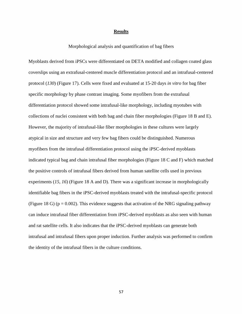
57
Results
Morphological analysis and quantification of bag fibers
Myoblasts derived from iPSCs were differentiated on DETA modified and collagen coated glass
coverslips using an extrafusal-centered muscle differentiation protocol and an intrafusal-centered
protocol (130) (Figure 17). Cells were fixed and evaluated at 15-20 days in vitro for bag fiber
specific morphology by phase contrast imaging. Some myofibers from the extrafusal
differentiation protocol showed some intrafusal-like morphology, including myotubes with
collections of nuclei consistent with both bag and chain fiber morphologies (Figure 18 B and E).
However, the majority of intrafusal-like fiber morphologies in these cultures were largely
atypical in size and structure and very few bag fibers could be distinguished. Numerous
myofibers from the intrafusal differentiation protocol using the iPSC-derived myoblasts
indicated typical bag and chain intrafusal fiber morphologies (Figure 18 C and F) which matched
the positive controls of intrafusal fibers derived from human satellite cells used in previous
experiments (15, 16) (Figure 18 A and D). There was a significant increase in morphologically
identifiable bag fibers in the iPSC-derived myoblasts treated with the intrafusal-specific protocol
(Figure 18 G) (p = 0.002). This evidence suggests that activation of the NRG signaling pathway
can induce intrafusal fiber differentiation from iPSC-derived myoblasts as also seen with human
and rat satellite cells. It also indicates that the iPSC-derived myoblasts can generate both
intrafusal and intrafusal fibers upon proper induction. Further analysis was performed to confirm
the identity of the intrafusal fibers in the culture conditions.

58
Table 4: Medium formulations used to differentiate intrafusal fibers from induced pluripotent
stem cells
Reagent Final Concentration/Amount Source Catalog Number
Differentiation Medium 1
DMEM
High
Glucose,
Pyruvate
N/A Invitrogen 11995-065
EGF
recombinant
human
protein
(hEGF)
10 ng/ml Invitrogen PHG0314
Insulin 10 µg/ml Sigma Aldrich I9278
Bovine
Serum
Albumin
(BSA)
500 µg/ml Sigma Aldrich A9418
Neuregulin 100 µg/ml R&D Systems 396-HB-050
Agrin 100 ng/ml R&D Systems 550-AG
Laminin 1 µg/ml ThermoFisher 23017015
Differentiation Medium 2
Neurobasal N/A ThermoFisher 21103-049
B27 1X ThermoFisher 17504001
Glutamax 1X ThermoFisher 35050-061
NaCl (4M) ~31.2 mM for 270-280 mOsm Sigma Aldrich S3014-1KG
GDNF 10 ng/ml Cell Sciences CRG400E
BDNF 20 ng/ml Cell Sciences CRB600D
CNTF 5 ng/ml Cell Sciences CRC400B

59
NT3 20 ng/ml Cell Sciences CRN500D
Reagent Final Concentration/Amount Source Catalog Number
NT4 20 ng/ml Cell Sciences CRN501C
Vitronectin 100 ng/ml Sigma Aldrich V8379-50ug
IGF-1 10 ng/ml Sigma Aldrich I3769-100ug
cAMP 1µM Sigma Aldrich A9501-1G
Shh 50 ng/ml R&D Systems 1845-SH-100
RA 0.1 µM Sigma Aldrich R2625
Agrin 100 ng/ml R&D Systems 550-AG
Laminin 1 µg/ml ThermoFisher 23017015
NGF 100 ng/ml R&D Systems 256-GF-100
NRG 100 ng/ml R&D Systems 396-HB-050

60
Table 5: Medium formulations used to differentiate extrafusal fibers from induced pluripotent
stem cell derived myoblasts
Reagent Final Concentration/Amount Source Catalog
Number
DK-HI Medium
DMEM/F12 (1:1) N/A Gibco 21041-025
Knockout Serum
Replacement (KSR)
150ul/ml ThermoFisher 10828028
HGF 10 ng/ml PeproTech 315-23-
20UG
IGF 2 ng/ml Sigma
Aldrich
I1271-MG
β-mercaptoethanol 99mM Gibco 21985-023
MEM Non-essential
Amino Acids
1X Gibco 11140-050
N2 Medium
DMEM/F12 (1:1) N/A Gibco 21041-025
Insulin-Transferrin-
Fetuin
1X Gibco 41400-45
L-Glutamine 1X Gibco 25030-081
N2 supplement 1X Gibco 17502-048

61
Figure 17: Cell culture protocol. Cells were differentiated into myoblasts from a previous
protocol and frozen into stocks. Cells were then expanded and treated with either intrafusal fiber
differentiation or extrafusal fiber differentiation.

62
Figure 18: Phase Contrast Analysis. Morphological comparison between Intrafusal fibers
derived from satellite cells and iPSC myoblasts treated with different differentiation protocols.
A) and D) Cultures of satellite cells treated with an intrafusal induction protocol. B) and E)
Cultures of iPSC myoblasts treated with an extrafusal-centered differentiation protocol C) and F)
Cultures of iPSC myoblasts treated with an intrafusal induction protocol. White arrowheads
show cells with intrafusal morphology. G) Quantification of morphologically assessed bag
fibers. (Error bars=SEM, p=0.002). All scale bars are 100 microns.

63
Immunocytochemical analysis
To investigate the role of NRG and other differentiation factors on the induction of intrafusal
fibers in iPSCs, we evaluated the phosphorylation of ErbB2 in cultures treated with and without
an intrafusal differentiation specific protocol. Immunocytochemistry was performed to evaluate
the level of phosphorylation of ErbB2 receptors to determine the phenotype of the muscle fibers.
IPSC-derived myoblasts were differentiated on DETA modified and collagen coated glass
coverslips using an extrafusal-centered muscle differentiation protocol and an intrafusal-centered
protocol. Cells were fixed at 15-20 days in vitro and evaluated for pErbB2 staining. Myofibers
found in both satellite-derived cultures and iPSC-derived cultures treated with the intrafusal
differentiation protocol exhibited typical pErbB2 staining. Numerous clusters of receptors
throughout the membrane were observed (Figure 19 A-E and 19 K-O). However, the iPSC-
derived myoblasts treated with extrafusal differentiation had significantly fewer clusters of
pErbB2 (Figure 19 F-J, P) (p=0.00054). This contrast indicates specific enhancement of the NRG
signaling pathway in these myofibers under intrafusal induction conditions. To further confirm
the increased induction of intrafusal fibers through NRG activation, cells were evaluated by S46
staining. S46 is a slow myosin heavy chain specific for intrafusal fibers (131). IPSC-derived
myoblast cultures treated with the extrafusal-specific protocol had significantly fewer s46
positive cells than the iPSC-derived myoblast cultures treated with the intrafusal differentiation
protocol (Figure 20 K) (p=0.03). These data indicate a lack of natural development of intrafusal
fibers using an extrafusal differentiation protocol and an increase in the amount of intrafusal
fibers using the intrafusal protocol due to an increased activation of the ErbB2 receptor mediated
pathway in response to NRG and other intrafusal differentiation factors.

64
Figure 19: Immunocytochemical Analysis of the myotubes induced under different conditions.
Molecular comparison between ErbB2 phosphorylation in Intrafusal fibers derived from satellite
cells and iPS cells treated with different differentiation protocols. A-E) Cultures of satellite cells
treated with an intrafusal induction protocol. F-J) Cultures of iPS cells treated with an extrafusal-
centered differentiation protocol K-O) Cultures of iPS cells treated with an intrafusal induction
protocol. P) Quantification of pERBb2 clustering within iPSC cultures. (Error bars are SEM,
***p=0.00054). All scale bars are 100 microns.

65
Figure 20: Immunocytochemical Analysis of the myotubes induced under different conditions.
Molecular comparison between s46 expression in Intrafusal fibers derived from iPS cells treated
with different differentiation protocols. (Error bars are SEM, *p=0.03) All scale bars are 100
microns.

66
Discussion
This work demonstrates the induction of intrafusal fibers from iPSCs that now enables a range of
investigations into various disease models of the spinal stretch reflex arc. Previous work has
demonstrated the differentiation of human muscle from iPSCs, however, this is the first study
targeting the differentiation of intrafusal fibers from iPSCs demonstrating successful
differentiation supported by morphological and immunocytochemical evidence. These cells can
now be integrated into existing “body-on-a-chip” testing platforms for a more thorough
evaluation of the neuromuscular reflex arc, neuromuscular diseases, and for predicting drug
efficacy and safety in clinical trials. For example, numerous compounds are known to mediate
specific effects on the muscle spindle, including its activity (132) development, (133) and the
ability to propagate pain to the central nervous system (134). Having a platform for functional
evaluation of these activities in an in vitro setting will allow for more through preclinical
evaluations in an attempt to reduce clinical trial failures.
Recent evaluations of the neuromuscular reflex arc have used “body-on-a-chip” testing platforms
to elucidate functionally relevant data for predictions of human physiology and drug efficacy,
safety, and toxicity. These systems have investigated the efferent neuromuscular processes of
exercise (135) and contraction (13) as well as afferent innervation of intrafusal fibers by sensory
neurons (14, 15). To enable these in vitro investigations to be more clinically relevant, iPSCs
show great promise due to their ability to model specific patient physiology, especially for
disease phenotypes. As shown in previous publications, iPSCs can be differentiated into
extrafusal fibers, (130) motoneurons, (128) and sensory neurons (129) but the differentiation of
intrafusal fibers from human iPSCs has not been reported. This work demonstrates the
differentiation of iPSC-derived intrafusal fibers, further advancing “body-on-a-chip” technology

67
to deliver disease-specific, or even patient-specific, results without sacrificing inappropriate
animal models.
In previous work, NRG has been used to induce the differentiation of intrafusal fibers from both
rat (34) and human cell lines (15). While these cells can be used to determine certain basic
aspects of physiology, the use of disease specific iPSCs is becoming more experimentally
significant for the progression of drug and disease studies into clinical trials. This work outlines
the induction of intrafusal fibers from iPSCs using the administration of NRG and other
intrafusal fiber inducing factors. Upon treatment with an intrafusal-specific protocol the average
quantity of iPSC-derived bag fibers per field of view increased from 0.76 ± 0.17 to 1.8 ± 0.27
(mean ± SEM) (Figure 18). Additionally, the morphologies present in the iPSC-derived
intrafusal fiber cultures more closely resembled those in previous publications (14-16, 34, 44)
(Figure 18). This data suggests NRG-mediated differentiation has occurred in the iPSC-derived
myoblast cultures. Immunocytochemistry for pERbB2 and S46 was used to further confirm the
activation of the NRG signaling pathway and the increased induction of intrafusal fibers. Each
of these markers have been previously used as an indicator for intrafusal fiber identity (16, 131).
An increase in the average level of phosphorylation of the ErbB2 receptors of individual
myotubes from 26.2 ± 2.84 to 67.5 ± 7.81 (mean ± SEM) (Figure 19) and an increase in the
number of myotubes positive for S46 staining from 0.73 ± 0.21 to 2.28 ± 0.26 (mean ± SEM)
(Figure 20), confirms increased differentiation of intrafusal fibers in a human iPSC-derived
culture using this protocol. A similar increase in both immunocytochemical markers
demonstrates that the intrafusal-specific differentiation protocol induced increased differentiation
of the iPSC-derived myotubes. A slightly lower increase derived from phase quantification could
be due to the fact that only bag fibers were counted for the intrafusal quantification under phase

68
contrast microscopy. It should be noted that the amount of intrafusal fibers present within muscle
tissue is highly variable and the reasons for which have yet to be fully understood(136-138).
Further evaluation of this differentiation system will drive current understanding toward a more
efficient differentiation process.
This work is the first evidence indicating that NRG can be used to drive human iPSC- derived
myoblasts toward the intrafusal fiber lineage. The incorporation of iPSC-derived intrafusal fibers
into existing neuromuscular testing platforms will allow for a more clinically relevant method for
disease pathology analysis and drug discovery testing. By incorporating iPSCs derived from
genetic mutant variants, it is now possible to identify the specific roles of these mutations on
muscle spindles; a first for neuromuscular diseases such as ALS and SMA where the
involvement of mechanosensation in disease progression is known but not yet fully elucidated
(139, 140).
Materials and Methods
Surface modification
Glass coverslips (18mm round, VWR, 48380-046) were cleaned and functionalized by a Harrick plasma
cleaner (model PDC-32G) by expose to an oxygen plasma from high purity oxygen gas. The coverslips
were submerged in a reaction solution of 1% v/v trimethoxysilyspropyldiethylenetriamine (DETA, United
Chemical Laboratories, Inc., T2910-KG) in dry toluene (VWR, BDH1151-4LG) and heated for 30
minutes to just below the boiling point of toluene (110.6°C). The vessel was then removed from heat and
allowed to cool at room temperature for 30 minutes. Next the coverslips were rinsed in 3 serial toluene
baths and submerged in dry toluene and heated for 30 minutes to just under the boiling point of toluene.
The surfaces were then removed and cured overnight (~15hrs) in an oven at approximately 110°C.
Surfaces were characterized by goniometry and X-ray photoelectron spectroscopy (XPS) to verify the

69
DETA functionalization according to Das et. al. 2006 (7). Goniometry characterization was performed by
contact angle goniometry with a 5 µl droplet of water. XPS characterization was performed with a
Thermo Scientific ESCALAB 220i-XL instrument with aluminum Kα X-rays and a 90° take off
angle.
Satellite cell culture
Satellite cells were cultured according to previously published techniques (68) (Figure 17, Table
4). Briefly, cells were plated at ~200 cells per mm2 and expanded in Lonza human skeletal
growth medium (Lonza, CC3160 or CC-3246 plus 10mM L-glutamine and CC-4139)
supplemented with 15% FBS on 18mm round coverslips that were DETA modified then
Collagen (ThermoFisher, A1048301, 60 ug/ml) coated until the cells reached ~80-90%
confluency. Cells were then given a full medium change into differentiation medium 1 and given
a half change of differentiation medium 1 two days after differentiation was initiated. After four
days of differentiation, cells were given a full medium change of differentiation medium 2 and
then a half change of differentiation medium 2 two days after the culture was switched to
differentiation medium 2. After four days in differentiation medium 2, cells were fed with a half
change of NBActiv4 (Brainbits, NB4-500) medium supplemented with 1X antibiotic/antimycotic
(ThermoFisher, 15240062) every other day until the culture was ready for analysis (10-15 days
after differentiation).
iPSC extrafusal cell culture
IPSCs were obtained from Coriell Institute for Medical Research (Camden, New Jersey). Cells
were cultured according to previously published techniques (130) until the myoblast stage and
then frozen as stocks in liquid nitrogen with 10% DMSO (Figure 17). Cells were then expanded

70
and differentiated as previously published.(130) Briefly, cells were plated at ~200 cells per mm2
and expanded in Myocult medium (Stemcell Technologies, 36253 and 5982) on 18mm
coverslips that were DETA modified then Collagen (ThermoFisher, A1048301, 60 ug/ml) coated
until cells reached ~80-90% confluency. Cells were then given a half medium change with DK-
HI supplemented with dexamethasone. Two days after feeding with DK-HI, cultures were given
a half medium change of N2 medium (Table 5). Cultures were fed every other day until the
culture was ready for analysis (10-15 days after differentiation).
iPSC intrafusal cell culture
IPSCs were obtained from Coriell Institute for Medical Research (Camden, New Jersey). Cells
were cultured according to previously published techniques (130) until the myoblast stage and
then frozen as stocks in liquid nitrogen with 10% DMSO. Myoblasts were then expanded as
previously published (130) and differentiated using a modified protocol. Myoblasts were
cultured according to previously published techniques (15). Briefly, myoblasts were plated at
~200 cells per mm2 and expanded in Myocult medium (Stemcell Technologies, 36253 and 5982)
on 18mm round coverslips that were DETA modified then Collagen (ThermoFisher, A1048301,
60 ug/ml) coated until cells reached ~80-90% confluency. Cells were then given a full medium
change into differentiation medium 1 and then a half change of differentiation medium 1 two
days after differentiation was initiated. After four days of differentiation, cells were given a full
medium change of differentiation medium 2 and then a half change of differentiation medium 2
two days after the culture was switched to differentiation medium 2. After four days in
differentiation medium 2, cells were fed with a half change of NBActiv4 (Brainbits, NB4-500)

71
medium supplemented with 1X antibiotic/antimycotic (ThermoFisher, 15240062) every other
day until the cultures were ready for analysis (10-15 days after differentiation).
Immunocytochemistry-pErbB2 and phalloidin
Cells were fixed with 4% paraformaldehyde for 15 minutes and then rinsed 2 times with
phosphate buffered saline (PBS) and stored in a third wash of PBS until immunocytochemical
treatment (~0-14 days). Before immunocytochemistry, cells were treated with a blocking
solution (donkey serum and 10% BSA in PBS) for 2 hours then treated with rabbit-anti-pErbB2
488 antibody (EMD Millipore, 16-235, 1:250 dilution) and Phalloidin 568 (Fisher Scientific,
A12380, 1:40 dilution) for 2 hours. Cells were rinsed with PBS for 5 minutes, then rinsed with
PBS and DAPI for 10 minutes and then rinsed with PBS for 15 minutes. Glass coverslips were
mounted onto glass slides using ProLongTM gold antifade mounting medium (ThermoFisher,
P36931). Slides were imagined using a Zeiss Axioskop 2 Mot plus spinning disk confocal
microscope.
Immunocytochemistry-S46 and phalloidin
Cells were fixed with 4% paraformaldehyde for 15 minutes and then rinsed 2 times with
phosphate buffered saline (PBS) and stored in a third wash of PBS until immunocytochemical
treatment (~0-14 days). Before immunocytochemistry, cells were permeabilized with 0.1%
Triton X-100 for 15 minutes and treated with a blocking solution (donkey serum and 10% BSA
in PBS) for 2 hours. Cells were then treated with Mouse-anti-S46 antibody (Developmental
Studies Hybridoma Bank, S46-s) overnight. The next day, cells were rinsed for 5, 10, and 15

72
minutes with PBS and treated with donkey-anti-mouse 488 Alexa Fluor® antibody
(ThermoFisher, A21202, 1:250 dilution) and Phalloidin 568 (1:40 dilution) for 2 hours. Cells
were then rinsed with PBS for 5 minutes, rinsed with PBS and DAPI for 10 minutes and rinsed
with PBS for 15 minutes. Glass coverslips were then mounted onto glass slides using
ProLongTM gold antifade mounting medium (ThermoFisher, P36931). Slides were imagined
using a Zeiss Axioskop 2 Mot plus spinning disk confocal microscope.
Image analysis
Quantification of intrafusal morphology was performed across three separate cultures for the two
iPSC-derived myoblast differentiation protocols. Within each culture, five random phase contrast
images were taken from each of the two replicate coverslips for each protocol, for a total of 30
images per condition. A nested statistical design was employed, in which the 5 replicate images
in each coverslip were nested in the independent coverslip samples (n = 6 coverslips). Images for
pErbB2 and S46 quantification were obtained similarly to those for the intrafusal morphology,
with n = 6 coverslips across 3 cultures and n = 8 coverslips across 4 cultures, respectively, each
with 5 replicate images. For each quantification, two tailed t-tests (α = 0.05, n = 6 or 8
coverslips) were performed to compare the numbers of intrafusal fibers per field of view,
numbers of pErbB2 clusters per myotube, and numbers of S46 positive myotubes per field of
view between the two differentiation protocols. Error bars in each figure are presented as
standard error of the mean (SEM).

73
CHAPTER FIVE: INVESTIGATION OF NOCICIEPTIVE MUSCLE
SPINDLE ACTIVITY
Introduction
The muscle spindle has largely been studied for its involvement in neuromuscular
communication efficiency and proprioception. However, increasing evidence suggests its
involvement in neuropathic pain, a term used to describe nerve damage resulting in the
perception of pain (141). This damage can result in sensitization of the nerve endings and
allodynia, or a painful response to a typically non painful stimulus (141). Many links exist
between neuropathic pain and afferent skeletal muscle activity (96, 142-147), some suggesting
nociceptor-mediated pain originating in the muscle spindle (17). The exact mechanisms involved
are of great research interest for as their investigation could lead to therapeutic innovations for
pain. Thus far, multiple afferent cells, including, intrafusal fibers (96, 143), nociceptors (144),
and other sensory neurons (143) have been linked to the propagation of pain signals from the
muscle tissue. To have a complete in vitro model of the human neuromuscular reflex arc, it is
important to have these functionalities represented in an in vitro platform.
Nociceptors are sensory neurons specialized for the perception of noxious stimuli (141). They
are present in a wide variety of tissues including skin, muscles, joints and fascia (148). Recent
research has allowed for the characterization of many nociceptive agonists including heat,
changes in pH, and specific molecules. Of particular interest to this work, capsaicin, a well
characterized nociceptive agonist is the molecule found in spicy peppers that gives them their
“hot” flavor (149). This molecule binds to transient potential receptor vanilloid-1 (TRPV1)
found on nociceptors(150). This binding opens the nonspecific TRPV1 receptor which allows for
a dramatic influx of calcium and sodium ions, evoking the perception of pain. However, if the

74
TRPV1 ion channel is exposed to capsaicin for a long enough time period, the associated neuron
gets flooded with calcium and can no send action potentials, leading to a sensation of numbness
(149). This mechanism is widely utilized in clinical settings as a method of delivering pain relief
with the topical application of capsaicin (151). In an in vitro setting, this mechanism can be used
to confirm likely nociceptive capabilities of the cell type in question. This work demonstrates the
use of capsaicin on human neural progenitor-derived sensory neurons to evaluate nociceptive
capability. Integration of these nociceptors into the existing platform for neuromuscular reflex
arc will incorporate a perspective on pain when evaluating neuromuscular activity in regard to
normal, diseased, and drug treatment conditions.
As described in Chapter 1, intrafusal fibers are the sensory component of skeletal muscle tissue.
These fibers form a collection called a muscle spindle which is innervated by both afferent and
efferent neurons. This spindle is embedded within skeletal muscle tissue and mediates afferent
muscular perception (44). However, recent research suggests its potential role in the propagation
of pain perception, including injury and inflammation, particularly in regard to the presence of
the TRPV1 ion channel (142, 145, 147, 152-155). This work investigates the potential for
intrafusal fibers of initiating the propagation of noxious stimuli to the CNS.
The confirmation of nociceptive activity in vitro is difficult to define. There is no method of
interrogating neurons for “pain” or how much of the sensation would be perceived by the CNS.
Therefore, it is important to carefully investigate and utilize a functional analog to demonstrate
the involvement of pain within an in vitro testing platform for the neuromuscular reflex arc.
First, nociceptive capability must be demonstrated in the cells themselves. Later, they can be
fully integrated into the existing neuromuscular testing platform. Here we demonstrate
nociceptive activity in neurons derived from commercially available human neural progenitor

75
(hnp1) cells and the possible nociceptive involvement of human intrafusal fibers. This is a first
step toward integrating more complex pain-linked disease states to the neuromuscular reflex arc
testing platform.
Results
Immunocytochemical analysis of hNP1-derived sensory neurons
To confirm the presence of nociceptive cells in hNP1-derived sensory neurons, we evaluated the
presence of nociceptive membrane channels on the membranes of the mature cells with
immunocytochemistry. HNP1 cells were expanded and differentiated using an adapted protocol
from previous methods (15). The differentiation protocol yields a mixed population of sensory
neurons, therefore further analysis was required to confirm nociceptive identity. Once the cells
reached maturity, they were fixed and evaluated by immunocytochemistry for the presence of
TRPV1, the receptor for capsaicin, and the high-affinity receptor tyrosine kinase A (TRKA) both
of which are found selectively on nociceptors(156). A correlation between TRPV1 and TRKA
expression was observed (Figure 21), suggesting that the cells positive for both markers are
indeed nociceptors. Functional analysis of the TRPV1 channel will further confirm their
nociceptive identity.

76
Figure 21: Immunocytochemical analysis of hNP1-derived neurons. Evaluation of TRPV1 and
TRKA expression in hNP1-derived neurons. All scale bars are 100 microns.
Functional evaluation of hNP1-derived neurons
To determine the nociceptive functionality of these cells, they were treated with capsaicin, a
known nociceptive agonist for the TRPV1 channel. HNP1 cells were expanded and differentiated
as with the immunocytochemical evaluation. Upon maturity, cells were replated onto custom
microelectrode arrays (cMEAs), allowed to recover for 3-5 days and evaluated using a
customized amplifier unit along with customized detection software. Upon application of 2 µM
capsaicin, a likely response was observed (Figure 22). This demonstrates a coordination with
neuronal firing and the administration of this nociceptive agonist, suggesting in vitro nociceptive
functional activity and further confirming the nociceptive population within the hNP1-derived
sensory neurons.

77
Figure 22: Raster plot demonstrating the electrophysiological response of hNP1-derived neurons
to capsaicin
Immunocytochemical evaluation of intrafusal fibers
To determine the potential involvement of intrafusal fibers in pain, these cells must be evaluated
for known mediators of nociception. Human skeletal muscle myoblasts were expanded and
differentiated according to previous methods. Upon maturation, cells were fixed and evaluated
by immunocytochemistry for the presence of TRPV1. The intrafusal cultures demonstrated a co-
staining of TRPV1 and S46, a known indicator of intrafusal fiber identity (131) (Figure 23). The
presence of a well-known nociceptive mediator both in the nuclei and on the cellular membrane
suggests a possible role of human muscle spindles in nociception.

78
Figure 23: Immunocytochemical analysis of human skeletal muscle. Cultures were evaluated for
TRPV1 and S46. All scale bars are 100 microns.
Discussion
This work demonstrates the potential for interrogation of pain on an in vitro neuromuscular
reflex arc testing platform. Evidence of immunocytochemical markers and the functional
evaluation of nociceptive activity within hNP1-derived sensory neurons suggests the
incorporation of these cells into an existing neuromuscular evaluation platform will allow for an
opportunity to evaluate pain in vitro. Additionally, the presence of TRPV1 on in multiple locales
(membrane and nuclei) of intrafusal fibers suggests a role of this cell type in nociception. Both

79
cell types can easily be integrated into existing neuromuscular testing platforms for a potential
new way to evaluate pain in vitro.
Materials and Methods
Surface preparation
Glass coverslips (18mm round, VWR, 48380-046) were cleaned and functionalized by a Harrick
plasma cleaner (model PDC-32G) by exposure to an oxygen plasma from high purity oxygen
gas. Coverslips and cMEAs were submerged in a reaction solution of 1% v/v
trimethoxysilyspropyldiethylenetriamine (DETA, United Chemical Laboratories, Inc., T2910-
KG) in dry toluene (VWR, BDH1151-4LG) and heated for 30 minutes on a hot plate to just
below the boiling point of toluene (110.6°C). The vessel was then removed from heat and
allowed to cool at room temperature for 30 minutes. Next the surfaces were rinsed in 3 serial
toluene baths and submerged in dry toluene and heated for 30 minutes on a hot plate to just under
the boiling point of toluene. The surfaces were then removed and cured overnight (~15hrs) in an
oven at approximately 110°C. Surfaces were characterized by goniometry and X-ray
photoelectron spectroscopy (XPS) to verify the DETA functionalization according to Das et. al.
2006 (7). Goniometry characterization was performed by contact angle goniometry with a 5 µl
droplet of water. XPS characterization was performed with a Thermo Scientific ESCALAB 220i-
XL instrument with aluminum Kα X-rays and a 90° take off angle. Surfaces were then sterilized
with 70% Ethanol and allowed to dry under sterile laminar flow. The cMEAs were then
assembled into custom polymethylmethacrylate and polydimethylsiloxane housing systems. The
surfaces were next coated with poly-ornathine (15 µg/ml, Sigma, P3655) overnight. The next
day, the surfaces were rinsed twice with PBS and coated with laminin (1 µg/ml Invitrogen

80
23017-015) and fibronectin (10 µg/ml EMD Millipore FC010) overnight. The next morning the
surfaces were rinsed twice with PBS and were ready for cell seeding.
Human neural progenitor cell expansion and differentiation
Human neural progenitor (STEMEZ TM hNP1, ) were obtained from Neuromics (Edina,
Minnesota) and expanded in RenCell medium (EMD Millipore, SCM005) supplemented with
basic fibroblast growth factor (20ng/ml, R&D, 233-FB) and epidermal growth factor (20ng/ml,
R&D, 236-EG) on Matrigel coated polystyrene (Corning Life Sciences, 354230). Matrigel was
incubated on polystyrene at a 1:100 dilution in DMEM : F12 (Gibco, 21041-025) for one hour at
room temperature. Cells were given a fill medium change every other day until ~90% confluence
was reached. Cells were then replated onto poly-ornathine/ fibronectin/ laminin, coated
coverslips with 0.05% trypsin and 0.2% trypsin inhibitor. Cells were fed every other day with
RenCell medium until ~90% confluency was reached. Differentiation was then initiated with a
full change of 100% KSR medium (knockout DMEM, ThermoFisher, 10829018; knockout
serum replacement, 150 µl/ml, ThermoFisher, 10828028; 2 mM L-glutamine, Gibco, 25030-081;
antibiotic/antimycotic, 1X, ThermoFisher, 15240-062; MEM Non-essential amino acids, 1X,
Gibco 11140-050; 55 µM β-mercaptoethanol, Gibco, 21985-023) supplemented with 100 nM
LDN (Tocris, 6053/10) and 10 µM SB431542 (Tocris, 1614/1) at day 1 of differentiation. On
day 3, cells were given a full medium change of 100% KSR medium supplemented with 100 nM
LDN, 10 µM SB431542, 3 µM CHIR99021 (Tocris, 4423/10), 10 µM SU5402 (Tocris, 3300),
and 10 µM DAPT (2643/10). On day 5, cells were given a full change of 75% KSR medium and
25% N2B Medium (DMEM/F-12, phenol red free, ThermoFisher, 21041-025; 200 µg/ml Human
Apo-transferrin, R&D, 3188-AT; 25 µg/ml Insulin, Millipore, 407709; 1.56 mg/ml D-(+)-
glucose, anhydrous, Sigma, G-7012) supplemented with 100 nM LDN, 10 µM SB431542, 3 µM

81
CHIR99021 , 10 µM SU5402 , and 10 µM DAPT. On day 7, cells were given a full change of
50% KSR medium and 50% N2B Medium supplemented with 100 nM LDN, 10 µM SB431542,
3 µM CHIR99021 , 10 µM SU5402 , and 10 µM DAPT. On day 9, cells were given a full change
of 25% KSR medium and 75% N2B Medium Supplemented with 100 nM LDN, 10 µM
SB431542, 3 µM CHIR99021 , 10 µM SU5402 , and 10 µM DAPT. On day 1, cells were given a
full change of 100% N2B Medium Supplemented with 100 nM LDN, 10 µM SB431542, 3 µM
CHIR99021 , 10 µM SU5402 , and 10 µM DAPT. .On day 13, cells were given a full medium
change with N2B medium supplemented with 25 ng/ml human-b-NGF (R&D, 256-GF), 25
ng/ml BDNF (Cell Sciences, CRB600B), and 25 ng/ml GDNF(CRG400B). On day 15, cells
undergoing electrophysiological analysis were replated onto Poly-ornathine/laminin/fibronectin
coated cMEAsCells were fed every 3-4 days in this final medium until cells were ready for
analysis ( ~5-15 days).
Electrophysiological Evaluation of hNP1 cells
Once hNP1 cells reached maturity (10-15 days post differentiation) cells were replated onto
custom cMEAs within housings and recovered for 3-5 days. The housings were then attached to
a custom amplifier circuit to observe and record electrical signals detected from the neurons.
First, 10 µl warmed medium was added as a negative control then capsaicin (1-2 µM ,Fisher
Scientific, ICN202757) was added at recorded time points and electrophysiological response was
recorded on a custom software program.

82
Human Intrafusal fiber differentiation
Human skeletal muscle was obtained from Lonza (CC-2580) and expanded in Lonza human
skeletal growth medium (CC-3160) supplemented with 15% FBS until ~70-80% confluence was
reached. Then the medium was fully switched to differentiation 1 medium (DMEM
(Invitrogen 11775-040), Insulin (Sigma I9278) at 10 µg/ml, BSA (Sigma A9418) at
500 µg/ml, and EGF (Invitrogen 13247-051) at 10 ng/ml)(36) and fed every 2 days for 4 days.
At this point, the medium was switched to differentiation 2 medium (described in detail in
Guo et al.(15)) and MNs were added to co-cultures at 50 cells per mm2. The cells were fed
every two days with differentiation 2 medium and maintained for four days. After this point,
the medium was given a half change with NBActive4 differentiation medium. The cells were
maintained in this medium for the remainder of the culture and fed every two days until 15–30
DIV total when they were analyzed via immunocytochemical analysis.
Immunocytochemical analysis
Upon maturation, cells were fixed with 4% paraformaldehyde for 15 minutes then rinsed three
times with PBS. At this point, cells were either stored in PBS for 0-14 days or incubated with
permeabilization solution for 15 minutes (Triton X-100) and then blocking solution (5% Donkey
serum +0.5% BSA in PBS) for 2 hours to prevent nonspecific binding. Cells were then
incubated with primary antibodies overnight (Rabbit anti TRKA,1:100 Abcam, ab8871 or
Guinea Pig-anti-TRPV1, 1:100, Invitrogen PA1-29770). The next day cells were rinsed three
times with PBS and incubated with secondary antibodies (Goat-anti-Guinea Pig-488 Alexa
Fluor, ThermoFisher A-11073 or Donkey-anti-Rabbi 568 AlexaFluor, ThermoFisher, A10042) in
a 1:250 dilution for 1-2 hours. Cells were then rinsed for 5minutes in PBS, 10 minutes in PBS

83
with DAPI (1:1000, ThermoFisher D1306), and 15 minutes with PBS then mounted onto glass
coverslips using ProLongTM gold antifade mounting medium. Slides were imagined using a Zeiss
Axioskop 2 Mot plus spinning disk confocal microscope.

84
CHAPTER SIX: PIEZOELECTRIC BIOMEMS CANTILEVER FOR
MEASUREMENT OF MUSCLE CONTRACTION AND FOR ACTUATION
OF MECHANOSENSITIVE CELLS
The work in this chapter was submitted for publication as follows. This work will also be
included in the doctoral dissertation of Elizabeth Coln.
Coln, E., Colón, A., Long, C., Narasimhan Sriram, N., Esch, M., Prot, J.M., Elbrecht, D., Wang,
Y., Jackson, M., Shuler, M., Hickman, J.J. Piezoelectric BioMEMS cantilever for measurement
of muscle contraction and for actuation of mechanosensitive cells. MRS Communications. June
11, 2019.
JJH, MLS, CJL conceived and designed the experiments; CJL, ME, JMP, and YW fabricated the
device; AC and DHE performed the experiments; EAC, AC, CJL, NNS, MJ analyzed the data;
EAC, AC, DHE, CJL, and JJH prepared the manuscript.
Introduction
Microcantilever sensors have been increasingly used in biomedical microelectromechanical
systems (bioMEMS) devices to measure small scale mechanical movement and are typically
silicon-based cantilevers due to availability and easy integration with silicon-based
technology.(157) One application of these microcantilever sensors is in the determination of
contractile force of cardiac and skeletal muscle tissues using in vitro “body-on-a-chip
systems”.(10, 158-162) These “body-on-a-chip” systems integrate biology and engineering to
measure the biochemical, mechanical, and physiological responses of organ analogues to predict
human response to drug and chemical exposure.(158, 159, 163, 164) The existing

85
microcantilever sensors in “body-on-a-chip” systems utilize imaging methods or optical laser
deflection of cantilevers for calculating contractility(157, 159) and rely on imaging methods or
expensive optical detection components.(165, 166) Since piezoelectric materials have high
sensitivity and the ability to convert mechanical energy to an electrical signal, they have been
used in a variety of microscale sensor systems such as pressure sensors and chemical
sensors,(167-169) and as biosensors to detect biological targets, such as microbes, proteins,
viruses, and nucleic acids.(170) Additionally, the ability of piezoelectric materials to convert
electrical signals to mechanical energy has led to their use as microactuators.(171) However,
these biosensor methods are based on shifts in the resonance frequencies to measure changes in
mass on the surface and are not suited for measuring dynamic force changes. Extending the use
of piezoelectric materials to cantilever-based muscle contraction bioMEMS devices would
enable a direct electrical readout of muscle contraction. Moreover, these cantilevers could also
be used in reverse as actuators to impose a stress on tissues attached to the device, specifically
the sensory portion of the reflex arc. The stretch reflex arc refers to how the central nervous
system senses and responds to peripheral stimuli to regulate skeletal movement and is composed
of skeletal muscle, the muscle spindle, motoneurons, and sensory neurons. The sensory portion is
composed of intrafusal fibers embedded in the muscle tissue of the muscle spindle and
proprioceptive sensory neurons.(172-174) Development of an in vitro sensory circuit of the
reflex arc would improve the overall understanding of the mechanosensitive feedback, allowing
opportunities to create more physiologically relevant systems and for investigation of
neuromuscular diseases.
This present study details the design and fabrication of a piezoelectric bioMEMS cantilever
which can act as either a sensing element for muscle contraction or as an actuator to apply

86
mechanical force to cells. This device incorporates a piezoelectric element composed of a thin
film of aluminum nitride (AlN) on top of the cantilever sandwiched between two platinum
electrodes. As a sensor, bending of the piezoelectric cantilever caused by contracting iPSC-
derived cardiac muscle grown on the cantilever surface produced a voltage across the
piezoelectric element, which was measured by an external amplifier. As an actuator, a periodic
voltage was applied to the piezoelectric cantilevers and the resultant bending was measured
using an optical beam displacement method. This bioMEMS system has the advantage of being
able to acquire a direct electrical signal from the cantilever with a simple system setup for high
content systems and may offer the advantage of high-throughput data collection by integration
with current robotic screening systems in drug discovery.
Experimental Details
Design and fabrication of piezoelectric cantilevers
Piezoelectric cantilever devices were fabricated at the Cornell Nanoscale Science & Technology
Facility (Ithaca, NY) using standard microfabrication processes. Each chip included 16
individual microscale cantilevers each 100 µm wide, 750 µm long, and 4 µm thick with a total
chip size of 17 mm x 14 mm. The 5-inch photomasks for each layer to produce the piezoelectric
cantilevers were designed using L-edit layout editor (Tanner). For production of the devices,
silicon oxide and silicon nitride layers were grown via Plasma Enhanced Chemical Vapor
Deposition (PECVD) (GSI PECVD System, Novanta, Inc., Bedford, MA); metal layers (titanium
and platinum) were deposited via electron-beam evaporation (CHA Mark 50 e-Beam Evaporator,
CHA Industries, Fremont, CA); the aluminum nitride (AlN) was sputtered using an aluminum
target in a nitrogen plasma environment (ATC Orion Sputter Deposition Tool, AJA International,

87
Inc., Scituate, MA); oxide and nitride layers were etched via reactive ion etching (RIE) (Oxford
PlasmaLab 80+ RIE System, Oxford Instruments, Concord, MA); the silicon device and handle
layers were etched via deep reactive ion etching (DRIE) (Unaxis 770 Deep Silicon Etcher,
Plasma-Therm, St. Petersburg, FL), and photolithography was performed using a Karl Suss MA6
Mask Aligner (Suss Microtec SE, Garching, Germany). The fabrication process, starting from a
silicon-on-insulator wafer with a 4 µm thick <100> silicon device layer, 1-2 µm buried oxide
layer, and 500 µm handle wafer, is shown in Figure 24, and focuses on the production,
patterning, and electrical isolation of the sandwich structure of AlN between two platinum
electrodes. A 1.5 µm thick silicon dioxide layer and 300 nm silicon nitride layer were deposited
via PECVD on the back and front (device) sides of the wafer, respectively. The piezoelectric
element consisting of an aluminum nitride layer sandwiched between two platinum electrodes
was produced by evaporation and liftoff of the bottom metal layer (10 nm titanium and 50 nm
platinum) (Figure 24A), followed by reactive sputtering of 300 nm of aluminum nitride (Figure
24B) and evaporation of a second electrode identical in composition and thickness to the first.
Both the aluminum nitride and top electrode layers were patterned via a liftoff process (Figure
24C). An insulation layer of silicon oxide and silicon nitride was deposited on top of the device
layer, including the piezoelectric element (Figure 24D). The insulation layer was patterned via
RIE to expose the contact pads and allow etching to define the cantilevers (Figure 24E). DRIE
was then used to define the cantilevers in the device layer silicon. To release the cantilevers and
open a window through the wafer to the cantilevers, the oxide on the backside of the wafer was
etched using RIE to create an etch mask for the DRIE process, which was used to perform a
through etch of the handle wafer to the cantilevers. Buffered hydrofluoric acid (HF) was used to
remove the buried oxide layer to fully release the cantilevers (Figure 24F). Each final cantilever

88
is 100 µm wide and 750 µm long, with a piezoelectric cantilever element 80 µm wide and 730
µm long on the top of the silicon cantilever. In the piezoelectric cantilever chip design (Figure
25A), each cantilever has an independent contact pad for the top and bottom platinum layers, and
these contact pads are connected to an external amplifier. A JEOL 6400 scanning electron
microscope (JOEL USA, Inc., Peabody, MA) was used to measure cantilever dimensions (Figure
25B).
Figure 24: Fabrication of piezoelectric cantilevers. (a) Ti/Pt bottom electrodes and contact pads
were deposited on an SOI wafer with insulation layers on the front and backside and patterned
via liftoff. (b) Piezoelectric AlN was reactively sputtered and patterned via liftoff. (c) Top metal
electrodes and contact pads were deposited and patterned via liftoff. (d) Top insulation layer
(ONO) was deposited via PECVD. (e) The insulation layer was patterned by RIE. (f) The front
and backside were etched via DRIE to define and release the cantilevers, followed by an HF dip
to remove the buried oxide layer.

89
Figure 25: Piezoelectric cantilever chip layout (a) Piezoelectric cantilever chip design showing
location of cantilevers, wires, and contact pads. (b) SEM image of piezoelectric cantilevers
showing top surface of platinum and SiO2 insulation. Because the cantilever chip was not coated
with a separate conductive layer, the piezoelectric element and platinum wires appear darker, as
the adjacent insulation layers charge slightly under the electron beam.
Surface modification of piezoelectric cantilevers
Piezoelectric cantilevers were chemically modified with a diethlyenetriamine (DETA)-
containing silane using previously published methods to enhance cell adhesion and functional
development.(159, 175-177) Following chemical modification, the chips were incubated in
human fibronectin (50 µg/mL) in phosphate buffered saline (PBS) at 37°C for 30 minutes just
prior to cell plating to adsorb the fibronectin to the DETA surface for cell attachment.
Cell culture on the piezoelectric cantilevers and housing system assembly
Commercially available, cryogenically preserved, human induced pluripotent stem cell (iPSC)
derived cardiomyocytes (iCell Cardiomyocytes, #CMC-100-110-005, Cellular Dynamics

90
International, Madison, WI) were plated onto piezoelectric cantilevers (2200 cells/mm2) in a
serum-free medium according to previously published methods that described culturing these
cells on non-piezoelectric bioMEMS cantilevers.(158-160, 176) These cardiomyocytes in our
serum-free culture system express a mature phenotype, have robust electrical and mechanical
function, and show appropriate functional changes in response to drug compounds.(159, 160,
178, 179) Every 2 days, one-half of the medium was replaced with fresh medium. The
piezoelectric chips with cultured cardiomyocytes were maintained for 7 days prior to assembling
into housings. System housings and gaskets were manufactured from 6 mm thick transparent
poly(methylmethacrylate) (PMMA) (McMaster-Carr, Elmhurst. IL) and 0.5 mm thick
poly(dimethylsiloxane) (Grace Bio-Labs, Bend, OR), respectively. Both housings and gaskets
were designed in Eagle (Autodesk) software and laser cut using a Versalaser PLS 75W laser
cutter (Universal Laser Systems, Scottsdale, AZ). The piezoelectric cantilevers were assembled
within the PMMA top and bottom plates, with the gaskets sealing the contact pads from the
cantilever area, which contained 500 µL of serum-free medium.
Contractile force measurement with piezoelectric cantilevers
A microelectrode array (MEA) amplifier (MEA 1040, Multichannel Systems, Reutlingen,
Germany) was interfaced to the piezoelectric cantilever chip in the housing via a custom printed
circuit board and was used to record the electrical output from the piezoelectric cantilevers. For
contractile force experiments, piezoelectric cantilever chips cultured with cardiomyocytes were
assembled into system housings with 500 µL serum-free medium over the piezoelectric
cantilevers. The force of the beating cardiomyocytes bent the cantilevers, producing a
mechanical stress in the piezoelectric aluminum nitride, in turn generating an electric potential

91
difference across the two electrodes (Figure 26C top panel). This voltage was measured by the
amplifier and recorded in real-time using a commercially available data acquisition software
package (MC_Rack, Multichannel Systems, Reutlingen, Germany), and the voltage data was
analyzed with Clampfit (Axon Instruments) software (Figure 26B and C top panel). To
demonstrate the ability of the piezoelectric cantilever sensors to detect changes in cardiomyocyte
function, the cardiomyocytes were dosed with 3 µM epinephrine to increase the beat frequency
during the recording (Figure 26C, bottom panel).
Figure 26: Sensing of muscle contraction using piezoelectric cantilevers. (a) Confocal image of
skeletal muscle on a piezoelectric cantilever, with myosin heavy chain shown in red and DAPI in
blue. (b) Schematic of recording setup for cardiomyocyte force measurements using piezoelectric
cantilevers. (c) Measurement of cardiomyocytes on piezoelectric cantilevers showing control
(top) and after treatment with addition of 3 µm epinephrine (bottom).
Immunocytochemical imaging of cardiomyocytes on piezoelectric cantilevers
Piezoelectric cantilevers were prepared for immunocytochemistry using methods that previously
described staining these cells on non-piezoelectric cantilevers and coverslips.(173, 177) Briefly,
cells on the piezoelectric cantilevers were fixed with a 4% (vol/vol) paraformaldehyde/PBS

92
solution for 15 minutes at room temperature, then rinsed 3X with PBS. Cardiomyocytes were
permeabilized for 15 minutes with 0.1% Triton X-100 and then blocked with 5% donkey serum
+ 0.5% bovine serum albumin in PBS for 1-2 hours at room temperature. Cells were incubated
overnight at 4°C with a mouse anti-myosin heavy chain primary antibody (Developmental
Studies Hybridoma Bank, Iowa City, IA) diluted in blocking buffer. The cells were rinsed with
PBS for 5 minutes, 0.01% Triton X-100 for 10 minutes, and PBS for 15 minutes. The cells were
incubated with Alexa Flour 594 (Invitrogen, Carlsbad, CA) secondary antibodies (1:250 diluted
in blocking buffer) for 1-2 hours at room temperature. The stained samples were then rinsed with
PBS for 5 minutes, 0.01% Triton X-100 and DAPI (Thermo Fisher P36931) for 10 minutes and
PBS for 15 minutes and imaged using a Zeiss LSM 510 confocal microscope (Figure 26 A).
Detection system for actuation of piezoelectric cantilevers
For actuation experiments, acellular piezoelectric cantilever chips were assembled into system
housings with 500 µL of PBS and interfaced through a custom printed circuit board (PCB) to a
Model 2100 Isolated Pulse Stimulator (A-M systems, Sequim, WA) (Figure 27A). A single
cantilever was stimulated at a time by connecting the two electrode pads to the stimulator
through the PCB. Two pulse shapes were applied to the piezoelectric cantilever: a monophasic
square pulse (5V, 150 ms duration, and 0.6 Hz), and a biphasic square pulse (±5V, 300 ms
duration, and 0.6 Hz). A previously described optical detection system (158-160, 177) was used
to measure the deflection of the piezoelectric cantilever. In this system, a 635 nm laser
(CPS635F, Thorlabs, Newton, NJ) and linear effect photodetector (PSD9, Thorlabs, Newton, NJ)
were located under a fixed stage and were aligned such that the laser was focused onto the tip of
the underside of the cantilever (Figure 27A). The laser was subsequently reflected onto the

93
center of the photodetector to measure the deflections of the cantilevers as a change in position
of the reflected laser spot on the photodetector. The deflection at the tip of the cantilever was
then determined using methods described in Pirozzi, et al. for this system.(180)
To understand the cantilever deflection when stimulated electrically as an actuator, a
mathematical relationship between the input pulses and the average deflection signals was
developed (Figure 27B and C). The deflection curves for the monophasic stimuli were averaged,
as were those for the biphasic stimuli. The induced strain in a piezoelectric material is
approximately linearly proportional to the electric field across the piezoelectric element, as
shown in Equation (1), where ε is the strain in the piezoelectric material, d is the piezoelectric
coefficient, and E is the electric field for the inverse effect.
휀 = 𝑑 × 𝐸 (1)
However, the electric field across the element is related not only to the voltage applied to the
electrode pads and geometric configuration, but also the resistive and capacitive elements in the
circuit, including those of the piezoelectric aluminum nitride element. The input voltage is
effectively filtered in a similar way as a low pass RC filter in producing the actuation. As all of
the input voltage changes used in this study were step functions (increasing and decreasing), the
differential equation for the effect of an RC filter has a simple time-domain solution, forming the
basis of Equation (2):
∆𝛿 = 𝐴∆𝑉𝑝𝑢𝑙𝑠𝑒(1 − 𝑒−𝑡 𝜏⁄ ) (2)
where Δδ is the change in position (deflection) of the tip of the cantilever, A is the parameter
relating the voltage across the AlN element to the cantilever deflection and is related to the
piezoelectric coefficient and physical layout of the device, ΔVpulse is the change in voltage of the
input step function, t is the time since the square wave initiation, and τ is the time constant of the

94
circuit. The measured deflection from the optical detection system was fit to Equation (2) using a
least-squares regression method to determine A and τ.
Figure 27: Actuation of piezoelectric cantilevers. (a) Schematic of optical deflection system. (b)
Piezoelectric cantilever actuation under a 5V monophasic 150 ms stimulation pulse. (c)
Piezoelectric cantilever actuation under a ±5V biphasic (total duration 300 ms) stimulation pulse.
Slight offset observed at end of actuation in (a) and (b) due to baseline drift in raw data (Figure
28).
Figure 28: Raw data from actuation of piezoelectric cantilevers. (a) Piezoelectric cantilever
actuation under a 5V monophasic 150 ms stimulation pulse. (b) Piezoelectric cantilever actuation
under a ±5V biphasic (total duration 300 ms) stimulation pulse.

95
Results and Discussion
Piezoelectric sensing for cardiomyocyte force measurements
To demonstrate that the piezoelectric cantilevers could produce electrical signals from
cardiomyocyte contraction, human iPSC-derived cardiac muscle was grown on the piezoelectric
cantilevers in the serum-free medium. Confocal microscopy of the cardiomyocytes on the
piezoelectric cantilevers (Figure 26A) shows the cardiomyocytes integrated on the top of the
cantilever surface, with a muscle-specific structural protein myosin heavy chain (red) and nuclei
(blue) visible. These cardiomyocytes formed a syncytium of contracting cardiomyocytes, and
this syncytium beat spontaneously due to the presence of pacemaker cells in the culture. Using
the direct piezoelectric effect, the piezoelectric cantilevers converted the mechanical strain
caused by the cardiomyocyte contraction into an electrical output. Figure 26B shows a simplified
schematic of the MEA amplifier system setup used for cardiomyocyte force measurements. The
real-time, continuous electrical signals generated by the piezoelectric AlN, in response to the
cardiomyocyte tissue bending of the cantilever, were recorded when the PCB was connected to
an external amplifier. To demonstrate that the system was capable of measuring changes in
cardiomyocyte function, 3 µM epinephrine was added to the medium to increase beat frequency
of the cardiomyocytes.(181) Upon addition of epinephrine, the change in cardiomyocyte beat
frequency was clearly observed, with an increase in frequency by a factor of more than two.
Figure 26C indicates clear peak shapes with measurable amplitude and frequency for both the
spontaneous cardiomyocyte contractions (top) and for the cardiomyocyte contractions induced
by the addition of epinephrine (bottom).

96
Actuation of piezoelectric cantilevers
The ability of the piezoelectric cantilevers to serve as actuators for applying mechanical stress to
tissue constructs was evaluated using an optical beam displacement method (Figure 27A) to
measure the deflection of the piezoelectric cantilever under a periodic waveform of applied
voltage. Using the inverse piezoelectric effect, when the electric signal was applied to the
piezoelectric aluminum nitride element, the aluminum nitride converted the electrical pulse into
a mechanical deformation, causing the cantilever to bend. The actuation of the piezoelectric
cantilevers was modeled electrically following a simple RC circuit,(182) and captures the
capacitance of the platinum/aluminum nitride/platinum stack as well as the bulk resistance of the
aluminum nitride and the resistance of the wires and other series elements in the circuit. Because
of the effective resistance and capacitance of the circuit, the shape of the input actuation signal
was not exactly reflected in the actuated cantilever beam. A transfer function model relating the
output deflection to the input voltage signal was determined to better understand the behavior of
the piezoelectric cantilevers as actuating elements and to help determine input functions to
produce desired deflection waveforms. Figures 27A and 27C show the recorded cantilever
deflection (black) of the 5V monophasic square wave input and 5V biphasic square wave,
respectively, and the modeled transfer function (red) for each stimulation input. In both the
monophasic and biphasic stimulations, the modeled transfer function very closely resembles the
stimulated actuation signal. There is a slight offset in the actuation signals for both the
monophasic and biphasic stimulations due the baseline drift in the raw data as shown in Figures
28A and 28B, respectively. Fitting these two models separately, the monophasic and biphasic
models had an average τ of 0.014s, and average A of 0.31 µm/V.

97
For use with mechanosensitive cells, such as intrafusal skeletal muscle fibers innervated by
sensory neurons as part of the stretch reflex arc, simple functions, such as square waves, are not
sufficient to mimic physiological strain profiles needed to trigger sensory neuron action
potentials. Instead, an isometric contraction profile would be desired to stretch the intrafusal
fibers to activate the mechanoreceptors in the sensory neuron’s axons to initiate an action
potential. The transfer function between time-dependent input voltage and time-dependent
deflection, developed here, could be used to generate the required input waveform to produce the
desired output deflection. Equation (3) relates the required applied voltage waveform to the
desired voltage across the piezoelectric element for an arbitrary waveform:
𝑉𝑎𝑝𝑝𝑙𝑖𝑒𝑑(𝑡) = 𝜏𝑑𝑉𝐴𝑙𝑁(𝑡)
𝑑𝑡+ 𝑉𝐴𝑙𝑁(𝑡) (3)
where Vapplied(t) is the input voltage waveform, τ is the time constant (0.014s), and VAlN(t) is the
voltage across the AlN element. As the deflection of the cantilever tip is related to the voltage
across the AlN element according to the inverse piezoelectric effect (Equation 4):
δ(t) = AVAlN(t), (4)
Equation (3) can be modified to relate the output deflection and voltage applied as time series
(Equation 5):
𝑉𝑎𝑝𝑝𝑙𝑖𝑒𝑑(𝑡) =1
𝐴(𝜏
𝑑𝛿(𝑡)
𝑑𝑡+ 𝛿(𝑡)) (5)
where δ(t) is the desired deflection of the tip of the cantilever, and A and τ are as in Equation (2).
For an arbitrary desired actuation profile (δ(t)) such as to mimic an isometric muscle contraction,
Equation (5) can be used to determine the input voltage waveform needed. This indicates that the
piezoelectric system could be used to stimulate intrafusal fibers to generate action potentials in
sensory neurons.

98
Conclusion
This work demonstrates the development of a new method using piezoelectric cantilevers to
measure contractile muscle force generation in human cells by acquiring a real-time, continuous
and direct electrical signal generated in the piezoelectric AlN, caused by the muscle tissue
bending the cantilever. This sensor was able to detect spontaneous cardiac muscle contraction
with enough sensitivity to observe changes in contraction frequency induced by the addition of
epinephrine. The piezoelectric cantilevers were additionally shown to function as actuators, with
a defined filtering effect of the input signal to cantilever deflection. The actuation experiments
enabled the determination of coefficients relating the input voltage waveform to cantilever
deflection, which can be used to determine the required input voltage waveforms to produce a
desired arbitrary actuation profile. The ability of these piezoelectric cantilevers to actuate opens
the opportunity for use with mechanosensitive cells, such as those in the sensory segment of the
stretch reflex arc to stretch intrafusal fibers. These piezoelectric cantilever devices, which were
designed to be incorporated into “body-on-a-chip” systems, have the potential to enable
functional experiments with mechanosensitive tissue structures such as those in the stretch reflex
arc, to incorporate these tissues into “body-on-a-chip” devices, and to increase the throughput of
measurements of cardiac and skeletal muscle mechanical function in “body-on-a-chip” devices
for basic physiological investigations, pharmaceutical compound development, toxin detection,
neurological and muscular disease modeling for therapeutic development, and predictive
toxicology.(183, 184) Integration of human skeletal muscle fibers on the piezoelectric cantilevers
would enable a more robust method to study the human central nervous system and improve
understanding of spinal cord injuries, diseases such as amyotrophic lateral sclerosis (ALS),

99
CHAPTER SEVEN: GENERAL DISCUSSION
Animal models have proven insufficient for demonstrating human, molecular biology, and
clinical response to drugs (1, 76). Movement toward more human-based preclinical testing
platforms is becoming more essential for accurate predictions of human biology and drug testing.
Also, many governing bodies have chosen to move away from animal models and move closer to
more ethical human-based in vitro testing platforms for drug safety evaluation and product
development. As a solution to these issues, “body-on-a-chip” technology has become
increasingly prevalent in human-based research. This technology integrates MEMS technology
with human-based cell culture to develop platforms for elucidating functional data based on
specific organ systems (77). Development of these systems to incorporate multiple facets of
human health enables the rapid and efficient progression of human disease research and drug
discovery.
Multiple in vitro platforms already exist for various investigative pathways. The neuromuscular
reflex arc, however, is a particularly challenging system to recapitulate in an animal-free
platform. In the in vivo reflex arc, both descending and ascending neuromuscular pathways
interact to form complex interactions with the CNS and the environment. While many of these
intracellular interactions and cell types have been demonstrated in other in vitro platforms, this
work describes the first functional evidence of γ-motoneuron innervation in vitro, the only
demonstration of intrafusal fiber differentiation from the increasingly clinically relevant iPSCs,
and insights as to how to incorporate complex aspects of afferent signaling into “body-on-a-
chip” technology.
The complexity of the neuromuscular reflex arc yields itself to misunderstandings, particularly
regarding cellular communication. Outside of this work, motoneuron innervation of extrafusal

100
fibers (13, 37, 82), sensory neuron innervation of intrafusal fibers (14, 15), and interactions
between motoneurons and sensory neurons (81) has been established. However, γ-motoneuron
innervation of intrafusal fibers had not been demonstrated. This important cellular interaction is
vital for the accurate sensation and proprioception of skeletal muscle (87). To establish a
complete model of the neuromuscular reflex arc, γ- motoneuron contribution must be considered.
This work demonstrates both molecular and functional evidence of human γ-motoneuron
innervation in vitro. Co-culture conditions were established for the survival of both human
motoneurons and intrafusal fibers (Figures 11, 12, and 15). Upon morphological analysis, these
cultures had developed close cell to cell interactions indicative of innervation (Figure 10 G). As a
confirmation of communication ability, the cultures were stained to confirm the identities of the
cell types involved and to establish molecular evidence of innervation (Figures 11 and 13).
Furthermore, functional evidence was established by stimulating the motoneurons to send signals
to the intrafusal fibers which they innervated (Figure 15). Collectively, these data show the in
vitro establishment of γ-motoneuron innervation of intrafusal fibers. This interaction can now
become integrated into current neuromuscular reflex arc testing platforms for a more detailed
perspective on neuromuscular responses to drug treatment, disease states, and development.
Current neuromuscular evaluation platforms can evaluate certain aspects of human biology such
as exercise (135) and therapeutics (124). However, certain neuromuscular diseases, such as ALS
and SMA, have genetic components that are difficult to functionally recapitulate in vitro or even
in animal models. Therefore iPSCs are gaining demand for their use in evaluation of genetic
diseases. In relation to the neuromuscular reflex arc, extrafusal fibers (130), motoneurons (128),
and sensory neurons (129) have been differentiated from iPSCs but this work is the first
demonstration of intrafusal fiber differentiation with morphological (Figure 18) and

101
immunocytochemical (Figures 19 and 20) evidence. Establishment of this cell line permits the
integration of these cells into neuromuscular reflex arc testing platforms for analysis of afferent
related disfunction, development, and drug response.
Several aspects of the afferent spindle signal, including the muscle spindle and nociceptors, can
be linked to the sensation of pain (141, 143, 144, 147, 148, 153). Therefore, it is vital to consider
these aspects when developing a preclinical model to evaluate potentially pain-related disease
states and drug testing. This work evaluates the potential for integrating nociceptors and
intrafusal fibers into a pain-centered neuromuscular reflex arc testing platform. Sensitization of
nociceptor endings within the muscle spindle has been of recent interest (17). Potential
interactions between nociceptors and other cells more frequently found in the muscle spindle
may possibly explain some of the more elusive aspects of neuropathic pain generation.
Establishment of neuropathic pain in vitro is a current challenge for pain research. While it is not
possible to interrogate cells for level of pain perception, neuropathic-related neuronal activity is
possible to demonstrate. This work shows the electrophysiological response of nociceptor marker
positive sensory neurons to a common nociceptive agonist, capsaicin, establishing a potential for
in vitro pain analysis (Figure 22). Additionally, the expression of pain-related receptors on
intrafusal fibers suggests a possible role in noxious afferent signaling (Figure 23).
Afferent signaling interrogation presents as a challenging aspect of neuromuscular reflex arc
research. While sensory neurons can receive and send signaling in a variety of ways (81),
intrafusal fibers must be mechanically stimulated (185). To overcome this challenge, aluminum
nitride, a piezoelectric material, can be integrated into existing cantilever systems for mechanical
stimulation of intrafusal fibers. This work outlines a method for developing the first
mechanically interrogative neuromuscular reflex arc testing platform, allowing for the

102
mechanical stimulation of intrafusal fibers and observation of subsequent signal propagation.
This advancement enhances the investigational capabilities of in vitro functional analysis,
particularly regarding complex neuromuscular interactions.
Collectively, these data demonstrate a significant advancement in the field of afferent
neuromuscular activity research. Integration of γ-motoneurons, iPSC-derived intrafusal fibers,
human-derived nociceptors, and mechanical interrogation into existing in vitro research allows
for substantial advancement in animal-free preclinical analyses.

103
LIST OF REFERENCES
1. C. M. Sakolish, M. B. Esch, J. J. Hickman, M. L. Shuler, G. J. Mahler, Modeling Barrier Tissues In Vitro: Methods, Achievements, and Challenges. EBioMedicine 5, 30-39 (2016).
2. C. Oleaga, C. Bernabini, A. S. Smith, B. Srinivasan, M. Jackson, W. McLamb, V. Platt, R. Bridges, Y. Cai, N. Santhanam, B. Berry, S. Najjar, N. Akanda, X. Guo, C. Martin, G. Ekman, M. B. Esch, J. Langer, G. Ouedraogo, J. Cotovio, L. Breton, M. L. Shuler, J. J. Hickman, Multi-Organ toxicity demonstration in a functional human in vitro system composed of four organs. Sci Rep 6, 20030 (2016).
3. C. Oleaga, A. Lavado, A. Riu, S. Rothemund, C. Carmona-Moran, K. Persaud, A. Yurko, J. Lear, N. Sriram Narasimhan, C. Long, F. Sommerhage, L. Richard Bridges, Y. Cai, C. Martin, M. Schnepper, A. Goswami, R. Note, J. Langer, S. Teissier, J. J. Hickman, Long‐Term Electrical and Mechanical Function Monitoring of a Human‐on‐a‐Chip System. (2018), vol. 29.
4. M. Stancescu, P. Molnar, C. W. McAleer, W. McLamb, C. J. Long, C. Oleaga, J. M. Prot, J. J. Hickman, A phenotypic in vitro model for the main determinants of human whole heart function. Biomaterials 60, 20-30 (2015).
5. Y. I. Wang, C. Oleaga, C. J. Long, M. B. Esch, C. W. McAleer, P. G. Miller, J. J. Hickman, M. L. Shuler, Self-contained, low-cost Body-on-a-Chip systems for drug development. Experimental biology and medicine (Maywood, N.J.) 242, 1701-1713 (2017).
6. D. E. C.J.L. C.W. McAleer, T. Sasserath, L.R. Bridges, J.W. Rumsey, C. Martin, M. Schnepper, Y. Wang, F. Schuler, A.B. Roth, C. Funk, M.L. Shuler, J.J. Hickman, Reconfigurable Multi-Organ System for the Evaluation of Anti-Cancer Therapeutics on Efficacy and Off-Target Toxicity. accepted to Sci. Trans. Med, (2019).
7. M. Das, C. A. Gregory, P. Molnar, L. M. Riedel, K. Wilson, J. J. Hickman, A defined system to allow skeletal muscle differentiation and subsequent integration with silicon microstructures. Biomaterials 27, 4374-4380 (2006).
8. M. B. Esch, A. S. Smith, J. M. Prot, C. Oleaga, J. J. Hickman, M. L. Shuler, How multi-organ microdevices can help foster drug development. Advanced drug delivery reviews 69-70, 158-169 (2014).
9. J. H. Sung, M. B. Esch, J. M. Prot, C. J. Long, A. Smith, J. J. Hickman, M. L. Shuler, Microfabricated mammalian organ systems and their integration into models of whole animals and humans. Lab on a chip 13, 1201-1212 (2013).
10. A. Smith, C. Long, K. Pirozzi, S. Najjar, C. McAleer, H. Vandenburgh, J. Hickman, A multiplexed chip-based assay system for investigating the functional development of human skeletal myotubes in vitro. Journal of Biotechnology 185, 15-18 (2014).
11. M. Arbab, S. Baars, N. Geijsen, Modeling motor neuron disease: the matter of time. Trends in Neurosciences 37, 642-652 (2014).
12. S. Schiaffino, A. C. Rossi, V. Smerdu, L. A. Leinwand, C. Reggiani, Developmental myosins: expression patterns and functional significance. Skeletal muscle 5, 1 (2015).
13. A. S. Smith, C. J. Long, K. Pirozzi, J. J. Hickman, A functional system for high-content screening of neuromuscular junctions in vitro. Technology 1, 37-48 (2013).

104
14. J. W. Rumsey, M. Das, A. Bhalkikar, M. Stancescu, J. J. Hickman, Tissue engineering the mechanosensory circuit of the stretch reflex arc: sensory neuron innervation of intrafusal muscle fibers. Biomaterials 31, 8218-8227 (2010).
15. X. Guo, A. Colon, N. Akanda, S. Spradling, M. Stancescu, C. Martin, J. J. Hickman, Tissue engineering the mechanosensory circuit of the stretch reflex arc with human stem cells: Sensory neuron innervation of intrafusal muscle fibers. Biomaterials 122, 179-187 (2017).
16. A. Colón, X. Guo, N. Akanda, Y. Cai, J. J. Hickman, Functional analysis of human intrafusal fiber innervation by human γ-motoneurons. Sci Rep 7, 17202 (2017).
17. J. V. Partanen, T. A. Ojala, J. P. Arokoski, Myofascial syndrome and pain: A neurophysiological approach. Pathophysiology : the official journal of the International Society for Pathophysiology 17, 19-28 (2010).
18. A. Maier, Development and regeneration of muscle spindles in mammals and birds. The International journal of developmental biology 41, 1-17 (1997).
19. H. H. Chen, S. Hippenmeyer, S. Arber, E. Frank, Development of the monosynaptic stretch reflex circuit. Current opinion in neurobiology 13, 96-102 (2003).
20. J. X. Liu, P. O. Eriksson, L. E. Thornell, F. Pedrosa-Domellof, Myosin heavy chain composition of muscle spindles in human biceps brachii. The journal of histochemistry and cytochemistry : official journal of the Histochemistry Society 50, 171-183 (2002).
21. E. R. Andrechek, W. R. Hardy, A. A. Girgis-Gabardo, R. L. Perry, R. Butler, F. L. Graham, R. C. Kahn, M. A. Rudnicki, W. J. Muller, ErbB2 is required for muscle spindle and myoblast cell survival. Mol Cell Biol 22, 4714-4722 (2002).
22. X. J. Chen, E. N. Levedakou, K. J. Millen, R. L. Wollmann, B. Soliven, B. Popko, Proprioceptive sensory neuropathy in mice with a mutation in the cytoplasmic Dynein heavy chain 1 gene. The Journal of neuroscience : the official journal of the Society for Neuroscience 27, 14515-14524 (2007).
23. P. J. Stapley, L. H. Ting, M. Hulliger, J. M. Macpherson, Automatic postural responses are delayed by pyridoxine-induced somatosensory loss. The Journal of neuroscience : the official journal of the Society for Neuroscience 22, 5803-5807 (2002).
24. K. G. Pearson, J. E. Misiaszek, M. Hulliger, Chemical ablation of sensory afferents in the walking system of the cat abolishes the capacity for functional recovery after peripheral nerve lesions. Experimental brain research 150, 50-60 (2003).
25. A. B. Sterman, H. H. Schaumburg, A. K. Asbury, The acute sensory neuronopathy syndrome: a distinct clinical entity. Annals of neurology 7, 354-358 (1980).
26. R. Forget, Y. Lamarre, Rapid elbow flexion in the absence of proprioceptive and cutaneous feedback. Human neurobiology 6, 27-37 (1987).
27. J. D. Cole, E. M. Sedgwick, The perceptions of force and of movement in a man without large myelinated sensory afferents below the neck. The Journal of Physiology 449, 503-515 (1992).
28. J. Cole, J. Paillard, in The Body and the Self, J. L. Bermudez, A. J. Marcel, N. M. Eilan, Eds. (MIT Press, 1995), pp. 245--266.
29. J. Izawa, S. E. Pekny, M. K. Marko, C. C. Haswell, R. Shadmehr, S. H. Mostofsky, Motor learning relies on integrated sensory inputs in ADHD, but over-selectively on

105
proprioception in autism spectrum conditions. Autism research : official journal of the International Society for Autism Research 5, 124-136 (2012).
30. A. Conte, N. Khan, G. Defazio, J. C. Rothwell, A. Berardelli, Pathophysiology of somatosensory abnormalities in Parkinson disease. Nature reviews. Neurology 9, 687-697 (2013).
31. G. Abbruzzese, A. Berardelli, Sensorimotor integration in movement disorders. Movement disorders : official journal of the Movement Disorder Society 18, 231-240 (2003).
32. M. H. Cameron, F. B. Horak, R. R. Herndon, D. Bourdette, Imbalance in multiple sclerosis: a result of slowed spinal somatosensory conduction. Somatosensory & motor research 25, 113-122 (2008).
33. E. B. Torres, R. W. Isenhower, J. Nguyen, C. Whyatt, J. I. Nurnberger, J. V. Jose, S. M. Silverstein, T. V. Papathomas, J. Sage, J. Cole, Toward Precision Psychiatry: Statistical Platform for the Personalized Characterization of Natural Behaviors. Frontiers in neurology 7, 8 (2016).
34. J. W. Rumsey, M. Das, J. F. Kang, R. Wagner, P. Molnar, J. J. Hickman, Tissue engineering intrafusal fibers: dose- and time-dependent differentiation of nuclear bag fibers in a defined in vitro system using neuregulin 1-beta-1. Biomaterials 29, 994-1004 (2008).
35. C. Jacobson, D. Duggan, G. Fischbach, Neuregulin induces the expression of transcription factors and myosin heavy chains typical of muscle spindles in cultured human muscle. Proc Natl Acad Sci U S A 101, 12218-12223 (2004).
36. X. Guo, K. Greene, N. Akanda, A. Smith, M. Stancescu, S. Lambert, H. Vandenburgh, J. Hickman, In vitro Differentiation of Functional Human Skeletal Myotubes in a Defined System. Biomaterials science 2, 131-138 (2014).
37. X. Guo, M. Gonzalez, M. Stancescu, H. Vandenburgh, J. J. Hickman, Neuromuscular junction formation between human stem cell-derived motoneurons and human skeletal muscle in a defined system. Biomaterials 32, 9602-9611 (2011).
38. X. Guo, S. Spradling, M. Stancescu, S. Lambert, J. J. Hickman, Derivation of sensory neurons and neural crest stem cells from human neural progenitor hNP1. Biomaterials 34, 4418-4427 (2013).
39. M. Das, J. W. Rumsey, C. A. Gregory, N. Bhargava, J. F. Kang, P. Molnar, L. Riedel, X. Guo, J. J. Hickman, Embryonic motoneuron-skeletal muscle co-culture in a defined system. Neuroscience 146, 481-488 (2007).
40. Q. Ma, Z. Chen, I. del Barco Barrantes, J. L. de la Pompa, D. J. Anderson, neurogenin1 is essential for the determination of neuronal precursors for proximal cranial sensory ganglia. Neuron 20, 469-482 (1998).
41. S. Williams, C. Jacobson, alpha-Dystroglycan is essential for the induction of Egr3, a transcription factor important in muscle spindle formation. Developmental neurobiology 70, 498-507 (2010).
42. L. F. Mavrinskaya, Development of muscle spindles in man. Neuroscience Translations 2, 529-535 (1968).
43. S. Hippenmeyer, N. A. Shneider, C. Birchmeier, S. J. Burden, T. M. Jessell, S. Arber, A role for neuregulin1 signaling in muscle spindle differentiation. Neuron 36, 1035-1049 (2002).

106
44. L. E. Thornell, L. Carlsson, P. O. Eriksson, J. X. Liu, C. Osterlund, P. Stal, F. Pedrosa-Domellof, Fibre typing of intrafusal fibres. Journal of anatomy 227, 136-156 (2015).
45. S. Schiaffino, C. Reggiani, Fiber types in mammalian skeletal muscles. Physiol Rev 91, 1447-1531 (2011).
46. D. L. McWhorter, J. M. Walro, S. A. Signs, J. Wang, Expression of alpha-cardiac myosin heavy chain in normal and denervated rat muscle spindles. Neuroscience letters 200, 2-4 (1995).
47. S. Britsch, The neuregulin-I/ErbB signaling system in development and disease. Advances in anatomy, embryology, and cell biology 190, 1-65 (2007).
48. W. G. Tourtellotte, C. Keller-Peck, J. Milbrandt, J. Kucera, The transcription factor Egr3 modulates sensory axon-myotube interactions during muscle spindle morphogenesis. Developmental biology 232, 388-399 (2001).
49. W. G. Tourtellotte, J. Milbrandt, Sensory ataxia and muscle spindle agenesis in mice lacking the transcription factor Egr3. Nat Genet 20, 87-91 (1998).
50. H. Ogawa, Y. Yamashita, in Progress in Brain Research, W. Hamann, A. Iggo, Eds. (Elsevier, 1988), vol. 74, pp. 63-68.
51. J. Van Hees, J. Gybels, C nociceptor activity in human nerve during painful and non painful skin stimulation. Journal of neurology, neurosurgery, and psychiatry 44, 600-607 (1981).
52. H. Wissing, H. A. Braun, K. Schäfer, in Progress in Brain Research, W. Hamann, A. Iggo, Eds. (Elsevier, 1988), vol. 74, pp. 99-107.
53. A. B. Vallbo, R. S. Johansson, Properties of cutaneous mechanoreceptors in the human hand related to touch sensation. Human neurobiology 3, 3-14 (1984).
54. J. M. Aimonetti, V. Hospod, J. P. Roll, E. Ribot-Ciscar, Cutaneous afferents provide a neuronal population vector that encodes the orientation of human ankle movements. The Journal of Physiology 580, 649-658 (2007).
55. A. B. Vallbo, K. E. Hagbarth, B. G. Wallin, Microneurography: how the technique developed and its role in the investigation of the sympathetic nervous system. J Appl Physiol (1985) 96, 1262-1269 (2004).
56. N. Kakuda, M. Nagaoka, Dynamic response of human muscle spindle afferents to stretch during voluntary contraction. The Journal of Physiology 513 ( Pt 2), 621-628 (1998).
57. N. Kakuda, Response of human muscle spindle afferents to sinusoidal stretching with a wide range of amplitudes. The Journal of Physiology 527 Pt 2, 397-404 (2000).
58. P. Malik, N. Jabakhanji, K. E. Jones, An Assessment of Six Muscle Spindle Models for Predicting Sensory Information during Human Wrist Movements. Frontiers in computational neuroscience 9, 154 (2015).
59. J. N. Sanes, K. H. Mauritz, M. C. Dalakas, E. V. Evarts, Motor control in humans with large-fiber sensory neuropathy. Human neurobiology 4, 101-114 (1985).
60. M. H. Cameron, S. Lord, Postural control in multiple sclerosis: implications for fall prevention. Current neurology and neuroscience reports 10, 407-412 (2010).
61. N. Jain, K. C. Catania, J. H. Kaas, Deactivation and reactivation of somatosensory cortex after dorsal spinal cord injury. Nature 386, 495-498 (1997).

107
62. R. W. van Deursen, G. G. Simoneau, Foot and ankle sensory neuropathy, proprioception, and postural stability. The Journal of orthopaedic and sports physical therapy 29, 718-726 (1999).
63. H. Z. Lefumat, R. C. Miall, J. D. Cole, L. Bringoux, C. Bourdin, J. L. Vercher, F. R. Sarlegna, Generalization of force-field adaptation in proprioceptively-deafferented subjects. Neuroscience letters 616, 160-165 (2016).
64. L. Bringoux, C. Scotto Di Cesare, L. Borel, T. Macaluso, F. R. Sarlegna, Do Visual and Vestibular Inputs Compensate for Somatosensory Loss in the Perception of Spatial Orientation? Insights from a Deafferented Patient. Frontiers in human neuroscience 10, 181 (2016).
65. J. Cole, Rehabilitation after sensory neuronopathy syndrome. Journal of the Royal Society of Medicine 91, 30-32 (1998).
66. M. Das, N. Bhargava, C. Gregory, L. Riedel, P. Molnar, J. J. Hickman, Adult rat spinal cord culture on an organosilane surface in a novel serum-free medium. In vitro cellular & developmental biology. Animal 41, 343-348 (2005).
67. M. Das, P. Molnar, H. Devaraj, M. Poeta, J. J. Hickman, Electrophysiological and morphological characterization of rat embryonic motoneurons in a defined system. Biotechnology progress 19, 1756-1761 (2003).
68. L. Thorrez, J. Shansky, L. Wang, L. Fast, T. VandenDriessche, M. Chuah, D. Mooney, H. Vandenburgh, Growth, differentiation, transplantation and survival of human skeletal myofibers on biodegradable scaffolds. Biomaterials 29, 75-84 (2008).
69. J. V. Hennessey, J. A. Chromiak, S. Della Ventura, J. Guertin, D. B. MacLean, Increase in percutaneous muscle biopsy yield with a suction-enhancement technique. J Appl Physiol (1985) 82, 1739-1742 (1997).
70. C. Powell, J. Shansky, M. Del Tatto, D. E. Forman, J. Hennessey, K. Sullivan, B. A. Zielinski, H. H. Vandenburgh, Tissue-engineered human bioartificial muscles expressing a foreign recombinant protein for gene therapy. Hum Gene Ther 10, 565-577 (1999).
71. G. Lee, S. M. Chambers, M. J. Tomishima, L. Studer, Derivation of neural crest cells from human pluripotent stem cells. Nature protocols 5, 688-701 (2010).
72. B. X. Gao, L. Ziskind-Conhaim, Development of ionic currents underlying changes in action potential waveforms in rat spinal motoneurons. J Neurophysiol 80, 3047-3061 (1998).
73. J. Liu, J. W. Rumsey, M. Das, P. Molnar, C. Gregory, L. Riedel, J. J. Hickman, Electrophysiological and immunocytochemical characterization of DRG neurons on an organosilane surface in serum-free medium. In vitro cellular & developmental biology. Animal 44, 162-168 (2008).
74. T. Xu, P. Molnar, C. Gregory, M. Das, T. Boland, J. J. Hickman, Electrophysiological characterization of embryonic hippocampal neurons cultured in a 3D collagen hydrogel. Biomaterials 30, 4377-4383 (2009).
75. R. Z. Sabirov, S. Morishima, in Patch Clamp Techniques: From Beginning to Advanced Protocols, Y. Okada, Ed. (Springer Japan, Tokyo, 2012), pp. 415-431.
76. G. J. Mahler, M. B. Esch, T. Stokol, J. J. Hickman, M. L. Shuler, Body-on-a-chip systems for animal-free toxicity testing. Alternatives to laboratory animals : ATLA 44, 469-478 (2016).

108
77. A. S. Smith, C. J. Long, B. J. Berry, C. McAleer, M. Stancescu, P. Molnar, P. G. Miller, M. B. Esch, J. M. Prot, J. J. Hickman, M. L. Shuler, Microphysiological systems and low-cost microfluidic platform with analytics. Stem cell research & therapy 4 Suppl 1, S9 (2013).
78. J. H. Sung, B. Srinivasan, M. B. Esch, W. T. McLamb, C. Bernabini, M. L. Shuler, J. J. Hickman, Using physiologically-based pharmacokinetic-guided "body-on-a-chip" systems to predict mammalian response to drug and chemical exposure. Experimental biology and medicine (Maywood, N.J.) 239, 1225-1239 (2014).
79. Neural Engineering: From Advanced Biomaterials to 3D Fabrication Techniques, (Springer International Publishing 2016, 2016), pp. 261-298
80. F. Zheng, F. Fu, Y. Cheng, C. Wang, Y. Zhao, Z. Gu, Organ-on-a-Chip Systems:
Microengineering to Biomimic Living Systems. Small 12, 2253-2282 (2016). 81. X. Guo, J. E. Ayala, M. Gonzalez, M. Stancescu, S. Lambert, J. J. Hickman, Tissue
engineering the monosynaptic circuit of the stretch reflex arc with co-culture of embryonic motoneurons and proprioceptive sensory neurons. Biomaterials 33, 5723-5731 (2012).
82. X. Guo, M. Das, J. Rumsey, M. Gonzalez, M. Stancescu, J. Hickman, Neuromuscular junction formation between human stem-cell-derived motoneurons and rat skeletal muscle in a defined system. Tissue engineering. Part C, Methods 16, 1347-1355 (2010).
83. M. Das, J. W. Rumsey, N. Bhargava, M. Stancescu, J. J. Hickman, A defined long-term in vitro tissue engineered model of neuromuscular junctions. Biomaterials 31, 4880-4888 (2010).
84. L. Kokontis, L. Gutmann, Current treatment of neuromuscular diseases. Archives of Neurology 57, 939-943 (2000).
85. M. S. Kinch, An analysis of FDA-approved drugs for neurological disorders. Drug discovery today 20, 1040-1043 (2015).
86. N. Stifani, Motor neurons and the generation of spinal motor neuron diversity. Frontiers in Cellular Neuroscience 8, (2014).
87. K. C. Kanning, A. Kaplan, C. E. Henderson, Motor neuron diversity in development and disease. Annual review of neuroscience 33, 409-440 (2010).
88. J. X. Liu, L. E. Thornell, F. Pedrosa-Domellof, Muscle spindles in the deep muscles of the human neck: a morphological and immunocytochemical study. The journal of histochemistry and cytochemistry : official journal of the Histochemistry Society 51, 175-186 (2003).
89. S. Ashrafi, M. Lalancette-Hébert, A. Friese, M. Sigrist, S. Arber, N. A. Shneider, J. A. Kaltschmidt, Wnt7A identifies embryonic gamma-motor neurons and reveals early postnatal dependence of gamma-motor neurons on a muscle spindle-derived signal. The Journal of neuroscience : the official journal of the Society for Neuroscience 32, 8725-8731 (2012).
90. I. J. Edwards, G. Bruce, C. Lawrenson, L. Howe, S. J. Clapcote, S. A. Deuchars, J. Deuchars, Na+/K+ ATPase alpha1 and alpha3 isoforms are differentially expressed in alpha- and gamma-motoneurons. The Journal of neuroscience : the official journal of the Society for Neuroscience 33, 9913-9919 (2013).

109
91. A. Enjin, N. Rabe, S. T. Nakanishi, A. Vallstedt, H. Gezelius, F. Memic, M. Lind, T. Hjalt, W. G. Tourtellotte, C. Bruder, G. Eichele, P. J. Whelan, K. Kullander, Identification of novel spinal cholinergic genetic subtypes disclose Chodl and Pitx2 as markers for fast motor neurons and partition cells. The Journal of comparative neurology 518, 2284-2304 (2010).
92. A. Friese, J. A. Kaltschmidt, D. R. Ladle, M. Sigrist, T. M. Jessell, S. Arber, Gamma and alpha motor neurons distinguished by expression of transcription factor Err3. Proceedings of the National Academy of Sciences 106, 13588-13593 (2009).
93. N. A. Shneider, M. N. Brown, C. A. Smith, J. Pickel, F. J. Alvarez, Gamma motor neurons express distinct genetic markers at birth and require muscle spindle-derived GDNF for postnatal survival. Neural development 4, 42 (2009).
94. A. Enjin, K. E. Leão, S. Mikulovic, P. Le Merre, W. G. Tourtellotte, K. Kullander, Sensorimotor function is modulated by the serotonin receptor 1d, a novel marker for gamma motor neurons. Molecular and Cellular Neuroscience 49, 322-332 (2012).
95. R. A. Powis, T. H. Gillingwater, Selective loss of alpha motor neurons with sparing of gamma motor neurons and spinal cord cholinergic neurons in a mouse model of spinal muscular atrophy. Journal of anatomy 228, 443-451 (2016).
96. J. P. Lund, S. Sadeghi, T. Athanassiadis, N. Caram Salas, F. Auclair, B. Thivierge, I. Arsenault, P. Rompre, K. G. Westberg, A. Kolta, Assessment of the potential role of muscle spindle mechanoreceptor afferents in chronic muscle pain in the rat masseter muscle. PloS one 5, e11131 (2010).
97. M. Das, P. Molnar, C. Gregory, L. Riedel, A. Jamshidi, J. J. Hickman, Long-term culture of embryonic rat cardiomyocytes on an organosilane surface in a serum-free medium. Biomaterials 25, 5643-5647 (2004).
98. M. S. Ravenscroft, K. E. Bateman, K. M. Shaffer, H. M. Schessler, D. R. Jung, T. W. Schneider, C. B. Montgomery, T. L. Custer, A. E. Schaffner, Q. Y. Liu, Y. X. Li, J. L. Barker, J. J. Hickman, Developmental Neurobiology Implications from Fabrication and Analysis of Hippocampal Neuronal Networks on Patterned Silane-Modified Surfaces. Journal of the American Chemical Society 120, 12169-12177 (1998).
99. M. Das, K. Wilson, P. Molnar, J. J. Hickman, Differentiation of skeletal muscle and integration of myotubes with silicon microstructures using serum-free medium and a synthetic silane substrate. Nature protocols 2, 1795-1801 (2007).
100. M. Das, J. W. Rumsey, N. Bhargava, M. Stancescu, J. J. Hickman, Skeletal muscle tissue engineering: a maturation model promoting long-term survival of myotubes, structural development of the excitation-contraction coupling apparatus and neonatal myosin heavy chain expression. Biomaterials 30, 5392-5402 (2009).
101. X. Guo, K. Johe, P. Molnar, H. Davis, J. Hickman, Characterization of a Human Fetal Spinal Cord Stem Cell Line NSI-566RSC and Its Induction to Functional Motoneurons. Journal of tissue engineering and regenerative medicine 4, 181-193 (2010).
102. A. S. T. Smith, C. J. Long, K. Pirozzi, J. J. Hickman, A functional system for high-content screening of neuromuscular junctions in vitro. Technology 1, 37-48 (2013).
103. M. Leu, E. Bellmunt, M. Schwander, I. Farinas, H. R. Brenner, U. Muller, Erbb2 regulates neuromuscular synapse formation and is essential for muscle spindle development. Development (Cambridge, England) 130, 2291-2301 (2003).

110
104. S. A. Jo, X. Zhu, M. A. Marchionni, S. J. Burden, Neuregulins are concentrated at nerve-muscle synapses and activate ACh-receptor gene expression. Nature 373, 158-161 (1995).
105. M. Oliveira Fernandes, W. G. Tourtellotte, Egr3-dependent muscle spindle stretch receptor intrafusal muscle fiber differentiation and fusimotor innervation homeostasis. The Journal of neuroscience : the official journal of the Society for Neuroscience 35, 5566-5578 (2015).
106. Y. Zhang, M. Wesolowski, A. Karakatsani, V. Witzemann, S. Kroger, Formation of cholinergic synapse-like specializations at developing murine muscle spindles. Developmental biology 393, 227-235 (2014).
107. T. A. Mays, J. L. Sanford, T. Hanada, A. H. Chishti, J. A. Rafael-Fortney, Glutamate receptors localize postsynaptically at neuromuscular junctions in mice. Muscle Nerve 39, 343-349 (2009).
108. M. Francolini, G. Brunelli, I. Cambianica, S. Barlati, A. Barbon, L. La Via, B. Guarneri, F. Boroni, A. Lanzillotta, C. Baiguera, M. Ettorre, M. Buffelli, P. Spano, F. Clementi, M. Pizzi, Glutamatergic reinnervation and assembly of glutamatergic synapses in adult rat skeletal muscle occurs at cholinergic endplates. J Neuropathol Exp Neurol 68, 1103-1115 (2009).
109. G. S. Bewick, B. Reid, C. Richardson, R. W. Banks, Autogenic modulation of mechanoreceptor excitability by glutamate release from synaptic-like vesicles: evidence from the rat muscle spindle primary sensory ending. The Journal of Physiology 562, 381-394 (2005).
110. K. E. Personius, B. S. Slusher, S. B. Udin, Neuromuscular NMDA Receptors Modulate Developmental Synapse Elimination. The Journal of neuroscience : the official journal of the Society for Neuroscience 36, 8783-8789 (2016).
111. H. Nishimaru, C. E. Restrepo, J. Ryge, Y. Yanagawa, O. Kiehn, Mammalian motor neurons corelease glutamate and acetylcholine at central synapses. Proc Natl Acad Sci U S A 102, 5245-5249 (2005).
112. A. K. Urazaev, S. T. Magsumov, G. I. Poletayev, E. E. Nikolsky, F. Vyskocil, Muscle NMDA receptors regulate the resting membrane potential through NO-synthase. Physiological research 44, 205-208 (1995).
113. M. Combes, P. Poindron, N. Callizot, Glutamate protects neuromuscular junctions from deleterious effects of beta-amyloid peptide and conversely: an in vitro study in a nerve-muscle coculture. Journal of neuroscience research 93, 633-643 (2015).
114. A. I. Malomuzh, M. R. Mukhtarov, A. Urazaev, E. E. Nikol'skii, F. Vyskochil, The effects of glutamate on spontaneous acetylcholine secretion processes in the rat neuromuscular synapse. Neuroscience and behavioral physiology 32, 577-582 (2002).
115. M. Lalancette-Hebert, A. Sharma, A. K. Lyashchenko, N. A. Shneider, Gamma motor neurons survive and exacerbate alpha motor neuron degeneration in ALS. Proc Natl Acad Sci U S A 113, E8316-e8325 (2016).
116. J. V. Hennessey, J. A. Chromiak, S. Dellaventura, J. Guertin, D. B. Maclean, Increase in percutaneous muscle biopsy yield with a suction-enhancement technique. Journal of Applied Physiology 82, 1739-1742 (1997).

111
117. C. Powell, J. Shansky, M. DelTatto, D. E. Forman, J. Hennessey, K. Sullivan, B. A. Zielinski, H. H. Vandenburgh, Tissue engineered human bioartificial muscles expressing a foreign recombinant protein for gene therapy. Human Gene Therapy 10, 565-577 (1999).
118. K. Takahashi, S. Yamanaka, Induction of pluripotent stem cells from mouse embryonic and adult fibroblast cultures by defined factors. Cell 126, 663-676 (2006).
119. I. A. Boyd, The Response of Fast and Slow Nuclear Bag Fibres and Nuclear Chain Fibres in Isolated Cat Muscle Spindles to Fusimotor Stimulation, and the Effect of the Intrafusal Contraction on the Sensory Endings. Quarterly Journal of Experimental Physiology and Cognate Medical Sciences 61, 203-253 (1976).
120. X. Guo, F. Sommerhage, C. McAleer, C. Martin, C. Long, Y. Wang, N. Santhanam, A. Colon, C. O. Sancho, J. Hickman, in Neural Engineering: From Advanced Biomaterials to 3D Fabrication Techniques, L. G. Zhang, D. L. Kaplan, Eds. (Springer International Publishing, Cham, 2016), pp. 261-298.
121. K. L. Pirozzi, C. J. Long, C. W. McAleer, A. S. Smith, J. J. Hickman, Correlation of embryonic skeletal muscle myotube physical characteristics with contractile force generation on an atomic force microscope-based bio-microelectromechanical systems device. Applied physics letters 103, 83108 (2013).
122. A. S. Smith, C. J. Long, K. Pirozzi, S. Najjar, C. McAleer, H. H. Vandenburgh, J. J. Hickman, A multiplexed chip-based assay system for investigating the functional development of human skeletal myotubes in vitro. Journal of biotechnology 185, 15-18 (2014).
123. A. S. Smith, C. J. Long, C. McAleer, N. Bobbitt, B. Srinivasan, J. J. Hickman, Utilization of microscale silicon cantilevers to assess cellular contractile function in vitro. Journal of visualized experiments : JoVE, e51866 (2014).
124. N. Santhanam, L. Kumanchik, X. Guo, F. Sommerhage, Y. Cai, M. Jackson, C. Martin, G. Saad, C. W. McAleer, Y. Wang, A. Lavado, C. J. Long, J. J. Hickman, Stem cell derived phenotypic human neuromuscular junction model for dose response evaluation of therapeutics. Biomaterials 166, 64-78 (2018).
125. M. Das, P. Molnar, C. Gregory, L. Riedel, J. J. Hickman, Long-term culture of embyonic rat cardiomyocytes on an organosilane surface in a serum free medium. Biomaterials 25, 5643-5647 (2004).
126. A. E. Volk, J. H. Weishaupt, P. M. Andersen, A. C. Ludolph, C. Kubisch, Current knowledge and recent insights into the genetic basis of amyotrophic lateral sclerosis. Medizinische Genetik : Mitteilungsblatt des Berufsverbandes Medizinische Genetik e.V 30, 252-258 (2018).
127. W. D. Arnold, D. Kassar, J. T. Kissel, Spinal muscular atrophy: diagnosis and management in a new therapeutic era. Muscle Nerve 51, 157-167 (2015).
128. F. Bianchi, M. Malboubi, Y. Li, J. H. George, A. Jerusalem, F. Szele, M. S. Thompson, H. Ye, Rapid and efficient differentiation of functional motor neurons from human iPSC for neural injury modelling. Stem cell research 32, 126-134 (2018).
129. M. Z. P. Guimaraes, R. De Vecchi, G. Vitoria, J. K. Sochacki, B. S. Paulsen, I. Lima, F. Rodrigues da Silva, R. F. M. da Costa, N. G. Castro, L. Breton, S. K. Rehen, Generation of iPSC-Derived Human Peripheral Sensory Neurons Releasing Substance P Elicited by TRPV1 Agonists. Frontiers in molecular neuroscience 11, 277 (2018).

112
130. J. Chal, Z. Al Tanoury, M. Hestin, B. Gobert, S. Aivio, A. Hick, T. Cherrier, A. P. Nesmith, K. K. Parker, O. Pourquie, Generation of human muscle fibers and satellite-like cells from human pluripotent stem cells in vitro. Nature protocols 11, 1833-1850 (2016).
131. J. Kucera, J. M. Walro, Origin of intrafusal fibers from a subset of primary myotubes in the rat. Anat Embryol (Berl) 192, 149-158 (1995).
132. D. R. Maxwell, E. A. Sumpter, A comparison of the actions of some drugs on decerebrate rigidity, muscle spindle activity and alpha-adrenoceptors. British journal of pharmacology 50, 355-363 (1974).
133. J. Kucera, J. M. Waldro, Treatment with beta bungarotoxin blocks muscle spindle formation in fetal rats. Development (Cambridge, England) 110, 483-489 (1990).
134. K. Yoshida, R. Kaji, T. Kubori, N. Kohara, T. Iizuka, J. Kimura, Muscle afferent block for the treatment of oromandibular dystonia. Movement disorders : official journal of the Movement Disorder Society 13, 699-705 (1998).
135. C. W. McAleer, A. S. Smith, S. Najjar, K. Pirozzi, C. J. Long, J. J. Hickman, Mechanistic investigation of adult myotube response to exercise and drug treatment in vitro using a multiplexed functional assay system. J Appl Physiol (1985) 117, 1398-1405 (2014).
136. L. C. Boyd-Clark, C. A. Briggs, M. P. Galea, Muscle spindle distribution, morphology, and density in longus colli and multifidus muscles of the cervical spine. Spine 27, 694-701 (2002).
137. U. Proske, The distribution and abundance of muscle spindles. Brain research bulletin 75, 502-503 (2008).
138. C. Osterlund, J. X. Liu, L. E. Thornell, P. O. Eriksson, Muscle spindle composition and distribution in human young masseter and biceps brachii muscles reveal early growth and maturation. Anat Rec (Hoboken) 294, 683-693 (2011).
139. R. M. Brownstone, C. Lancelin, Escape from homeostasis: spinal microcircuits and progression of amyotrophic lateral sclerosis. J Neurophysiol 119, 1782-1794 (2018).
140. H. K. Shorrock, T. H. Gillingwater, E. J. N. Groen, Molecular Mechanisms Underlying Sensory-Motor Circuit Dysfunction in SMA. Frontiers in molecular neuroscience 12, 59 (2019).
141. S. P. Cohen, J. Mao, Neuropathic pain: mechanisms and their clinical implications. BMJ (Clinical research ed.) 348, f7656 (2014).
142. M. K. Chung, J. Lee, J. Joseph, J. Saloman, J. Y. Ro, Peripheral group I metabotropic glutamate receptor activation leads to muscle mechanical hyperalgesia through TRPV1 phosphorylation in the rat. The journal of pain : official journal of the American Pain Society 16, 67-76 (2015).
143. N. F. Capra, C. K. Hisley, R. M. Masri, The influence of pain on masseter spindle afferent discharge. Archives of oral biology 52, 387-390 (2007).
144. M. S. Gold, G. F. Gebhart, Nociceptor sensitization in pain pathogenesis. Nature medicine 16, 1248-1257 (2010).
145. A. B. M. a. K. R. Bley, Ed., Turnung up the Heat on Pain: TRPV1 Receptors in Pain and Inflammation, (Burkhäuser, 2005), pp. 248.
146. K. A. Pollak, J. D. Swenson, T. A. Vanhaitsma, R. W. Hughen, D. Jo, A. T. White, K. C. Light, P. Schweinhardt, M. Amann, A. R. Light, Exogenously applied muscle metabolites

113
synergistically evoke sensations of muscle fatigue and pain in human subjects. Experimental physiology 99, 368-380 (2014).
147. J. Y. Ro, J. S. Lee, Y. Zhang, Activation of TRPV1 and TRPA1 leads to muscle nociception and mechanical hyperalgesia. Pain 144, 270-277 (2009).
148. L. Colloca, T. Ludman, D. Bouhassira, R. Baron, A. H. Dickenson, D. Yarnitsky, R. Freeman, A. Truini, N. Attal, N. B. Finnerup, C. Eccleston, E. Kalso, D. L. Bennett, R. H. Dworkin, S. N. Raja, Neuropathic pain. Nature reviews. Disease primers 3, 17002 (2017).
149. M. J. Caterina, M. A. Schumacher, M. Tominaga, T. A. Rosen, J. D. Levine, D. Julius, The capsaicin receptor: a heat-activated ion channel in the pain pathway. Nature 389, 816-824 (1997).
150. K. Bolcskei, Z. Helyes, A. Szabo, K. Sandor, K. Elekes, J. Nemeth, R. Almasi, E. Pinter, G. Petho, J. Szolcsanyi, Investigation of the role of TRPV1 receptors in acute and chronic nociceptive processes using gene-deficient mice. Pain 117, 368-376 (2005).
151. P. Anand, K. Bley, Topical capsaicin for pain management: therapeutic potential and mechanisms of action of the new high-concentration capsaicin 8% patch. British journal of anaesthesia 107, 490-502 (2011).
152. P. Cavuoto, A. J. McAinch, G. Hatzinikolas, A. Janovska, P. Game, G. A. Wittert, The expression of receptors for endocannabinoids in human and rodent skeletal muscle. Biochem Biophys Res Commun 364, 105-110 (2007).
153. K. Wang, L. Arendt-Nielsen, P. Svensson, Capsaicin-induced muscle pain alters the excitability of the human jaw-stretch reflex. Journal of dental research 81, 650-654 (2002).
154. L. Vyklicky, K. Novakova-Tousova, J. Benedikt, A. Samad, F. Touska, V. Vlachova, Calcium-dependent desensitization of vanilloid receptor TRPV1: a mechanism possibly involved in analgesia induced by topical application of capsaicin. Physiological research 57 Suppl 3, S59-68 (2008).
155. S. Lotteau, S. Ducreux, C. Romestaing, C. Legrand, F. Van Coppenolle, Characterization of functional TRPV1 channels in the sarcoplasmic reticulum of mouse skeletal muscle. PloS one 8, e58673 (2013).
156. X. Fang, L. Djouhri, S. McMullan, C. Berry, K. Okuse, S. G. Waxman, S. N. Lawson, trkA is expressed in nociceptive neurons and influences electrophysiological properties via Nav1.8 expression in rapidly conducting nociceptors. The Journal of neuroscience : the official journal of the Society for Neuroscience 25, 4868-4878 (2005).
157. J. Fritz, Cantilever biosensors. Analyst 133, 855-863 (2008). 158. C. Oleaga, C. Bernabini, A. S. T. Smith, B. Srinivasan, M. Jackson, W. McLamb, V. Platt, R.
Bridges, Y. Cai, N. Santhanam, B. Berry, S. Najjar, N. Akanda, X. Guo, C. Martin, G. Ekman, M. B. Esch, J. Langer, G. Ouedraogo, J. Cotovio, L. Breton, M. L. Shuler, J. J. Hickman, Multi-Organ toxicity demonstration in a functional human in vitro system composed of four organs. Scientific Reports 6, 20030 (2016).
159. M. Stancescu, P. Molnar, C. W. McAleer, W. McLamb, C. J. Long, C. Oleaga, J.-M. Prot, J. J. Hickman, A phenotypic in vitro model for the main determinants of human whole heart function. Biomaterials 60, 20-30 (2015).
160. C. Oleaga, A. Riu, S. Rothemund, A. Lavado, C. W. McAleer, C. J. Long, K. Persaud, N. S. Narasimhan, M. Tran, J. Roles, Investigation of the effect of hepatic metabolism on off-

114
target cardiotoxicity in a multi-organ human-on-a-chip system. Biomaterials 182, 176-190 (2018).
161. J. Deng, Y. Qu, T. Liu, B. Jing, X. Zhang, Z. Chen, Y. Luo, W. Zhao, Y. Lu, B. Lin, Recent organ-on-a-chip advances toward drug toxicity testing. Microphysiological Systems 2, (2018).
162. A. M. Ghaemmaghami, M. J. Hancock, H. Harrington, H. Kaji, A. Khademhosseini, Biomimetic tissues on a chip for drug discovery. Drug Discovery Today 17, 173-181 (2012).
163. M. B. Esch, A. S. Smith, J.-M. Prot, C. Oleaga, J. J. Hickman, M. L. Shuler, How multi-organ microdevices can help foster drug development. Advanced Drug Delivery Reviews 69, 158-169 (2014).
164. J. H. Sung, B. Srinivasan, M. B. Esch, W. T. McLamb, C. Bernabini, M. L. Shuler, J. J. Hickman, Using physiologically-based pharmacokinetic-guided “body-on-a-chip” systems to predict mammalian response to drug and chemical exposure. Experimental biology and medicine 239, 1225-1239 (2014).
165. J. H. Lee, K. S. Hwang, J. Park, K. H. Yoon, D. S. Yoon, T. S. J. B. Kim, Bioelectronics, Immunoassay of prostate-specific antigen (PSA) using resonant frequency shift of piezoelectric nanomechanical microcantilever. 20, 2157-2162 (2005).
166. D. Isarakorn, M. Linder, D. Briand, N. J. M. S. De Rooij, Technology, Evaluation of static measurement in piezoelectric cantilever sensors using a charge integration technique for chemical and biological detection. 21, 075801 (2010).
167. V. Mohammadi, S. Mohammadi, F. Barghi, in Piezoelectric Materials and Devices-Practice and Applications. (IntechOpen, 2013).
168. J. Ali, J. Najeeb, M. A. Ali, M. F. Aslam, A. J. J. B. B. Raza, Biosensors: their fundamentals, designs, types and most recent impactful applications: a review. 8, (2017).
169. P. Sangeetha, A. V. Juliet, MEMS Cantilever Based Immunosensors for Biomolecular Recognition. International Journal of Computer Technology Electronics Engineering 2.
170. B. N. Johnson, R. Mutharasan, Biosensing using dynamic-mode cantilever sensors: a review. Biosensors and Bioelectronics 32, 1-18 (2012).
171. S. Tadigadapa, K. Mateti, Piezoelectric MEMS sensors: state-of-the-art and perspectives. Measurement Science Technology 20, 092001 (2009).
172. M. Das, C. A. Gregory, P. Molnar, L. M. Riedel, K. Wilson, J. J. Hickman, A defined system to allow skeletal muscle differentiation and subsequent integration with silicon microstructures. Biomaterials 27, 4374-4380 (2006).
173. A. Colón, X. Guo, N. Akanda, Y. Cai, J. J. S. r. Hickman, Functional analysis of human intrafusal fiber innervation by human γ-motoneurons. 7, 17202 (2017).
174. J. W. Rumsey, M. Das, A. Bhalkikar, M. Stancescu, J. J. Hickman, Tissue engineering the mechanosensory circuit of the stretch reflex arc: sensory neuron innervation of intrafusal muscle fibers. Biomaterials 31, 8218-8227 (2010).
175. M. Das, K. Wilson, P. Molnar, J. J. Hickman, Differentiation of skeletal muscle and integration of myotubes with silicon microstructures using serum-free medium and a synthetic silane substrate. Nature Protocols 2, 1795 (2007).

115
176. A. Natarajan, M. Stancescu, V. Dhir, C. Armstrong, F. Sommerhage, J. J. Hickman, P. Molnar, Patterned cardiomyocytes on microelectrode arrays as a functional, high information content drug screening platform. Biomaterials 32, 4267-4274 (2011).
177. K. Wilson, M. Das, K. J. Wahl, R. J. Colton, J. J. Hickman, Measurement of contractile stress generated by cultured rat muscle on silicon cantilevers for toxin detection and muscle performance enhancement. PloS one 5, e11042 (2010).
178. C. Oleaga, A. Lavado, A. Riu, S. Rothemund, C. A. Carmona‐Moran, K. Persaud, A. Yurko, J. Lear, N. S. Narasimhan, C. J. Long, Long‐Term Electrical and Mechanical Function Monitoring of a Human‐on‐a‐Chip System. Advanced Functional Materials 29, 1805792 (2019).
179. C. J. L. C.W. McAleer, D. Elbrecht, T. Sasserath, L.R. Bridges, J.W. Rumsey, C. Martin, M. Schnepper, Y. Wang, F. Schuler, A.B. Roth, C. Funk, M.L. Shuler, J.J. Hickman, Reconfigurable Multi-Organ System for the Evaluation of Anti-Cancer Therapeutics on Efficacy and Off-Target Toxicity. accepted to Sci. Trans. Med., (2019).
180. K. Pirozzi, C. Long, C. McAleer, A. Smith, J. Hickman, Correlation of embryonic skeletal muscle myotube physical characteristics with contractile force generation on an atomic force microscope-based bio-microelectromechanical systems device. Applied Physics Letters 103, 083108 (2013).
181. A. Grosberg, P. W. Alford, M. L. McCain, K. K. Parker, Ensembles of engineered cardiac tissues for physiological and pharmacological study: heart on a chip. Lab on a Chip 11, 4165-4173 (2011).
182. W. Liu, Z. Feng, R. Liu, J. Zhang, The influence of preamplifiers on the piezoelectric sensor’s dynamic property. Review of Scientific Instruments 78, 125107 (2007).
183. A. P. Haring, H. Sontheimer, B. N. Johnson, Microphysiological human brain and neural systems-on-a-chip: potential alternatives to small animal models and emerging platforms for drug discovery and personalized medicine. Stem Cell Reviews and Reports 13, 381-406 (2017).
184. C. Luni, E. Serena, N. Elvassore, Human-on-chip for therapy development and fundamental science. Current opinion in biotechnology 25, 45-50 (2014).
185. N. L. Strominger, R. J. Demarest, L. B. Laemle, in Noback's Human Nervous System, Seventh Edition: Structure and Function. (Humana Press, Totowa, NJ, 2012), pp. 143-154.