development of a mreb-targeted real-time pcr method for
TRANSCRIPT
HAL Id: hal-02864334https://hal.archives-ouvertes.fr/hal-02864334
Submitted on 2 Feb 2021
HAL is a multi-disciplinary open accessarchive for the deposit and dissemination of sci-entific research documents, whether they are pub-lished or not. The documents may come fromteaching and research institutions in France orabroad, or from public or private research centers.
L’archive ouverte pluridisciplinaire HAL, estdestinée au dépôt et à la diffusion de documentsscientifiques de niveau recherche, publiés ou non,émanant des établissements d’enseignement et derecherche français ou étrangers, des laboratoirespublics ou privés.
Distributed under a Creative Commons Attribution - NonCommercial - NoDerivatives| 4.0International License
Development of a mreB-targeted real-time PCR methodfor the quantitative detection of Vibrio harveyi in
seawater and biofilm from aquaculture systemsJulia Mougin, Roxane Roquigny, Marie-Agnès Travers, Thierry Grard, Maryse
Bonnin-Jusserand, Cédric Le Bris
To cite this version:Julia Mougin, Roxane Roquigny, Marie-Agnès Travers, Thierry Grard, Maryse Bonnin-Jusserand,et al.. Development of a mreB-targeted real-time PCR method for the quantitative detection ofVibrio harveyi in seawater and biofilm from aquaculture systems. Aquaculture, Elsevier, 2020, 525,pp.735337. �10.1016/j.aquaculture.2020.735337�. �hal-02864334�
Development of a mreB-targeted real-time PCR method for the quantitative detection of Vibrio
harveyi in seawater and biofilm from aquaculture systems
Julia Mougina, Roxane Roquignya, Marie-Agnès Traversb,c, Thierry Grarda, Maryse Bonnin-Jusserand a,1
, Cédric Le Brisa,⁎,1
a Univ. Littoral Côte d'Opale, UMR 1158 BioEcoAgro, TERRA Viollette, USC Anses, INRAe, Univ. Lille,
Univ. Artois, Univ. Picardie Jules Verne, Univ. Liège, Yncréa, F-62200 Boulogne-sur-Mer, France b Laboratoire de Génétique et Pathologie des Mollusques Marins, SG2M-LGPMM, Ifremer, F-17390 La
Tremblade, France c IHPE, Université de Montpellier, CNRS, Ifremer, Université de Perpignan Via Domitia, F-34090
Montpellier, France
*1 corresponding author [email protected]
Abstract
Vibrio harveyi is a particularly problematic Gram-negative bacterium because it can form biofilms on
aquaculture facility surfaces, leading to resistance of bacteria against antibiotics and water sanitizers.
A SYBR Green I quantitative real-time PCR method was developed to detect V. harveyi directly from
environmental samples, including seawater and biofilm. Specific primers targeting the mreB gene were
designed. The exclusivity and inclusivity of the newly designed primers were evaluated using a panel
of 85 bacteria: 58 V. harveyi from multiples origins and 27 non-V. harveyi isolates, and compared with
two pairs of primers targeting the topA and toxR genes that were designed previously. All sets of
primers were able to distinguish V. harveyi from closely related species belonging to the Harveyi clade.
However, the mreB primers showed better inclusivity and were thus used to develop the real-time PCR
assay. A quantification curve was obtained from pure culture of V. harveyi and exhibited excellent
efficacy with detection levels as low as 5 genome copies in the PCR reaction. After selection of the
extraction kit allowing the best DNA quantity and purity, validation was performed on both seawater
and biofilm samples collected from a fish farm. The presence of inhibitors in the DNA templates was
evaluated and a 10-fold dilution of template DNA was recommended in order to avoid their effects.
The assay was able to detect V. harveyi from environmental samples, confirming the validity of the
method. This real-time PCR method will help to evaluate the dynamics of V. harveyi in aquaculture
facilities. Suitable prophylactic control measures could be designed using this method, instead of the
use of curative methods such as antibiotics
Keywords
Vibrio harveyi ; Quantitative detection ; mreB gene ; Seawater ; Biofilm ; Aquaculture
1. Introduction
Vibrio are Gram-negative bacteria ubiquitous in aquatic environments (Farmer et al., 2005). Some of
them, such as Vibrio harveyi, are known to be the causative agents of vibriosis, a fatal hemorrhagic
septicemia disease affecting aquatic animals (Austin and Zhang, 2006). Vibriosis caused by V. harveyi is
a major concern in aquaculture. More particularly, this species is a well-known pathogen of shrimps,

mollusks and crustaceans (Sawabe et al., 2007b; Soto-Rodriguez et al., 2012; Travers et al., 2008).
Infections have also been reported in fishes such as seabass, Dicentrarchus labrax (Pujalte et al., 2003).
Recently, Vendramin et al. (2016) identified V. harveyi as an emerging problem in reared seabass, an
economically important fish species in Europe (FAO 2005–2020). Importantly, Bourne et al. (2006)
highlighted that biofilms on rearing tanks may act as potential reservoirs for V. harveyi. Biofilms of V.
harveyi are even more dangerous to reared animals given that they can enhance the resistance of
bacteria to antibiotics or water sanitizers (Karunasagar et al., 1996; Thompson et al., 2004a). This
phenomenon leads to persistence of V. harveyi in aquaculture tanks. Early diagnosis and monitoring of
V. harveyi prevalence are therefore major concerns in seabass farming systems.
Conventional culture-based and biochemical methods for the detection and identification of V.
harveyi are unreliable, laborious, and time-consuming (Bonnin-Jusserand et al., 2017). On the contrary,
polymerase chain reaction (PCR)-based methods offer a rapid, reliable, and more specific analysis
(Cano-Gomez et al., 2009). A number of conventional PCR methods have already been reported for the
detection of V. harveyi in farming systems (Conejero and Hedreyda, 2003; Kim et al., 2014; Oakey et
al., 2003; Pang et al., 2006). However, to our knowledge, only two studies have described real-time
PCR methods for the quantification of V. harveyi in seawater coming from an abalone farm (Fukui and
Sawabe, 2008; Schikorski et al., 2013). These studies did not process complex samples such as biofilms,
and did not include a wide range of environmental isolates or several closely V. harveyi-related species
belonging to the Harveyi clade. They also stated that improvements in their protocols remain to be
made in order to increase the sensitivity of their methods. Furthermore, Fukui and Sawabe (2008)
highlighted the lack of specificity of their designed oligonucleotides, leading to an unreliable estimation
of the amount of V. harveyi in seawater.
Identification of reliable target genes of V. harveyi remains difficult due to genome similarity
between species belonging to the Harveyi clade (Gomez-Gil et al., 2004). The standard target 16S rRNA
gene, commonly used, is not suitable to distinguish these species from one another. For instance, V.
harveyi, V. campbellii, and V. rotiferianus share more than 99% sequence similarity in the 16S rRNA
gene (Gomez-Gil et al., 2003; Gomez-Gil et al., 2004). Cano-Gomez et al. (2011) performed a multilocus
sequence analysis (MLSA) based on the 16S rRNA, mreB, ftsZ, pyrH, rpoA and topA genes, and results
showed that the concatenation of both the mreB and topA loci was sufficiently accurate to differentiate
between V. harveyi-related species. Therefore, using only the mreB (rod shape-determining gene,
subunit B) or topA (topoisomerase I) gene could potentially enable discrimination by PCR. In addition,
Pascual et al. (2010) performed an MLSA study based on the 16S rRNA, gyrB, pyrH, rctB, recA, rpoD and
toxR genes. They suggested that the individual toxR (transmembrane transcriptor regulator) gene has
high discriminatory power and could be suitable to differentiate between V. harveyi-related species.
Oligonucleotides targeting the topA and toxR genes have already been designed (Cano-Gomez et al.,
2015; Pang et al., 2006). Their specificity still needs to be clarified since the studies did not include a
wide range of environmental isolates or several closely V. harveyi-related species. However, no
oligonucleotide targeting the mreB gene of V. harveyi has been described so far.
This study reports the development of a specific SYBR Green I realtime PCR method for the detection
and quantification of V. harveyi in both seawater and biofilm samples from aquaculture tanks. The
optimized method provides a common protocol to extract V. harveyi DNA from both of these complex
environmental samples, while minimizing the impact of PCR inhibitors. The specificity of the previous
topA and toxR and the newly designed mreB oligonucleotides was assessed. Thereafter, in view of the
results obtained, the real-time PCR protocol was established and optimized using the mreB target gene.
Finally, the detection and quantification of indigenous V. harveyi in seawater and biofilm from a fish
farm were performed.
2. Materials and methods
2.1. Bacteria strains and culture methods
A total of 85 Vibrio strains were used in this study (Table 1): (i) 38 collection strains were obtained
from the Belgian Coordinated Collections of Microorganisms (BCCM/LMG), the German Collection of
Microorganisms and Cell Cultures (DSMZ), the Spanish Type Culture Collection (CECT), and the
Collection of Institut Pasteur (CIP), (ii) 35 isolates were obtained from a fish farm raising seabass
Dicentrarchus labrax. Bacteria were isolated from the spleen of moribund fish, plated on marine agar
(MA), (Difco Laboratories, Detroit, MI, USA), incubated at 25 °C overnight and then identified by matrix-
assisted laser desorption ionization - time of flight mass spectrometry (MALDI TOF MS) using the Bruker
Biotyper database by a veterinary laboratory, and (iii) 12 isolates were isolated by the French Research
Institute for the Exploitation of the Sea (IFREMER) from oysters (Crassostrea gigas) and abalone
(Haliotis tuberculata). All bacteria were maintained in LuriaBertani broth supplemented with 20% NaCl
(LBS), with a final pH of 7.2 ± 0.2, and with 20% glycerol and stored at −80 °C until use. Bacteria were
plated on thiosulfate-citrate-bile salts-sucrose (TCBS) agar (Biokar Diagnostics, Beauvais, France) and
incubated overnight at growth temperature, in order to check the purity of the isolates. A single colony
was then plated on LBS agar and incubated overnight at growth temperature. This temperature was 37
°C, except for LMG 11216T, LMG 25266T, Vh2, Vh3, Vt1, Vt2 and Vg1, for which the growth temperature
was 25 °C.
2.2. Isolation of bacteria from seawater samples
Seawater volumes of 1 L from aquaculture tanks were collected 20 cm below the water surface.
Samples were maintained on ice during transport to the laboratory and processed within 2 h. They
were filtered through 0.45 μm-pore-size nitrocellulose filters (Sartorius, Goettingen, Germany). If the
samples contained many aggregates and filters clogged, several filters were used. They were then
immersed in 25 mL of LBS with 20% glycerol and stored at −80 °C until analysis. Subsequently, samples
were defrosted at ambient temperature and vortexed for 3 min in order to free bacteria from filters.
Filters were removed and the liquid was centrifuged at 8000 ×g for 10 min. The cell pellet was
resuspended in 25 mL of physiological water (0.9% NaCl) and centrifuged at 8000 ×g for 10 min.
Supernatant was removed and DNA was extracted from cell pellets.

2.3. Isolation of bacteria from biofilm samples
Pieces of concrete (5 cm × 2.5 cm) were made with the same materials as the walls of aquaculture
tanks and immersed 20 cm below the water surface in these tanks. The objective was to mimic the
biofilm formation occurring on the walls of aquaculture tanks, on pieces of concrete that can easily be
retrieved. The concrete pieces, still immersed in seawater, were maintained on ice during transport to
the laboratory and processed within 2 h. Bacteria from biofilm were collected by swabbing 2.34 cm2 of
concrete surface using a sterile stainless-steel jig. The procedure used to remove biofilm from concrete
has previously been described by Mougin et al. (2019), and was modified slightly. Briefly, two swabs
(150C, Copan, Brescia, Italy) were used twice for each piece of concrete and immersed into 3 mL of LBS
with 20% glycerol. The tube containing the two swabs was then stored at −80 °C until analysis.
Subsequently, samples were defrosted at ambient temperature and vortexed for 3 min in order to free
bacteria from swabs. Swabs were removed and the liquid was centrifuged at 8000 ×g for 10 min. The
cell pellet was resuspended in 3 mL of physiological water (0.9% NaCl) and centrifuged at 8000 ×g for
10 min. Supernatant was removed and DNA was extracted from cell pellets.
2.4. DNA extraction
DNA templates were extracted from pure culture, isolated from LBS agar, using the DNeasy
Blood&Tissue kit (Qiagen, Hilden, Germany) and an automated nucleic acid extractor QIAcube Connect
(Qiagen, Hilden, Germany), according to the manufacturer's instructions. The final elution volume was
100 μL. The extracted DNA was then stored at −20 °C until use.
In order to prepare DNA templates from environmental samples, five DNA extraction kits were
tested: the DNeasy PowerWater kit (no. 1), the DNeasy PowerBiofilm kit (no. 2), the DNeasy
Blood&Tissue kit (no. 3) (Qiagen, Hilden, Germany), the Extracta DNA Prep for PCRTissue kit (no. 4)
(Quantabio, Beverly, MA, USA), and the Arcis DNA Sample Prep kit (no. 5) (Arcis Biotechnology,
Daresbury, United Kingdom). For each kit, the protocol was carried out according to the manufacturer's
instructions. Extracted DNA was resuspended in a final volume of 100 μL and stored at −20 °C.
2.5. Design of primers, specificity, and annealing temperature
Three V. harveyi-specific pairs of primers were tested in this study (Table 2): two pairs of primers
targeting the topA and toxR genes (CanoGomez et al., 2015; Pascual et al., 2010), and one newly
designed pair of primers targeting the mreB gene (Table 2).
In order to design the mreB primers, two alignments were performed. The first alignment allowed
the identification of conserved regions shared by V. harveyi strains: 69 mreB gene sequences of V.
harveyi, obtained from the GenBank database (National Center for Biotechnology Information, NCBI),
were aligned using Mega X software version 10.0.1 (supplemental data, appendix A). The second
alignment allowed the identification of divergent regions between V. harveyi and
V. harveyi-related species: 9 mreB gene sequences of V. harveyi-related species were aligned with one
mreB gene sequence of V. harveyi using MultAlin (Corpet, 1988) (supplemental data, appendix B). The
target sequence was chosen from the conserved region of V. harveyi and the divergent region between
V. harveyi and V. harveyi-related species. In silico analyses for melting temperature (Tm) estimation,
secondary structures, and potential dimer formation of mreB, topA and toxR were performed using
Oligo Calculator version 3.27 (Kibbe, 2007) and Primer3Plus (Rozen and Skaletsky, 2000).
The annealing temperatures (Ta) of all sets of primers were optimized using a temperature gradient
ranging from 55 °C to 65 °C with the iCycler™ Thermal Cycler (Bio-Rad Laboratories, Hercules, CA, USA)
(data not shown).
The in silico specificity of the primers was verified using the nucleotide BLAST search program with the
GenBank database (NCBI). The experimental specificity was evaluated using conventional PCR.
Exclusivity and inclusivity tests were performed in triplicate with a panel of V. harveyi (n = 58) and non-
V. harveyi organisms (n = 27) (Table 1). Exclusivity was validated by a lack of PCR products, and
inclusivity was validated by a single band and expected product sizes on gel electrophoresis.
All primers were synthetized by TIB MOLBIOL (TIB MOLBIOL Syntheselabor GmbH, Berlin, Germany)
and were suspended in nuclease-free water to reach a final concentration of 10 μM and stored at
−20°C.
2.6. Conventional PCR conditions
The PCR reaction mixture contained 2.5 μL of 10× PCR buffer, 0.5 μL of 10 mM dNTP, 0.5 μL of each
primer (10 μM), 0.125 μL of 5 units.μL−1 HotStarTaq DNA polymerase (Qiagen, Hilden, Germany), 2 μL
of template DNA (equilibrated to 25 ng.μL−1), and 18.875 μL of nuclease-free water to a final volume of
25 μL. The PCR reaction was run on a Thermal Cycler (Applied Biosystems, Forster City, CA, USA), under
the following conditions: 5 min at 94 °C, followed by 30 cycles of 1 min at 94 °C, 1 min at the optimized
Ta, and 1 min at 72 °C. The final cycle was followed by an additional 7 min of extension at 72 °C. All PCR
experiments were carried out in triplicate and contained a positive control (LMG 4044 V. harveyi DNA),
a negative control (CIP 70.67 V. campbellii DNA), and a no template control (NTC), DNA-free. The size
of the PCR product was verified by ethidium bromide agarose gel electrophoresis (2%).
2.7. Real-time PCR conditions
The reaction mixture contained 10 μL of 2× Master Mix LightCycler® 480 SYBR Green I Master (Roche
Diagnostics, France), 1 μL of each mreB primer (10 μM), 5 μL of template DNA, and 3 μL of water to a
final volume of 20 μL. The quantitative PCR reaction was run on a LightCycler® 480 (Roche Diagnostics,
France), under the following conditions: 5 min at 95 °C, followed by 40 cycles of 10 s at 95 °C, 20 s at
55 °C, and 10 s at 72 °C. The melting curve analysis was then performed under the following conditions:
5 s at 95 °C, 1 min at 65 °C, and an increase of the temperature from 65 °C to 97 °C at 0.3 °C.s−1. All
quantitative PCR experiments were performed in triplicate and included a positive control (LMG 4044
V. harveyi DNA), a negative control (CIP 70.67 V. campbellii DNA), and a no template control (NTC),
DNA-free. Lack of specific amplification was defined for CT values ≥35. The baseline of PCR was
automatically set by the system, and data analysis was carried out with LightCycler® 480 SW 1.5.1
software.
2.8. Sequencing conditions
PCR amplification products were generated for three pairs of primers targeting the ftsZ (cell division
protein FtsZ gene), recA (recombination and DNA repair protein gene) and gyrB (uridylate kinase gene)
genes (Table 2), screened from the literature (Sawabe et al., 2007a; Teh et al., 2010). The PCR reaction
mixture contained 25 μL of 2× Platinum™ Green Hot Start PCR Master Mix (Invitrogen, Carlsbad, CA,
USA), 1 μL of each primer (10 μM), 5 μL of template DNA (equilibrated to 25 ng.μL−1), and 18 μL of
nuclease-free water to a final volume of 50 μL. The PCR reaction was run on a Thermal Cycler (Applied
Biosystems, Forster City, CA, USA), under the following conditions: 5 min at 95 °C, followed by 30 cycles
of 1 min at 95 °C, 2 min 15 s at the optimized Ta, and 1 min 15 s at 72 °C. The final cycle was followed
by an additional 7 min of extension at 72 °C. The PCR products of expected size were then sequenced
from both sides (forward and reverse), using Sanger sequencing by GenoScreen (Lille, France). Analysis
was performed using the nucleotide BLAST search program with the GenBank database (NCBI). For
whole-genome sequencing, organisms were sent to GenoScreen on LBS agar after incubation at growth
temperature. Analysis was performed with CLC Genomics Workbench version 20.0.2. Raw sequences
were paired and trimmed using default parameters and then assembled into contigs using the default
parameters of the Genome Finishing Module. A distance tree was generated using the neighbor joining
algorithm, with these contigs and different genome sequences of Vibrio spp. available on NCBI.
2.9. Real-time PCR validation
For the sensitivity of the DNA detection assay, pure V. harveyi DNA was extracted and serially diluted
in nuclease-free water. The purity and the quantity of the extracted DNA were determined using a
DeNovix DS-11 spectrophotometer (Clinisciences, Nanterre, France) and a Qubit 3.0 fluorometer
(dsDNA high-sensitivity assay), (Invitrogen, Carlsbad, CA, USA), respectively. The number of genome
copies was estimated from the DNA quantity measured, considering the genome size of V. harveyi
equal to 5.67 × 10⁶ bp (Brankatschk et al., 2012; Urbanczyk et al., 2013). The calibration curve was
plotted using the means of CT values of 20 replicate series versus the logarithm of each dilution factor.
The presence of PCR inhibitors in the extracted DNA templates was verified by adding an amount of
125 ng of pure V. harveyi DNA into template DNA obtained from 3 different seawater and 3 different
biofilm samples, in order to reach a final concentration of 106 genome copies per reaction. A 10-fold
dilution series of inoculated DNA template and pure DNA, prepared in nuclease-free water, were then
amplified by real-time PCR.
In order to verify whether the assay enabled detection and quantification of indigenous V. harveyi
from environmental samples, real-time analysis of 4 seawater and 4 biofilm samples collected from a
fish farm was performed. Genome copy equivalent (GE) concentrations were estimated by standard
curve, taking into account the dilution factor of the assay.
3. Results
3.1. Specificity of the real-time PCR assay
First, the exclusivity of the mreB, topA and toxR primers was confirmed by performing conventional
PCR on a panel of 23 non-V. harveyi collection strains including, at least one reference strain for each
of the 10 species, and 4 non-V. harveyi (Table 1). These organisms were mostly species in the Harveyi
clade, genetically close to V. harveyi. Second, the inclusivity of the three pairs of primers was
determined by testing a panel of 15 V. harveyi collection strains, including the reference strain LMG
4044T. Then, 43 V. harveyi isolates obtained from reared seabass (n = 31), abalone (H. tuberculata) (n
= 7), and oyster (C. gigas) (n = 5) were tested. Among these 58 organisms, only 53, 52 and 48 showed
positive results for the mreB, topA and toxR primers, respectively (Table 3.A). In fact, for all the primers,
no PCR product was detected from the LMG 10946 and LMG 10947 collection strains and from the Vh2,
Vh15 and Vh28 isolates. Moreover, the results diverged between the mreB, topA and toxR primers for
5 isolates. PCR products were detected from Vh5, Vh8, Vh22 and Vh29 isolates using the mreB and
topA primers, while no PCR product was detected using the toxR primers. PCR products were detected
from the Vh25 isolate using the mreB primers, while no PCR product was detected using the topA and
toxR primers. As misidentifications could have occurred, identification of these 10 organisms required
confirmation.
In order to clearly identify these 10 organisms (LMG 10946, LMG 10947, Vh2, Vh5, Vh8, Vh15, Vh22,
Vh25, Vh28 and Vh29), Sanger sequencing was performed targeting three reference genes: ftsZ, recA,
and gyrB. The non-V. harveyi isolates Vt1, Vt2, Vg1 and Vp1 were also sequenced. When the sequencing
results were divergent between the three genes, whole-genome sequencing was performed. Analysis
of Vh5, Vh8, Vh22, Vh25 and Vh29 sequences showed high identity to V. harveyi (supplemental data,
Appendix C). However, LMG 10946, LMG 10947, Vh2, Vh15, Vh28, Vt1, Vt2, Vg1 and Vp1 sequences
exhibited high identity to non-V. harveyi species (supplemental data, appendix C and D). PCR results
obtained with the mreB primers fitted with these data, whereas those obtained with topA and toxR did
not (Table 3.B).
Ultimately, the mreB, topA and toxR primers exhibited 100%, 98.1% and 90.6% inclusivity,
respectively. The lack of PCR product detected from all the non-V. harveyi strains (n = 27) revealed
100% exclusivity for all primers. The specificity of the mreB primers was confirmed by sequencing PCR
products obtained from the LMG 4044T reference strain. Moreover, analysis of the melting curve,
obtained from real-time PCR experiments, highlighted only one peak with a melting temperature (Tm)
of 83.70 ± 0.10 °C (data not shown).
Detection of PCR products using 16S rRNA primers from all tested strains confirmed that DNA
extraction was properly performed. The inclusion of blank samples with no template DNA confirmed
non-specific reactions between the components.
3.2. Sensitivity and quantification limits of the assay
Assay sensitivity was determined using 10 independent dilution series of purified LMG 4044T V.
harveyi DNA, ranging from 2, 5, and 10 to 107 genome copies per reaction, in duplicate. The lowest
number of genome copies detected in at least 95% of the 20 replicates was 5 genome copies per
reaction, corresponding to the limit of detection (LOD) for pure DNA of V. harveyi.

The linear regression showed accurate correlation (R = 0.9907) between the logarithm of genome
copies per reaction and the threshold cycle (CT) value (Fig. 1). The correlation was linear over a range
of 5 to 107 genome copies per reaction, confirming that the limit of quantification (LOQ) corresponded
to 5 genome copies per reaction. Real-time PCR efficiency was calculated from the slope of the linear
portion of the calibration curve, according to the formula: E = 10–1/slope − 1 and was equal to 99.9%.
3.3. Quantitative detection of V. harveyi in environmental samples
In order to obtain DNA templates with optimal quantity and purity from both seawater and biofilm
samples, five extraction kits were compared (Table 4). To check the integrity of extracted DNA and the
potential presence of PCR inhibitors, two conventional PCRs were carried out. The first targeted the
16S rRNA V4V5 hypervariable region found in most bacteria and archaea, and the second targeted 16S
rRNA found in most Vibrio species. Both PCR analyses indicated the potential presence of PCR inhibitors
when using no. 3 and no. 1 kits. In fact, no PCR product was detected from seawater and biofilm DNA
templates extracted with the no. 3 kit. PCR products were only detected from seawater DNA templates
extracted with the no. 1 kit using 16S rRNA V4V5 primers. These results highlighted that the no. 3 kit
was not suitable for DNA extraction from either of our samples, and the no. 1 kit was not suitable for
DNA extraction from biofilm samples. The seawater and biofilm DNA templates extracted with no. 4
and no. 5 kits were cloudy solutions. That is why the PCR analyses were not carried out and the two
kits were not selected. PCR products were detected from seawater and biofilm extracted DNA
templates with the no. 2 kit, using both primers. Moreover, the highest quantity of extracted DNA was
obtained with the no. 2 kit. These results indicated that the no. 2 kit – DNeasy PowerBiofilm kit (Qiagen)
– is suitable for the extraction of Vibrio DNA from both our environmental samples and was therefore
selected.
Even though this chosen kit seemed to reduce the amount of coextracted inhibitors, some of them
could still subsist in the environmental extracted DNA templates. This is why the effect of these
inhibitors was evaluated. A known amount of pure V. harveyi DNA was added to 3 different seawater
and 3 different biofilm DNA templates. These DNA templates were extracted with the no. 2 kit, and
previously identified as negative for the target gene. A series of 10-fold dilutions, ranging from 102 to
106 genome copies per reaction of pure DNA template, was prepared and used as a standard. Series of
10-fold dilutions (102 to 106 genome copies per reaction) of inoculated seawater and biofilm DNA
templates were then prepared and amplified by realtime PCR. The results showed a significant increase
in the CT value for the less diluted seawater and biofilm DNA templates (106) compared to the CT value
of pure DNA (Fig. 2). The data obtained confirmed that seawater and biofilm samples contained
inhibitors affecting assay efficiency. Nevertheless, the results indicated that the effect of inhibitors was
avoided by 10-fold dilution since no increase in CT value was observed for the 10-fold diluted DNA
template (102 to 105). Therefore, the DNA templates extracted from both samples must be 10fold
diluted in order to avoid the influence of real-time PCR inhibitors.
The real-time PCR assay was validated by detecting and quantifying indigenous V. harveyi by real-
time PCR from 4 seawater and 4 biofilm samples, collected from a seabass farm. The results showed
specific amplification from both seawater and biofilm samples (Table 5). However, a negative result
was obtained from one biofilm sample, meaning that the quantity of V. harveyi was below the limit of
detection of the assay. The concentration of indigenous V. harveyi ranged from 1.23 to 2.68 × 10²
GE.mL−1 in seawater, and from 2.84 × 10² to 5.00 × 10³ GE.cm−2 in biofilm samples.
4. Discussion
As Vibrio harveyi is a major pathogen in aquaculture, its early detection and quantification are a key
focus for aquaculture farms. Development of reliable identification tools is complex since species
belonging to the Harveyi clade are genetically and phenotypically similar. Whole-genome and
fingerprinting techniques exhibited high discriminatory power. Nevertheless, these later are expensive,
timeconsuming and thus not suitable to provide a rapid diagnosis in a context of vibriosis outbreaks
(Cano-Gomez et al., 2009). On the contrary, real-time PCR is a rapid and reliable method that is
commonly used. Previous studies have highlighted the need for more V. harveyispecific primers and
accurate detection sensitivity in order to prevent vibriosis in aquaculture facilities (Fukui and Sawabe,
2008; Schikorski et al., 2013).
In this work, a V. harveyi-specific quantitative real-time PCR was developed. The fluorescent reporter
SYBR Green I was used since it has the advantage of not involving the design of an oligonucleotide
probe. The protocol was established with a newly designed pair of primers targeting the mreB gene,
which is a single-copy gene that is stable in the genome (Cano-Gomez et al., 2011). The present real-
time PCR allowed for discrimination between V. harveyi and closely related species belonging to the
Harveyi clade, such as V. alginolyticus, V. azureus, V. campbellii, V. jasicida, V. mytili, V. natriegens, V.
owensii (synonym of V. communis), V. parahaemolyticus, and V. rotiferianus (Hoffmann et al., 2012;
Sawabe et al., 2007a; Urbanczyk et al., 2013).
Additional information was found regarding the exclusivity and inclusivity of the topA and toxR
primers previously designed by CanoGomez et al. (2015) and Pang et al. (2006), respectively. The lack
of inclusivity of both sets of primers suggested sequence heterogeneities in the topA and toxR genes
of epidemiologically distinct strains within V. harveyi species. Importantly, Vibrio species are known for
high genomic plasticity (Rowe-Magnus et al., 2006). This is consistent with the study carried out by
Conejero and Hedreyda (2003), who developed a V. harveyi-specific conventional PCR targeting the
toxR gene. The validation experiments revealed false-negative results for two V. harveyi isolates: STD
3–101 and VIB 391. Both these organisms were isolated from shrimps while other organisms that
showed positive results were isolated from fishes. This shows that the high genomic plasticity of V.
harveyi can lead to false-negative results. Nevertheless, in the present study, the mreB gene appeared
relatively well conserved since no falsenegative results were recorded for our isolates.
Discriminating between species belonging to the Harveyi clade is complex and misidentifications can
occur. Our results showed that the V. harveyi-LMG 10946 and LMG 10947 collection strains have
previously been identified incorrectly. According to our data, LMG 10946 was identified as V. campbellii
and LMG 10947 as V. owensii. Thompson et al. (2001) had studied both strains, among others, using
fluorescent amplified fragment length polymorphism (FAFLP) genotyping. The strains were distributed
in the same cluster as the LMG 4043 and LMG 11659 collection strains identified as V. owensii.
Therefore, these results indicated genome similarities with non-V. harveyi species, suggesting potential
misidentification. Furthermore, Hoffmann et al. (2012) reported previous incorrect identifications for
several collection strains, by performing an MLSA study based on the ftsZ, mreB, rctB, rpoD, topA and
toxR genes. They confirmed that strains LMG 16862 and LMG 16863, first identified as V. harveyi, were
actually V. campbellii, and LMG 4043 was V. owensii. They also demonstrated that V. communis and V.
owensii are synonyms. In our study, negative PCR results were obtained using the mreB, topA and toxR
primers for all the collection strains previously mentioned, confirming the accurate exclusivity of all
sets of primers.
The present study clearly demonstrates the complexities of processing environmental samples from
aquaculture systems. Our method was optimized to quantify the population of indigenous V. harveyi
in complex environmental samples: seawater and biofilm from aquaculture. Detection of target DNA
in tanks was even more problematic due to the high diversity and abundance of organic matter. The
challenge was to provide accurate DNA yield, while preserving DNA quality. Filtration is a common
method used to process aquatic samples (Staley et al., 2015). This method makes it possible to
concentrate bacteria and thereby increase DNA yield, without previous enrichment steps, which can
be time-consuming (Akkermans et al., 1995). Likewise, swabbing has been widely used to collect
environmental biofilm samples from concrete (De Muynck et al., 2010). However, environmental
samples often contain various PCR-inhibitors leading to false-negative results or inaccurate
quantification (Wilson, 1997). That is why our study reports the DNA extraction efficiency for five
distinct DNA extractions kits. We found that the DNA extraction kit allowing the best DNA yield and
purity was the DNeasy PowerBiofilm kit (Qiagen), which was therefore selected to perform the assay.
Nevertheless, this optimization was not sufficient and a few inhibitors remained in extracted template
DNA. The effects of these inhibitors can be avoided by a simple widely used method, which consists in
diluting template DNA of environmental samples (Wilson, 1997). However, the target DNA is also
diluted, and this strategy reduces detection sensitivity. The real-time PCR detection limit for V. harveyi
derived from pure culture was 5 genome copies per PCR reaction. Although complex environmental
matrices can affect sensitivity, the assay detection limit for environmental samples could not be
determined due to background V. harveyi levels in seawater and biofilm samples. However, performing
this assay in aquaculture systems did enable the detection and quantification of indigenous V. harveyi
in unseeded samples. The present method is therefore applicable in aquaculture facilities.
The concentration of indigenous V. harveyi detected in seawater samples ranged from 1 to 102
GE.mL−1. Higher bacteria abundances were obtained in the study carried out by Zhou et al. (2007). They
quantified the abundance of V. alginolyticus in environmental seawater by real-time PCR. The
concentration ranged from 102 to 103 CFU.mL−1. Likewise, Saulnier et al. (2009) quantified the
abundance of V. aestuarianus in seawater by real-time PCR. The concentrations found ranged from 101
to 102 cells.mL−1. The volume of the treated sample could explain this difference. Importantly, in these
previous studies, volumes of 1 mL or less of seawater were analyzed by real-time PCR, while in the
present study, 1 L of seawater was first concentrated and then analyzed. In this way, the concentration
of bacteria by filtration allowed us to improve the detection limit. Nevertheless, the concentration of
large volumes of water can lead to a significant loss of the target organism during the filtration
procedure (Akkermans et al., 1995; Fukui and Sawabe, 2008). The concentration of indigenous V.
harveyi detected in biofilm samples ranged from 101 to 103 GE.cm−2. This value makes sense when
compared to the findings reported by Shikuma and Hadfield (2010). They quantified the abundance of
V. cholerae in seawater and biofilm samples isolated from harbors by realtime PCR. High abundance of
V. cholerae was detected and the concentration obtained was around 103 GE.cm−2. They also
highlighted that the concentration in seawater samples was lower than in biofilm, and was
approximately 101 GE.mL−1. Thus, our method allows for accurate quantification of bacteria in
environmental samples.
5. Conclusion
To conclude, the present method is a useful molecular tool, allowing
for direct quantification of V. harveyi in seawater and biofilm samples from aquaculture. The mreB
primers designed showed high specificity for V. harveyi strains isolated from various organisms,
achieving discrimination between V. harveyi and closely related species belonging to the Harveyi clade.
This tool will be used in a forthcoming study with the aim of monitoring seasonal changes in V. harveyi
abundance in aquaculture facilities.
Funding
Julia Mougin would like to thank the Hauts-de-France regional council and ULCO for their financial
support of her PhD studies. This work was funded by the French government, Ifremer and the region
Hauts-de-France in the framework of the CPER 2014–2020 MARCO project. This work was also
supported by a grant from FEAMP (PFEA470017FA1000006) as part of the LUVIBAR project.
Declaration of Competing Interest
The authors declare that they have no known competing financial interests or personal relationships
that could have appeared to influence the work reported in this paper.
Acknowledgements
The authors thank Aquanord-Ichtus as a contributor to the LUVIBAR project and especially Guillaume
Tielie, Anabelle Duhamel and Céline Doyen. The authors are also grateful to Thomas Brauge and
Stéphanie Copin for their scientific assistance. The authors thank the National Reference Laboratory
for mollusk diseases for sharing V. harveyi strains, and the French ministry DGAL founding the NRL.
Finally, the authors thank the veterinary laboratory LABOCEA for sharing V. harveyi strains isolated
from Aquanord-Ichtus.
References
Akkermans, A.D., Van Elsas, J.D., De Bruijn, F.J., 1995. Molecular Microbial Ecology Manual. Springer.
Austin, B., Zhang, X.H., 2006. Vibrio harveyi: a significant pathogen of marine vertebrates and
invertebrates. Lett. Appl. Microbiol. 43, 119–124. https://doi.org/10.1111/j.
1472-765X.2006.01989.x.
Bonnin-Jusserand, M., Copin, S., Le Bris, C., Brauge, T., Gay, M., Brisabois, A., Grard, T., Midelet-Bourdin,
G., 2017. Vibrio species involved in seafood-borne outbreaks (Vibrio cholerae, V. parahaemolyticus
and V. vulnificus): review of microbiological versus recent molecular detection methods in seafood
products. Crit. Rev. Food Sci. Nutr. 59, 597–610. https://doi.org/10.1080/10408398.2017.1384715.
Bourne, D.G., Høj, L., Webster, N.S., Swan, J., Hall, M.R., 2006. Biofilm development within a larval
rearing tank of the tropical rock lobster, Panulirus ornatus. Aquaculture 260, 27–38.
https://doi.org/10.1016/j.aquaculture.2006.06.023.
Brankatschk, R., Bodenhausen, N., Zeyer, J., Bürgmann, H., 2012. Simple absolute quantification
method correcting for quantitative PCR efficiency variations for microbial community samples. Appl.
Environ. Microbiol. 78, 4481–4489. https://doi. org/10.1128/AEM.07878-11.
Cano-Gomez, A., Bourne, D.G., Hall, M.R., Owens, L., Høj, L., 2009. Molecular identification, typing and
tracking of Vibrio harveyi in aquaculture systems: current methods and future prospects.
Aquaculture 287, 1–10.
Cano-Gomez, A., Høj, L., Owens, L., Andreakis, N., 2011. Multilocus sequence analysis provides basis
for fast and reliable identification of Vibrio harveyi-related species and reveals previous
misidentification of important marine pathogens. Syst. Appl.
Microbiol. 34, 561–565. https://doi.org/10.1016/j.syapm.2011.09.001.
Cano-Gomez, A., Høj, L., Owens, L., Baillie, B.K., Andreakis, N., 2015. A multiplex PCRbased protocol for
identification and quantification of Vibrio harveyi-related species. Aquaculture 437, 195–200.
https://doi.org/10.1016/j.aquaculture.2014.10.050.
Conejero, M., Hedreyda, C., 2003. Isolation of partial toxR gene of Vibrio harveyi and design of toxR-
targeted PCR primers for species detection. J. Appl. Microbiol. 95, 602–611.
https://doi.org/10.1046/j.1365-2672.2003.02020.x.
Corpet, F., 1988. Multiple sequence alignment with hierarchical clustering. Nucleic Acids Res. 16,
10881–10890. https://doi.org/10.1093/nar/16.22.10881.
De Muynck, W., De Belie, N., Verstraete, W., 2010. Antimicrobial mortar surfaces for the improvement
of hygienic conditions. J. Appl. Microbiol. 108, 62–72. https://doi.org/ 10.1111/j.1365-
2672.2009.04395.x.
FAO, 2005-2020. National Aquaculture Sector Overview. (Vue générale du secteur aquacole national
- France. Département des pêches et de l’aquaculture de la FAO).
Farmer, J.R., Michael Janda, J., Brenner, F.W., Cameron, D.N., Birkhead, K.M., 2005. Vibrio Pacini 1854,
411 al. In: Brenner, D.J., JT, K.N. Staley, Garrity, G.M. (Eds.), Bergey’s Manual of Systematics of
Archaea and Bacteria. Springer, New York, pp. 494–546.
Fukui, Y., Sawabe, T., 2008. Rapid detection of Vibrio harveyi in seawater by real-time PCR. Microbes
Environ. 23, 172–176.
Gomez-Gil, B., Thompson, F., Thompson, C., Swings, J., 2003. Vibrio rotiferianus sp. nov., isolated from
cultures of the rotifer Brachionus plicatilis. Int. J. Syst. Evol. Microbiol.
53, 239–243. https://doi.org/10.1099/ijs.0.02430-0.
Gomez-Gil, B., Soto-Rodŕiguez, S., García-Gasca, A., Roque, A., Vazquez-Juarez, R., Thompson, F.L.,
Swings, J., 2004. Molecular identification of Vibrio harveyi-related isolates associated with diseased
aquatic organisms. Microbiology 150, 1769–1777. https://doi.org/10.1099/mic.0.26797-0.
Hoffmann, M., Monday, S., Fischer, M., Brown, E., 2012. Genetic and phylogenetic evidence for
misidentification of Vibrio species within the Harveyi clade. Lett. Appl.
Microbiol. 54, 160–165. https://doi.org/10.1111/j.1472-765X.2011.03183.x.
Karunasagar, I., Otta, S., Karunasagar, I., 1996. Biofilm formation by Vibrio harveyi on surfaces.
Aquaculture 140, 241–245. https://doi.org/10.1016/0044-8486(95) 01180-3.
Kibbe, W.A., 2007. OligoCalc: an online oligonucleotide properties calculator. Nucleic Acids Res. 35,
W43–W46. https://doi.org/10.1093/nar/gkm234.
Kim, M.S., Cho, J.Y., Choi, H.S., 2014. Identification of Vibrio harveyi, Vibrio ichthyoenteri, and
Photobacterium damselae isolated from olive flounder Paralichthys olivaceus in Korea by multiplex
PCR developed using the rpoB gene. Fish. Sci. 80, 333–339. https://doi.org/10.1007/s12562-014-
0702-5.
Mougin, J., Copin, S., Bojolly, D., Raguenet, V., Robert-Pillot, A., Quilici, M.-L., MideletBourdin, G., Grard,
T., Bonnin-Jusserand, M., 2019. Adhesion to stainless steel surfaces and detection of viable but non
cultivable cells of Vibrio parahaemolyticus and Vibrio cholerae isolated from shrimps in seafood
processing environments: Stayin’alive? Food Control 102, 122–130.
https://doi.org/10.1016/j.foodcont.2019.03.024.
Oakey, H., Levy, N., Bourne, D., Cullen, B., Thomas, A., 2003. The use of PCR to aid in the rapid
identification of Vibrio harveyi isolates. J. Appl. Microbiol. 95, 1293–1303.
https://doi.org/10.1046/j.1365-2672.2003.02128.x.
Pang, L., Zhang, X.H., Zhong, Y., Chen, J., Li, Y., Austin, B., 2006. Identification of Vibrio harveyi using
PCR amplification of the toxR gene. Lett. Appl. Microbiol. 43, 249–255.
https://doi.org/10.1111/j.1472-765X.2006.01962.x.
Parada, A.E., Needham, D.M., Fuhrman, J.A., 2016. Every base matters: assessing small subunit rRNA
primers for marine microbiomes with mock communities, time series and global field samples.
Environ. Microbiol. 18, 1403–1414. https://doi.org/10.
1111/1462-2920.13023.
Pascual, J., Macián, M.C., Arahal, D.R., Garay, E., Pujalte, M.J., 2010. Multilocus sequence analysis of
the central clade of the genus Vibrio by using the 16S rRNA, recA, pyrH, rpoD, gyrB, rctB and toxR
genes. Int. J. Syst. Evol. Microbiol. 60, 154–165. https://doi.org/10.1099/ijs.0.010702-0.
Pujalte, M., Sitjà-Bobadilla, A., Macián, M., Belloch, C., Alvarez-Pellitero, P., Pérez-
Sánchez, J., Uruburu, F., Garay, E., 2003. Virulence and molecular typing of Vibrio harveyi strains
isolated from cultured dentex, gilthead sea bream and European sea bass. Syst. Appl. Microbiol. 26,
284–292. https://doi.org/10.1078/ 072320203322346146.
Quince, C., Lanzen, A., Davenport, R.J., Turnbaugh, P.J., 2011. Removing noise from pyrosequenced
amplicons. BMC Bioinf. 12, 38. https://doi.org/10.1186/1471-210512-38.
Rowe-Magnus, D.A., Zouine, M., Mazel, D., 2006. The adaptive genetic arsenal of pathogenic Vibrio
species: the role of integrons, the biology of vibrios. Am. Soc.
Microbiol. 95–111.
Rozen, S., Skaletsky, H., 2000. Primer3 on the WWW for general users and for biologist programmers.
In: Bioinformatics methods and protocols: Methods in Molecular Biology. Springer, pp. 365–386.
Saulnier, D., De Decker, S., Haffner, P., 2009. Real-time PCR assay for rapid detection and quantification
of Vibrio aestuarianus in oyster and seawater: a useful tool for epidemiologic studies. J. Microbiol.
Methods 77, 191–197. https://doi.org/10.1016/j. mimet.2009.01.021.
Sawabe, T., Kita-Tsukamoto, K., Thompson, F.L., 2007a. Inferring the evolutionary history of vibrios by
means of multilocus sequence analysis. J. Bacteriol. 189, 7932–7936.
https://doi.org/10.1128/JB.00693-07.
Sawabe, T., Inoue, S., Fukui, Y., Yoshie, K., Nishihara, Y., Miura, H., 2007b. Mass mortality of Japanese
abalone Haliotis discus hannai caused by Vibrio harveyi infection.
Microbes Environ. 22, 300–308.
Schikorski, D., Renault, T., Paillard, C., Bidault-Toffin, A., Tourbiez, D., Saulnier, D., 2013. Development
of TaqMan real-time PCR assays for monitoring Vibrio harveyi infection and a plasmid harbored by
virulent strains in European abalone Haliotis tuberculata aquaculture. Aquaculture 392, 106–112.
https://doi.org/10.1016/j. aquaculture.2013.02.005.
Shikuma, N.J., Hadfield, M.G., 2010. Marine biofilms on submerged surfaces are a reservoir for
Escherichia coli and Vibrio cholerae. Biofouling 26, 39–46. https://doi.org/
10.1080/08927010903282814.
Soto-Rodriguez, S.A., Gomez-Gil, B., Lozano, R., del Rio-Rodríguez, R., Diéguez, A.L., Romalde, J.L., 2012.
Virulence of Vibrio harveyi responsible for the “bright-red” syndrome in the Pacific white shrimp
Litopenaeus vannamei. J. Invertebr. Pathol. 109, 307–317.
https://doi.org/10.1016/j.jip.2012.01.006.
Staley, C., Gould, T.J., Wang, P., Phillips, J., Cotner, J.B., Sadowsky, M.J., 2015. Evaluation of water
sampling methodologies for amplicon-based characterization of bacterial community structure. J.
Microbiol. Methods 114, 43–50. https://doi.org/ 10.1016/j.mimet.2015.05.003.
Teh, C., Chua, K., Thong, K.L., 2010. Simultaneous differential detection of human pathogenic and
nonpathogenic Vibrio species using a multiplex PCR based on gyrB and pntA genes. J. Appl. Microbiol.
108, 1940–1945. https://doi.org/10.1111/j.13652672.2009.04599.x.
Thompson, F.L., Hoste, B., Vandemeulebroecke, K., Swings, J., 2001. Genomic diversity amongst Vibrio
isolates from different sources determined by fluorescent amplified fragment length polymorphism.
Syst. Appl. Microbiol. 24, 520–538. https://doi.org/ 10.1078/0723-2020-00067.
Thompson, F.L., Iida, T., Swings, J., 2004a. Biodiversity of vibrios. Microbiol. Mol. Biol.
Rev. 68, 403–431. https://doi.org/10.1128/MMBR.68.3.403-431.2004.
Thompson, J.R., Randa, M.A., Marcelino, L.A., Tomita-Mitchell, A., Lim, E., Polz, M.F., 2004b. Diversity
and dynamics of a North Atlantic coastal Vibrio community. Appl.
Environ. Microbiol. 70, 4103–4110. https://doi.org/10.1128/AEM.70.7.4103-4110. 2004.
Travers, M.-A., Le Goïc, N., Huchette, S., Koken, M., Paillard, C., 2008. Summer immune depression
associated with increased susceptibility of the European abalone, Haliotis tuberculata to Vibrio
harveyi infection. Fish Shellfish Immunol 25, 800–808. https:// doi.org/10.1016/j.fsi.2008.08.003.
Urbanczyk, H., Ogura, Y., Hayashi, T., 2013. Taxonomic revision of Harveyi clade bacteria (family
Vibrionaceae) based on analysis of whole genome sequences. Int. J. Syst.
Evol. Microbiol. 63, 2742–2751. https://doi.org/10.1099/ijs.0.051110-0.
Vendramin, N., Zrncic, S., Padrós, F., Oraic, D., Le Breton, A., Zarza, C., Olesen, N.J., 2016. Fish health in
Mediterranean aquaculture, past mistakes and future challenges. Bull. Eur. Assoc. Fish Pathol. 36,
38–45.
Wilson, I.G., 1997. Inhibition and facilitation of nucleic acid amplification. Appl. Environ. Microbiol. 63,
3741.
Zhou, S., Hou, Z., Li, N., Qin, Q., 2007. Development of a SYBR green I real-time PCR for quantitative
detection of Vibrio alginolyticus in seawater and seafood. J. Appl.
Microbiol. 103, 1897–1906. https://doi.org/10.1111/j.1365-2672.2007.03420.x.
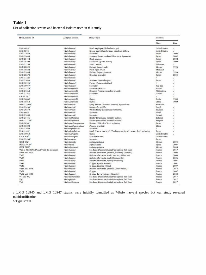
Table 1
List of collection strains and bacterial isolates used in this study
a LMG 10946 and LMG 10947 strains were initially identified as Vibrio harveyi species but our study revealed
misidentification.
b Type strain.
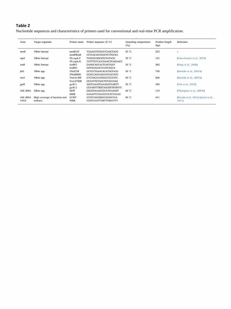
Table 2 Nucleotide sequences and characteristics of primers used for conventional and real-time PCR amplification.
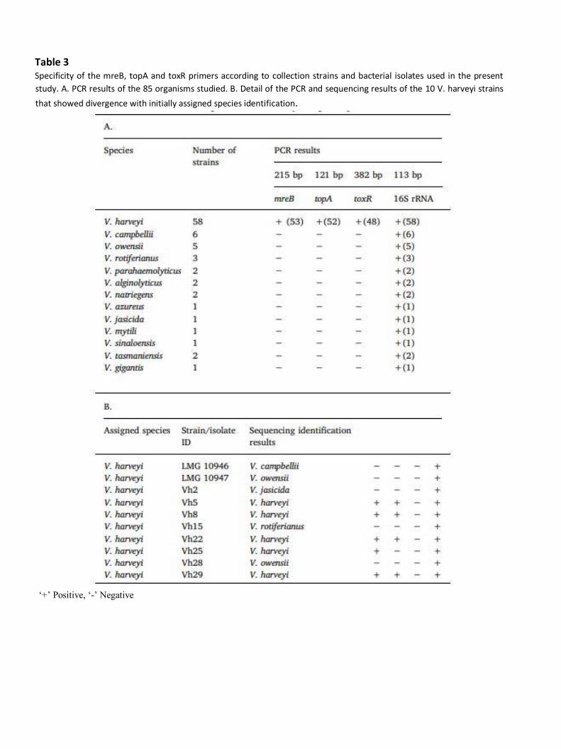
Table 3 Specificity of the mreB, topA and toxR primers according to collection strains and bacterial isolates used in the present
study. A. PCR results of the 85 organisms studied. B. Detail of the PCR and sequencing results of the 10 V. harveyi strains
that showed divergence with initially assigned species identification.
‘+’ Positive, ‘-’ Negative
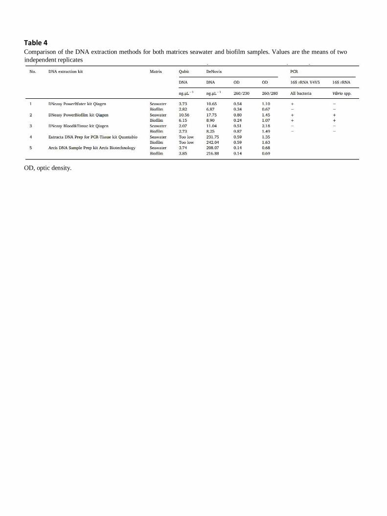
Table 4 Comparison of the DNA extraction methods for both matrices seawater and biofilm samples. Values are the means of two
independent replicates
OD, optic density.

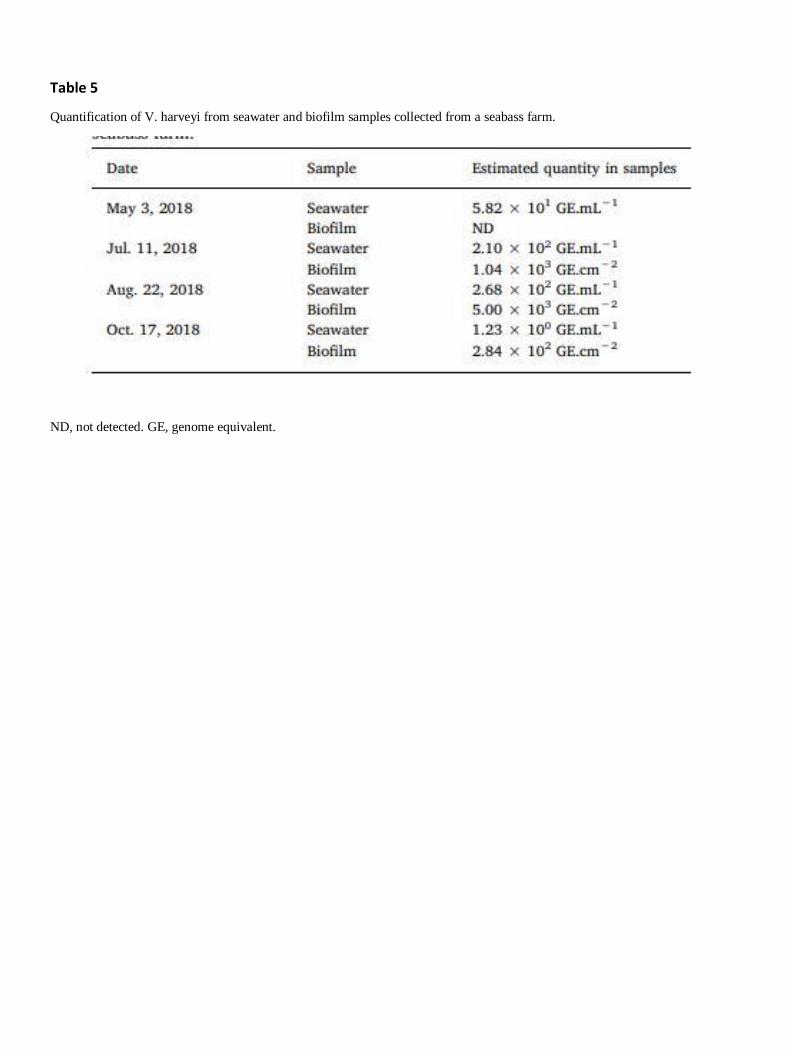
Table 5
Quantification of V. harveyi from seawater and biofilm samples collected from a seabass farm.
ND, not detected. GE, genome equivalent.
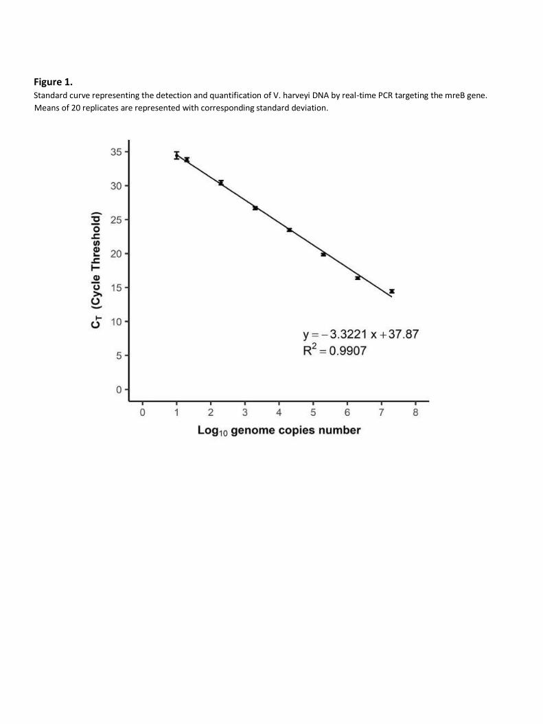
Figure 1. Standard curve representing the detection and quantification of V. harveyi DNA by real-time PCR targeting the mreB gene.
Means of 20 replicates are represented with corresponding standard deviation.
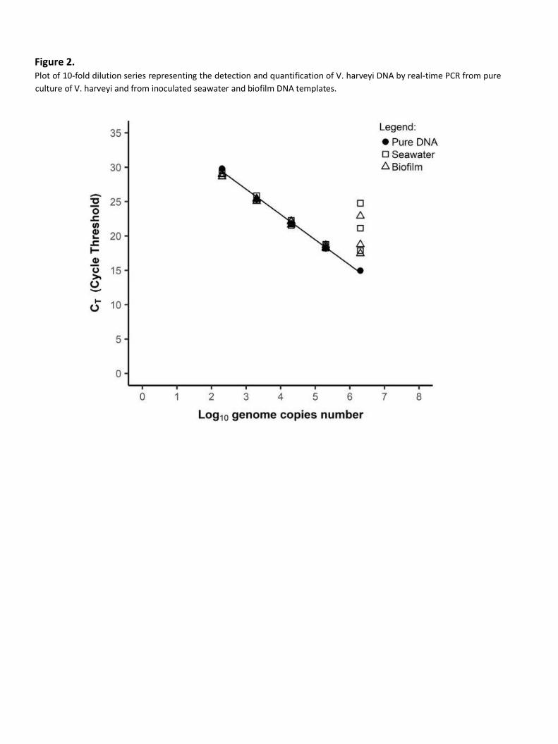
Figure 2. Plot of 10-fold dilution series representing the detection and quantification of V. harveyi DNA by real-time PCR from pure
culture of V. harveyi and from inoculated seawater and biofilm DNA templates.