development of a spreadsheet and a database for - … of a spreadsheet and a database for 1 ... •...
TRANSCRIPT

1Development of a spreadsheet and a database for
recording, reporting and archiving data from quality
assurance tests of radiological equipment
Samuel Kuttner, Medical Physicist
Department of radiology
UNN, Tromsø, Norway
2012

2HistoryFeb 2009
2 Radiography/CT
Feb 2010
1 Radiography/CT
Sep 2011
1 Radiography/CT
Jan 2012
1 MR1 NM0,5 Radiography/ CT

3
GravdalGravdalGravdalGravdal
KirkenesKirkenesKirkenesKirkenes
LongyearbyenLongyearbyenLongyearbyenLongyearbyen
MosjMosjMosjMosjøenenenen
SandnessjSandnessjSandnessjSandnessjøenenenen
NordreisaNordreisaNordreisaNordreisa
FinnsnesFinnsnesFinnsnesFinnsnes
Number of modalities
Radiography: 67
C-arms 57
CT 15
Workstation monitors~50
Total 189
Northern Norway Regional Health
Authority (Helse Nord RHF)

4Background• Prioritied tasks
• Establish procedures for quality assurance (QA):
• Radiography
• Computed Tomography
• Workstation monitors
• Challenges
• No exsisting procedures – everything had to be ”invented”
• Many modalities (~190 pc)
• 190 constancy controls annually
• 190 reports annually
• Lot of time for report writing!
• Large amount of data to be handled
• We didn’t want this!
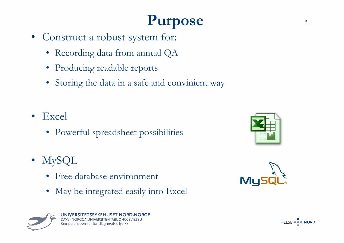
5Purpose• Construct a robust system for:
• Recording data from annual QA
• Producing readable reports
• Storing the data in a safe and convinient way
• Excel
• Powerful spreadsheet possibilities
• MySQL
• Free database environment
• May be integrated easily into Excel

MySQL
relational
database
structure
Equipment table
QA session table
QA data table(Partly shown)
Table
Columns
Relations

List of
equipment
Matching by relation

8Automated excel-sheetEnter ID#Query result
Scan parameters

CTDI100 measurements
Scan parameters Measurement dataMySQL query & Trend analysis

10ResultsTolerance
level
Measurement
data Conclusion

11Trend analysis
Baseline values
(MySQL queries)
Measurement
dataDeviations
Conclusion

12Automatically
generated report

How to add data to MySQL?
1. , = .
2. Copy values
3. Paste
4. Ctrl+Shift+Enter

How to query data from MySQL?
• MySQL-editor command line
• One line syntaxes
• Must copy/paste data manually
• Microsoft query (Excel)
• Visual representation of commands
• Returns data to Excel directly

Example queries• Half value layer (HVL)
• Thickness of aluminium required to half the intensity of an x-ray beam
• Plot ALL measured HVL [mmAl], 70kVp, small focal, 2011Measured HVLs in 2011
0
0,5
1
1,5
2
2,5
3
3,5
4
4,5
1 4 7 10 13 16 19 22 25 28 31 34 37 40 43 46 49 52 55 58
Modality #
HV
L [
mm
Al]
Top 6 highest HVLs
0
0,5
1
1,5
2
2,5
3
3,5
4
4,5
Varian Acuity Philips BV
Libra
Philips Libra
9
Philips BV
Libra
Philips BV
Libra
Philips BV
Libra
Modality #
HV
L [
mm
Al]
HVL of two identical modalities
0
0,5
1
1,5
2
2,5
3
3,5
4
4,5
Varian Acuity Hospital 1 Varian Acuity Hospital 2
Modality #
HV
L [
mm
Al]

HVL trend analysis• Stationary radiology systems
• Period: 2009-2011
• Increase of HVL over time
HVL trend
2,5
2,6
2,7
2,8
2,9
3
3,1
3,2
3,3
2009 2010 2011
Year
HV
L [
mm
Al]

CT dose in air• 120 kVp, head mode, normalized to mAs, all scanners
• Higher dose for small collimations, due to lower geometric efficiency
Measured CTDI100 air, isocenter, head mode
0
0,2
0,4
0,6
0,8
1
1,2
0 5 10 15 20 25 30 35 40Total collimation, NxT [mm]
CT
DI 1
00/
mA
s [m
Gy
/m
As]
Dual slice 4 slice 16 slice 64 slice
• Dual slice
• Constant dose for all total total collimations.
• Multi slice
• Dose increase with decreasing total collimation
• Due to finite size of the focal spot increases the penumbra.
Larger part of the beam for smaller collimations.

HU of water and air• All kVps, Body mode, all scanners in 2011
Measured HU of water in 2011, Body mode, all 80-140kVp
-8
-6
-4
-2
0
2
4
6
8
1 3 5 7 9 11 13 15 17 19 21 23 25 27 29 31 33 35 37 39
Scanner #
CT
DI 1
00/
mA
s [m
Gy/
mA
s]
Measured HU of air in 2011, Body mode, all 80-140kVp
-1060
-1050
-1040
-1030
-1020
-1010
-1000
-990
-980
-970
-960
38 16 9 3 18 11 7 13 34 21 24 27 28 25 23 33 35 37 30 31
Scanner #
CT
DI 1
00/
mA
s [m
Gy/
mA
s]
Water
Air

High voltage measurements
• Measured on radiography and fluoroscopy systems, 2009-2012
• Deviation of measured kVp from nominal values
• 5% tolerance level seems OK!
Deviation of measured kVp from nominal value, 2009-2012
0 %
5 %
10 %
15 %
20 %
25 %
30 %
1 97 193 289 385 481 577 673 769 865 961 1057 1153 1249 1345 1441 1537 1633 1729 1825 1921 2017 2113
Measurement #
Ab
solu
te v
alu
e o
f d
evia
tio
n [
%]
5% of measurements outside 5% tolerance
level

20Ambient light measurements
Same room:
Light on Light offBakgrunnslys
0
20
40
60
80
100
120
140
B2.64
9 (N
evro
gr. 3
)
Ultr
alydg
rans
knin
g (B3.6
88) v
enstr
e
Ultr
alydg
rans
knin
gen
(B3.6
88) h
oyre
Røntg
engr
ansk
ning
stas
jon
3
B2.64
3 (D
emor
om-A
nne-
May
)
Røntg
engr
ansk
ning
stas
jon
2
Røntg
engr
ansk
ning
stas
jon
1
Røntg
engr
ansk
ning
stas
jon
6
Røntg
engr
ansk
ning
stas
jon
4
B3621
(Bar
ngr.
Thore
sten)
Røntg
engr
ansk
ning
stas
jon
5
Røntg
engr
ansk
ning
stas
jon
12
Røntg
engr
ansk
ning
stas
jon
10
Røntg
engr
ansk
ning
stas
jon
11
Røntg
engr
ansk
ning
stas
jon
7
Røntg
engr
ansk
ning
stas
jon
9
Røntg
engr
ansk
ning
stas
jon
8
B3621
(Bar
ngr.
Lill-So
fie)
B2.64
9 (N
evro
gr. T
orgrim
)
Und
ervis
ningsro
m (B
3-61
6)
B3.62
2 (T
horax
gr. S
igne)
B2.64
9 (N
evro
gr. G
ry)

21
Non-diagnostic monitor
Luminance ratio (LR) • LR = Lmaks/Lmin
• LR > 250 (TG-18)
• MDView193 performs better!
MDView 193

22Conclusions
• A comprehensive system for recording, reporting and
archiving data from quality assurance tests of radiological
equipment was developed
• Excel and MySQL are excellent tools for the handling of dose
and image quality data generated from QA tests
• Benchmarking
• Purchasing processes
• Problem solving
• National databases
• Still missing a really user friendly interface

23
Thank you
for your attention!
Reference litterature• IEC, 61223-3-5, Part 3-5: Acceptance tests, 2004
• IEC, 61223-2-6, Part 2-6: Constancy tests, 2006
• ImPACT, Information Leaflet No.1: CT scanner
Acceptance Testing (2001)
• IPEM Report 32. Part III (2003)
• IPEM Report 91 (2005)
• AAPM report 39 (1993)
• AAPM report no.74 (2002)
• AAPM Task group 18 report (2005)
• AAPM Task group 66 report (2003)
• CT manufacturer manuals
• Phantom manuals