development of an e-learning system for teaching ... · pylori (hp)-associated gastritis, gastric...
TRANSCRIPT
REVIEW ARTICLE
Development of an e-learning system for teaching endoscopistshow to diagnose early gastric cancer: basic principlesfor improving early detection
Kenshi Yao1• Noriya Uedo2
• Manabu Muto3• Hideki Ishikawa4
Received: 7 October 2016 / Accepted: 1 December 2016 / Published online: 28 December 2016
� The Author(s) 2016. This article is published with open access at Springerlink.com
Abstract We developed an internet e-learning system in
order to improve the ability of endoscopists to diagnose
gastric cancer at an early stage. The efficacy of this system
at expanding knowledge and providing invaluable experi-
ence regarding the endoscopic detection of early gastric
cancer was demonstrated through an international multi-
center randomized controlled trial. However, the contents
of the system have not yet been fully described in the
literature. Accordingly, we herein introduce the contents
and their principles, which comprise three main subjects:
technique, knowledge, and experience. Since all the
e-learning contents and principles are based on conven-
tional white-light endoscopy alone, which is commonly
available throughout the world, they should provide a good
reference point for any endoscopist who wishes to devise
learning materials and guidelines for improving their own
clinical practice.
Keywords Endoscopic diagnosis � Gastric cancer � E-learning � International multicenter randomized controlled
trial
Introduction
In many countries, gastric cancer is not diagnosed until it is
at an advanced stage. Advanced endoscopic imaging such
as magnifying endoscopy with narrow-band imaging is
generally used in Japan [1] and some Western countries
[2], but is still rarely used in developing countries, where
the prevalence of gastric cancer is high. Conventional
white-light endoscopy is a standard endoscopy technique
throughout the world. However, to date, there has been no
standardized learning system based on conventional white-
light endoscopy which could be commonly applied to
screening endoscopy for the detection of early gastric
cancer.
We constructed an e-learning system and finalized a
global study to test its usefulness through a randomized
controlled trial [3]. In that trial, after receiving a pre-test,
participants were randomly allocated to either an e-learning
or a non-e-learning group. Only those in the e-learning
group gained access to the e-learning system. Two months
after the pre-test, both groups received a post-test. Five
hundred fifteen endoscopists from 35 countries were
assessed for eligibility, and 332 were enrolled in the study,
with 166 allocated to each group. Of these, 151 participants
in the e-learning group and 144 in the non-e-learning group
were included in the analysis. The mean improvement rate
(standard deviation) in the e-learning and non-e-learning
groups was 1.24 (0.26) and 1.00 (0.16), respectively
(P\ 0001, t test). We have already reported that the
e-learning system was useful for improving the endoscopic
Conference presentation 88th Annual Meeting of the Japan GastricCancer Association, March 18, 2016, Beppu City, Japan.
& Kenshi Yao
1 Department of Endoscopy, Fukuoka University Chikushi
Hospital, 1-1-1 Zokumyouin, Chikushino,
Fukuoka 818-8502, Japan
2 Department of Gastrointestinal Oncology, Osaka Medical
Center for Cancer and Cardiovascular Diseases, Osaka, Japan
3 Department of Therapeutic Oncology, Graduate School of
Medicine, Kyoto University, Kyoto, Japan
4 Department of Molecular-Targeting Cancer Prevention,
Graduate School of Medical Science, Kyoto Prefectural
University of Medicine, Kyoto, Japan
123
Gastric Cancer (2017) 20 (Suppl 1):S28–S38
DOI 10.1007/s10120-016-0680-7
detection of early gastric cancer, irrespective of pre-test
score, endoscopist experience, or geographical area [3].
However, to date, the contents of this e-learning system
have not been fully described in the literature.
Accordingly, the aim of this article is to demonstrate the
contents of the e-learning system so that medical practi-
tioners can obtain information regarding what they need to
learn and how they should teach endoscopy practice in
order to improve the detection of early gastric cancer using
white-light endoscopy alone.
E-learning contents
In order to improve the endoscopic detection of early
gastric cancer, doctors need to acquire basic principles that
comprise the three keys to detection: technique, knowl-
edge, and experience [4, 5]. Accordingly, the e-learning
contents were devised as follows [3]:
1. Technique
• Lecture (video clips and slides).
1. Ideal preparation with mucolytic and defoam-
ing agents.
2. Use of anticholinergic agents.
3. Avoiding blind spots:
• Gastric wall should be distended by air
insufflation.
• Mucus and bubbles should be rinsed off
using irrigating water with defoaming
agent.
• Entire stomach should be mapped: system-
atic screening of the stomach (SSS).
2. Knowledge
• Test (ten questions without answers).
• Lecture (video clips and slides).
1. General: normal appearance and chronic
gastritis.
2. Detection: awareness of signs of suspicious
lesions.
3. Awareness of multiple synchronous lesions.
4. Characterization using the GUP system: G
gastritis-like lesions, U ulcerative lesions, and
P polypoid lesions.
• Ten (ten questions with answers and
descriptions).
3. Experience: 100 cases for EGC detection training
• Mock test (ten cases with scores and no answers).
• Random version of the 100 cases.
• Systematic version of the 100 cases.
• Random version of the 100 cases.
• Mock test (ten cases with scores and answers).
Technique
Video clips and slides were uploaded in order to explain
the requisite endoscopic technique for improving the
detection of early gastric cancer.
Ideal preparation with mucolytic and defoaming agents
The correct preparation for endoscopic examination is
mandatory to minimize time and effort during the proce-
dure, i.e., the removal of mucus and froth from the mucosal
surface. Thirty minutes before the procedure, patients are
asked to drink a mixture of water with mucolytic and
defoaming agents. The formula in Japan is 100 mL of
water with 20,000 U Pronase� (Kaken Pharmaceutical,
Tokyo, Japan), 1 g of sodium bicarbonate, and 10 mL of
dimethylpolysiloxane (20 mg/mL, Horii Pharmaceutical
Ind., Osaka, Japan). However, Pronase� is not available
for use in all countries. An alternative mixture comprises
100 mL of water mixed with 2 mL of acetylcysteine
(200 mg/mL Parvolex�, Celltech, Slough, UK; or Muco-
myst, Bristol-Myers Squibb, New York, NY, USA), and
0.5 mL activated dimethicone (40 mg/mL, Infacol�, For-
est Laboratories, Dartford, UK).
Use of anticholinergic agents
In the normal physiological state, the gastric wall is always
moving due to peristalsis. In order to detect subtle mucosal
changes, we need to carefully scan the entire mucosal
surface. However, peristalsis makes it difficult for the
endoscopist to obtain static observations. Accordingly,
intravenous or intramuscular administration of an anti-
cholinergic agent such as 10–20 mg of scopolamine
butylbromide (Buscopan�) is recommended just before
insertion of the endoscope, even for upper gastrointestinal
(GI) screening endoscopies. If there are contraindications
to the use of anticholinergic agents (e.g., cardiovascular
disease), 1 mg of glucagon can be administered to inhibit
peristalsis.
Avoiding blind spots
During the endoscopy, in order to avoid blind spots, a
standardized approach to map the entire stomach needs to
be employed. A basic technique for avoiding blind spots
involves the following procedures: (1) distending the gas-
tric wall by air insufflation, (2) rinsing mucus and froth off
Development of an e-learning system for teaching endoscopists how to diagnose early gastric… S29
123
the gastric mucosa by irrigating with water and a
defoaming agent, and (3) mapping the entire stomach. The
need for these steps is explained below.
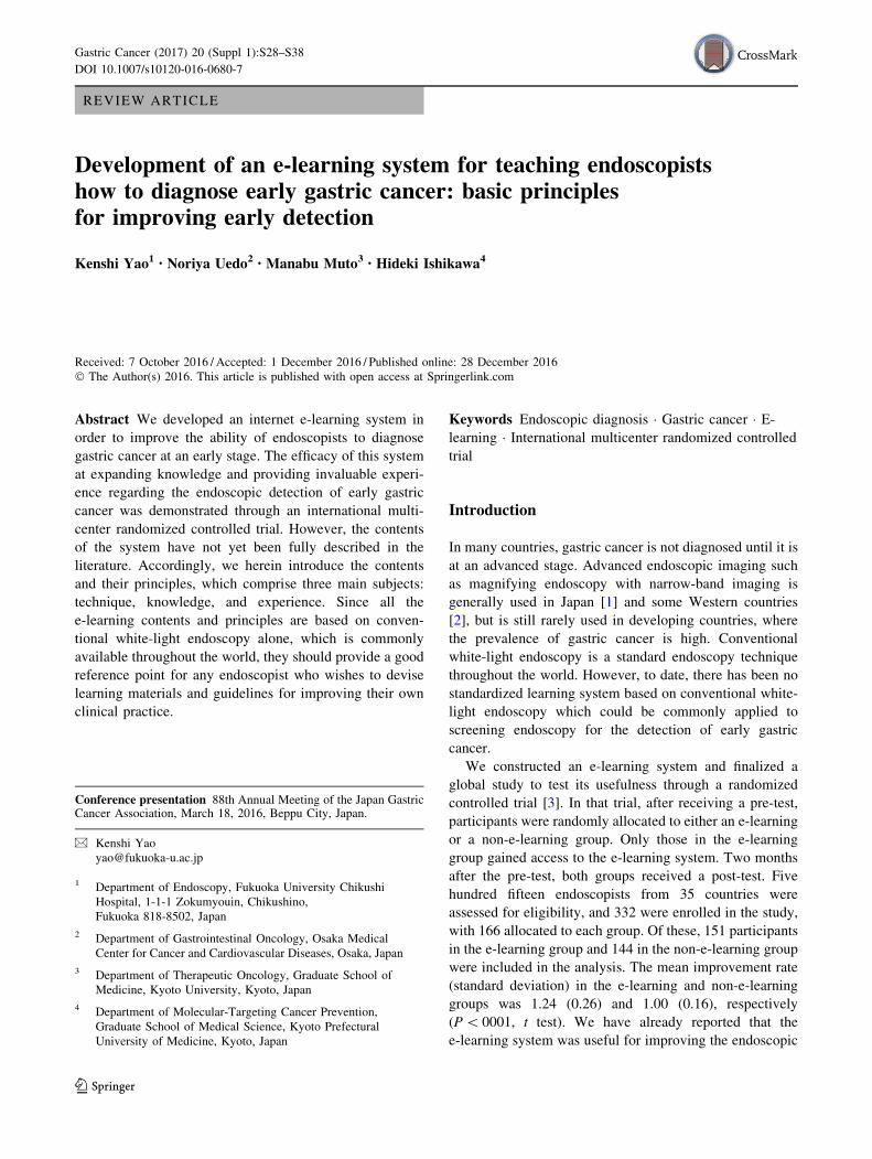
1. If the gastric wall is not fully distended, we may fail to
detect a cancer on the greater curvature, even a rather
advanced cancer. The lecture demonstrates a very good
example of advanced gastric cancer on the greater
curvature of the lower gastric body. With insufflation
of a small amount of air, the appearance is normal
(Fig. 1a). However, when more air is insufflated and
the gastric wall together with the greater curvature
with gastric folds is distended, a distinct lesion,
suggestive of advanced gastric cancer, becomes evi-
dent (Fig. 1b).
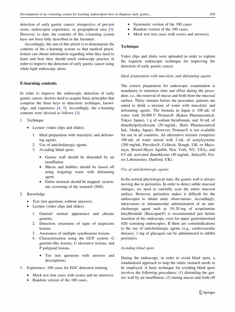
2. A short video clip of an endoscopy examination is
included in the video lecture in order to demonstrate
why the mucus and froth need to be rinsed off. Even
after patients drink a mixture of water with mucolytic
and defoaming agents, cases with copious mucus and
froth within the stomach are sometimes encountered
(Fig. 2a). However, when the mucus and froth are
rinsed off the gastric mucosal surface through irriga-
tion with water and a defoaming agent, even subtle
lesions can be detected (Fig. 2b).
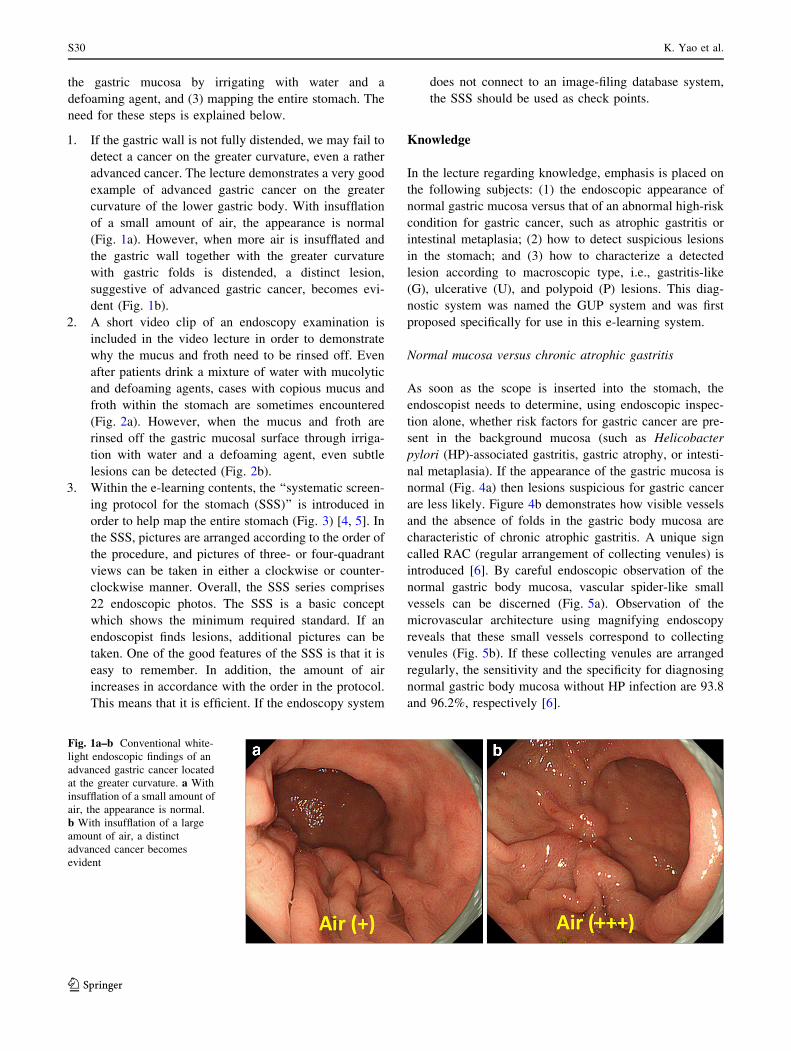
3. Within the e-learning contents, the ‘‘systematic screen-
ing protocol for the stomach (SSS)’’ is introduced in
order to help map the entire stomach (Fig. 3) [4, 5]. In
the SSS, pictures are arranged according to the order of
the procedure, and pictures of three- or four-quadrant
views can be taken in either a clockwise or counter-
clockwise manner. Overall, the SSS series comprises
22 endoscopic photos. The SSS is a basic concept
which shows the minimum required standard. If an
endoscopist finds lesions, additional pictures can be
taken. One of the good features of the SSS is that it is
easy to remember. In addition, the amount of air
increases in accordance with the order in the protocol.
This means that it is efficient. If the endoscopy system
does not connect to an image-filing database system,
the SSS should be used as check points.
Knowledge
In the lecture regarding knowledge, emphasis is placed on
the following subjects: (1) the endoscopic appearance of
normal gastric mucosa versus that of an abnormal high-risk
condition for gastric cancer, such as atrophic gastritis or
intestinal metaplasia; (2) how to detect suspicious lesions
in the stomach; and (3) how to characterize a detected
lesion according to macroscopic type, i.e., gastritis-like
(G), ulcerative (U), and polypoid (P) lesions. This diag-
nostic system was named the GUP system and was first
proposed specifically for use in this e-learning system.
Normal mucosa versus chronic atrophic gastritis
As soon as the scope is inserted into the stomach, the
endoscopist needs to determine, using endoscopic inspec-
tion alone, whether risk factors for gastric cancer are pre-
sent in the background mucosa (such as Helicobacter
pylori (HP)-associated gastritis, gastric atrophy, or intesti-
nal metaplasia). If the appearance of the gastric mucosa is
normal (Fig. 4a) then lesions suspicious for gastric cancer
are less likely. Figure 4b demonstrates how visible vessels
and the absence of folds in the gastric body mucosa are
characteristic of chronic atrophic gastritis. A unique sign
called RAC (regular arrangement of collecting venules) is
introduced [6]. By careful endoscopic observation of the
normal gastric body mucosa, vascular spider-like small
vessels can be discerned (Fig. 5a). Observation of the
microvascular architecture using magnifying endoscopy
reveals that these small vessels correspond to collecting
venules (Fig. 5b). If these collecting venules are arranged
regularly, the sensitivity and the specificity for diagnosing
normal gastric body mucosa without HP infection are 93.8
and 96.2%, respectively [6].
Fig. 1a–b Conventional white-
light endoscopic findings of an
advanced gastric cancer located
at the greater curvature. a With
insufflation of a small amount of
air, the appearance is normal.
b With insufflation of a large
amount of air, a distinct
advanced cancer becomes
evident
S30 K. Yao et al.
123
How to detect and characterize suspicious lesions
in the stomach
The diagnostic process can be divided into two steps:
detection and characterization. Detection requires good
endoscopic technique and thorough knowledge. The lecture
describes the procedure for improving the detection and
characterization of early gastric cancers.
Detection In order to detect early gastric cancer, endo-
scopists need to be aware of the signs of suspicious lesions.
The Paris classification of macroscopic appearance was
Fig. 2a–b A video clip which
demonstrates the need to rinse
mucus and froth off the mucosal
surface during endoscopic
examination. a When copious
mucus and froth are present, we
are unable to detect subtle
lesions. b After the mucus and
froth have been rinsed off the
gastric mucosal surface by
irrigating with water and a
defoaming agent, we are able to
detect a subtle lesion of early
gastric cancer
Fig. 3 Systematic screening protocol for the stomach (SSS). The SSS
should be initiated as soon as the scope is inserted into the gastric
antrum. In the antegrade view, endoscopic images of four quadrants
of the gastric antrum, body, and middle–upper body are obtained and
then, in the retroflex view, images of the four quadrants of the gastric
fundus and cardia, and three quadrants of the gastric middle–upper
body and incisura are taken. Overall, the SSS comprises 22
endoscopic images. A anterior wall, G greater curvature, L lesser
curvature, P posterior wall, Q quadrant
Development of an e-learning system for teaching endoscopists how to diagnose early gastric… S31
123

proposed in 2003 with reference to Japanese systems of
classification [7]. The Paris classification is now commonly
used worldwide. However, this classification is not very
practical in a clinical setting when detecting and charac-
terizing suspicious lesions at screening endoscopy.
Accordingly, the diagnostic system was modified to make
it more suitable for adoption by endoscopists in their own
clinical practice. Briefly, the visible endoscopic findings
that endoscopists need to be concerned about can be divi-
ded into just three types: gastritis-like lesions [G: gastritis
vs superficial cancer (0 IIa, IIb, IIc)], ulcerative lesions [U:
peptic ulcer vs superficial cancer with ulceration (0 III)],
and polypoid lesions [P: polyp vs polypoid cancer (0 I)].
Based on this concept, which seems to be readily endorsed
worldwide, endoscopists attempt to detect the presence of
suspicious lesions. Provisionally, this diagnostic system
was named ‘‘the GUP system’’ [3]. This is an original,
brand new idea, developed specifically for this e-learning
project. Instructions based on this GUP system are given
throughout the e-learning course.
For the purpose of detection, the first thing that needs to
be mastered is how to be aware of the presence of suspi-
cious lesions. The most common markers for detection are
(1) surface change and (2) color change. Other markers,
namely abrupt changes in the vascular/mucosal pattern,
changes in light reflection, and spontaneous bleeding, are
also of importance. Early gastric cancers of both the
polypoid and ulcerative types are easily detected by surface
change alone, provided that the endoscopist follows the
SSS protocol with optimum preparation. On the other hand,
superficial mucosal lesions that mimic gastritis—that is,
gastritis-like lesions (G)—are very difficult to detect, even
with optimum preparation and the best technique.
Accordingly, endoscopists need to be particularly aware of
the key signs for detecting superficial gastritis-like
neoplasia.
The first key marker is surface change. Figure 6a shows
an endoscopic photo of a very small early gastric cancer of
the superficial depressed type. Since this lesion is very
small and only shows subtle changes, with morphology
Fig. 4a–b Conventional white-
light endoscopic findings for the
gastric body. a Normal gastric
mucosa without HP infection.
b Gastric mucosa with chronic
atrophic gastritis associated
with HP infection. Visible
vessels and an absence of folds
in the gastric body mucosa are
characteristic of chronic
atrophic gastritis
Fig. 5 a Conventional white-
light endoscopic findings for the
gastric body. Regular
arrangement of collecting
(RAC) venules is present in
normal mucosa. b Magnifying
white-light endoscopic findings
for the gastric body. The small
vascular-spider-like vessels in acorrespond to regularly
arranged venules (arrow)
S32 K. Yao et al.
123

mimicking an erosive gastritis lesion, it is very difficult to
detect this lesion. However, it is essential to be aware that
such subtle surface changes can be important signs of early
gastric cancer. The second important marker is color
change. A well-demarcated area with color change is a
useful indicator of a gastritis-like cancer. Figure 6b pre-
sents the endoscopic findings of superficial depressed type
which mimics gastritis (gastritis-like lesion). In the back-
ground atrophic mucosa, a clear vascular pattern located in
the deeper part of the mucosa and submucosa can be
visualized. If the background vascular pattern is traced, it
soon becomes apparent that there is a well-demarcated area
where the vascular pattern is abruptly interrupted at the
margin of the lesion. This is indeed a useful finding for
early detection.
Characterization For this e-learning system, consider-
able effort was directed into creating very simple criteria
Fig. 6a–b Examples of endoscopic markers for detection by con-
ventional white-light endoscopy. a Marker, surface change: a very
small early gastric cancer of the superficial depressed type (gastritis-
like lesion) as indicated by an arrow in the gastric antrum. b Marker,
color change: a superficial depressed type which mimics gastritis
(gastritis-like lesion) in the gastric body. Arrows show a well-
demarcated reddened lesion with abrupt interruption of the vascular
pattern in the background mucosa
Fig. 7a–b Examples of the
GUP system and its criteria for
characterization by
conventional white-light
endoscopy with
chromoendoscopy (gastritis-like
lesion). a Erosive gastritis.
b Small early gastric cancer of
superficial depressed type (0
IIc)
Development of an e-learning system for teaching endoscopists how to diagnose early gastric… S33
123
for making a diagnosis based on conventional white-
light endoscopic appearance alone. For characterization,
two distinct markers, namely color and surface mor-
phology, should be applied to the interpretation of con-
ventional endoscopic findings. Chromoendoscopy using
indigo carmine is useful for enhancing the surface pat-
tern (Fig. 7). The differential diagnosis is made using the
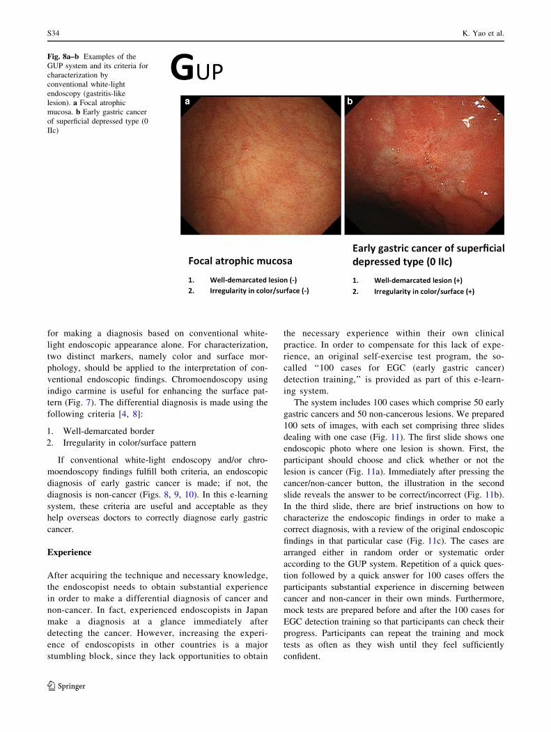
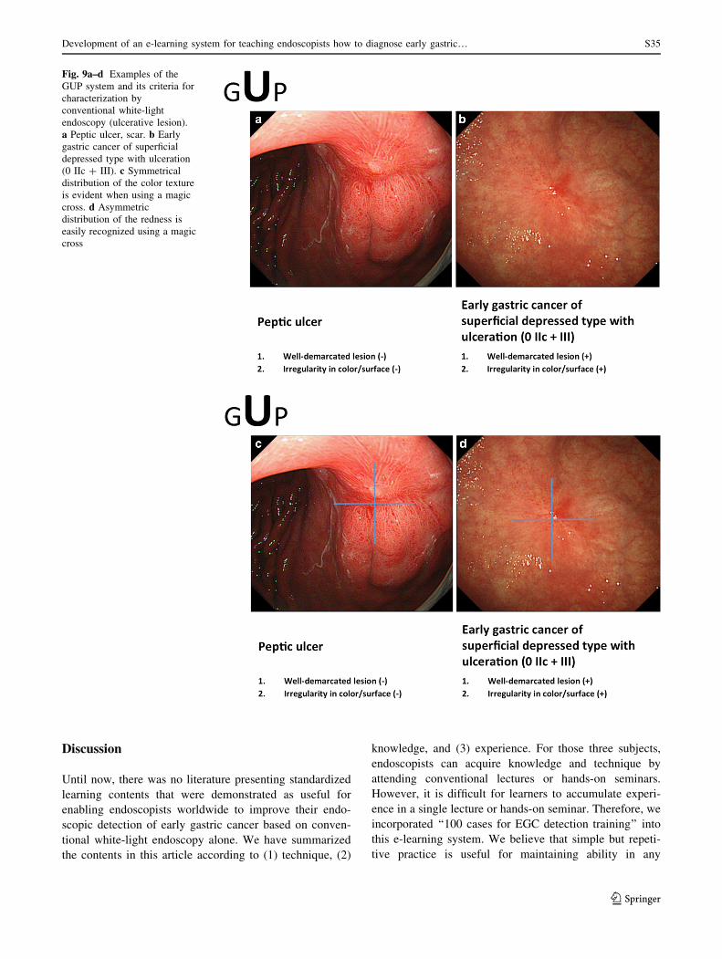
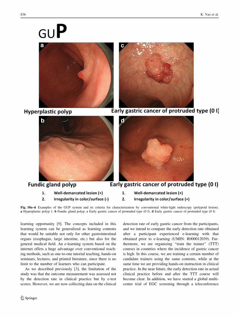
following criteria [4, 8]:
1. Well-demarcated border
2. Irregularity in color/surface pattern
If conventional white-light endoscopy and/or chro-
moendoscopy findings fulfill both criteria, an endoscopic
diagnosis of early gastric cancer is made; if not, the
diagnosis is non-cancer (Figs. 8, 9, 10). In this e-learning
system, these criteria are useful and acceptable as they
help overseas doctors to correctly diagnose early gastric
cancer.
Experience
After acquiring the technique and necessary knowledge,
the endoscopist needs to obtain substantial experience
in order to make a differential diagnosis of cancer and
non-cancer. In fact, experienced endoscopists in Japan
make a diagnosis at a glance immediately after
detecting the cancer. However, increasing the experi-
ence of endoscopists in other countries is a major
stumbling block, since they lack opportunities to obtain
the necessary experience within their own clinical
practice. In order to compensate for this lack of expe-
rience, an original self-exercise test program, the so-
called ‘‘100 cases for EGC (early gastric cancer)
detection training,’’ is provided as part of this e-learn-
ing system.
The system includes 100 cases which comprise 50 early
gastric cancers and 50 non-cancerous lesions. We prepared
100 sets of images, with each set comprising three slides
dealing with one case (Fig. 11). The first slide shows one
endoscopic photo where one lesion is shown. First, the
participant should choose and click whether or not the
lesion is cancer (Fig. 11a). Immediately after pressing the
cancer/non-cancer button, the illustration in the second
slide reveals the answer to be correct/incorrect (Fig. 11b).
In the third slide, there are brief instructions on how to
characterize the endoscopic findings in order to make a
correct diagnosis, with a review of the original endoscopic
findings in that particular case (Fig. 11c). The cases are
arranged either in random order or systematic order
according to the GUP system. Repetition of a quick ques-
tion followed by a quick answer for 100 cases offers the
participants substantial experience in discerning between
cancer and non-cancer in their own minds. Furthermore,
mock tests are prepared before and after the 100 cases for
EGC detection training so that participants can check their
progress. Participants can repeat the training and mock
tests as often as they wish until they feel sufficiently
confident.
Fig. 8a–b Examples of the
GUP system and its criteria for
characterization by
conventional white-light
endoscopy (gastritis-like
lesion). a Focal atrophic
mucosa. b Early gastric cancer
of superficial depressed type (0
IIc)
S34 K. Yao et al.
123
Discussion
Until now, there was no literature presenting standardized
learning contents that were demonstrated as useful for
enabling endoscopists worldwide to improve their endo-
scopic detection of early gastric cancer based on conven-
tional white-light endoscopy alone. We have summarized
the contents in this article according to (1) technique, (2)
knowledge, and (3) experience. For those three subjects,
endoscopists can acquire knowledge and technique by
attending conventional lectures or hands-on seminars.
However, it is difficult for learners to accumulate experi-
ence in a single lecture or hands-on seminar. Therefore, we
incorporated ‘‘100 cases for EGC detection training’’ into
this e-learning system. We believe that simple but repeti-
tive practice is useful for maintaining ability in any
Fig. 9a–d Examples of the
GUP system and its criteria for
characterization by
conventional white-light
endoscopy (ulcerative lesion).
a Peptic ulcer, scar. b Early
gastric cancer of superficial
depressed type with ulceration
(0 IIc ? III). c Symmetrical
distribution of the color texture
is evident when using a magic
cross. d Asymmetric
distribution of the redness is
easily recognized using a magic
cross
Development of an e-learning system for teaching endoscopists how to diagnose early gastric… S35
123
learning opportunity [9]. The concepts included in this
learning system can be generalized as learning contents
that would be suitable not only for other gastrointestinal
organs (esophagus, large intestine, etc.) but also for the
general medical field. An e-learning system based on the
internet offers a huge advantage over conventional teach-
ing methods, such as one-to-one tutorial teaching, hands-on
seminars, lectures, and printed literature, since there is no
limit to the number of learners who can participate.
As we described previously [3], the limitation of the
study was that the outcome measurement was assessed not
by the detection rate in clinical practice but by e-test
scores. However, we are now collecting data on the clinical
detection rate of early gastric cancer from the participants,
and we intend to compare the early detection rate obtained
after a participant experienced e-learning with that
obtained prior to e-learning (UMIN: R000012039). Fur-
thermore, we are organizing ‘‘train the trainer’’ (TTT)
courses in countries where the incidence of gastric cancer
is high. In this course, we are training a certain number of
candidate trainers using the same contents, while at the
same time we are providing hands-on instruction in clinical
practice. In the near future, the early detection rate in actual
clinical practice before and after the TTT course will
become clear. In addition, we have started a global multi-
center trial of EGC screening through a teleconference
Fig. 10a–d Examples of the GUP system and its criteria for characterization by conventional white-light endoscopy (polypoid lesion).
a Hyperplastic polyp 1. b Fundic gland polyp. c Early gastric cancer of protruded type (0 I). d Early gastric cancer of protruded type (0 I)
S36 K. Yao et al.
123
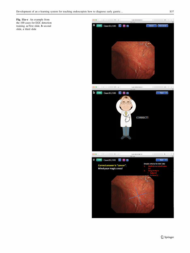
Fig. 11a–c An example from
the 100 cases for EGC detection
training. a First slide, b second
slide, c third slide
Development of an e-learning system for teaching endoscopists how to diagnose early gastric… S37
123

system using the learning contents described in this article
[JSPS Core-to-Core Program (B. Asia-Africa Science
Platforms)]. Another limitation was that the opportunity to
partake in this e-learning course was limited to participants
who were registered with this trial. However, we are pur-
suing the possibility of opening up this e-learning system
using an official internet website as a platform so that any
endoscopist in any country can access this system freely.
In conclusion, good practice based on good technique,
knowledge, and adequate experience improves the ability
of a doctor to detect gastric cancer or other cancers at an
early stage. The aim of introducing the contents of this
e-learning system in this article is to enable any endo-
scopist in any country to benefit from such practice. This
e-learning system is likely to contribute to improved
human health and welfare by reducing the mortality from
gastrointestinal cancer.
Acknowledgements This trial was supported by the Central
Research Institute of Fukuoka University (I) and the JSPS Core-to-
Core Program (B. Asia-Africa Science Platforms). We wish to thank
Miss Katherine Miller (Royal English Language Centre, Fukuoka,
Japan) for correcting the English used in this article.
Compliance with ethical standards
Ethical standards All procedures were conducted in accordance
with the ethical standards of the responsible committees on human
experimentation (both institutional and national) and with the Hel-
sinki Declaration of 1964 and later versions. Informed consent was
obtained from all participants before they were included in the study.
Funding The Central Research Institute of Fukuoka University
(I) and the JSPS Core-to-Core Program (B. Asia-Africa Science
Platforms).
Disclosure of potential conflicts of interest The authors hereby
declare that they have no conflict of interest.
Open Access This article is distributed under the terms of the
Creative Commons Attribution 4.0 International License (http://
creativecommons.org/licenses/by/4.0/), which permits unrestricted
use, distribution, and reproduction in any medium, provided you give
appropriate credit to the original author(s) and the source, provide a
link to the Creative Commons license, and indicate if changes were
made.
References
1. Ezoe Y, Muto M, Uedo N, et al. Magnifying narrowband imaging
is more accurate than conventional white-light imaging in
diagnosis of gastric mucosal cancer. Gastroenterology.
2011;141(6):2017–25. doi:10.1053/j.gastro.2011.08.007.
2. Kaltenbach T, Sano Y, Friedland S, et al. American Gastroentero-
logical Association. American Gastroenterological Association
(AGA) Institute technology assessment on image-enhanced
endoscopy. Gastroenterology. 2008;134(1):327–40 (Epub 2007Oct 30).
3. Yao K, Uedo N, Muto M, et al. Development of an e-learning
system for the endoscopic diagnosis of early gastric cancer: an
international multicenter randomized controlled trial. EBioMedi-
cine. 2016;9:140–7. doi:10.1016/j.ebiom.2016.05.016.
4. Yao K. The endoscopic diagnosis of early gastric cancer. Ann
Gastroenterol. 2013;26(1):11–22.
5. Veitch AM, Uedo N, Yao K, et al. Optimizing early upper
gastrointestinal cancer detection at endoscopy. Nat Rev Gastroen-
terol Hepatol. 2015;12(11):660–7. doi:10.1038/nrgastro.2015.128.
6. Yagi K, Nakamura A, Sekine A. Characteristic endoscopic and
magnified endoscopic findings in the normal stomach without
Helicobacter pylori infection. J Gastroenterol Hepatol.
2002;17:39–45.
7. The Paris endoscopic classification of superficial neoplastic
lesions. Esophagus, stomach, and colon: November 30 to Decem-
ber 1, 2002. Gastrointest Endosc. 2003;58(6 Suppl):S3–43.
8. Yao K, Nagahama T, Matsui T, et al. Detection and characteri-
zation of early gastric cancer for curative endoscopic submucosal
dissection. Dig Endosc. 2013;25(Suppl 1):44–54. doi:10.1111/den.
12004.
9. Mabe K, Yao K, Nojima M, et al. An educational intervention to
improve the endoscopist’s ability to correctly diagnose small
gastric lesions using magnifying endoscopy with narrow-band
imaging. Ann Gastroenterol. 2014;27(2):149–55.
S38 K. Yao et al.
123