development of blastomeres of mouse eggs isolated at...
TRANSCRIPT

/ . Embryol. exp. Morph. Vol. 18, 1, pp. 155-180, August 1967 155With 4 plates
Printed in Great Britain
Development of blastomeres of mouse eggsisolated at the 4- and 8-cell stage
ByANDRZEJ K. TARKOWSKI1 & JOANNA \ V R 6 B L E W S K A 1
From the Department of Embryology, University of Warsaw
Organization of a mammahan egg at the beginning of development has beenthe subject of several experimental as well as descriptive cytological and cyto-chemical investigations.
The regulative capacities of blastomeres isolated at the 2-cell stage have beenstudied in the rat (Nicholas & Hall, 1942), rabbit (Seidel, 1952,1960) and mouse(Tarkowski, 1959a, b; Mulnard, 1965a, b). Investigations by Seidel andTarkowski have shown that although the majority of 1/2 blastomeres canregulate and develop into smaller but otherwise normal blastocysts, som e of themgive rise to purely trophoblastic vesicles devoid of the inner cell mass. In themouse, Tarkowski (1959 a, b) observed in addition in a few cases multicellularmorulae which had not undergone cavitation. The findings of Tarkowski havebeen recently confirmed by Mulnard (1965 a, b) who studied cytochemicallymouse 1/2 blastomeres developing in vitro. At least some of the blastocystsdeveloped from 1/2 blastomeres are capable of further normal development—many embryos have been collected and several young were born in both therabbit (Seidel, 1952, 1960) and the mouse (Tarkowski, 1959a, b). The youngproved to be quite normal and fertile.
In the rabbit, single blastomeres of the 4-cell stage develop into blastocystsor trophoblastic vesicles (Seidel, 1956, 1960). However, the incidence of thelatter forms is much higher among 1/4 blastomeres than among those originatingfrom the 2-cell stage. The most advanced embryo which developed from atransplanted 1/4 blastomere attained the stage of five somites and was quitenormal (Seidel, 1956, 1960). Similar investigations on the mouse have been per-formed on a very limited scale (Tarkowski, 1959 a, b) and allowed only theconclusion that a 1/4 blastomere is still able to develop into a blastocyst com-posed of a trophoblast and the inner mass.
The conclusions drawn by Seidel and Tarkowski on the basis of their experi-mental investigations concerning the organization of the mammalian egg wereslightly different. Seidel explained his results by assuming the existence in theundivided egg of an organizing centre whose presence, in whole or at least in
1 Authors' address: Department of Embryology, University of Warsaw, Warsaw 64,Poland.

156 A. K. TARKOWSKI & J. WR6BLEWSKA
part, in the isolated blastomere is essential for the formation of a typical blasto-cyst. In a given blastomere lack of the cytoplasm from the organizing centre wouldlead, according to Seidel, to the development of a trophoblastic vesicle. Thefact that trophoblastic vesicles attain higher incidence among forms developedfrom 1/4 blastomeres than among those developed from 1/2 blastomeres agreeswell with this hypothesis.
Tarkowski tried to correlate his findings with the results of cytochemicalinvestigations carried out by Dalcq, Mulnard and their collaborators on rodenteggs (for review see Dalcq (1957), Mulnard (1961); also discussion in this paper)and concluded that the regulative capacities of blastomeres of the 2-cell stage areto a certain degree limited by the character of the inherited cytoplasm. The factthat the normal proportions between the trophoblast and the inner mass arenot always reproduced in the resulting forms would seem to confirm theexistence in the undivided egg of cytoplasmic territories of different and alreadydetermined fate.
However, certain difficulties have been encountered in interpreting the resultsin terms of Dalcq and Mulnard's conceptions and an auxiliary hypothesis hadto be proposed (Tarkowski, 1959Z?; see also discussion). In order to clarify thematter and verify the conclusions presented by Tarkowski we have decided toextend the investigations to later stages, i.e. to the 4-cell stage which, so far,has not been extensively studied, and to the 8-cell stage which has not beenstudied in this respect at all.
The present experiments have been devised in such a way as to allow data tobe collected on the developmental capacities of all blastomeres constituting theegg. With the exception of the investigations of Nicholas & Hall (1942) and someof the experiments carried out by Mulnard (19656) who separated the first twoblastomeres from each other, in all other investigations the development of 1 /2and 1/4 blastomeres was studied after the other(s) were destroyed with a needleinside the zona pellucida. Even if one assumes that the material obtained in thisway is a random sample, the conclusions concerning the organization of the eggand the mechanisms responsible for the first cellular differentiation in develop-ment (inner mass cells versus trophoblastic cells) are inevitably indirect, and cannot be as clear-cut as when the development of all blastomeres of the egg canbe followed. However, in order to study the development of all the blastomeresof an egg, culture in vitro is indispensable. Such an experiment requires a removalof the zone pellucida and a handling of naked blastomeres. Recovery of naked1/2 mouse blastomeres transferred to the oviduct is, for unknown reasons,practically null (A. K. Tarkowski, unpublished results). This finding, togetherwith the even smaller size of 1/4 and 1/8 blastomeres, renders transplantationof the latter not worth trying. Consequently, the procedure which we had toadopt consisted in separating the 4- to 8-cell eggs into individual blastomeresand their subsequent culturing in vitro.

Mouse blastomere development 157
MATERIALS AND METHODS
The eggs used in these experiments were obtained from spontaneously ovulat-ing females belonging to inbred strains A, CBA-p, CBA-T6T6 and an outbredalbino strain. In one case only ovulation was induced with gonadotropins, theeggs collected being just after the second cleavage.
Autopsy was performed during the third day (first day taken as the day whenvaginal plug is found). Depending on the time of day and the strain used thestage of the recovered eggs varied from the late 4-cell stage to the late 8-cellstage, i.e. just preceding the fourth cleavage.
The eggs were washed out from the oviducts with Ringer solution containing0-1 % of bovine plasma albumin, fraction V (Armour). At least one egg was leftin Ringer solution (control egg I) and the others placed in 0-5 % pronase(Calbiochem) in Ringer solution in order to remove the zona pellucida (Mintz,1962). When it was noticed that the zonae became very thin the eggs were putback into Ringer solution and the zonae removed by gently sucking the eggsinto a mouth-controlled micropipette. This last step was introduced in order toprevent the blastomeres from being submitted to direct action of the enzyme.One naked egg (sometimes two or three) was selected as control egg II and leftin Ringer solution. The remaining eggs were placed in 002 % Versene in cal-cium and magnesium-free Dulbecco solution. The time required for separationof blastomeres varied from 2 to 30 min and presumably depended mainly on thephase of the intermitotic period of the treated eggs. Blastomeres which have justdivided are joined together by a broad cytoplasmic bridge and the separationenforced upon them may lead to their disruption. On the contrary, blastomereswhich are just before the next division fall apart very easily. Subtle pipetting ofthe eggs was usually carried out in order to hasten the separation. Isolatedblastomeres were washed in Ringer solution and placed in culture medium.
Culture of blastomeres was carried out in drops of medium under liquidparaffin (Tarkowski, 1961, 1963, 1965—the latter paper describes slight modi-fications introduced recently). The medium employed was that devised byBrinster (1963). Drops of a few millimetres in diameter were placed on thebottom of a siliconized ('Repelcote', Hopkin and Williams) glass Petri dishesor small plastic dishes. The culture dishes were usually prepared several hours inadvance and placed in a desiccating chamber filled with alveolar air. A con-tainer with physiological saline was always kept in the desiccating chamber.
Each of the two control eggs (see above) and each of the isolated blastomereswere placed in separate drops. If blastomeres from several eggs were placed inthe same dish the two complete eggs served as controls for all sets of isolatedblastomeres. All procedures, from autopsy to placing the eggs and blastomeresinto the incubator were carried out at room temperature and lasted for 30-45min. The duration of culture varied from 36 to 48 h. During this period thecontrol eggs can develop into well-formed blastocysts.

158 A. K. TARKOWSKI & J. WR6BLEWSKA
The forms developed from isolated blastomeres were fixed in Heidenhein'sfixative (saturated aqueous solution of mercuric chloride, 2 parts; distilledwater, 2 parts; formalin, 1 part) according to Sembrat (1955). After washing indistilled water the forms were mounted in a drop of egg albumin on a slide, bythe method described by Dalcq (1952) and Mulnard (1955). The slide was thenprocessed through 50 % alcohol containing Lugol, 50 % alcohol, stained withEhrlich haematoxylin, differentiated in slightly acidified 50 % alcohol and sub-sequently in 50 % alcohol alkalized with ammonium, dehydrated, cleared inxylene and mounted in Canadian balsam.
From control eggs preparations enabling precise counting of nuclei were pre-pared according to the air-drying method of Tarkowski (1966).
RESULTS
(1) Introductory remarks and classification of the material
In examining the forms developed from isolated blastomeres we have beenconcerned first with their morphology and, secondly, with the number of con-stituting cells (i.e. number of interphase nuclei and metaphase plates). Theclassification presented below comprises all forms irrespective of whether theyhave developed from 1/4, 2/8, or 1/8 blastomeres.
After the blastomeres which had not cleaved at all or had undergone only onecleavage and degenerated have been excluded, all the remaining forms weredivided into completely integrated and non-integrated ones, i.e. those composedof loosely connected cells or vesicles. Among the integrated forms, whosedevelopment was in our opinion least disturbed, the following types werediscerned:
I. Trophoblastic vesicle (Figs. 3, 4, 12, 28, 36, 50). All cells contribute to thewall of the vesicle and the inner cell mass is absent altogether. The wall of thevesicle is of the same thickness throughout the surface. All cells have undergonedifferentiation in a trophoblastic direction.
II. False blastocyst (Figs. 2, 5, 8, 11, 18, 25, 26, 27,33, 34,35, 43, 44,46,48).Among nuclei which are randomly distributed all over the surface of the vesicle,one may discern a group of nuclei lying closely together. The cells to whichthese nuclei belong contribute to the wall of the vesicle but are usually slightlythicker and not so distended as the remaining cells. When such a vesicle isexamined from the side the thickening may imitate an inner cell mass (Figs. 5,8, 11,33,43).
III. Blastocyst—a more or less ndrmal copy of a blastocyst developed from thewhole egg (Figs. 1, 6, 7, 9, 10, 13, 14, 16, 17, 22, 23, 24, 32, 39, 40, 41, 42, 49).It possesses a number of inner mass cells (sometimes as few as only one) coveredfrom outside by the overlying trophoblastic cells.
IV. Morula (Figs. 21, 29, 37, 51). In some of these forms very small cavitiesor intracellular vacuoles are visible.

Mouse blastomere development 159Among non-integrated forms the following types were,discerned:(1) A group of cells attached to a vesicle or a group of vesicles (Figs. 19,
45, 47).(2) A group of small trophoblastic vesicles, each consisting of several cells
(Fig. 54), or a group of very strongly vacuolated and loosely connected singlecells (Fig. 55). Single cells with one enormous vacuole inside resemble at firstsight a vesicle composed of several cells.
(3) A group of loosely connected cells (Figs. 15, 38). Small vacuoles cansometimes be seen in the cytoplasm of some or all the contributing cells.
Whether a given form should be considered as integrated or non-integrateddid not usually cause much difficulty. Accurate classification of the non-integrated forms into the three above-mentioned types is not of essentialimportance, since the whole group can be considered as developing ratherabnormally. All the available evidence clearly indicates that such an abnormalway of development must be caused simply by a deleterious treatment of blasto-meres during manipulations and/or unfavourable conditions of culturing anddoes not reflect any inherent peculiarities of blastomeres developing in isolation.However, as far as integrated forms are concerned, the proper description oftheir morphology and consequently an accurate classification is of utmostimportance for disclosing the developmental capacities of single blastomeres.This part of the work presented the greatest difficulty. It is obvious that becauseof the minute dimensions of the collected forms some errors in classifying wereinevitable. In several cases it was simply impossible to decide for certain whethera given form should be assigned to one or the other of the two neighbouringtypes. The main difficulties were as follows: (1) Deciding whether a given vesiclebelongs to type I or type II, especially when the supposed false inner mass isviewed from above. (2) Deciding whether some cells are or are not covered fromoutside by other cells and consequently whether the form should be consideredas representing type III or type II, respectively. (3) Deciding whether a formwith a very small cavity should be considered as a blastocyst or as a morula. Bydefinition it is already a blastocyst, but if in the same culture other blastomereshave developed large vesicles or into blastocysts, extremely poor cavitation insuch a form should not be overlooked. Being aware that in some cases classi-fication must have been inevitably subjective and unprecise we think that ingeneral it does characterize properly both the diversity of material and the maintrends of development of isolated blastomeres.
After the whole material had been divided into integrated and non-integratedforms and classified into different types, a selection of poorly developed formswas carried out. We considered as poorly developed all forms originating from1/4 or 2/8 blastomeres and composed of less than 8 cells and all forms originatingfrom 1/8 blastomeres and composed of less than 4 cells. Since a 4-cell stage of1/8 form or an 8-cell stage of 1/4 or 2/8 forms correspond to a 32-cell stage of awhole egg, i.e. a stage representing a transition from morula to blastocyst, the

160 A. K. TARKOWSKI & J. WR6BLEWSKA
developmental capacity of blastomeres which have reached this number of cellscan be conclusively and definitely determined. All data presented in the tableshave, therefore, been calculated twice—for the whole material and for theselected material. Since the underdeveloped forms have usually been en-countered in those cultures where the development of control eggs (see below)was also extremely poor, it seems sound to assume that the blastomeres fromwhich they have developed must have been damaged in some way duringmanipulations or have been subjected to very unfavourable conditions duringculture.
(2) Development of control whole eggsThe conditions of culturing, and to a lesser degree the effect of harmful
treatment during manipulations, can be to some extent evaluated from thestate of control eggs. At the end of culturing the appearance of the livingcontrol eggs was recorded (large blastocysts, small blastocysts, irregular blasto-cysts with non-incorporated cells, regular or irregular morulae, early cleavagestages). Next the number of cells was estimated.
Judging from the morphology of control eggs and from the number of cellsof which they were composed, the conditions of individual cultures must havevaried greatly. The range of variation in the number of cells was very wide(from 8 to 80 cells) and their morphology varied from degenerating cleavingeggs or irregular morulae to large swollen blastocysts. Many of the irregularitiesobserved in the development of isolated blastomeres (for instance, lack of inte-gration into one entity, poor cavitatibn, or cavitation in forms displayingretarded cleavage) correspond to abnormalities displayed by control eggs andcan be thus attributed simply to improper conditions of culturing.
All figures are reproduced at a magnification of x 700. The type of blastomere from whicha form has developed is given in parentheses.
PLATE 1
Figs. 1-4. Four sister forms developed from one egg.Fig. 1. A 14-cell blastocyst (2/8). The inner cell mass is composed of only two cells.Fig. 2. A 14-cell false blastocyst (2/8). The false inner mass occupies the upper right part ofthe wall of the vesicle and lies slightly below the plane of focusing.Fig. 3. An 8-cell trophoblastic vesicle (2/8).Fig. 4. A 17-cell trophoblastic vesicle (1/4).Figs. 5-7. Three sister forms developed from one egg.Fig. 5. A 16-cell false blastocyst (1/4).Fig. 6. An 11 -cell early blastocyst (1/4).Fig. 7. A 16-cell blastocyst (1/4).Figs. 8-10. Three sister forms developed from one egg.Fig. 8. A 12-cell false blastocyst (2/8).Fig. 9. A 24-cell very regular blastocyst (2/8).Fig. 10. A 24-cell very regular blastocyst (2/8) with the inner mass clearly covered from thetop by flat trophoblastic cells.

/. Embryol. exp. Morph., Vol. 18, Part 1 PLATE 1
A. K. TARKOWSKI & J. WROBLEWSKA facing p. 160

J. Embryol. exp. Morph., Vol. 18, Part 1 PLATE 2
J*•A
11 1620
12
17 21
i 13
22
14
,15
A. K. TARKOWSKl & J. WROBLEWSKA
19 23

Mouse blastomere development 161In calculating the average cell number of control eggs only those eggs were
taken into account which developed into blastocysts and reached 16 or morecells. The average cell number of control eggs cultured in zona equals 45-4(25 specimens), that of control eggs cultured without zona, 44-6 (22 specimens).The difference is negligible and shows that the lack of the zona pellucida has nodetectable effect on multiplication of cells during development of such eggs invitro. It is interesting to note that cleavage of isolated blastomeres was not toany extent inferior in comparison with whole eggs. This can be concluded fromthe fact that the average number of cells in vesicular forms (types I, II and III)developed from isolated blastomeres and multiplied by a factor of 4 (1/4 and 2/8blastomeres) or a factor of 8 (1/8 blastomeres) is even higher than the averagenumber of cells in control eggs. The corresponding values for 1/4, 2/8 and 1/8forms equal 49-7, 51-9 and 54-7 respectively.
(3) Development of 1J4 blastomeres
A total of 42 forms developed from 1/4 blastomeres have been obtained.Eggs from which the blastomeres originated were at the 4- to 7-cell stage. Fromsome of these eggs blastomeres 2/8 or 1/8 were also cultured but these will bedealt with under separate headings. From 4 eggs all four forms are representedby preparations, from the remaining ones only three, two or one form. Theresults are presented in Table 1.
PLATE 2
Figs. 11-15. Five sister forms developed from one egg.Fig. 11. A 10-cell false blastocyst (2/8).Fig. 12. A 6-cell trophoblastic vesicle (2/8).Fig. 13. An 8-cell blastocyst (1/8).Fig. 14. A 6-cell blastocyst (1/8) with all nuclei gathered on one pole.Fig. 15. A 4-cell non-integrated form (1/8) composed of loosely attached cells. Each cellcontains one big and several small vacuoles.Figs. 16-19. Four sister forms developed from one egg.Fig. 16. A 26-cell blastocyst (1/4).Fig. 17. A 16-cell blastocyst (1/4). Only two cells are in the inner mass.Fig. 18. An 18-cell false blastocyst (1/4). The false inner mass is seen from above as a groupof nuclei lying in the central part of the wall of the vesicle.Fig. 19. A 9-cell nonintegrated form (1/8) composed of a loose aggregation of eight cellsattached to one cell containing an enormous intracellular vacuole.Figs. 20-23. Four sister forms developed from one egg.Fig. 20. A very young 14-cell blastocyst (2/8). A group of enveloped cells can be clearlydistinguished from flat enveloping cells. The main part of a small blastocoelic cavity liesslightly below the plane of focusing and only a part of it is seen on the figure.Fig. 21. An 11-cell morula (1/4). Some cells contain small vacuoles.Fig. 22. An early 15-cell blastocyst (2/8) with a big inner mass and a small cavity.Fig. 23. A 14-cell blastocyst (2/8).
I I JEEM l8

162 A. K. TARKOWSKI & J. WR6BLEWSKA
Of the 42 forms 35 have divided into 8 or more cells. Among 7 poorly developedforms 4 developed into trophoblastic vesicles, 1 into a false blastocyst, 1 into amorula and 1 into a group of loosely connected cells. The majority of the formscollected (88 %) are integrated. The average number of cells of the integratedforms is slightly higher than that of the non-integrated ones. The average cellnumbers of integrated forms representing types I, II and III are very similar to eachother. Among integrated forms blastocysts are most common; they are followedby trophoblastic vesicles and false blastocysts. Morulae are represented in about13 % of cases only. Considering generally the ability of cells descended from 1/4blastomeres to form vesicles or to accumulate fluid inside their cytoplasm, onlyone non-integrated form and five integrated ones (morulae) did not display thisability (14-3 %).
The different types of forms developed from 1/4 blastomeres are representedin the following figures: trophoblastic vesicle (Fig. 4); false blastocyst (Figs. 5,18); well-developed blastocyst (Figs. 7, 16, 17); early blastocyst (Fig. 6);morula (Fig. 21).
(4) Development of pairs of 1/8 blastomeres (2/8 blastomeres)
As far as cytoplasmic material is concerned a pair of 1/8 blastomeres corre-sponds strictly to a 1/4 blastomere. There is no doubt that in each case a 2/8pair was composed of two sister blastomeres originating from one maternal 1/4blastomere. However, 2/8 blastomeres should be treated separately for tworeasons. First, they originate from a later developmental stage which may becharacterized by a higher degree of cellular differentiation. Secondly, because ofthe more advanced stage of development of the maternal egg, their ability todevelop in vitro is less impaired than in the case of blastomeres from youngerstages. These factors can express themselves not only in the different rate ofcleavage of 1 /4 and 2/8 blastomeres but also in different morphology of formsdeveloped from them.
Pairs of 1/8 blastomeres have been obtained from 36 eggs at 5- to 8-cell stage.In some cases both 2/8 and 1/4 blastomeres were cultured, in others both 2/8and 1/8 blastomeres. From 1 egg all four forms are represented on preparations,from 16 eggs, three forms, from 9 eggs, two forms and from 10 eggs, one form.Altogether 80 2/8 forms were available, but only 61 reached 8 or more cells.All data concerning the development of 2/8 blastomeres are presented in Table 2.
The integrated forms amount to about 80 % of the total. Among the integratedforms trophoblastic vesicles are most common; they are followed by blastocystsand false blastocysts. Morulae occur in about 10 % of cases. The average cellnumber of the integrated forms is higher than that of the non-integrated ones.The average cell numbers of the vesicular forms (calculated for the wholematerial including underdeveloped forms) increase from type I to type III.However, if only those forms which have reached 8 or more cells are taken intoaccount, the average cell number of trophoblastic vesicles becomes close to that

Tab
le 1
. D
evel
opm
ent
of 1
J4 b
last
omer
es
Typ
e of
deve
lopm
ent
Inte
grat
ed
form
sT
roph
obla
stic
ves
icle
(I)
Fal
se b
last
ocys
t (I
I)B
last
ocys
t (I
H)
Mor
ula
(IV
)
Tot
al
Non
-int
egra
ted
form
sG
rou
p of
cel
ls a
ttac
hed
to a
ves
icle
(s)
Gro
up
of v
esic
les
Loo
se a
ggre
gati
on o
fce
lls
Tot
al Tot
al
No
. of
form
s
11 7 14 5
37 2 2 1 5
42
2 7, 3 5, 9, 9, 7
x4
,2
xx
8,
2x
10 12
All
form
s
No
. of
cel
ls i
npa
rtic
ular
for
ms
5,6
,8,
15
,3x
16
, 17
,25
8, 1
1, 1
2. 1
6, 1
82
x9
,2x
11
,6x
16
,26
8,2
x1
1 — — —
Ave
rage
no.
ofce
lls
12
011
-413
-3 8-6
11-9
— — — 9-4
11-6
Gen
eral
inci
denc
e(%
)
26-2
16-7
33-3
11-9
88-1
— — — 11-9
—
Inci
denc
eam
ong
inte
grat
edfo
rms
(%)
29-7
18-9
37-8
13-5
— — — — — —
For
ms
com
pose
d
No
. of
form
s
7 6 14 4 31 2 2 0 4 35
Ave
rage
no.
ofce
lls
161
12-2
13-9 9-5
13-2
— — — 10
0
12-8
of 8
or
mor
e ce
lls
Gen
eral
inci
denc
e(%
)
20
017
14
00
11-4
88-6
— — — 11-4
—
Inci
denc
eam
ong
inte
grat
edfo
rms
(%)
22-6
19-3
45-2
12-9
— — — — — —
^̂ ^̂ 1 | a as

164 A. K. TARKOWSKI & J. WR6BLEWSKA
of false blastocysts. This is due to the fact that the integrated forms which havecleaved poorly develop most often into trophoblastic vesicles. The average cellnumber in morulae is clearly lower than in any type of vesicular forms andcorresponds rather to the average cell number of the non-integrated forms.
The following figures illustrate forms developed from 2/8 blastomeres:trophoblastic vesicle (Figs. 3, 12); false blastocyst (Figs. 8, 11); blastocyst(Figs. 1, 9, 10, 22, 23); non-integrated form (a group of trophoblastic vesicles)(Fig. 54).
Comparison of data presented in Tables 1 and 2 indicates that the develop-ment of 1/4 and 2/8 blastomeres in the same culture conditions differs in tworespects. First, 2/8 blastomeres cleave slightly better than 1/4 blastomeres.Secondly, incidence of the three types of vesicles (types I, II and III) is differentin each series: while among 1/4 blastomeres blastocysts are most common,among 2/8 blastomeres trophoblastic vesicles predominate. It is quite possiblethat in vivo or under optimal conditions of culturing in vitro these differenceswould disappear.
(5) Development of 1J8 blastomeres
Altogether 144 forms were available for study; 122 forms reached 4 or morecells. Data concerning the development of 1/8 blastomeres are presented inTable 3.
The average cell number in the whole of the material equals 6-2, and afterexcluding 2- and 3-cell forms, 6-9. As far as multiplication of cells is concernedsingle 1/8 blastomeres develop as well or even better than the whole pairs(compare Tables 2 and 3). It should be emphasized, however, that many of the1/8 blastomeres failed to divide and degenerated (these are not included in theTables) and a high proportion have undergone only one cleavage division (2-cellforms). From the morphological point of view development of the 1/8 blasto-meres was inferior to that of the 2/8 or 1/4 blastomeres; integrated forms arerepresented only in 65 % of cases (72 % in the selected group). Among integratedforms trophoblastic vesicles predominate (about 45 %). False blastocysts areare also very common (about 30 %) while blastocysts account for only slightlymore than 15 % of cases. Morulae have been encountered in about 8 % of casesonly. The average cell number in particular types of integrated forms followsthe pattern characteristic for 2/8 forms, i.e. it increases from type I to type III,while morulae have the lowest number of cells. Most of the 2-cell and 3-cellforms belong to the non-integrated category. Some of them have formed,however, regular vesicles and were included into type I of integrated forms.
Different forms developed from 1/8 blastomeres are represented in thefollowing figures: trophoblastic vesicle (Figs. 28, 36, 50); false blastocyst(Figs. 25, 26, 27, 33, 34, 35, 43, 44, 46, 48); blastocyst (Figs. 13, 14, 24, 32, 39,40, 41, 42, 49); morula (Figs. 29, 37, 51); non-integrated forms (Figs. 15, 19,30, 38, 45, 47, 55).

Tab
le 2
. D
evel
opm
ent
of 2
/8 b
last
omer
es
All
form
s F
orm
s co
mpo
sed
of 8
or
mor
e ce
lls
Inci
denc
e In
cide
nce
amon
g am
ong
Ave
rage
G
ener
al
inte
grat
ed
Ave
rage
G
ener
al
inte
grat
edT
ype
of
No.
of
No.
of
cells
in
no. o
f in
cide
nce
form
s N
o. o
f no
. of
inci
denc
e fo
rms
^de
velo
pmen
t fo
rms
part
icul
ar f
orm
s ce
lls
(%)
(%)
form
s ce
lls
(%)
(%)
o
Inte
grat
ed f
orm
s >̂
Tro
phob
last
ic v
esic
le (I
) 24
3
,5x
4,6
,2x
8,2
x9
,2x
11
, 10
-6
300
381
17
13-3
27
-9
33-3
<>
12
,13
,2x
14
,15
,3x
16
, g
2x
17
,20
£•Fa
lse
blas
tocy
st (
II)
15
5,6
,8,1
0,1
1,4
x1
2,1
4,1
5,
12-5
18
-7
23-8
13
13
-5
21-3
25
-5
§2
x1
6,1
8,2
0 q
Bla
stoc
yst
(III
) 16
8
,12
,3x
14
,2x
15
,4x
16
, 1
70
20
0 25
-4
16
17
0 26
-2
31-4
T
O
19,2
1,2x
24,2
8 g*
Mor
ula(
IV)
8 2
x4
,6,8
,9,1
0,1
2,1
6 8-
5 10
0 12
-7
5 1
10
8-2
9-8
JgT
otal
63
—
12
-4
78-7
—
51
14
-3
83-6
—
^
Non
-int
egra
ted
form
s S
Gro
up o
f ce
lls a
ttac
hed
6 6
,7,8
,9,1
1,1
6 9-
5 7-
5 —
4
11
0 6-
5 —
§
to a
ves
icle
(s)
^G
roup
of
vesi
cles
5
2x
8,1
1,1
2,1
5 10
-8
6-3
—
5 10
-8
8-2
—L
oose
agg
rega
tion
of
6 4
,5,6
,2x
7,1
3 7
0 7-
5 —
1
—
1-6
—ce
lls Tot
al
17
—
90
21-3
—
10
11
1 16
-4
—T
otal
80
-
11-7
—
—
61
13
-8
—
—
UJ

ON
ON
Tab
le 3
. Dev
elop
men
t of
1/8
blas
tom
eres
Typ
e of
deve
lopm
ent
Inte
gra
ted f
orm
sT
rophobla
stic
ves
icle
(I)
Fal
se b
last
ocys
t (I
I)
Bla
stoc
yst
(III
)M
oru
la (
TV
)
To
tal
Non-i
nte
gra
ted
for
ms
Gro
up o
f ce
lls
atta
ched
to a
ves
icle
(s)
Gro
up o
f ve
sicl
esL
oose
agg
rega
tion
of
cell
s
To
tal
Tota
l
No.
of
form
s
42 28 16 7
93 13 16 22 51 144
All
for
ms
No.
of
cel
ls i
npar
ticu
lar
form
s
4 x
2,
3, 1
0 x
4, 5
x 5
, 3
x 6
,4
x7
, 1
1x
8,
2x
10
, 11
, 12
4, 3
x 5
, 5
x 6
, 3
x 7
, 9
x 8
,2
x9
, 3
x1
0,
2x
11
2x
6,
7, 1
0x
8,
11
,2x
12
4 x
4,
7, 2
x 8
—
3, 4
, 5,
2 x
6,
7, 3
x 8
, 2
x 9,
10,
112,
3,
7x
4,
5, 3
x6
, 2
x8
, 10
12x2
, 2
x3
, 2
x4
, 2
x5
;2
x 7
, 2
x 8
— —
Ave
rage
no.
ofce
lls 60
7-6
8-4
5-6
6-9
7-2
51
3-5
50
6-2
Gen
eral
inci
denc
e(%
)
29-2
19-4
11
14-
9
64-6 90
11
115
-3
35-4
—
Inci
denc
eam
ong
inte
grat
edfo
rms
(%)
45-2
301
17-2 7-5
— — — — — —
Fo
rms
No
. of
form
s
37 28 16 7 88 12 14 8 34 122
com
pose
d
Ave
rage
no. o
fce
lls
6-5
7-6
8-4
5-6
71
7-6
5-5
60
6-3
6-9
of 4
or
mor
e ce
lls
Gen
eral
inci
denc
e(%
)
30-3
23
0
131
5-7
72-1 9-8
11-5 6-6
27-9
—
Inci
denc
eam
ong
inte
grat
edfo
rms
(%)
42-0
31-8
18-2 80
— — — — — —
H o CO o wr w >

Mouse blastomere development 167
(6) Description of cases in which development of all blastomeres ofan egg has been recorded
Although in the majority of cases all blastomeres of the egg had been placedin culture, complete sets of developmental forms have been obtained only in alimited number of cases. This was because of the degeneration of some blasto-meres or their loss during the making of permanent preparations.
So far, in the description of the material we were concerned separately with1/4, 2/8 and 1/8 blastomeres and not with the number of forms obtained fromeach egg. Since from several eggs different types of blastomeres have beenobtained and cultured (for instance 2/8 and 1/8, 1/4 and 2/8, 1/4 and 1/8) thenumber of complete sets obtained is larger than it might appear from the pre-ceding description.
Altogether from 17 eggs all blastomeres have developed and are representedon preparations. In the description which follows the structure and the numberof cells (in parentheses) of each form are given.
(1) Eight 1/8 forms (Figs. 24-31): one trophoblastic vesicle (7), three falseblastocysts (8, 10, 10), one blastocyst (7), one morula (10), a group of vesicles(10) and a 3-cell form with three nuclei and small vacuoles in the cytoplasm.
(2) One 2/8 form and six 1/8 forms: 2/8: false blastocyst (11); 1/8: fourtrophoblastic vesicles (2, 2, 7, 8), one false blastocyst (4), a group of looselyconnected cells (8).
(3) One 2/8 form and six 1/8 forms: 2/8: morula (16); 1/8: one trophoblasticvesicle (4), two false blastocysts (8, 9), two blastocysts (12,12), a group of vesicles(4).
(4) Two 2/8 forms and four 1/8 forms: 2/8: one trophoblastic vesicle (20),one false blastocyst (20); 1/8: four trophoblastic vesicles (8, 8, 10, 10).
(5) Two 2/8 forms and four 1/8 forms: 2/8: one trophoblastic vesicle (16),one blastocyst (16); 1/8: three trophoblastic vesicles (5, 5, 5), one false blasto-cyst (11).
(6) Three 2/8 forms and two 1/8 forms: 2/8: two blastocysts (12, 16), onenon-integrated form composed of vesicles and non-vacuolated cells (16);1 /8: two trophoblastic vesicles (4, 8).
(7) Three 2/8 forms and two 1/8 forms: two trophoblastic vesicles (11, 17),one blastocyst (28); 1/8: two trophoblastic vesicles (5, 8).
(8) Three 2/8 forms and two 1/8 forms: 2/8: one trophoblastic vesicle (13),one false blastocyst (5), one blastocyst (16); 1/8: one trophoblastic vesicle (4),a 2-cell form with enormous intracellular vacuoles.
(9) Three 2/8 forms and two 1/8 forms: 2/8: one trophoblastic vesicle (13),two blastocysts (8,14); 1/8: one trophoblastic vesicle (5), a group ofvesicles (8).
(10) Three 2/8 forms and two 1/8 forms: 2/8: one trophoblastic vesicle (14),two non-integrated forms composed of groups of vesicles (8, 15); 1/8: one

168 A. K. TARKOWSKI & J. WR6BLEWSKA
trophoblastic vesicle (7), a non-integrated form composed of a group of cellsattached to one cell with enormous vacuole (7).
(11) Four 2/8 forms: two false blastocysts (12, 12), one morula (9), a non-integrated form composed of highly vacuolated cells (8).
(12) Three 2/8 forms and one 1/4 form: (Figs. 20-23): 2/8: one very youngand two young blastocysts (14, 15, 15); 1/4: a morula (11).
(13) Three 2/8 forms and one 1/4 form (Figs. 1-4): 2/8 one trophoblasticvesicle (8), one false blastocyst (14), one blastocyst (14); 1/4: a trophoblasticvesicle (17).
(14) One 2/8 form and three 1/4 forms: 2/8: a trophoblastic vesicle (4): 1/4:one trophoblastic vesicle (4), one blastocyst (16), one morula (11).
(15) Four 1/4 forms: two false blastocysts (8, 8), two morulae (8, 8).(16) Four 1/4 forms: one trophoblastic vesicle (16), three blastocysts (16,
16, 16).(17) Four 1/4 forms: two trophoblastic vesicles (16, 16), one blastocyst (8),
a group of vesicles (12).Several sets were also available in which only one or two blastomeres were
lacking. Some of these sets are presented in the plates and the details can befound in the description of figures.
In most of the above-described cases all blastomeres of the egg have de-veloped into regular vesicular forms. This demonstrates conclusively that up tothe 8-cell stage all blastomeres possess the ability to differentiate into tropho-blastic cells. It would seem, therefore, that all cases of sporadic development of
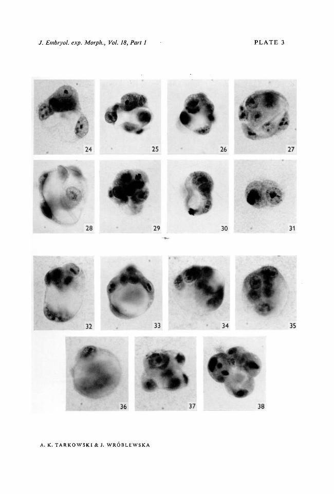
PLATE 3
Figs. 24-31. Eight sister forms developed from one egg.Fig. 24. A 7-cell blastocyst (1/8). The inner mass is covered from the top by one tropho-blastic cell.Fig. 25. A 10-cell false blastocyst (1/8).Fig. 26. A 10-cell false blastocyst (1/8).Fig. 27. An 8-cell false blastocyst (1/8). The false inner mass is seen from above.Fig. 28. A 7-cell trophoblastic vesicle (1/8).Fig. 29. A 10-cell morula (1/8). One cell contains numerous vacuoles.Fig. 30. A 10-cell non-integrated form composed of three vesicles (1/8). Only the biggestvesicle resembling a blastocyst is pictured.Fig. 31. A 2-cell form (1/8) with three nuclei. Small vacuoles are present in both cells.Figs. 32-38. Seven sister forms developed from one egg.Fig. 32. An 8-cell blastocyst (1/8).Fig. 33. An 8-cell false blastocyst (1/8).Fig. 34. An 8-cell false blastocyst (1/8). It possesses two separate blastocoelic cavities.Fig. 35. A 6-cell false blastocyst (1/8). Four cells whose nuclei lie closely together (seen fromabove) constitute supposedly the false inner mass.Fig. 36. A 4-cell trophoblastic vesicle (1/8).Fig. 37. An 8-cell morula (1/8).Fig. 38. A non-integrated form composed of highly vacuolated cells. Eight pycnotic nuclei.
/. Embryol. exp. Morph., Vol. 18, Part 1 PLATE 3
25 26 27
28 29 30 31
32 33 34 35
36 37 38
A. K. TARKOWSKI & J. WROBLEWSKA

/. Embryol. exp. Morph., Vol. 18, Part 1 PLATE 4
47 50
A. K. TARKOWSKl & J. WROBLEWSKA

Mouse blastomere development 169some blastomeres into morulae or non-integrated aggregations of cells may beregarded as resulting from injuries inflicted during manipulations and/orsuboptimal conditions of culturing. It should be added that morulae wereobserved more often among 1/4 and 2/8 forms than among 1/8 forms. Accordingto the hypothesis that the mechanism underlying the formation of inner masscells and trophoblast is ooplasmic segregation, the reverse should be true.Another conclusion which can be drawn from the above data is that the typeof development of blastomeres is extremely variable, both among individualsets and among forms belonging to one set, and that it cannot be predicteda priori.
(7) Abnormal development of isolated blastomeres
Abnormal development displayed by some of the isolated blastomeres hasunexpectedly provided useful information about certain peculiarities of cleavagecells.
Tn normal development of the mouse the blastocoel begins to appear when the
PLATE 4
Figs. 39-45. Seven sister forms developed from one egg.Fig. 39. An 8-cell blastocyst (1/8).Fig. 40. An 8-cell blastocyst (1/8).Fig. 41. An 8-cell blastocyst (1/8).Fig. 42. A 6-cell blastocyst (1/8) with two cavities separated by a bridge composed of fourcells. These cells would presumably have become the inner mass had the two cavities joinedtogether.Fig. 43. An 8-cell false blastocyst (1/8).Fig. 44. A 6-cell false blastocyst (1/8).Fig. 45. A 6-cell irregular non-integrated form with all cells highly vacuolated.Fig. 46. A 10-cell false blastocyst (1/8). Six nuclei on one pole, two on the other.Fig. 47. A 7-cell non-integrated form (1/8) resembling at first sight a typical blastocyst. Infact this is a group of six cells (two enveloped and four enveloping) attached to one cellcontaining an enormous intracellular vacuole.Fig. 48. A 6-cell false blastocyst (1/8) with a false inner mass seen from above. The nuclei ofthe two other cells lie slightly below the plane of focusing.Fig. 49. An early 8-cell blastocyst (1/8). The inner mass cells are clearly covered from outsideby an enveloping cell.Fig. 50. An 8-cell trophoblastic vesicle (1/8).Fig. 51. A 4-cell morula (1/8) composed of one enveloped and three flattened envelopingcells.Fig. 52. A 2-cell vesicle (1/8).Fig. 53. A 1/8 blastomere which has not divided in culture and has chromosomes scatteredin the cytoplasm. Note numerous vacuoles in the cytoplasm.Fig. 54. An 11-cell aggregation of small vesicles developed from a pair of 1/8 blastomeres(2/8).Fig. 55. A non-integrated form (1/8) composed of four loosely connected cells, each con-taining one enormous vacuole.

170 A. K. TARKOWSKI & J. WR6BLEWSKA
the 5th cleavage division. Normally, the ability of the cleavage cells to secretethe blastocoelic fluid is not displayed before this stage. It would seem that acertain number of cleavages is required before the cells attain the state ofdifferentiation enabling them to undertake this physiological activity. Examina-tion of the development of isolated blastomeres shows that under unfavourableexperimental conditions, mitotic divisions (cleavages) and secretion of theblastocoelic fluid are not impaired to the same degree. Poor culture conditionsact in the first instance deleteriously on cell division; many blastomeres divideonly once or twice. However, most of the 4-cell forms originating from 1/4 and2/8 blastomeres have developed into regular trophoblastic vesicles and so didsome of the 2-cell forms originating from 1/8 blastomeres (Fig. 52). Theseblastomeres have undergone only four cleavage divisions and although the 5thcleavage has been inhibited the secretion of the blastocoelic fluid has not been.In one case a 1 /8 blastomere has been observed which had not divided at all andhad chromosomes scattered in a very highly vacuolated cytoplasm (Fig. 53).Vacuolization of the cytoplasm which in normal development represents avisible sign of secretory activity (see below) can, thus, be displayed by a blasto-mere which has not gone further than the third cleavage. Secretion of the blasto-coelic fluid seems, therefore, to represent a cytoplasmic activity which a cellundertakes after a certain definite period of time and irrespective of the numberof nuclear cycles. This puzzling phenomenon certainly merits further in-vestigation.
In histological preparations or in whole permanent mounts of normalblastocysts developing in vivo or in vitro small vacuoles can always be seen inthe trophoblastic cells. The contents of these vacuoles are presumably con-tinuously discharged into the intercellular spaces thus leading to the formationof the blastocoelic cavity and preventing the accumulation of large amounts offluid in the cytoplasm. In our experiments some of the blastomeres developedinto non-integrated forms composed of loosely attached cells or groups of cells.In such cases a single cavity could not, of course, be formed, the end productof development being a group of tiny vesicles (Fig. 54) or a group of highlyvacuolated cells (Fig. 55). It is quite understandable that the cells whose entireor nearly entire surface is exposed to the surrounding medium cannot secretethe synthesized fluid back into the environment and must accumulate it in thecytoplasm. Consequently, one enormous vacuole can be formed, the cytoplasmand the nucleus being pushed to the periphery of the cell (Figs. 19, 47, 55). Thecleavage cells can, thus, start to produce the fluid irrespective of whether theyadhere to each other or not and, consequently, whether they have or have not apossibility of discharging it from the cytoplasm. It would seem, therefore, thata prerequisite of a formation of a blastocyst from the whole egg or a vesicularform (of any type) from an isolated blastomere is a close adhesion of cleavagecells and, consequently, the creation of an intercellular environment. Once theegg has about 32 cells, i.e. when all or nearly all blastomeres have completed

Mouse blastomere development 171
opposing surfaces of the cells become exposed to different conditions (inter-cellular versus external) the cells themselves must inevitably become polarizedand can start secreting the blastocoelic fluid into the interior of the aggregation.
DISCUSSION
The results of the experiments described above made it clear that the inter-pretation proposed by one of us (Tarkowski, 1959 a, b) and by Mulnard (1965 a)to explain the development of 1/2 blastomeres cannot be extended to 1/4 and1/8 blastomeres and that a completely new outlook on mechanisms governingearly development and responsible for divergent differentiation of cleavage cellsin an embryonic (inner mass) or an extra-embryonic (trophoblast) direction isneeded.
In their work on the development of 1/2 blastomeres of the mouse bothTarkowski and Mulnard interpreted the results in the light of conceptionselaborated by Dalcq, Mulnard and their co-workers (for reviews see Dalcq,(1957) and Mulnard (1961)). On the basis of cytochemical studies on earlymammalian development, the Belgian authors postulate that the uncleaved eggis characterized by polarity and bilateral symmetry and that the cytoplasm fromthe 'dorsal' and 'ventral' zones of the egg is segregated during cleavage untilit is in the cells of inner mass and trophoblast, respectively. Both Tarkowski andMulnard suggested that the great variation in the size of the inner mass (numberof cells) displayed by blastocysts developed from 1/2 blastomeres was due tovariable position of the first cleavage in relation to the plane of bilateral sym-metry and, consequently, to variable and unequal distribution of cytoplasmfrom each zone to the daughter blastomeres. Multicellular morulae and tro-phoblastic vesicles were considered as representing the extremes of this scaleof variation and originating from eggs in which this distribution was extremelyunequal, i.e. the plane of cleavage deviated very considerably from the plane ofsymmetry.
According to a segregation hypothesis, in a random sample of 1/2 forms onewould expect to encounter in similar proportions forms in which trophoblasticcells predominate and forms composed mainly of 'dorsal' cytoplasm, i.e.morulae or very poorly cavitated blastocysts. However, this was not found tobe true. Multicellular morulae were observed very rarely and the blastocysts, incomparison with control blastocysts developing from whole eggs, are charac-terized by a marked preponderance of trophoblastic cells over inner mass cells.In this situation Tarkowski (19596) had to propose an auxiliary hypothesis,namely that when 1/2 blastomeres develop in isolation the formation oftrophoblastic cells is privileged and occurs at the expense of the 'dorsal'material.
However, the results of the present work and the recent findings by Mulnard(19656) invalidate the above interpretation. Mulnard described the develop-

172 A. K. TARKOWSKI & J. WR6BLEWSKA
ment of trophoblastic vesicles from two blastomeres separated from each otherat the 2-cell stage. If it were true that the fate of cytoplasm occupying differentregions of the uncleaved egg is already determined, then the two sister blasto-meres should develop into forms displaying complementary structure. This wasnot the case. Mulnard himself is very cautious in considering these results asevidence against the conception that ooplasmic segregation plays an importantrole in early mammalian development. He is inclined to think that the conditionsof the experiment itself (separation of blastomeres, sticking of naked blastomeresto the glass, and so on) might modify and disturb the development of blasto-meres and lead to the origin of purely trophoblastic structures from the cyto-plasm of the whole egg. Although such a caution is certainly justified it seemsdoubtful whether his results could be ascribed only to the technical inade-quacies of the experiment. It is worth stressing that formation of trophoblasticvesicles from 1/2 blastomeres does not occur only for naked blastomeres buthas been also observed among blastomeres left inside zona pellucida anddeveloping both in vivo (Tarkowksi, 1959 a, b) and in vitro (Mulnard,1965/a).
According to Dalcq and Mulnard's conceptions one would expect that formsdeveloped from blastomeres isolated at later stages should also display a com-plementary structure and should deviate more drastically from the normal typeof development, i.e. from a blastocyst developed from the whole egg. However,in our experiments devoted to stages IV and VIII such a phenomenon has notbeen observed. The majority of blastomeres isolated at these two stages developinto vesicular forms in which all, or the bulk of, cells behave like trophoblasticcells. Among forms developed from 1/4, 2/8, and 1/8 blastomeres the incidenceof morulae is very similar and ranges about 10 %. If development into morulaeresults from the intrinsic properties of certain blastomeres then the incidenceof these particular forms should be higher among 1/8 forms than among 1/4and 2/8 forms because of a more advanced segregation of the cytoplasm of theegg among individual blastomeres. One could also expect that 1/8 blastomeresshould develop into forms representing extremes of the scale of variation, suchas morulae and trophoblastic vesicles, while intermediate forms like falseblastocysts and blastocysts would be very rare or lacking altogether. The picturewe observe is not in accord with these expectations. If one agrees that morulaerepresent badly developing forms and that the occurrence of non-integratedforms also results from poor culture conditions (or injury during manipulations),then it can be suggested that all blastomeres at the 4- and 8-cell stage have anability to develop into regular vesicular forms (types I, II or III). Such a con-clusion seems to be well corroborated by the fact that in several cases all blasto-meres of the egg followed this type of development.
On the ground of the results of our present work as well as of those of otherauthors we want to put forward a hypothesis which seems to explain satis-factorily both normal development (or, strictly speaking, the mechanisms

Mouse blastomere development 173responsible for the formation of a blastocyst and the differentiation of blasto-meres into inner mass and trophoblastic cells), and the development of blasto-meres isolated at the 2-, 4- and 8-cell stage.
When a mouse egg passes from morula to blastocyst it is composed of about30 cells. A certain number of these cells occupy the interior of the morula andare completely separated from outside by other cells. The latter become flattenedand represent predecessors of the trophoblastic cells. It seems reasonable toassume that the conditions in which external and internal cells find themselvesare diametrically different. The internal cells, being completely cut off from theexterior, develop in a micro-environment created by the external cells. In ouropinion, the position of a cell in the morula and, a consequence of the position,the different environmental conditions play a decisive role in the differentiationof cells in one of the two directions (trophoblast versus inner mass). For theformation of the inner mass it is necessary that certain blastomeres shouldbecome isolated from the exterior before the moment when blastocoelic fluidstarts to accumulate between the cells.
As far as development of isolated blastomeres is concerned, the later the stagefrom which the blastomere has been taken, the smaller the number of cells at thetime of cavitation. Consequently the number of blastomeres which can becomeenveloped by other blastomeres is more and more reduced. While in the caseof a 1/2 blastomere the number of cells in the morula at the beginning ofblastulation is still large enough for the formation of nearly normal blastocystsin the majority of cases (although the number of embryonic cells is clearlyreduced in comparison with blastocysts developing from the whole egg),development of blastocysts from 1/4 and 2/8 blastomeres takes place only inabout 30-40 % of cases, and from 1/8 blastomeres only in about 15 % of cases.This decrease in the incidence of blastocysts is accompanied by an increase inthe incidence of trophoblastic vesicles and false blastocysts. It would seem,therefore, that all blastomeres at the 4- and 8-cell stage possess the ability todifferentiate into trophoblastic cells. Differentiation into inner mass cells wouldnot be inherent in any blastomere but would represent an alternative route ofdevelopment requiring an intercellular environment. Putting this the other wayround, up to the 8-cell stage any blastomere of the egg can give rise to innermass cells if at the proper time the descending cells find themselves shielded byother cells.
It is worthwhile to refer in this connexion to the experiments carried out bySeidel (1956, 1960), who found that in the rabbit trophoblastic vesicles developmore often from 1/4 than from 1/2 blastomeres. This observation confirms ourresults obtained with mouse eggs and can be explained in a similar way. Seidel'smaterial consisted of blastomeres left inside zona pellucida and developing invivo. Neither lack of zona nor development in vitro can be made responsible,therefore, for the results which we have obtained in our experiments.
An experimental approach therefore provides evidence that at least up to the

174 A. K. TARKOWSKI & J. WROBLEWSKA
8-cell stage the fate of blastomeres is still labile and that it becomes fixed at laterstages. Mintz (1964a, b, 1965) came to a similar conclusion on the basis of herexperiments on fusion of eggs and claims that this lability extends to even laterstages (see also Tarkowski, 1965). She has shown that when morulae are fusedtogether there is no selective sorting out of cells in the resulting aggregationsand, consequently, those of the originally external cells which occupy the' sticking surface' become secondarily internal and differentiate into inner masscells.
It is rather strange that the precocious cytochemical differentiation of themammalian egg, described by Belgian authors, should play a decisive morpho-genetic role in normal development and have no effect upon development ofisolated blastomeres. However, it should be stressed in this connexion that inspite of the fact that many interesting and important data on cytochemistry ofmammalian eggs and early stages of development have been accumulated, clear-cut evidence of cytoplasmic continuity between different regions of the uncleavedegg and the components of the resulting blastocyst is very limited. Perhaps themost convincing proof in this respect is provided by studies of Mulnard (1955)on acid phosphatase in early development of the rat. However, using the samemethods the above author was unable to obtain similar results with the mouse,in which acid phosphatase cannot be demonstrated until the time when the eggenters the fourth cleavage division (Mulnard, 1965a). Increase in the contentand/or activity of this enzyme which from this stage on is found in the internalcells may be equally well interpreted as a sign and result of their differentiationcaught in statu nascendi. Although some technical modifications introducedrecently by Dalcq (1966) allowed him to reveal acid phosphatase in oocytes andearly cleavage stages of the mouse, these investigations too do not provide aclear picture of the continuity of 'dorsal' and 'ventral' cytoplasm throughcleavage and thus do not fill the gap between the uncleaved egg and blastocyst.In the opinion of the present authors, the evidence in favour of ooplasmicsegregation in mammals is not conclusive, at any rate not as far as the mouse isconcerned.
Appearance of bilateral symmetry in mammalian development as early as inthe oocyte, as postulated by Dalcq & Mulnard, seems to the present authorsvery strange and problematical. Why should the mammalian egg display sucha type of symmetry? Early appearance of bilateral symmetry is understandablein the case of amphibians or other animals in which the plane of symmetry ofthe egg becomes the plane of symmetry of the embryo. However, even in theamphibian egg the visible signs of bilateral symmetry do not appear until afterfertilization. In mammals, where early development is subordinated, first of allto the elaboration of the embryonic and extra-embryonic material and where theembryo proper is formed much later from the part of the embryonic shield, thebilateral symmetry of the egg would have nothing in common with the bilateralsymmetry of the embryo itself. One wonders at the biological sense of an early

Mouse blastomere development 175
appearance of bilateral symmetry in the mammalian egg, a symmetry of veryshort duration and of no meaning in later development.
Another aspect of the structure of the egg is its polarity. This is an universalpeculiarity of all eggs and it is sound to assume a priori that the cytoplasm of themammalian egg also displays some differences along the polar axis. Has thisgradient any influence on the fate of individual blastomeres originating fromdifferent llevels' of the egg? There is no evidence either for or against such, adependency simply because in isolation experiments we do not know from whatpart of the egg a given blastomere originates. From our experiments it is evidentthat up to the 8-cell stage each blastomere can develop into a vesicular form. Itis only the structure of the vesicle (trophoblastic vesicle, false blastocyst,blastocyst) that is variable. What causes the development of a given blastomereinto one or another type of vesicle ? Is it possible that the variable structure of theresulting forms is due to cytoplasmic inequality of blastomeres associated withthe polar differentiation of the egg ? At present we are not able to offer a con-clusive answer to this question, the available evidence speaking rather againstsuch a dependency.
A normal or a false blastocyst developed from an isolated blastomere, as wellas a blastocyst developed from a whole egg, display polarity, as on one polethere is an inner mass. This polarity manifests itself for the first time when asmall cavity (or cavities) appears on one side of the morula. Is this polarityprimary or secondary? In the case of a blastocyst developed from a whole eggit would be tempting to assume that this polarity develops against a back-ground of a pre-existing primary polarity of the egg. However, there is noevidence either for of against such an assumption. The only trace of the animalpole is the position of the second polar body. It is very doubtful, however,whether at this stage the polar body still occupies its original position; very oftenit is no longer present. No data are available on the relation between the polaraxis of the blastocyst and the polar axis of the undivided egg and speciallydevised experiments would be necessary to elucidate this question.
A blastocyst developed from an isolated blastomere imitates a blastocystdeveloped from a whole egg and in its formation similar mechanisms must beinvolved to those of normal development. A trophoblastic vesicle does notdisplay any polarity. This type of vesicle must originate from a group of cellswhich at the moment when they start secreting blastocoelic fluid through theirinnermost surfaces are exposed to the exterior to the same degree. With theincrease of the cavity the cells are pushed out to the peripheries of the aggrega-tion, which thus becomes a trophoblastic vesicle. Cell divisions which followcan only increase the number of cells constituting the wall of the vesicle butcannot lead to the formation of the inner mass. The most interesting and obscuresituation is represented by a false blastocyst (type II) which like a normalblastocyst displays also polarity. In these forms some of the cells, though theycontribute to the wall of the vesicle, are packed more tightly together and are

176 A. K. TARKOWSKI & J. WR6BLEWSKA
less distended than the remaining ones. It may be that the shape and behaviourof these cells is a result of their lower activity in secretion of the blastocoelicfluid. One could speculate that when the cavity first appeared in these forms,the cells, or just a cell, in question were partially covered by other cells andconsequently their character and behaviour became ultimately intermediatebetween the external (trophoblastic) and internal (inner mass) cells. This seemsa probable, although completely hypothetical explanation of the origin of suchforms.
This proposed origin of trophoblastic vesicles, false blastocysts and blasto-cysts would imply that at the time when blastocoelic fluid starts to accumulatethe forms are composed of differing numbers of cells—the lowest in presumptivetrophoblastic vesicles, the highest in presumptive blastocysts. If a blastomere isable to divide into a relatively high number of cells before starting secretoryactivity, the possibility that some cells will become enveloped completely byothers is increased. If at this crucial moment the number of cells is still low atrophoblastic vesicle will inevitably be formed. No data on the cell number at thetime of cavitation are available. However, we know the cell number of alreadydeveloped forms. As can be seen from Tables 1, 2 and 3 in forms originatingfrom 2/8 and 1/8 blastomeres the average cell number is lowest in trophoblasticvesicle, higher in false blastocyst and highest in blastocysts, which confirms ourhypothesis. Among 1/4 forms the average cell number in the three types ofvesicles is similar. However, this sample is small, hence the significance ofaverages is limited.
The crucial problem of why blastomeres develop into one of the three typesof vesicles remains unresolved. We are inclined to think that the factors involvedare twofold: first, the size of blastomere related to the stage of development atwhich it is separated and, secondly, some unknown factors, varying from experi-ment to experiment, in treating and culturing the blastomeres. The differencesin the development of blastomeres originating from different developmentalstages can be explained satisfactorily in the way proposed by us. Variation in thestructure of forms originating from the same stage is, however, more difficultto elucidate. Evidence that external rather than internal (i.e. primary differencesin developmental capacities) factors play a major role is provided by the obser-vation that while in some cases all or nearly all sister blastomeres develop intotrophoblastic vesicles, in others blastocysts predominate. Generally speakingthe incidence of various types of vesicles among sister blastomeres is not con-stant and changes from egg to egg.
SUMMARY
1. Mouse eggs at stages from 4- to 8-cell were disaggregated into individualblastomeres; the blastomeres were subsequently cultured in vitro for 36-48 h.
2. The material collected consisted of 42 forms developed from 1/4 blasto-meres, 80 forms developed from pairs of 1/8 blastomeres (2/8 blastomeres) and

Mouse blastomere development 111144 forms developed from 1/8 blastomeres. In 17 cases development of all sisterblastomeres constituting the egg has been observed.
3. The majority of 1/4, 2/8 and 1/8 blastomeres develop into vesicular forms.Three types of vesicles have been discerned: (a) trophoblastic vesicle, a purelytrophoblastic structure deprived of the inner mass cells; (b) false blastocyst, atrophoblastic vesicle whose wall is thicker on one pole (false inner mass);(c) blastocyst, a small copy of a blastocyst developed from the whole egg.
4. Development of some blastomeres into irregular non-integrated forms orinto morulae results presumably from harmful treatment during manipulationsand/or suboptimal conditions during culturing.
5. Incidence of blastocysts decreases from about 40 % among forms developedfrom 1/4 blastomeres to about 30 % among 2/8 forms and to about 15 % among1/8 forms. This decrease is accompanied by a slight increase in the incidence offalse blastocysts and a very marked increase in the incidence of trophoblasticvesicles.
6. A hypothesis is formulated according to which the differentiation of blasto-meres into trophoblastic and inner mass cells is achieved epigenetically anddepends in the first instance on the position occupied by blastomeres in themorula. Up to the 8-cell stage all blastomeres possess the ability to differentiatein a trophoblastic direction. Differentiation into inner mass cells is not inherentand represents an alternative route of differentiation requiring an intercellularenvironment.
7. With the decreasing size of the blastomere at the time of separation (i.e.advancing stage of development) the number of cells attained by it by the time ofcavitation decreases. Consequently the number of cells which can become en-veloped by other cells becomes smaller and smaller. This leads to the decreasein the size of inner mass and finally, in extreme cases, to the development ofpurely trophoblastic vesicles.
8. It remains obscure why blastomeres originating from the same stagedevelop either into trophoblastic vesicles or into false blastocysts or into blasto-cysts. The available evidence does not suggest the existence of a primarydevelopmental inequality of blastomeres and seems to indicate that the type ofdevelopment is governed to a great extent by some unknown conditions of cultur-ing, varying from experiment to experiment. It seems that a decreasing rate ofcleavages increases the chances of development of trophoblastic vesicles.
9. Production of blastocoelic fluid is a cytoplasmic activity which a cleavagecell undertakes after a certain definite period of time and irrespective of thenumber of the nuclear cycles. Suppression of cleavage does not necessarilylead to the suppression of this activity. If the cleavage cells do not adhere closelyto one another blastocoelic fluid cannot be discharged from the cells andaccumulates in large amounts in the cytoplasm.

178 A. K. TARKOWSKI & J. WROBLEWSKA
RESUME
Developpement de blastomeres (Vceufs de souris, isoles auxstades a 4 et 8 blastomeres
1. Des oeufs de souris, aux stades 4 a 8 blastomeres, ont ete desagreges et lesblastomeres isoles ont ete ensuite cultives in vitro pendant 36 a 48 h.
2. Le materiel obtenu consistait en 48 germes provenant de blastomeres 1/4,80 germes provenant de paires de blastomeres 1/8 (2/8), et 144 germes provenantde blastomeres 1/8. Dans 17 cas, on a observe le developpement de tous lesblastomeres freres formant l'ceuf.
3. La majorite des blastomeres 1/4, 2/8 et 1/8 se developpent en formesvesiculaires. On a distingue trois types de vesicules: (a) vesicule trophoblastique— structure purement trophoblastique privee des cellules de la masse interne;(b) faux blastocyste, — une vesicule trophoblastique dont la paroi est plusepaisse a un pole (fausse masse interne); (c) blastocyste — une copie reduited'un blastocyste provenant d'un oeuf entier.
4. Le developpement de quelques blastomeres en germes irreguliers nonintegres, ou en morulas, resulte probablement d'un traitement lesant au coursdes manipulations et (ou) de conditions sub-optimales au cours de la culture.
5. La proportion de blastocystes obtenus s'abaisse depuis 40 % environ parmiles germes developpes a partir de blastomeres 1/4 jusqu'a environ 30 % parmiles germes 2/8 et environ 15% parmi les germes 1/8. Cette diminution estaccompagnee d'un leger accroissement de la frequence des faux blastocystes etd'un accroissement tres marque de celle des vesicules trophoblastiques.
6. On formule une hypothese selon laquelle la differenciation des blasto-meres en cellules trophoblastiques et internes est epigenetique et depend enpremier lieu de la position occupee par les blastomeres dans la morula. Jusqu'austade 8, tous les blastomeres possedent la capacite de se differencier dans le senstrophoblastique. La differenciation en cellules de la masse interne n'est pasintrinseque et represente un mode alternatif de differenciation necessitant unmilieu intercellulaire.
7. Au fur et a mesure que diminue la taille du blastomere au moment de sonisolement (c'est-a-dire que le stade de developpement est plus avance) le nombrede cellules qu'il comporte au moment de creusement diminue. En consequence,le nombre des cellules qui peuvent etre enveloppees par d'autres cellules devientde plus en plus faible. Ceci mene a la diminution de la taille de la masse interneet finalement, dans les cas extremes, au developpement de vesicules purementtrophoblastiques.
8. On ne sait pas pourquoi des blastomeres issus du meme stade se develop-pent soit en vesicules trophoblastiques, soit en faux blastocystes, ou encore enblastocystes. Les resultats acquis ne suggerent pas l'existence d'une inegaliteembryogenique primaire des blastomeres et semblent indiquer que le type dedeveloppement est dirige a un degre eleve par quelques facteurs de culture

Mouse blastomere development 179inconnus, variables d'une experience a l'autre. II semble qu'un taux de segmenta-tion en diminution augmente les chances de developpement de vesiculestrophoblastiques.
9. La production de liquide blastocelien represente une activite cytoplasmi-que qu'entreprend une cellule de segmentation apres un certain laps de temps,defini, et sans rapport avec le nombre de cycles nucleaires. La suppression desclivages ne conduit pas necessairement a la suppression de cette activite. Si lescellules de segmentation n'adherent pas etroitement l'une a l'autre, le liquideblastocelien ne peut pas etre rejete des cellules et s'accumule en grandes quantitesdans le cytoplasme.
REFERENCESBRINSTER, R. L. (1963). A method for in vitro cultivation of mouse ova from two-cell to
blastocyst. Exp. Cell Res. 32, 205-8.DALCQ, A. M. (1952). L'oeuf des Mammiferes comme objet cytologique (avec une technique
de montage in toto et ses premiers resultats). Bull. Acad. r. Med. Belg 6e Serie 17, 236-64.DALCQ, A. M. (1957). Introduction to General Embryology, pp. 103-28. London: Oxford
University Press.DALCQ, A. M. (1966). Detection des enzymes de dephosphorylation dans les oeufs de rat et de
souris fixes au formol et traites in toto. Arch. Biol. {Liege), 77, 205-344.MINTZ, B. (1962). Experimental study of the developing mammalian egg: removal of the zona
pellucida. Science 138, 594-5.MINTZ, B. (1964a). Synthetic processes and early development in the mammalian egg. / . exp.
Zool. 85-100.MINTZ, B. (19646). Formation of genetically mosaic mouse embryos, and early development
of 'lethal (/12/12)-normal' mosaics. J. exp. Zool. 157, 273-92.MINTZ, B. (1965). Experimental genetic mosaicism in the mouse. In Preimplantation Stages
of Pregnancy. A Ciba Foundation Symposium (ed. G. E. W. Wolstenholme and M.O'Connor), pp. 194-207. London: J. and A. Churchill.
MULNARD, J. (1955). Contribution a la connaissance des enzymes dans l'ontogenese. Lesphosphomoncesterases acide et alcaline dans le developpement du Rat et de la Souris.Arch. Biol. {Liege), 66, 525-685.
MULNARD, J. (1961). Problemes de structure et d'organisation morphogenetique de l'oeufdes Mammiferes. In Symposium on the Germ Cells and Earliest Stages of Development,pp. 639-88. Milan: Fondazione A. Baselli, Instituto Lombardo.
MULNARD, J. (1965 a). Aspects cytochemiques de la regulation in vitro de l'oeuf de souris apresdestruction d'un des blastomeres du stade II. 1. La phosphomoncesterase acide. Bull.Acad. r. Med. Belg. 2e serie, 5, 31-67.
MULNARD, J. (19656). Studies of regulation of mouse ova in vitro. In Preimplantation Stagesof Pregnancy. A Ciba Foundation Symposium (ed. G. E. W. Wolstenholme and M.O'Connor), pp. 123-38. London: J. and A. Churchill.
NICHOLAS, J. S. & HALL, B. V. (1942). Experiments on developing rats. II. The develop-ment of isolated blastomeres and fused eggs. / . exp. Zool. 90, 441-59.
SEIDEL, F. (1952). Die Entwicklugspotenzen einen isolierten Blastomere des Zweizellen-stadiums im Saugetierei. Naturwissenschaften 39, 355-6.
SEIDEL, F. (1956). Nachweis eines Zentrums zur Bildung der Keimscheibe im Saugetierei.Naturwissenschaften 43, 306-7.
SEIDEL, F. (1960). Die Entwicklungsfahigkeiten isolierter Furchungszellen aus dem Ei desKaninchens Oryctolagus cuniculus. Roux Arch. EntwMech. 152, 43-130
SEMBRAT, K. (1955). Studies on the cleavage of the rat eggs. Zoologica Poloniae 6,139-84.
TARKOWSKI, A. K. (1959a). Experiments on the development of isolated blastomeres ofmouse eggs. Nature, Lond. 184, 1286-7.

180 A. K. TARKOWSKI & J. WR6BLEWSKA
TARKOWSKI, A. K. (19596). Experimental studieson regulation in the development of isolatedblastomeres of mouse eggs. Acta Theriologica 3, 191-267.
TARKOWSKI, A. K. (1961). Mouse chimaeras developed from fused eggs. Nature, Lond. 190,857-60.
TARKOWSKI, A. K. (1963). Studies on mouse chimeras developed from eggs fused in vitro.NCI Monogr. 11, 51.-71.
TARKOWSKI, A. K. (1965). Embryonic and postnatal development of mouse chimeras. InPreimplantation Stages of Pregnancy. A Ciba Foundation Symposium (ed. G. E. W.Wolstenholme and M. O'Connor), pp. 183-93. London: J. and A. Churchill.
TARKOWSKI, A. K. (1966). An air-drying method for chromosome preparations from mouseeggs. Cytogenetics 5, 394-400.
{Manuscript received 15 February 1967)