development of fungal leather-like material from bread waste
TRANSCRIPT

Development of Fungal Leather-like Material from Bread Waste
Final Submission (2021.06.10)
Master Programme in Resource Recovery
Industrial Biotechnology
Egodagedara Ralalage Kanishka Bandara Wijayarathna

i
MAIN INFORMATION
Programme: MSc degree in Resource Recovery in major of Industrial
Biotechnology
Swedish title: Utveckling av svampläderliknande material från bröd svinn
English title: Development of fungal leather-like material from bread
waste
Year of publication: 2021
Authors: Egodagerada Ralalage Kanishka Bandara Wijayarathna
Supervisor: Main-supervisor - Akram Zamani, Co-supervisor - Amir
Mahboubi Soufiani.
Examiner: Dan Åkesson
Keywords: Leather, Fungal leather, Leather-like material, Food waste,
Bread waste, Fungal material, Sustainable material, Filamentous fungi

ii
ABSTRACT
Food waste and fashion pollution are two of the significant global environmental issues
throughout the recent past. In this research, it was investigated the feasibility of making a
leather-like material from bread waste using biotechnology as the bridging mechanism. The
waste bread collected from the supermarkets were used as the substrate to grow filamentous
fungi species Rhizopus Delemar and Fusarium Venenatum. Tanning of fungal protein fibres
was successfully performed using vegetable tanning, confirmed using FTIR and SEM images.
Furthermore, glycerol and a biobased binder treatment was performed for the wet-laid fungal
microfibre sheets produced. Overall, three potential materials were able to produce with tensile
strengths ranging from 7.74 ± 0.55 MPa to 6.92 ± 0.51 MPa and the elongation% from 16.81
± 1.61 to 4.82 ± 0.36. The binder treatment enhanced the hydrophobicity even after the glycerol
treatment, an added functional advantage for retaining flexibility even after contact with
moisture. The fungal functional material produced with bread waste can be tailored
successfully into leather substitutes using an environmentally benign procedure.

iii
ACKNOWLEDGEMENT
Studying biotechnology was a real challenge that I faced in my life starting from scratch with
zero biology knowledge. Before anything, I must thank Amir Mahboubi Soufiani for those
interesting 4 lectures on bio-ethanol which planted the first seed of interest. Then, it was Jorge
Ferreira and Agneta Hultstrand who clearly and simply taught the basics of biology and finally,
Mohammad Taherzadeh and Akram Zamani for teaching the complex analysis parts in the bio-
process design.
Regarding the thesis, the first thank goes to Mohammad Taherzadeh for encouraging me to mix
my previous education, textile engineering knowledge with biotechnology and giving me the
“Tip” on the project “Sustainable Fungal Textile”.
This thesis project was conducted as a part of the project “Sustainable Fungal Textiles, a new
approach for re-use food waste” which is financed by VINNOVA, Sweden.
I should say how lucky I am to have Akram Zamani as my supervisor. From the first email I
sent her about asking for a thesis, up to date she was there every single time I wanted her help.
I never expected a supervisor to be this friendly and accessible. So, thank you so much Akram,
hands down the best supervisor one can have. I would also like to thank Amir again for being
an extra source of knowledge and ideas for this project and for helping present the research
work both in the thesis and the manuscript. I also take the opportunity to thank my examiner
Dan Åkesson for the valuable comments and guidance received to develop the research work.
My sincere gratitude goes to all the lab technicians Marlen, Jonas, Ville, Kristina, and Haike
from the textile department for timely attending to all the needs of my lab work. Also, a big
thank goes to everyone who worked in the labs for all the sharing and caring helps. Special
thank goes to Sajjad for helping me on the 4l reactor cultivations, Taner and Neda for helping
on 26l reactor cultivations, Sofie and Mohsen for helping on HPLC work, Clarrise for all the
tips given throughout the lab work, Faranak for all the bits of help on polymer analysis and
lastly Jorge for that awesome cultivation of 1.3m3 bioreactor. Both myself and my friend
Ghasem were lucky enough to be the last students trained by Jorge on bioreactor cultivations.
Big love goes to my buddy Ghasem for being an understanding friend and for all the cherished
moments we had both in and outside the labs. A huge thank goes to Sneha for being such a nice
companion throughout the thesis, especially in the polymer lab.
Finally, my loving wife Nathaliya, thank you for your unconditional love, motivation and
positive thoughts on the thesis which showered on me every single day. None of this would be
possible without you being my backbone. A huge love goes to my daughter Tenuki and son
Keymitha especially for baring my absences and for watching videos on fungi and
microorganisms with me and sharing your interesting thoughts with me.
The scanning electron microscopy (SEM) was performed by Karin Adolfsson from the KTH
Royal Institute of Technology, Stockholm.

iv
TABLE OF CONTENTS
MAIN INFORMATION ............................................................................................................. i
ABSTRACT ............................................................................................................................... ii
ACKNOWLEDGEMENT ....................................................................................................... iii
LIST OF FIGURES .................................................................................................................. vi
LIST OF TABLES ................................................................................................................. viii
1. INTRODUCTION ................................................................................................................. 1
1.1 BACKGROUND AND PROBLEM DESCRIPTION ......................................................... 1
1.2 PURPOSE AND LIMITATIONS........................................................................................ 1
1.3 LITERATURE REVIEW .................................................................................................... 2
1.3.1 FOOD WASTE & BREAD WASTE ............................................................................ 2
1.3.2 FASHION INDUSTRY & LEATHER INDUSTRY .................................................... 3
1.3.3 FILAMENTOUS FUNGI ............................................................................................. 4
1.3.4 TANNINS ..................................................................................................................... 5
1.3.5 WET-LAID PROCESS ................................................................................................. 5
1.3.6 FUNGI RESEARCHED FOR FASHION INDUSTRY ............................................... 6
1.4 ETHICAL ASPECTS OF THE PROJECT.......................................................................... 7
2.MATERIALS AND METHOD .............................................................................................. 9
2.1 MATERIALS ................................................................................................................... 9
2.1.1 FUNGI ........................................................................................................................... 9
2.1.2 SUBSTRATE .............................................................................................................. 10
2.1.2.1 Dry Ground Bread .................................................................................................... 10
2.1.2.2 Wet Ground Bread ................................................................................................... 11
2.1.2.3 Hydrolysis of Bread ................................................................................................. 11
2.1.3 OTHER MATERIALS AND CHEMICALS .............................................................. 11
2.2 METHOD DESCRIPTION ............................................................................................ 12
2.2.1 CULTIVATION OF FUSARIUM VENENATUM ...................................................... 12
2.2.1.1 Preparation of agar plates for Fusarium Venenatum ................................................ 12
2.2.1.2 Assessment on Optimum Cultivation Conditions .................................................... 12
Experiment 1. The physical nature of the substrate ............................................................. 12
Experiment 2. The different sizes of sieving on dry ground bread ...................................... 13
Experiment 3. Effect on wet ground bread storage – -18°C and 4°C .................................. 14
Experiment 4. Effect on growth temperature ....................................................................... 14
Experiment 5. Effect on phosphorous as a nutrient supplement .......................................... 14
2.2.1.3 SCALING UP OF F. VENENATUM CULTIVATION ........................................... 15

v
Protocol for 4L reactor ......................................................................................................... 16
Protocol for 26 reactors ........................................................................................................ 19
2.2.2 CULTIVATION OF RHIZOPUS DELEMAR ............................................................ 21
2.2.3 BIOMASS PRE-TREATMENTS ............................................................................... 23
2.2.3.1 NaOH treatment ....................................................................................................... 23
2.2.3.2. Ca(OH)2 Treatment ................................................................................................. 23
2.2.3.3. Tannin treatment ..................................................................................................... 23
2.2.3.4 Ca(OH)2 and Tannin treatment ................................................................................ 24
2.2.4 MAKING FILMS FROM BIOMASS ........................................................................ 24
2.2.5 FILM DRYING PROCESS ........................................................................................ 25
2.2.6 POST-TREATMENTS OF FILMS ............................................................................ 26
2.2.6.1 Glycerol post-treatment ............................................................................................ 27
2.2.6.2 Biobased binder post-treatment ................................................................................ 27
2.3 ANALYSIS ON BIOMASS YIELD.............................................................................. 29
2.4 ANALYSIS ON BIOMASS CULTIVATION METABOLITES .................................. 29
2.5 TENSILE TEST ............................................................................................................. 29
2.6 WATER CONTACT ANGLE ....................................................................................... 30
2.7 FOURIER TRANSFORM INFRARED SPECTROSCOPY (FTIR) ............................ 30
2.8 THERMO GRAVIMETRIC ANALYSIS (TGA) ......................................................... 31
2.9 SCANNING ELECTRON MICROSCOPE (SEM) AND FLUIDSCOPE™
SCANNING (oCelloScope) ................................................................................................. 31
2.9.1 FLUIDSCOPE™ SCANNING (oCelloScope ANALYSIS) ...................................... 31
2.9.2 SEM ANALYSIS ........................................................................................................ 31
2.10 DENSITY MEASUREMENT ..................................................................................... 31
2.11 TESTING THE BEST ALTERNATIVES OF FILM PREPARATION ON F.
VENENATUM BIOMASS ................................................................................................... 32
2.12 MAKING A PRODUCT WITH THE FILMS ............................................................. 33
3. RESULTS AND DISCUSSION ................................................................................... 34
3.1 F. VENENATUM BIOMASS YIELD ANALYSIS WITH DRY WEIGHT ............. 34
3.2 CHANGES IN FUNGAL METABOLITES DURING FUNGAL CULTIVATION .... 35
3.2.2 RHIZOPUS DELEMAR CULTIVATION .................................................................. 38
3.3 ANALYSIS OF PROPERTIES OF R. DELEMAR FILMS ........................................... 39
3.3.1.1 Tensile tests of leather-like material ........................................................................ 42
3.3.2 WATER CONTACT ANGLE (θ°) ............................................................................. 47
3.3.3 FOURIER TRANSFORM INFRARED SPECTROSCOPY ...................................... 48

vi
3.3.5 SCANNIN ELECTRON MICROSCOPY (SEM) ...................................................... 50
3.3.6 FLUIDSCOPE™ ANALYSIS (oCelloScope) ............................................................ 51
3.4 FILMS MAKING WITH F. VENENATUM .................................................................. 52
3.5 OCELLOSCPE IMAGES OF F. VENENATUM BIOMASS ........................................ 53
3.6 ATTEMPT TO MAKE A PRODUCT WITH THE FILMS .......................................... 54
4. CONCLUSION .................................................................................................................... 55
5. PROPOSED FUTURE WORK ........................................................................................... 56
REFERENCES ........................................................................................................................ 57
LIST OF FIGURES
Figure 1 Asexual life-cycle of a typical Aspergillus species; Adapted from (Svanström,
2013). (Shows the life cycle of a filamentous fungus.) ............................................................. 4
Figure 2. Industrial wet-laid non-woven production process diagram (www.edana.org) ......... 6
Figure 3 The graphical expression on the experimental procedure of the laboratory work
carried out in the project. ........................................................................................................... 9
Figure 4: Grinding of dried bread using the rotary mill ........................................................... 10
Figure 5: Disk grinder used to make wet ground bread as substrate ....................................... 11
Figure 6: 4L bench top bio reactor ........................................................................................... 15
Figure 7: 26l bioreactor ............................................................................................................ 18
Figure 8: The 1.3m3 bioreactor (A) and the biomass of Rhizopus Delemar (B) harvested from
that............................................................................................................................................ 22
Figure 9: The process of biomass film making (A) concentration adjusted biomass and water
solution, (B) the vacuum funnel arrangement with the vacuum pump, (C) the solution is in
the top container, ready to pass through the filter membrane, (C) the prepared film on the
membrane ................................................................................................................................. 25
Figure 10: Film drying methods (glycerol bath (A), frame drying (B), freeze-drying (D),
Normal drying (D)) .................................................................................................................. 26
Figure 11: 20% glycerol bath ................................................................................................... 27
Figure 12: Application of the biobased binder Lotus® using the applicator ZUA 2000......... 27
Figure 13: Schematic presentation on thickness test positions ................................................ 30
Figure 14: 5A Dog bone shape (adapted from Appels et al. (2020)) ....................................... 30

vii
Figure 15: The suspension began to leak from the joint due to the pressure and clogged
membrane ................................................................................................................................. 32
Figure 16: Ethanol concentration change with the time (key: exp.= experiment) ................... 36
Figure 17: Glucose concentration change with the time (key: exp.= experiment) .................. 37
Figure 18: Sugar mix concentration change with the time (key: exp.= experiment) ............... 37
Figure 19: Rhizopus Delemar cultivation metabolites of 1.3 m3 Reactor ............................... 38
Figure 20: The ultimate tensile strength on drying methods (Key: No treatment = untreated
biomass, Tannin = Tannin pre-treated biomass, Ca(OH)2 + Tannin = Ca(OH)2 and tannin pre-
treated biomass, Lyocell + Tannin = Biomass and Lyocell composite treated with tannin,
Lyocell + Ca(OH)2 + Tannin = Biomass and Lyocell composite treated with both Ca(OH)2
and tannin, ND = Normal drying, ETOH + ND = Ethanol treated and normal drying, ND +
20& Gly = Normal dried films with glycerol post-treatment, ETOH + frame dry = Ethanol
treated with drying on frame) .................................................................................................. 40
Figure 21: Freeze dried sample with a popped air bubble ....................................................... 40
Figure 22 :The stretch % on drying method (Key: same as figure 20) .................................... 41
Figure 23: Comparison between 10% and 20% glycerol concentrations (Key- RD= Rhizopus
Delemar, ND= Normal drying) ................................................................................................ 42
Figure 24: Stretch % (Key: RD Neat = films from untreated biomass, RD Tannin = Films
from tannin treated biomass) ................................................................................................... 44
Figure 25: Tensile test results (Key: RD Neat = films from untreated biomass, RD Tannin =
Films from tannin treated biomass) ......................................................................................... 44
Figure 26: Elastic modulus of different samples (Key: RD Neat = films from untreated
biomass, RD Tannin = Films from tannin treated biomass) .................................................... 45
Figure 27: Stress strain curves plotted with raw data .............................................................. 46
Figure 28 : Young's modulus vs density plot of fungal films prepared using R. Delemar.
(Adapted from Granta design CES EduPack 2019 and (Appels et al., 2020)) ........................ 46
Figure 29: Water contact angle ((Key: RD Neat) = films from untreated biomass, RD Tannin
= Films from tannin treated biomass) ...................................................................................... 47
Figure 30: FTIR graphs of RD Tannin and RD Neat (Key: RD= Rhizopus Delemar) ........... 48
Figure 31: TGA graphs of RD Neat (A) and RD Tannin (B) .................................................. 49
Figure 32: SEM images of RD Neat at X900 (A-1), RD Neat at X400(A-2), RD Tannin at
X1000 (B-1), RD Tannin at X400 (B-2), RD Neat + Glycerol at X1000 (C-1), RD Neat +
Glycerol at X350 (C-2), RD Tannin + Glycerol at X1000 (D-1), RD Tannin + Glycerol at
X400 (D-2), RD Neat + Glycerol + Binder at X1000 (E-1), RD Neat + Glycerol + Binder at

viii
X300 (E-2), RD Tannin + Glycerol + Binder at X1100 (F-1), RD Tannin + Glycerol + Binder
at X400 (F-2)............................................................................................................................ 50
Figure 33: oCelloScope images of RD Neat (A) and RD Tannin (B) ..................................... 51
Figure 34: FV Tannin film after dried ..................................................................................... 52
Figure 35: RD Tannin films in glycerol bath (A) and after dried with glycerol treatments (B)
.................................................................................................................................................. 52
Figure 36: oCelloScope images of tannin treated F. Venenatum biomass, ground (A) and non-
ground (B) ................................................................................................................................ 53
Figure 37: Making a pouch using RD Tannin + Glycerol + Binder films ............................... 54
Figure 38: Making a coin wallet from RD Neat + Glycerol + Binder films ............................ 54
LIST OF TABLES
Table 1: The properties assessment on biobased binders received from Organoclick AB ...... 28
Table 2: Yield of F. Venenatum biomass in different substrates ............................................. 35
Table 3: Tensile strength, Elongation, Elastic Modulus and Density of the films prepared
(Key: RD – Rhizopus Delemar, Neat – Untreated biomass, Tannin – Tannin treated biomass,
Glycerol and Binder – film post treatments) ............................................................................ 43

1
1. INTRODUCTION
1.1 BACKGROUND AND PROBLEM DESCRIPTION
In 2011, the Food and Agriculture Organisation of the United Nations (FAO) published a report
mentioning that one-third of the food produced for human consumption is wasted or lost (FAO,
2011). In addition to that, in 2013, the same organisation mentioned that 1.6 G. Tonnes of food
is wasted in a year, which accounts for a carbon footprint of 3.3 G. Tonnes of CO2 equivalent
even without considering the land use for cultivation (FAO, 2013). Bakery products that
contain the highest fraction of bread hold a substantial portion of household food waste,
according to the Waste and Resources Action Plan (WRAP), which accounts for 800,000
tonnes per year in the UK (WRAP, 2009). At the same time, Brancoli et al. (2019) have shown
that 80,410 tonnes of bread are wasted in Sweden annually.
On the other hand, the fashion industry is one of the most polluting industries globally,
contributing to 5% of all landfill and 20% of all freshwater pollution (Fiber2Fashion, 2012).
Leather is one of the earliest thing humankind used to cover their bodies, and the leather
industry has evolved with the advancement of the fashion industry (Quilleriet, 2004). However,
the leather industry has a hidden dark side due to poorly maintained animal farms, the working
conditions in a tannery, and the harmful effluents of the tanning industry. Due to those factors,
leather alternatives have been in attraction over the recent past. Biobased and biodegradable
material as a leather alternative is a timely requirement for advancing the path towards circular
economy and sustainability.
When the global food waste issue and the demand for biobased leather alternatives, connected
through biological means, it can be recognised that fungi could be a promising solution. This
project aims to investigate the possibility of producing a material to be used as a leather
alternative, from filamentous fungi that will be grown on bread waste. Filamentous fungi are
known for their ability to produce different hydrolytic enzymes, making them the leading
supplier of enzymes for lignocellulosic hydrolysis (Parachin et al., 2011). In addition to that,
filamentous fungi can also produce compounds with antimicrobial activities (Svahn et al.,
2012). The fungal cell wall mainly contains linear polysaccharides such as chitin, chitosan and
glucan (Bartnicki-Garcia, 1968). Vegetable tannin is a plant-based, biodegradable compound
that could enhance the stability of fungal material just as the animal hide. Using these properties
and materials will be possible to give a solution for the ethical and environmental problems in
the leather industry.
1.2 PURPOSE AND LIMITATIONS
This project aims to find a solution to reduce food waste as well as leather industry issues while
checking the feasibility of using fungi to produce biobased leather-like materials. It is part of a
larger ongoing project at the University of Borås, which is called “sustainable fungal textiles,
a new approach for re-use of food waste”. If using materials made by fungal biomass in leather

2
applications will be successful, it will be a ground-breaking achievement towards sustainability
in the fashion industry.
However, complications and limitations are expected throughout this process. The following
questions were used as a guide to the research.
1. What will be the best condition for fungal (F. Venenatum) growth on bread waste?
2. Will the enzymatic hydrolysis of bread enhance the fungal growth?
3. Is it possible to produce a material with mechanical properties comparable to natural
leather using fungal biomass and the wet-laid process?
4. How can the properties of the outcome be enhanced to use it as a potential leather-like
material?
5. To check the possibility of producing a pilot product from the materials with the best
properties.
1.3 LITERATURE REVIEW
1.3.1 FOOD WASTE & BREAD WASTE
Food, food scarcity and food waste have become global issues throughout recent years. The
Food and Agriculture Organisation of the United Nations (FAO) continuously conducts
research and publishes information on food security and how to defeat hunger. Wasting food
plays a vital role when it comes to the battle against food scarcity. Most of the time, the wasted
food is entirely edible. Globally, one-third of the food produced for human consumption is
wasted or lost (FAO, 2011). The carbon footprint of food waste better explains the criticalness
of the issue. In 2007, the produced but not eaten food contributed to a 3.3 G tonne of CO2
equivalent and ranked as the third top greenhouse gas (GHG) emitter after the USA and China
(FAO, 2013).
As the Latin motto of FAO, “fiat panis” says, “let there be bread”, bread has become the most
widely consumed food all around the world (Lohman, 2019). Similar to the consumption,
wastage of bread has also increased with time. Annual bread waste in the UK is about 800,000
tonnes, and in Sweden, this is around 80,410 tonnes per year(WRAP, 2009;Brancoli et al.,
2019). Gmoser et al. (2019b) claim that 7-10% of the global bread production is wasted
annually. Bread waste has been used in several value addition projects especially using
microbiology. One such successful idea was to go back to the roots and use bread waste as a
substrate to produce beer (Bondloll, 2016). At the same time, in Scandinavian countries, bread
waste is used as a substrate for ethanol production as a fuel (Melikoglu and Webb, 2013).
However, the landfills are still where most of the bread waste ends (Melikoglu et al., 2013).
An array of research articles on upcycling bread waste by the cultivation of fungi have been
published recently. Few of them can be listed as new food products or protein supplements
(Gmoser et al., 2020), producing colour pigments (Gmoser et al., 2019b), bioethanol and
animal feed production(Nair et al., 2017). In addition to that, forming films using chitosan rich

3
fibrous cell walls of Rhizopus Delemar and investigating mechanical and chemical properties
of those films was carried out by Köhnlein (2020) during his thesis project at the University of
Borås.
1.3.2 FASHION INDUSTRY & LEATHER INDUSTRY
The fashion industry has become the second-largest polluter after the oil industry because of
concepts like fast fashion (Conca, 2015). In their report, The Boston Consulting Group (2017)
describes that in 2015 the global fashion industry has contributed 1715 million tonnes of CO2
emissions and 92 million tonnes of garments and textiles have become waste, mostly ended up
either incineration or landfills. Conca (2015) further describes that over 70 million trees are cut
annually to produce regenerated cellulose like viscose and rayon, while cotton is the largest
pesticide-consuming crop globally. Furthermore, Jacometti (2019) points out the clothing
production has doubled during the last two decades and the importance of sustainable practices
in the fashion industry.
Leather is the earliest apparel of humankind which has evolved significantly within the fashion
industry (Quilleriet, 2004). The tanner was a position in the social hierarchy and, his task was
to convert animal skins and hides into refined material to protect humans. According to The
Food and Agriculture Organisation of the United Nations (FAO), between 2012 and 2014, the
average annual production of bovine hides is 6.531 million tonnes which accounts for 6255
million USD (FAO, 2016). The leather manufacturing process consisted of more than 20 steps,
and harmful chemicals are used in some of those steps. The waste generated from tanneries
were classified as hazardous waste in May 1980 due to the presence of chromium (Cr), which
uses in modern tanning, lead (Pb) and sulphides (Chaney, 1983).
The other catastrophic side of the leather industry is animal farms and the hygienic conditions
of the employers in the leather processing industries. Corradini et al. (2016) describe the
world’s largest leather and meat producer (JBS, Brazil) as slaughters 100,000 cattle, 70,000
pigs and 25,000 lambs every day. The leather consumed as a luxury material comes from all
these animal farms, which uses more than 40% of the annual cereal production, which comes
from almost 1/3 of the 14 billion hectares of arable land available globally as feed (Corradini
et al., 2016). So, when the environmental impact is considered, the direct effects from tanneries
and the indirect effect from animal farms should be considered to understand the criticalness
of the current situation.
Due to these environmental problems, people tend to turn back towards the vegetable tannins,
which stabilise the animal hides and skins from putrefaction since ancient time. Due to the
ethical and environmental concerns of the leather industry and the increasing demand for
leather in fashion, there is a considerable research interest in leather alternatives throughout the
recent past (Jones et al., 2021).

4
1.3.3 FILAMENTOUS FUNGI
The tree of life describes two different cell types prokaryotes (single cellular) and eukaryotes
(single or multicellular) (Madigan, 2014). Prokaryotes further divide into two kingdoms as
bacteria and archaea, while Eukaryotes further split into five kingdoms: Protozoa, Chromista,
Fungi, Plantae, and Animalia (Ruggiero et al., 2015). Fungi are an ancient kingdom. Most fungi
reproduce asexually by spores and consist of tubular cells called hyphae (Carris et al., 2012).
They are heterotrophs1 and absorb nutrients directly via cell walls. Some fungi, which use
living organisms to obtain carbon and energy, are called biotrophs2 , and others who do the
same thing with dead organisms are called saprotrophs3 (Carris et al., 2012). Fungi have
divided into several groups known as phyla. The number of phyla and the classification changes
with time because the kingdom of fungi is under research throughout. According to the current
taxonomic arrangements of fungi, there are six different phyla namely, Ascomycota,
Basidiomycota, Blastocladiomycota, Chytridiomycota, Glomeromycota and
Neocallimastigomycota (Money, 2016a). Fungi have unicellular and multicellular organisms.
Yeast is a unicellular fungus, and multicellular fungi are called filamentous fungi.
Reproduction of filamentous fungi takes place with the use of microscopic particles known as
spores. Spores can be either sexual such as basidiospores, ascospores and zygospores or asexual
such as conidia and sporangiospores. These spores travel passively with the help of air and
water. Some fungi contain biomechanical mechanisms to release spores out (Money, 2016b)
and land on any surface. From there onwards, germination and fungal mycelium growth begin.
From the mycelium or the roots like a network of interconnected hyphae branches, more spores
will pop up. This is the life cycle of a filamentous fungus, as shown in figure 1.
Figure 1 Asexual life-cycle of a typical Aspergillus species; (Adapted from (Svanström,
2013)). (Shows the life cycle of a filamentous fungus.)
1 Heterotrophs are organisms which obtain carbon and energy from other organisms 2 & 3 Biotrophs and saprotrophs can be considered as two sections under heterotrophs

5
In this thesis, two specific fungus types are used, namely Rhizopus Delemar and Fusarium
Venenatum. Rhizopus Delemar, earlier a section of Rhizopus Oryzae (Abe et al., 2007) belongs
to the recently abandoned fungi phyla Zygomycota (Spatafora et al., 2016), and newly
categorized as in Mucoromycota. Fusarium Venenatum is already commercialized as a vegan
food Quorn™ (www.quorn.com/) with specific process improvements.
1.3.4 TANNINS
Tannins are the fourth most abundant compounds in plant cells after cellulose, hemicelluloses,
and lignin, which dominates the secondary metabolites of a plant (Bule et al., 2020). They are
polyphenols consist of carbon, hydrogen and oxygen. The role of tannins in a plant is to
maintain the defence mechanism from insects (Covington, 2009;Kochhar, 2016). Covington
(2009) further explains about three fractions of tannins extracted from trees.
1. Non-tans; molecular weight < 500
This fraction consists of low tanning power; however, useful in the penetration properties of
tannin due to high solubility.
2. Tans; 500 < molecular weight < 3000
The fraction is effectively used in the tanning process.
3. Gums; molecular weight > 3000
This fraction does not penetrate leather because of the high molecular weight. Using aggressive
conditions to increase the tannin extraction yield will be useless due to the increased extraction
of gums.
Tannins are used in the leather industry by utilizing their ability to bind and precipitate the
collagen proteins in animal hides or skin (Bule et al., 2020). This process converts the animal
hides to stable, smooth, flexible, and resilient towards heat and microorganisms’ leather. It is
a vast group of compounds, and they are divided into two main segments, hydrolysable tannins
and condensed tannins (Khanbabaee and van Ree, 2001).
Tannic acid (C76H52O46) (TA) is one such type of hydrolysable tannins which is known for
antimicrobial, antioxidant, anti-enzymatic and astringent properties (Zhou et al., 2014;Aelenei
et al., 2009). Aelenei et al. (2009) further describe that TA can be used as a medication for
diarrhoea. However, if consumed in larger quantities, TA would inhibit iron absorption since
it can reduce the effectiveness of the digestive enzymes (Chung et al., 1998). Nam et al. (2001)
has done an animal study to check the usage of TA as a treatment for cancer, and they have
shown the anticarcinogenic activities of TA towards chemically induced cancer.
1.3.5 WET-LAID PROCESS
The wet-laid technology is originated from paper production. Following four steps take place
in wed laid process (figure 4),
1. Fibres are mixed with water and make a homogeneous suspension.

6
2. Fibre suspension is laid on a perforated screen to form a sheet.
3. Excess water is drained, allowing fibres to arrange more tightly.
4. The sheet formed is heat dried, and fibres bonded either mechanically or chemically.
(Mao and Russell, 2015;Montefusco, 2005)
Figure 2. Industrial wet-laid non-woven production process diagram (www.edana.org)
In papermaking, commonly short length cellulose fibres are used where hydrogen bonds
dominate the bonding and inter fibre friction (Przybysz et al., 2016;Lindström et al., 2005).
The preferred fibre length in papermaking is between 1- 4 mm (Riley, 2012). However, when
a paper is wet, it loses all its strength because due to the re-wetting, hydrogen bonds are either
broken or weakened. So, according to EDANA, a sheet to be identified as a non-woven the
bonding of fibres should be by a mechanical or a chemical mean (EDANA).
By considering the fibre characteristics, bonding and structure, papers are expected to be weak,
stiff, inextensible and smooth. In contrast, textiles are expected to be firmer, softer, and more
porous, especially with an increased drape that describes the factors like stiffness, thickness,
and flexural rigidity of a fabric (Malik and Roy, 2012;Choudhary and Bansal, 2017).
Furthermore, Malik and Roy (2012) describe the challenges in producing textile-like material
from wet-laid non-woven derived from papermaking technology.
1.3.6 FUNGI RESEARCHED FOR FASHION INDUSTRY
With recent research and innovations, fungi have become a promising substitute for producing
biocompatible materials in the fashion industry. Several types of research have been done,
significantly to develop dyes for colouration. Palomino et al. (2020)s research on red pigments
from Scytalidium cuboideum, Sharma et al. (2012)s research on textile dyes using Trichoderma
virens, Alternaria alternata and Curvularia lunata, Sudha et al. (2014)s research on obtaining
pigments from Penicillium vinaceum and Rhizopus spp., Rajendran et al. (2013)s research on
yellow stains from Thermomyces sp., Gmoser et al. (2019a)s research on optimized post-
treatment to enhance pigments, are few examples for researches carried on dyes.
7
The primary area of interest in this project is material development using fungal biomass, which
has been vastly researched over recent years. García and Prieto (2019)s research on potential
material for footwear using Komagataeibacter, a cellulose producing bacteria, and Appels et
al. (2020)s work on creating a fungal material with vacuum filtration and post-treat it with
glycerol to obtain materials with tunable properties using Schizophyllum commune are two of
the related and essential examples.
On the other hand, some developed materials using biotechnology and biobased building
blocks have already commercialised too. One playmaker in that category is Mylo™ from Bolt
threads, USA (https://boltthreads.com), a material developed using mycelium grown on
sawdust and other organic substances. The other is ZOA™ from Modern Meadow, USA
(https://modernmeadow.com) who has developed collagen, the same protein present in animal
hides using genetically modified yeast. Another commercialised leather substitute is Piñatex
from the UK based ananas-anam (https://ananas-anam.com), which use pineapple fibres as raw
material. Another successful research company in the Netherlands called Neffa
(https://neffa.nl/mycotex/), has developed a material (MycoTEX®) using fungal mycelia,
which has the potential to be used as a substitute for woven and knitted textiles in the future.
The company has grown fungi schizophyllum commune, in Petri dishes for one and a half weeks
until they become fully grown. Then, only the fungal mycelium (without spores) is harvested
as round shaped patches. Those patches are overlapped and layered to make the patented fabric.
This current project will be carried out as a continuation of Köhnlein (2020)´s master thesis
project. Köhnlein (2020) thoroughly investigated the cultivation of Rhizopus Delemar and
forming sheets using the fungal biomass. One major issue experienced in that project was large
bread particles that were not consumed by the fungus and remain in the sheets, thus affecting
the sheets' properties. In this current project, while performing some process improvements to
remove the bread particles as much as possible, it will be investigated the enhancement of
mechanical properties with tannin pre-treatment, different post treatments and by producing
composites with cellulose fibers. Moreover, it will be investigated what is the best condition to
gain the maximum (or optimum) fungal growth for Fusarium Venenatum and, that will be
stepped up to obtain higher biomass quantity to form sheets with different post-treatments.
1.4 ETHICAL ASPECTS OF THE PROJECT
The whole project was carried out with one main goal: to produce a material that can be used
as a replacement in the fashion industry material using bread waste. Therefore, the two critical
aspects of that goal were finding a solution for the fashion industry pollution and finding a
solution to one of the most considerable fractions of food waste, the bread. During the project,
it was discovered that fungal biomass could be an attractive raw material to be used to produce
leather-like material by performing a tanning process using the same conditions as in literature
with vegetable tannins. Following that path led to a certain level of success in producing a
material with leather-like properties; thus, the base for ground-breaking research findings was
set.
Throughout the recent past, global warming, climate change, sustainable living, etcetera were
hot topics worldwide. The united nations have set 17 sustainable goals to follow, expecting

8
relief from the climate emergency upon success in 2030 (UN, 2015). Biodegradability is an
essential factor when it comes to the category of sustainable materials. Biobased production is
an assurance of biodegradability. The material developed in this project is biobased and
environmentally friendly processes used without any harmful chemicals. Thus, it is convinced
that creating such a material is a crucial factor in sustainable production and consumption.
When it comes to leather alternatives, the most popular materials are made with petroleum
polymers, such as poly vinyl chloride (PVC)(Meyer et al., 2021). Fungal material development
towards leather substitutes has a promising future (Jones et al., 2021). If the research and
development of the material produced during this project will follow a proper path, it is assured
that it will confirm the above claim in the future.

9
2.MATERIALS AND METHOD
Figure 3 The graphical expression on the experimental procedure of the laboratory work
carried out in the project.
This section contains the information on different materials and descriptive explanations on the
other procedures used throughout the laboratory work.
2.1 MATERIALS
2.1.1 FUNGI
Two edible and generally regarded as safe (GRAS) fungi strains were used in this project. The
zygomycetes fungus Rhizopus Delemar (CBS 145940) which was originally isolated from
leaves used in tempeh production, (Centraalbureau voor Schimmelcultures, Utrecht, The
Netherlands) was used as one fungus strain, and Fusarium venenatum (ATCC® 20334™,
American Type Culture Collection, Manassas, VA, USA) a micro fungus which uses to
produce the commercial meat alternative Quorn™, was used as the second fungus strain.
Collection of waste bread,
Drying and grinding
Rhizopus Delemar biomass grown on bread waste
Grinding the biomass and performing
Tannin treatment
Forming films by wet-laying fungal microfibres
Post Treatments
Glycerol
Biobased Binder

10
2.1.2 SUBSTRATE
The substrate which was used for all the fungal cultivations was waste bread collected from
nearby supermarkets. The bread which was just after the “best before date” was collected as it
is in big bags and the ones which contained fruits, seeds, and garlic or mustard cream had to
sort out as to omit potential problems when it comes to films preparation since it was not sure
whether the fungus was able to consume those. Brown bread was also opted out as they contain
different seeds. Moreover, the hard brown crust was also removed, and the white inner parts
were collected; however, the whole was used in soft buns. The substrate preparation from waste
bread was done several ways, mainly for the Fusarium Venenatum cultivation conditions
experiments.
2.1.2.1 Dry Ground Bread
The substrate for the scale-up cultivation of R. Delemar was prepared as described in this
section. The sorted bread was broken into smaller parts ca. 3 to 4 cm and let them dry at room
temperature for 48 hrs by laying flat on laboratory tables. Next, the dried bread was ground
using a rotary milling machine (SM 100, Retsch, Haan, Germany) to a powder with a particle
size of a maximum of 3mm. The ground bread was collected into sealable plastic bags and
stored at -18°C until further use.
Figure 4: Grinding of dried bread using the rotary mill

11
2.1.2.2 Wet Ground Bread
For wet grinding of bread, the waste bread collected from the supermarkets were broken into
small pieces and were soaked in water with a ratio of 2:3 in bread to water. Then the soaked
bread and water mixture was sent through a disk grinder (MKCA6-5J, Masuko Sangyo, Japan).
The gap between the grinder disks was adjustable with a scale that shows +1 related to +100µm
of the gap between the discs and vice-versa. The bread grinding was done in 1 repetition of +1
and 5 repeats of the -1 gap. Finally, the bread suspension was collected, and the dry weight was
measured. For storage, the slurry was divided into segments, and one part was stored in a
freezer at -18°C, and the other was stored at +4°C.
Figure 5: Disk grinder used to make wet ground bread as substrate
2.1.2.3 Hydrolysis of Bread
The hydrolysis of bread was done with α-amylase (Spezyme CL WB, Genencor), 4µl per 1
gram of bread at 70°C for two hours in a water bath (Grant Instruments (Cambridge) Ltd, UK).
After that, the solution was filtered using a sieve to remove the solids. Then, the filtered liquid
was centrifuged (Fresco 21, Thermo Fisher Scientific, USA) at 8000G for 5 minutes to further
remove the suspended solids. The supernatant was collected carefully and was used as substrate
in flasks for Fusarium Venenatum cultivation.
2.1.3 OTHER MATERIALS AND CHEMICALS
Throughout the project, several chemicals were used. Agar, HCl, NaOH pellets, Ca(OH)2,
K2HPO4 and peptone from Sigma Aldrich, ethanol, and glycerol from VVR chemicals, and

12
glucose from Fisher chemicals was readily available the lab. The vegetable tannin compound
used was extracted from chest wood and was purchased from Vinofirm, Belgium. Biobased
binders OrganoClick Lotus®, Lily®, Oak®, and Oley® were kindly provided by OrganoClick
AB, Sweden as a help to the research work.
2.2 METHOD DESCRIPTION
2.2.1 CULTIVATION OF FUSARIUM VENENATUM
In this section, the procedure followed to cultivate the fungal strain Fusarium Venenatum will
be explained. At the beginning of the project, one of the research questions was to check the
conditions to gain optimum fungal growth. To get the answers, numerous experiments were
carried out. Finally, it was possible to understand the behaviour of the fungi in bread as the
substrate. Scaling up the fungal cultivation with the acquired results was one of the most
significant achievements of this project.
2.2.1.1 Preparation of agar plates for Fusarium Venenatum
First, Agar medium was prepared by mixing 20g/l glucose, 4g/l peptone and 17g/l agar using
a magnetic stirrer. The pH adjustment was made to 5.5 using o.5M NaOH and 0.5M HCl. Then
the medium was sterilised using an autoclave (VX95, system, Germany) at 121°C temperature
for 20 minutes. Aseptic conditions were maintained in all the steps of inoculations using 100%
ethanol to clean and by working around a flame. After cooling down to ca. 60°C to 50°C, the
agar medium was poured into Petri dishes (hereafter called agar plates) and kept overnight to
check on contaminations. On the next day, the agar plates were inoculated with 0.2ml of spores’
suspension which was prepared by putting 10ml of sterile water to a previously prepared agar
plate that had healthily grown F. Venenatum and scraping the spores with a sterile loop. Then,
the 0.2ml of spore suspension was spread out around the agar plate using an L shaped pure
spreader. After that, the agar plates were kept in an incubator at 30°C for 2 days until the fungus
was fully grown. Finally, the well and healthily grown fungi agar plate were sealed with
parafilm and kept at 4°C until further use.
2.2.1.2 Assessment on Optimum Cultivation Conditions
To obtain the optimum cultivation condition, several sets of experiments were done with
varying different parameters. Finally, the optimum conditions were acquired, and the scaling
up of fungal cultivation was successfully done until the 26L bioreactor. All the experiments
were conducted in triplicates.
Experiment 1. The physical nature of the substrate
In this experiment, the fungal growth was assessed on the physical appearance of the bread
substrate. Sieved dry ground bread, hydrolysed bread and wet ground bread were the three

13
variables, and fungal growth was measured to select the most suitable alternative. The ground
bread was obtained from the previous project of Köhnlein (2020) and sieved using an ordinary
kitchen sieve with the pore size of ca. 2mm with the expectation of eliminating the larger brown
particles. The other two substrates (wet ground bread and hydrolysed bread) were prepared as
per the procedures mentioned earlier (Section 2.1.2.x). The substrate in each flask was planned
to adjust to 4% concentration. For the dry ground bread, mixing 8g from the sieved bread with
200ml of water was done. For the wet ground bread, the calculated volume was added from the
ground bread slurry and mixed with water to achieve the 4% concentration. However, in
hydrolysis, the attention was adjusted before the hydrolysis step, as after hydrolysis, some
segment of bread was planned to filter out. Therefore, the supernatant, which was collected by
centrifuging 200ml, was filled in each flask.
After the preparation, the pH was adjusted to 5.5 using 0.5M HCl and 0.5 NaOH solutions.
Then, the shake flasks were closed using cotton plugs and covered using Aluminium foils.
Next, the shake flasks were sterilised by heat treatment at 121°C for 20 min using an autoclave
(VX95, Systec, Germany). After the sterilisation, the shake flasks were taken out from the
autoclave and let cool down until the required cultivation temperature.
For the inoculation of shake flasks, the agar plates prepared previously were used. 10ml of
sterile water was poured into a previously prepared agar plate and the spores were scraped
carefully using a sterile loop. The spore suspension was collected into a sterile falcon tube and
from that 4ml were added to each shake flask. Aseptic conditions were maintained by cleaning
the working surface and hands with 100% ethanol and working near a flame. The cultivation
was carried out for two days at 30°C with 100rpm rotation in the water bath shakers (Grant
Instruments (Cambridge) Ltd, UK). After the cultivation, the biomass was filtered out using a
kitchen sieve and washed twice with tap water while purging the unconsumed bread particles.
Then, the dry weight was calculated by letting the biomass be dried at 70°C overnight in the
oven (Termaks, Sweden).
Experiment 2. The different sizes of sieving on dry ground bread
The effect on fungal growth with different sieving was evaluated in this experiment. To prepare
the substrate for this experiment the dry ground bread from Köhnlein (2020)’s project was used
as the same as the earlier experiment. The bread was sieved using two sieves with pore sizes
of 500µm and 1000µm. Five different batches of substrate were prepared as follows,
• Original bread (not sieved)
• Coarse bread (the remaining in the 1000µm sieve)
• Medium bread (the remaining in the 500µm sieve)
• Fine bread (the bread particles which passed through the 500µm sieve)

14
• Fine + Medium bread (the bread passed through the 1000µm sieve only)
To prepare the flasks, 8g from each fraction was mixed with 200ml of water inside the shake
flasks. Inoculation and cultivation were done as the same procedure described in the previous
section (Experiment 1). After 48hr, the fungal biomass was harvested, filtered with a sieve, and
washed twice with tap water to purge the unconsumed bread particles. Finally, the dry weight
was measured by drying the biomass in an oven at 70°C overnight.
Experiment 3. Effect on wet ground bread storage – -18°C and 4°C
The wet ground bread prepared as the substrate (Section 2.1.2.2) was stored at both -18°C and
4°C. Since there could be a change in nutrients or digestibility due to freezing and thawing of
the substrate it was decided to check the effect on the fungal growth. The shake flasks were
prepared by adding the calculated amount from both bread solutions according to the dry
weight and mixing with the required amount of water to adjust the concentration to 4%. Fungal
inoculation, cultivation and harvesting with dry weight measurements were done as the same
procedures described in previous sections (Experiment 1 and 2). However, a noticeable change
occurred in agar plates inoculation.
The temperature which the predecessor of the project in hand, Köhnlein (2020), used to
cultivate F. Venenatum throughout his master’s thesis was 30°C. So, the same temperature was
continued in previous experiments and used a separate incubator as F. Venenatum was not a
healthy microorganism, thus prone to contaminations. Controlling the temperature of that
incubator was creating significant issues on fixing the temperature at a required value. One
cultivation of agar plates was taken place in the incubator without supervision due to the
inability of lab access on a red -day. On the following day, the incubator's temperature has
dropped to 26°C, and the fungus has shown considerably better and healthier growth.
Experiment 4. Effect on growth temperature
To confirm the hypothesis on temperature, which took place in agar plates incubation of the
above experiment 3, another set of shake flasks trial was done. Triplicate experiments were
done using original bread as the substrate ( Experiment 2) to compare 26°C as the accidental
observation and 30°C as the temperature used by Köhnlein (2020). The shake flasks
preparation, sterilisation, fungus inoculation, cultivation and harvest were done as described in
earlier steps (Experiment 1). The dry weight was measured to analyse the effect of the
hypothesis.
Experiment 5. Effect on phosphorous as a nutrient supplement
As a nutrient, the importance of phosphorous for fungal growth was evaluated with another set
of shake flask experiments; for this wet ground bread was used with the concentration and pH
adjustment as described above (Experiment 3). KH2PO4 (Sigma-Aldrich) was used as the
source of phosphorous. For one set of shake flasks 1g/l of KH2PO4 was used and for the other
set 2 g/l of KH2PO4 was used. The sterilization of shake flasks, inoculation, cultivation and

15
harvesting of fungi was done as previously explained (Experiment 1). The dry weight
measurements were done to check the truthiness of the hypothesis.
2.2.1.3 SCALING UP OF F. VENENATUM CULTIVATION
According to the results obtained from different experiments mentioned in section 2.2.2.2
(Which will be elaborated in the “results and discussion” section) and considering the
feasibility on scale-up, the optimum conditions were selected as follows,
• Substrate = Wet ground bread without any additional nutrients (-18°C or 4°C)
• Cultivation temperature = 26 °C
Even though the dry ground bread gave a higher yield, it was decided to continue with the wet
ground bread as the dark bread particles were crushed and suspended in the solution. Since,
unlike R. Delemar, F. Venenatum biomass contained soft, thin, and fragile fibres thus, it was
expected to reduce the unconsumed bread particles as much as possible to gain homogenised
fungal biomass.
The next step on scaling up was with a 4L bench top bioreactor (Belach Bioteknik AB,
Sweden). The following protocol was prepared and developed with the practical experience
gained throughout the two successful sessions and several trial experimental works.
Figure 6: 4L bench top bio reactor

16
Protocol for 4L reactor
• pH probe calibration
o The calibration should be done before everything because the pH probe must
be autoclaved with the reactor. Once the reactor is autoclaved the pH probe
should not be removed due to the risk of contamination.
o The calibration is done using the software.
o Fix the pH probe to the control unit press calibrate and collect the reading
while the sensor is in pH 4 buffer. Enter the value at the box near pH 4.
o Follow the same step again with pH 7 buffer and enter the value at the box
near pH 7.
o Now the pH probe is calibrated and ready to use in the reactor.
• Clean all the parts of the reactor and assemble them properly
o Remove the top screws and disassemble all the parts. Clean them with soap
and water.
o Fix all the parts properly making sure that the glass cylinders rest on the “o”
rings perfectly.
• Fill the reactor with water and check for any leakages
o Fill the reactor (about 3/4 of the volume) with water and check whether there
are any leakages near the o rings.
o Remove the water let it dry and fill it with the substrate
• Prepare the substrate according to the required concentration and pour it into the
reactor using a funnel.
• Autoclave the reactor and the condenser
o Keep one screw at the top lid of the reactor open, to insert the temperature
probe of the autoclave. Keep the removed screw on the lid when the reactor is
placed inside the autoclave so that it will be easy to screw it when the
temperature probe is taken out just after the autoclaving process.
o The condenser should disassemble from the reactor and autoclave.
o Cover all the openings with foil paper. (air tube, sampler tube, top of the pH
probe, condenser end).
o Autoclave two to three funnels and enough water for the inoculation and
substrate adjustment.
o When the autoclaving is finished take them out and prepare for inoculation
o Clean hands with ethanol.
o Open the autoclave.

17
o While the reactor is inside the autoclave remove the temperature probe and
screw the cap.
o Take the reactor and condenser out carefully without removing the foil caps.
• Inoculation
o Clean the table and hands with ethanol.
o Light up the Bunsen burner.
o Keep the previously prepared healthily grown agar plates around the flame.
o Pour 10 ml of autoclaved water into each of the agar plates and carefully
scrape the spores using a loop.
o Collect the spore suspension into a falcon tube. The volume of required spore
suspension was 70ml as the working volume of the reactor was 3.5l.
o Once the requires volume is collected, fix the flame closer to the reactor lid
using a holder.
o Carefully remove a screw cap in the lid, spray ethanol and insert an autoclaved
funnel.
o Carefully pour the spore suspension into the reactor using the funnel.
o Remove the funnel and screw the cap tight again.
• Fix the reactor to the control unit
o Place the reactor on a scale and fix the flame near the reactor using a holder
o Connect the pH probe, air tube to the control unit.
o Insert the temperature probe into the specially designed metal tube fixed in the
reactor lid.
• Run the software
o Chose the correct reactor.
o Turn on the air by clicking the air button in the software. Turn on the air valve
fixed on the table.
o Turn on the temperature control by clicking the button and set the required
temperature. Turn on the hot and cold water taps to supply hot and cold water
to the control unit.
o Control the aeration using the gauge in the control unit.
• Sample and data collection
o Record the initial pH, Weight of the total reactor.
o Fix a syringe to the sample collection tube and collect the initial sample.

18
o Record the pH and collect samples several times within the process (after 5hr,
after 15 hr, after 24 hr and after 48hr).
• Harvesting the biomass
o When the fermentation time is finished, remove all the connection to the
control unit.
o Remove two screw caps in the reactor lid and pour the broth into a big beaker.
o Filter the biomass using a sieve followed by washing twice with tap water to
purge the unconsumed bread particles.
o Finally, pack the biomass into a sealable plastic bag and store it at -18°C until
further use.
The Next scale-up step for F. Venenatum was the 26l bioreactor (Bioengineering, Wald,
Switzerland). The working volume of the reactor was 20l. Thus the substrate was prepared with
wet ground bread (800g dry weight) mixing with water to get the required 4% concentration.
No pH adjustment was made on this scale. The sterilisation of the substrate was done using the
autoclave by diving the 20l into 4 x 5l fractions for a better heat transfer. The preculture was
prepared with two 300ml shake flasks with a working volume of 200ml, and sterilisation,
vaccination and cultivation were done as described in the above sections; however, the shake
flask cultivation (pre-culture) was carried out only for 24hr. During the project, the F.
Venenatum cultivation on the 26l reactor was successfully done twice with several trial
attempts. Thus, another protocol was prepared with the gathered practical experience.
Figure 7: 26l bioreactor

19
Protocol for 26 reactors
• Dismantling the reactor for cleaning
o Release and remove the connection of the condenser with the reactor (The
condenser does not need to be cleaned frequently).
o Carefully remove the barometer and keep it in a safe place.
o Loosen and remove the outlet air filter (top ceramic filter) carefully
o Remove the clamp which connects the foam breaker and the blue motor.
Carefully remove the motor and take it down as this is very heavy.
o Remove the foam breaker from the reactor
o Remove the top cover of the reactor. This part too is very heavy
o Remove the inlet air filter (bottom ceramic filter) carefully
o Remove the sampling valve, the sparger and substrate pumping hose
o Dismantle the hose after every 3 or 4 runs and clean thoroughly. Check the air
filter and replace it if necessary
o To clean the sparger connect it to the pressurise air and clean it thoroughly
before fixing it again.
o Wash and clean all the parts and wash the reactor itself using the pressure
washer
• Assembling the reactor (Always start from the top)
o Fix the reactor top (Always tight the opposite bolts simultaneously)
o The screw knobs on the reactor top cover should be at the same level
o Fix the foam breaker followed by the motor
o Fix the outlet air filter (spring top, ceramic filter bottom)
o Fix the inlet air filter (Ceramic filter top, spring bottom)
o Fix the knob at the opening which the substrate pump connects for sterilisation
o Fix the sparger and sampling valve
o Tight all the connectors and finally fix the barometer
• Sterilisation of the reactor
o Close the valve 417 at the rear side of the reactor

20
o Close the valve 351 at the front left side of the reactor
o Open valve 363, which is on the right side of the valve 351
o Close the valve 372 at the front bottom of the reactor
o Open the air valve (green) near the compressor
o Open the condensate valve (black) near the wall
o From the temperature control panel of the reactor select sterilisation and select
on
o Open the air valve (black) at the back of the reactor
o Press and hold the sample valve using the leg and let the steam come out until
the temperature reaches 70°C to 80°C
o The temperature should increase up to 130°C
o Check the two pipes on either side of the reactor which the steam circulates with
the heat safe glove, if the sterilisation happens perfectly the pipes should
become warmer
o The sterilisation will take place for 20 mins
o After the sterilisation finish wait until the temperature decreases to 100°C
o Turn all the valves (417,351,372 and 363) in the opposite direction
o Press the controller panel to confirm the sterilisation
o Turn off the air valve (black) at the rear side of the reactor and wait until it cools
down
• Inoculation and cultivation
o Prepare the substrate and preculture, sterilise and keep them ready near the
reactor
o Sterilise the pumping hose with two separate bags at the two ends and the air
filter covering with aluminium foil. Use the instruments cycle in the autoclave.
o To maintain aseptic conditions clean hands with 100% ethanol and use flame
near the working area
o Remove the bag of the hose which is at the end that connects to the reactor,
remove the knob on the reactor and quickly finish connecting the hose to the
reactor
o Pour the preculture into the sterilised substrate bottle and add antifoam if
requires

21
o Remove the other bag of the hose and fix it to the sterilised substrate bottle
o Fix the pressurised air supply to the air valve of the bottle and feed the substrate
to the reactor while shaking to stop sedimentation
o When all the substrate is fed quickly close the valve on the hose and close the
air valve too.
o Use the computer and start aeration 20l which is equivalent to 1VVM
o Increase the temperature step by step (only 2° at a time) until the desired
temperature.
• Harvesting the biomass
o Open the valve on the hose and let the biomass broth comes back to the bottle
o Close the computer and control panel
o Filter the biomass using a sieve and wash with tap water to purge the
unconsumed bread particles
o Pack the biomass into sealable plastic bags and store at -18°C until further use
The cultivation of F. Venenatum was completed with two runs of 4l bioreactor and two runs of
26l reactor. The harvested biomass was stored in a freezer at -18°C until further use.
2.2.2 CULTIVATION OF RHIZOPUS DELEMAR
The preparation of agar plates for Rhizopus Delemar was slightly different from Fusarium
Venenatum. The agar medium was prepared, sterilised, and poured into Petri dishes with
maintaining the aseptic conditions by cleaning the working surface and hands with 100%
ethanol and doing the experiment near a flame. To inoculate the agar plates, 20ml of sterile
water was poured into a previously prepared agar plate containing healthily grown R. Delemar
and carefully scraped the spores with a sterile L shape spreader. Then, the spore suspension
was collected to a sterile falcon tube, and 0.1ml was used to inoculate one agar plate. The
spores were spread around the agar plate using another sterile L spreader. Finally, the
inoculated agar plates were kept in an incubator at 35°C for 2 to 3 days and the healthily grown
plates were sealed with paraffin tapes and stored at four °C until further use.
As the next step, inoculation of Erlenmeyer flasks was done. For that, 250ml Erlenmeyer flasks
with working volumes of 100 ml were used. First, four flasks with 100ml water and 4g of dry
ground bread (4% concentration) were prepared with pH adjustment to 5.5 using 0.5M NaOH
and 0.5M HCl. Then, the flasks were sterilised using the autoclave (VX95, Systec, Germany)
at 121°C for 20 mins. Next, the flasks were inoculated with 2ml of spore suspension, prepared
with the agar plates with the same procedure explained above. After that, the flasks were kept
in a rotary water bath shaker (Grant Instruments (Cambridge) Ltd, UK) at 35°C and 125 rpm

22
in shaking for 24hr. After 24hr, they were used as the preculture for the 26L bioreactor
(Bioengineering, Wald, Switzerland).
The next step in the biomass cultivation scale-up process was the cultivation in the 26L
bioreactor. The reactor was prepared as per the protocol described previously. As the substrate,
20 L of ultrapure (MilliQ, Sigma-Aldrich, USA) and 800g of dry ground bread powder were
sterilised using the autoclave at 121°C for 20 min and just before the inoculation mixed under
sterile conditions while the reactor was sterilised separately using the control panel. The
preculture of 4 Erlenmeyer flasks was used to inoculate the medium. Then, the bottle which
contained substrate and preculture was connected and loaded into the reactor. The temperature
was set to 35°C and aeration was set to 1VVM. The descriptive information on running the 26l
reactor is available in the previous section on the protocol of the 26l reactor. The cultivation
was carried out for 24hr.
The final step of this scaled-up cultivation was to run the 1.3m3 reactor. As the substrate, 40kg
dry ground bread powder and 1000L of water were loaded to the reactor. Then the substrate
including the reactor was sterilised in-situ at 80°C for 1 hr. Then the harvested broth from 24hr
cultivation in the 26 L reactor was used as the pre-inoculum for the 1.3m3 bioreactor. The pH
was measured throughout to check on contaminations. The regulation of pH was done using
10M NaOH. After 48 hr fungal cultivation, the biomass was harvested using a compact
mechanical sieve (Russell Finax Ltd, UK) and washed twice to purge the unfixed particles.
Finally, the biomass was stored at -18°C in a freezer for further use.
Figure 8: The 1.3m3 bioreactor (A) and the biomass of Rhizopus Delemar (B) harvested from
that.
(A) (B)

23
2.2.3 BIOMASS PRE-TREATMENTS
Biomass treatments were first done on Rhizopus Delemar since some methods were already
tested in Köhnlein (2020)’s master thesis. Some of the treatments were tested in the current
project having the plan to move forward with the best one or two treatments with Fusarium
Venenatum. In this section, the treatments will be explained one by one.
2.2.3.1 NaOH treatment
Since the R. Delemar biomass harvested from the 1.3m3 reactor was collected into several
plastic bags for each bag dry weight measurement was required to do at the beginning. The
treatment solution was prepared as 30ml of solution
1g dry weight of the biomass . 100g from the wet biomass was
taken into a beaker and according to the dry biomass weight percentage calculations, water was
added to achieve the required concentration. Then depending on the total volume of water in
the biomass and water slurry, NaOH was added to reach a concentration of 4g/l. Then,
everything was thoroughly mixed using a glass rod. Next, the biomass, water and NaOH
mixture was autoclaved at 121°C for 20 mins.
To neutralise the alkali pH of the solution after the autoclave first the biomass was filtered
using a sieve. Then, performed washing and sieving several times for the solids that remained
on the sieve which is known as alkali-insoluble material (AIM), until the pH becomes neutral
according to a litmus indicator.
2.2.3.2. Ca(OH)2 Treatment
Another treatment was done with a less strong base Ca(OH)2. Same as previous treatment 100g
from wet biomass was taken and according to the dry weight calculations waster was added to
obtain the required concentration (30ml of solution
1g dry weight of the biomass). Then the mixture was ground
using a kitchen grinder (Bosch, Germany) 5 times 1 minute each. After that, 1g/l Ca(OH)2 was
added and mixed thoroughly with a glass rod. Next, the sample was kept in a water bath shaker
at room temperature and let to mix for 24 hr.
After the treatments, the solution was filtered using a sieve and washed once with tap water
followed by another sieve filtration. Neutralization was not mandatory as it was safe to work
with the AIM as it is.
2.2.3.3. Tannin treatment
The next pre-treatment on the biomass was done using tannin. One set of treatments were done
directly on biomass. As usual biomass water slurry concentration was adjusted with dry weight
calculations. At the beginning of the experiment’s tannin treatment was done with 5g/l
concentration and continued for 1 day in a water bath shaker at room temperature.
However, in the latter part, the concentration was increased to 15g/l and treatment was
continued for 8 days in a water bath shaker at a temperature of 25°C according to the literature
24
referred (McLaughlin et al., 1946). Moreover, with the information learnt from the literature,
the pH of the biomass slurry was adjusted to 3.5 using 0.1M NaOH and 0.1M HCL before the
commencement of the treatment.
At the end of the treatment, the biomass was separated by filtration using a sieve followed by
washing once with tap water to remove the unfixed tannin.
2.2.3.4 Ca(OH)2 and Tannin treatment
This treatment was only carried out during the earlier tannin treatment conditions. The biomass
was treated with Ca(OH)2 as described earlier (section 2.2.3.2) and after one day 5g/l of tannin
was added to the same mixture. Then, tannin treatment was continued for one day in a water
bath shaker at room temperature.
At the end of the treatments, the biomass was separated by filtration using a sieve followed by
washing once with tap water to remove the unfixed tannin. Also, neutralisation was not
required as the resulting material was safe to handle.
2.2.4 MAKING FILMS FROM BIOMASS
Producing fungal films from differently treated biomass was performed using the wet-laid
technique with the help of a vacuum funnel (Sterlitech, USA). Circle-shaped films with a
diameter of 100mm were made using the equipment. After each treatment, the dry weight was
measured in the final biomass solution and prepared a biomass suspension with a concentration
of 1g/L. Then, according to the planned grammage (GSM) of the films, the required volume of
the suspension was calculated using the dry weight data. Then, that volume of suspension was
passed through a membrane (Spectra Mesh® woven filters - Nylon) of pore size 30µm. Initial
film drying was done using 130gsm blotting papers. After that, pressing the films was done
with 12kN for 5 minutes using a bench press (Rondol technology, UK). The drying of films
was tested with different methods which will be described in the next section.

25
Figure 9: The process of biomass film making (A) concentration adjusted biomass and water
solution, (B) the vacuum funnel arrangement with the vacuum pump, (C) the solution is in the
top container, ready to pass through the filter membrane, (C) the prepared film on the
membrane
2.2.5 FILM DRYING PROCESS
From the beginning of the film making a proper drying that retains the circle shape of the film
was a huge challenge. There were several methods attempted which will be described below to
find a solution,
Normal drying – The film placed on a plexiglass sheet and the edges were hold using a plastic
circle ring which has almost the same diameter with the help of a ca. 3kg weight. This assembly
could be used to stack and dry about 5 films once.
(A)
(D)
(C)
(B)

26
Ethanol and normal drying – The films were first put into a pure ethanol bath and soaked for
15 minutes. After that, the film was taken out and extra ethanol was removed with blotting
papers followed by normal drying as mentioned earlier.
Freeze drying – The film in wet form, was kept inside a freezer at -18°Cover night and directly
transferred into a freeze dryer ((FreeZone 2.5L, Labconco, USA). By this the film becomes dry
in a freezing atmosphere with a vacuum.
Normal drying and 20% glycerol bath – The films were dried as described above and the dried
sample then soaked in a 20% glycerol bath for 1 hr. After that, the samples were taken out and
the excess water was removed using blotting papers. Finally, the samples were let dry at room
temperature without any support.
Ethanol and frame drying – The films were soaked in pure ethanol for 15 mins followed by
drying with sandwiching the film between two plastic rings and clamping to resist the shrinkage
and wrinkles.
Figure 10: Film drying methods (glycerol bath (A), frame drying (B), freeze-drying (D),
Normal drying (D))
The flexural properties were analysed with these different drying methods used and finally
concluded by selecting the normal drying and normal drying with a 20% glycerol bath.
2.2.6 POST-TREATMENTS OF FILMS
With the physical appearance, properties understood by touching and flexural rigidity
measurements (tensile test data) many pre-treatments and film drying methods were omitted.
The final pre-treatment which appeared to be interesting was the tannin treatment. Moreover,
the interesting post-treatment was glycerol treatment. Therefore, films from untreated biomass
and tannin treated biomass was continued further with glycerol post-treatment to enhance the
mechanical characteristics of the films. In addition to that, after the biobased binder was
received, another post-treatment was done to further enhance the mechanical properties.
(A) (B) (C) (D)

27
2.2.6.1 Glycerol post-treatment
After completely dried the films were treated with glycerol. A glycerol bath with 20%
concentration was prepared and the films were soaked in that for 1 hr. After that, the samples
were dewatered initially using 130gsm blotting papers (Ahlstrom-Munksjö Falun AB, Sweden)
followed by drying at room temperature under a weight in a stacked arrangement to prevent
shrinkage.
Figure 11: 20% glycerol bath
2.2.6.2 Biobased binder post-treatment
When the research was progressed, there was an interest in using a biobased binder to further
enhance the binding nature of the fungal material. A company was found, luckily from Sweden
itself. After several discussions, the company agreed to send 4 different samples which have
been commercialised as non-woven binders. In the following table, the binders have compared
their properties to select one to proceed with the post-treatment experiment.
Figure 12: Application of the biobased binder Lotus® using the applicator ZUA 2000

28
Table 1: The properties assessment on biobased binders received from Organoclick AB
Binder name Lotus® Oak® Oley® Lily®
Pro
per
ties
an
d P
ara
met
ers
Use Hydrophobic.
Improves dry and
wet strength.
Hydrophilic.
Improves dry and
wet strength.
Increase material
stiffness.
Improve barrier
properties such as oil
repellence of fibre base
compounds
Hydrophilic.
Improves dry and
wet strength.
Increase material
stiffness.
Content Aqueous emulsion
of modified
biopolymers and
plant compounds.
The aqueous
formulation of
modified
biopolymers and
plant compounds.
The aqueous
formulation of modified
biopolymers and plant
compounds.
The aqueous
formulation of
modified
biopolymers and
plant compounds.
Feasible
fibres/materials
Cellulose (i.e.,
Cotton) Nonwoven
or paper (wet and
dry).
Cellulose (i.e.,
Cotton) Nonwoven
or paper (air-laid).
Cellulose (i.e., Cotton)
Nonwoven or paper.
Cellulose (i.e.,
Cotton) Nonwoven
or paper.
Application Dilute to less than
9% (initial
concentration 18%)
and spray /apply
(4-12g/m2 of dry
matter)
Dilute to less than
7% (initial
concentration 14%)
and spray /apply
(4-12g/m2 of dry
matter)
Dilute the original
solution to 50% and
knife coat or
impregnation (for 3-
10g/m2)
Dilute to less than
10% and spray
/apply (4-12g/m2 of
dry matter)
Drying 100 °C to 150 °C
Dry strength > 100
°C Wet
strength > 140 °C
100 °C to 180 °C
Dry strength > 100
°C Wet
strength > 140 °C
100 °C to 180 °C
Above 150 °C is
preferred.
100 °C to 180 °C
Dry strength > 100
°C Wet
strength > 140 °C
Hydrophobic
nature
Hydrophobicity
will achieve within
1-5 days, check
with a water drop.
Water repellence can be
achieved if Lotus is
applied above Olea.
Most of the properties and characteristics are almost equal except for the hydrophobicity
mentioned in the binder Lotus®. Hydrophobicity was an interesting property since there was a
requirement to retain the glycerol in the sheet even when it is contacted water. Therefore, for
the binder post-treatment biobased binder Lotus® was selected to apply on both sides of the
films, just after the glycerol treatment in wet form with the help of an applicator (ZUA 2000,
Zehntner GmbH, Switzerland). For the heat drying, different techniques were tried i.e., an oven
with high heat, autoclave, and a furnace with a very slow heating rate. However, the films were
very brittle after the heat treatment thus, the drying of the films was done at room temperature
without any support.

29
2.3 ANALYSIS ON BIOMASS YIELD
Biomass yield analysis was performed mainly with the fungal cultivations of F. Venenatum
since R. Delemar cultivation was well established and the purpose of that scale up cultivation
was to obtain biomass for film making. All the experiments of biomass yield were performed
in triplicates. First, the weight of the empty aluminium cups was measured. Then, wet biomass
was put into the aluminium cups and measurement of weight was done, followed by keeping
the aluminium cups with web biomass in a ventilated oven at 70°C overnight. The dried
biomass samples were kept in a desiccator until cool down and the weight of the aluminium
cup with the dried biomass was measured. The dry weight percentage was achieved from the
following equation hence the biomass yield determination was done. In the equation, m(dry
biomass & Al cup), m(wet biomass & Al cup), and m(Al cup) are referred to dry biomass weighted with
Aluminium cup, wet biomass weighted with aluminium cup and the empty aluminium cup
weight.
Dry weight percentage = m(dry biomass + Al cup)- m(Al cup)
m(wet biomass+Al cup)-m(Al cup) × 100%
2.4 ANALYSIS ON BIOMASS CULTIVATION METABOLITES
During all the experiments samples were collected to analyse the metabolites during fungal
growth. A high-performance liquid chromatography (HPLC) analysis was done to obtain the
information. While the experiments were in progress 1.5ml samples were collected in
triplicates and centrifuge them using a microcentrifuge (Heraeus™ Fresco™ 21, Thermo
Fisher Scientific, USA) at 14000 rpm for 8 minutes. The supernatant was transferred into
another sampling tube and stored at -18°C until the analysis. On the day of analysis, the samples
were taken out and let thaw followed by another centrifuge using the same microcentrifuge at
14000 rpm for 8 minutes. Then the supernatant was collected using a 1ml syringe and fill it to
an HPLC vail while passing it through a filter with the pore size of 0.2µm. The column used
for the HPLC analysis was an ion-exchange column based on hydrogen-ion (Aminex HPX-
87H, Bio-Rad, Hercules, USA). The working temperature was 60°C with 0.6 ml/min of 0.05M
H2SO4 as mobile phase with a refractive index (RI) detector.
2.5 TENSILE TEST
For the tensile test first, the film thickness was determined by measuring 5 places as shown in
Figure 13 with a gauge (Mitutoyo Mexicana S.A. de C.V., Mexico). Then the films were cut
into type 5A dog bone shape (Figure 14) according to SS-EN ISO 527-2 (2012) by using the
standard die of a sample cutter (Elastocon, Sweden). After that, the test samples were
conditioned in a climate chamber (Memmert GmbH, Germany) for 24 hr at 23±2°C and 50±4%
relative humidity (RH) corresponding to the ISO-130 (2005). The following day, the samples
were tested for tensile strength with a tensile testing machine (H10KT, Tinius Olsen, USA)
using a load cell of 100N. The ultimate tensile strength (UTS, MPa) was calculated according

30
to the equation given below, elongation (%) and elastic modulus (GPa) were obtained from the
computer in five repetitions, to compare the flexural properties.
𝑈𝑙𝑡𝑖𝑚𝑎𝑡𝑒 𝑡𝑒𝑛𝑠𝑖𝑙𝑒 𝑠𝑡𝑟𝑒𝑛𝑔𝑡ℎ (𝑈𝑇𝑆) = 𝑇ℎ𝑒 𝑚𝑎𝑥𝑖𝑚𝑢𝑚 𝑙𝑜𝑎𝑑 𝑎𝑡 𝑏𝑟𝑒𝑎𝑘
(𝑇ℎ𝑖𝑐𝑘𝑛𝑒𝑠𝑠 × 𝑤𝑖𝑑𝑡ℎ 𝑜𝑓 𝑡ℎ𝑒 𝑠𝑎𝑚𝑝𝑙𝑒 )
Figure 13: Schematic presentation on thickness test positions
Figure 14: 5A Dog bone shape (adapted from Appels et al. (2020))
2.6 WATER CONTACT ANGLE
To determine the water repellence nature in films a water contact angle test was done using an
optical tensiometer (Attention Theta, Biolin Scientific, Sweden). A water drop of 2.9 µl with
ultrapure (MiliQ, Sigma-Aldrich) water was placed on the film and a video was recorded using
a camera for10 seconds. The water contact angle was measured with the video images. The
mean values of the contact angles from different films were obtained from the software (One
Attention, Biolon Scientific, Sweden).
2.7 FOURIER TRANSFORM INFRARED SPECTROSCOPY (FTIR)
There was a requirement to show the reaction between tannin and fungal biomass. For that. an
analysis was done using an FTIR (Nicolet iS10, Thermo Fisher Scientific, USA). Extra

31
moisture was removed from the films using a desiccator for 24hr before the test. A background
spectrum was acquired to eliminate errors due to atmospheric humidity and other factors. The
sample test runs were performed in duplicates and the spectra were collected for each sample
between the wave lengths of 600 to 4000 cm-1. The analysis was done to check the absorbance
related to functional groups hence the nature of the reaction was assessed.
2.8 THERMO GRAVIMETRIC ANALYSIS (TGA)
To determine the thermal decomposition of the material with the measurement of the weight
loss was performed using a TGA analysis with Q500 TA instruments (Waters LLC, USA).
Approximately 10mg was taken from each batch of films and heat-treated in the furnace from
room temperature to 800°C with a rate of 10°C/min in a nitrogen atmosphere. The experiment
was run in duplicates and the graphs on percentage weight loss against the temperature were
obtained. This experiment was also conducted to check the properties of films from untreated
biomass and tannin treated biomass only.
2.9 SCANNING ELECTRON MICROSCOPE (SEM) AND
FLUIDSCOPE™ SCANNING (oCelloScope)
To visually examine the fungal fibres in film production biomass solutions, microscopic image
analysis with the Fluid Scope™ scanning technology using an oCelloScope (BioScience
Solutions, Denmark) and to analyse the surface properties of the produced fungal films an SEM
analysis (S-4800, Hitachi, Tokyo, Japan) was done.
2.9.1 FLUIDSCOPE™ SCANNING (oCelloScope ANALYSIS)
Only untreated biomass and tannin treated biomass was tested to check the effect of tannin
treatments for the fungal fibres. A volume of 1ml was taken from the solutions which prepared
to make films and diluted 100 times to have a better image from the equipment. A sample tray
with 24 wells (Sigma – Aldrich) was used and 1ml from the diluted solution was transferred
into one well. The images were captured from the camera.
2.9.2 SEM ANALYSIS
For the SEM, the films from each batch were sent to the KTH Royal Institute of Technology,
Stockholm. Images were captured using an ultra-high resolution field emission scanning
electron microscope, utilizing a voltage of 2.0 kV. As the sample preparation, the films were
coated with 3 nm palladium/platinum and attached to a carbon tape on the SEM stub.
2.10 DENSITY MEASUREMENT
From each batch of films, 7 specimens of 1cm x 1cm were carefully cut out and measured
weight. The samples kept in a desiccator for 24hr before the experiment to remove extra

32
moisture. With the weight and the thickness acquired while performing the tensile test, the
density of each film was calculated.
2.11 TESTING THE BEST ALTERNATIVES OF FILM PREPARATION
ON F. VENENATUM BIOMASS
There were several attempts to make films from the fungal biomass of F. Venenatum. However, due to the thinness of the fungal microfibers, the attempts were unsuccessful. The sheets were made as explained in the previous section 2.2.4 passing a 1g/l biomass suspension with the volume adjusted according to the required GSM, through the filter membrane. First, the filtration was giving problems as the 30µm filter membrane which was used to make films from R. Delemar, got clogged so the suspension was stuck and started to leak through
the joints due to the pressure (
Figure 15).
Figure 15: The suspension began to leak from the joint due to the pressure and clogged
membrane
As a solution for this, the concentration of the suspension increased to 3g/l from earlier used
1g/l, to have a low amount of water in the top container. By doing that it was possible to make
a sheet on the membrane however due to the thinness the handling was hard at the beginning.
Nonetheless, with some practice making a film on the membrane and handling it was done
without much problem.
For film making tannin treated biomass (FV Tannin) was only tried at the beginning. Because
of the thinness, two batches of tannin treatments were done for ground and unground biomass.
As the same as earlier the pH was adjusted to 3.5 using 1M HCl followed by mixing of tanning
15g/l. then the suspension was mixed thoroughly and let the treatment run for 8 days at 25 °C.
The sheets were prepared using 3g/l suspension with a volume calculated according to the
required grammage (GSM). The prepared sheets were tried to dry with normal drying followed
by glycerol post-treatments.

33
2.12 MAKING A PRODUCT WITH THE FILMS
It was proposed to test the workability of the films by attempting to make a product. For this
purpose, films from untreated biomass (RD Neat) and tannin treated biomass (RD Tannin) with
both glycerol and binder treatments were used. First the films underwent the glycerol treatment
followed by the binder treatment. However, after the binder treatment the films were
overlapped one on another, in order to produce a continues sheet of fungal films. The whole
sheet was let to dry for two days at the room temperature and used as the raw material to
produce a mobile phone pouch and a coin wallet.

34
3. RESULTS AND DISCUSSION
In the beginning, the main goal of the project was to produce a material that has the potential
to be used in a fashion application using bread waste and filamentous fungi. However, within
the process a hypothesis was raised to check the success of the reaction of fungal protein
fibres of F. Venenatum and R. Delemar (Ferreira et al., 2013;Marhendraswari et al.,
2020;Wiebe, 2002) with tannin in order to create a material in par with leather. Both fungal
strains were successfully cultivated using waste bread as the substrate. Films were prepared
using the harvested biomass with vacuum filtration. R. Delemar films were able to produce
promising results with some post treatments while films from F. Venenatum were hard to
handle due to the thin nature of the fungal fibres. Here in this section the results acquired
throughout the thesis work are presented, analysed in detail, and compared to other relevant
literature.
3.1 F. VENENATUM BIOMASS YIELD ANALYSIS WITH DRY WEIGHT
One main objective of the thesis was to finalize the optimum cultivation conditions for F.
Venenatum growing on bread waste while removing the unconsumed bread particles in order
to scale up the cultivation. Some experiments performed in Köhnlein (2020)’s work were
repeated to have a clear understanding. Different batches of shake flask experiments were
organized by changing different parameters, and the dry weights of harvested biomass were
acquired in order to calculate the biomass yield. Moreover, the pH of the medium at the
harvest was also recorded. By comparing those data with the intention of removing the bread
particles, a substrate preparation method and temperature were selected.
As described for experiment 4, when shake flask experiment was done at the accidental
temperature 26°C, the highest biomass yield was acquired. This temperature is in the range
of the suppliers recommended temperature (25°C to 30°C) (ATCC® 20334™). However,
different literatures have reported different temperatures between the 25°C to 30°C for their
cultivation conditions (Wiebe, 2002;Hosseini et al., 2009;Gordon et al., 2001). It could be
assumed that the optimum cultivation temperature can be dependent on the type of substrate.
Hence, a future work is proposed to further investigate the optimum growth temperature for
this specific fungal strain on bread waste.
Even though the initial cultivation pH was adjusted to 5.5 the observed final pH readings were
between 4.20 and 4.95 except the hydrolysed bread experiment which had a pH reading of
6.13 ± 0.09. This higher pH in hydrolysed bread medium must have a relationship with the
lower biomass yield.
Even though in experiment 1, the wet ground bread showed a lower yield compared to dry
ground bread, a lower amount of visible unconsumed bread particles was noticed in the
harvest. Moreover, the same wet ground bread substrate prepared for the experiment 3,
showed a higher yield than the dry ground bread harvest in experiment 1. A reason can be
assumed as the condition of the spores taken from the agar plates which were incubated at
30°C (experiment 1) and 26°C (experiment 3). Because the agar plates used for experiment 3
were incubated at 26°C (the accidental temperature experiment), the better growth of spores
may have been improved hence the biomass yield of the experiment 3 was higher ( Table 2).

35
Based on the above-mentioned observations, it was decided to continue 26°C as the
cultivation temperature and to use wet ground bread waste as substrate for the scaling up the
cultivation of F. Venenatum using 4 l and 26 l bioreactors.
Table 2: Yield of F. Venenatum biomass in different substrates
Sample
Yield of biomass
(𝑔 𝑑𝑟𝑦 𝑏𝑖𝑜𝑚𝑎𝑠𝑠
𝑔 𝑏𝑟𝑒𝑎𝑑 (𝑠𝑢𝑏𝑠𝑡𝑟𝑎𝑡𝑒)) Final pH
Exp. 01
Dry Ground Sieved B-read 0.181 ± 0.033 4.94 ± 0.05
Hydrolyzed Bread 0.050 ± 0.004 6.13 ± 0.09
Wet Ground Bread 0.124 ± 0.008 4.66 ± 0.25
Exp. 02
Fine Bread 0.121 ± 0.006 4.21 ± 0.16
Medium Bread 0.138 ± 0.009 4.25 ± 0.03
Coarse Bread 0.099 ± 0.006 4.34 ± 0.11
Fine + Medium Bread 0.126 ± 0.004 4.23 ± 0.14
Original Bread 0.128 ± 0.007 4.18 ± 0.11
Exp. 03
Frozen Bread 0.187 ± 0.007 4.21 ± 0.16
Room Temp. Bread 0.187 ± 0.003 4.25 ± 0.03
Exp. 04
Original Bread 30°C 0.125 ± 0.005 4.21 ± 0.08
Original Bread 26°C 0.203 ± 0.003 4.83 ± 0.01
Exp. 05
1M K2HPO4 + Bread 0.180 ± 0.018 4.70 ± 0.08
2M K2HPO4 + Bread 0.189 ± 0.018 4.80 ± 0.13
3.2 CHANGES IN FUNGAL METABOLITES DURING FUNGAL
CULTIVATION
3.2.1 FUSARIUM VENENATUM CULTIVATION
Fusarium Venenatum was cultivated in 2 replicates using the 4 l bench top bubble column
bioreactor and the 26 l bubble column bioreactor. The yields of the biomass from all the 4
cultivation experiments are as follows,
• 4l bioreactor 1st experiment – 0.1475 g dry biomass
g dry bread
• 4l bioreactor 2nd experiment – 0.1689 g dry biomass
g dry bread

36
• 26l bioreactor 1st experiment – 0.1095 g dry biomass
g dry bread
• 26l bioreactor 2nd experiment – 0.084 g dry biomass
g dry bread
It can be clearly seen that the biomass yield of 4 l bench top bioreactor experiments is higher
than the 26 l reactor. The last experiment of the 26 l reactor has the lowest yield and it seems
like the fungi was under stress which confirmed by the HPLC results of higher ethanol yield
(Figure 16). Moreover, with the HPLC data it can be clearly seen the enzymatic reactions of
the fungus by breaking complex sugars and converting them into glucose ( Figure 17 and Figure
18). Even at the end of the cultivation there were enough glucose (nutrients) for the fungus to
consume and grow. Additionally, there was an issue in controlling the temperature especially
in this last cultivation of 26 l reactor. Even though the temperature was adjusted in the computer
to 26°C, the panel fixed to the reactor was showing lower temperatures, thus it was required to
regulate the temperature several times. This also could be a reason why the fungus went under
stress at the end of the cultivation.
Figure 16: Ethanol concentration change with the time (key: exp.= experiment)
The remaining concentration of glucose at the end of cultivation of the second experiment of
26 l reactor is higher than the other two experiments which further explains the low growth of
biomass in that experiment. The reason for the variable increments in glucose concentration
after 23 hr could be due to the imbalance rates of starch hydrolysis.
0,000
0,500
1,000
1,500
2,000
2,500
3,000
3,500
4,000
0 5 10 15 20 25 30 35 40 45 50
Co
nce
ntr
atio
n (
g/l)
Cultivation time (h)
Ethanol
4L 2nd exp. 26L 1st exp. 26L 2nd exp.

37
-0,100
0,100
0,300
0,500
0,700
0,900
1,100
1,300
1,500
0 5 10 15 20 25 30 35 40 45 50
Co
nce
ntr
atio
n (
g/l)
Cultivation time (h)
Glucose
4L 2nd exp. 26L 1st exp. 26L 2nd exp.
Figure 17: Glucose concentration change with the time (key: exp.= experiment)
0,000
0,200
0,400
0,600
0,800
1,000
1,200
1,400
1,600
0 5 10 15 20 25 30 35 40 45 50
Co
nce
ntr
atio
n (
g/l)
Cultivation time (h)
Sugar mix
4L 2nd exp. 26L 1st exp. 26L 2nd exp.
Figure 18: Sugar mix concentration change with the time (key: exp.= experiment)

38
3.2.2 RHIZOPUS DELEMAR CULTIVATION
R. delemar was cultivated on bread waste throughout the whole scale-up process until the 1.3
m3 reactor for one time in order to gain the fungal biomass. Therefore, the assessment of fungal
growth and metabolites was performed only once using the HPLC. The hike in the glucose
concentration resembles the enzymatic hydrolysis of starch which has taken place parallelly
with fungal growth. The acetic acid concentration was 0 g/l and the ethanol concentration at
the harvest was 0.943 g/l which confirm a fungal cultivation without any contaminations. There
were remaining glucose with a concentration of 11.41 g/l which indicates that there was surplus
of carbon source in the reactor for the fungi to grow.
In R.Delemar cultivation the sugar mix concentration was almost constant throughout the
cultivation. The difference in sugar mix concentration between F. Venenatum cultivations and
R. delemar cultivation from 1.394 g/l to 0.551 g/l is assumed to be due to the reason of
hydrolysis of some sugar contained parts of bread (e.g., lignocellulose) while grinding and heat
treating in the autoclave. Since dried ground bread was use directly in the 1.3m3 reactor
followed by sterilisation at 80°C for 1 hr, this hydrolysis may have not taken place in R.
Delemar cultivation.
Figure 19: Rhizopus Delemar cultivation metabolites of 1.3 m3 Reactor
0,0
2,0
4,0
6,0
8,0
10,0
12,0
14,0
16,0
18,0
20,0
0 5 10 15 20 25 30 35 40 45 50
Co
nce
ntr
atio
n (
g/l)
Cultivation time (h)
Rhizopus Delemar Cultivation in 1.3m3 Reactor
Glucose Ethanol Sugar mix

39
3.3 ANALYSIS OF PROPERTIES OF R. DELEMAR FILMS
3.3.1 TENSILE TEST
The films from variously pre-treated R. Delemar fungal biomass were produced with wet-laid
technique, followed by different post treatments to enhance the properties. Tensile strength and
elongation tests are the most important tests when defining the mechanical properties of a novel
material (Meyer et al., 2021;ISO-527-2, 2012;ISO-3376, 2020). Hence, after the film
preparation with any of the finishing method explained in Section 2.2.5 FILM DRYING
PROCESS the tensile test was done in order to check the mechanical properties of the films.
The films produced with untreated biomass and dried using the normal drying method showed
the maximum tensile strength among all other films (19.9 ± 0.9 MPa) ( Figure 20). In all the
batches of films prepared with fungal materials of different pre-treatments and composites, the
normal drying films have been able to possess a considerably higher tensile strength. The
reason for this could be the facilitative nature of water on making hydrogen bonds between the
fungal fibres (Przybysz et al., 2016). However, the elongation of normal dried films was very
low. Thus, for characterisation they were considered as strong but brittle films. No matter how
important the tensile strength was without a considerable level of a elongation, a material will
not be suitable to be deemed as a leather-like material.
On the other hand, the films made from tannin pre-treated biomass and normal drying followed
by a 20% glycerol treatment, showed the highest elongation of 15.8. However, the tensile
strength of these films were low (2.72 ± 0.21 MPa) which made them to be categorised as
ductile materials. Glycerol here has worked as a plasticiser and reduced the brittleness of the
material thus increasing the flexibility and stretchability (Appels et al., 2020).
The films soaked in ethanol coagulation bath followed by normal drying and the same
treatment with frame drying formed films with low elongation and comparatively lower tensile
strength than films from all the other drying methods. The reason for this could be that ethanol
is making the hydrogen bonds weaker as in contrast the water (Przybysz et al., 2016).
Therefore, the films were characterised as weak and brittle thus it was decided not to proceed
with drying with ethanol.
Freeze drying produced films with lower tensile strength (2.2 to 4.1 MPa) and moderate
elongation (3 to 4.85). Moreover, the films were spongy in physical appearance and in some
cases contained air bubbles ( Figure 21). After considering the mechanical properties and
physical appearance it was decided to omit freeze drying as there were not much improvement
in using an energy consuming method especially when it comes to scale up.

40
Figure 21: Freeze dried sample with a popped air bubble
0
2
4
6
8
10
12
14
16
18
20
ND ETOH + ND Freeze dry ND + 20% GLY ETOH + Framedry
Ultimate Tensile Strength (MPa)
No treatment
Tannin
Ca(OH)2 + Tannin
Lyocell + Tannin
Lyocell + Ca(OH)2 + Tannin
Figure 20: The ultimate tensile strength on drying methods (Key: No treatment = untreated
biomass, Tannin = Tannin pre-treated biomass, Ca(OH)2 + Tannin = Ca(OH)2 and tannin pre-
treated biomass, Lyocell + Tannin = Biomass and Lyocell composite treated with tannin,
Lyocell + Ca(OH)2 + Tannin = Biomass and Lyocell composite treated with both Ca(OH)2 and
tannin, ND = Normal drying, ETOH + ND = Ethanol treated and normal drying, ND + 20&
Gly = Normal dried films with glycerol post-treatment, ETOH + frame dry = Ethanol treated
with drying on frame)

41
The effectiveness of glycerol was tested with 10% glycerol and 20% glycerol from a separate
experiment using 400gsm films made with untreated biomass ( Figure 23). The 20% glycerol
treated films showed 7.5 elongation % while the 10% treated films showed only 6.1 elongation.
In contrast the tensile strength of 20% glycerol treated films was 10.8 ± 1.2 MPa and the same
of 10% films was 13.1 ± 0.8 MPa. Therefore, the 10 % glycerol treated films were more brittle
than 20% glycerol treated films. The main purpose of glycerol treatment was to reduce the
brittleness and enhance flexibility of the film thus, this could be clearly seen as glycerol
concentration was increased.
Figure 22 :The elongation % on drying method (Key: same as figure 20)
0
2
4
6
8
10
12
14
16
18
ND ETOH + ND Freeze dry ND + 20% GLY ETOH + Framedry
Elongation %
No treatment
Tannin
Ca(OH)2 + Tannin
Lyocell + Tannin
Lyocell + Ca(OH)2 + Tannin

42
Figure 23: Comparison between 10% and 20% glycerol concentrations (Key- RD= Rhizopus
Delemar, ND= Normal drying)
With the evaluations explained above, the final decision of drying method to continue forward
was, normal drying with 20% glycerol treatment. Twenty percent glycerol treatment was
selected because the purpose was to reduce the ductility as much as possible while trying to
enhance the tensile strength using the biobased binder treatment.
3.3.1.1 Tensile tests of leather-like material
Due to the better tensile strength the films made with untreated biomass were selected as
potential materials to experiment further. Moreover, due to the shown higher elongation with
the glycerol treatment the films produced from tannin treated biomass were also selected to
continue further with the research work on developing a material with leather-like properties.
The goal of these experiments was to reduce the brittleness of untreated biomass films and on
the other hand increase the tensile strength of the tannin treated films with the help of different
post-treatments. As expounded in previous sections, the glycerol treatment was a better
discovery on brittleness reduction and the elasticity showed a significant increment in tannin
treated biomass films with 15.86 and more than 100% increment in untreated biomass films
from 2.04 to 4.82. In their article ( Figure 25), Appels et al. (2020) have reported different
elongation % with different concentrations of glycerol. For 8% glycerol the elongation % was
14.9 ± 1.7 and for 16% glycerol the value was 23.7 ± 1.7. The findings in this project lies in
between those two figures even though the glycerol concentration was 20%. The difference in
the fungal strain and applied method could be the reasons for those variations.
RD Neat (ND + 20% glycerol)
RD Neat (ND + 10% glycerol)
4
4,5
5
5,5
6
6,5
7
7,5
8
0 2 4 6 8 10 12 14 16
Elo
nga
tio
n %
Ultimate tensile strength MPa
Comparison of glycerol bath concentrations

43
Moreover, to further enhance the tensile strength of the films which were more flexible, a
biobased binder treatment was performed. With that, the elongation was significantly increased
until 11.49 on untreated biomass sheets with a 138.3% increment and also the tannin treated
biomass films shown an increment of 6.4% to 16.0.
To reiterate, the tensile strength was at the highest in films made with the untreated biomass
however, the samples were brittle. With the glycerol treatments even though the films got
enhanced in elongation, the tensile strength got reduced by 61.0% to 7.74 ± 0.55 MPa in
untreated biomass films and by 71.7 % to 2.27 ± 0.21 MPa in tannin treated biomass films (
Figure 24). Nonetheless, the biobased binder treatment decreased the tensile strength of
untreated biomass films slightly to 7.10 ± 0.6 MPa and in contrast, increased the same in tannin
treated biomass films to 6.92 ± 0.51 MPa. In comparison, Appels et al. (2020) have reported
tensile strengths as 6.4 ± 0.7 and 3.6 ± 0.3 in 8% and 16% glycerol treated films, respectively.
The tannin treated films with only glycerol treatment showed a lower tensile strength.
However, the tensile strength of untreated biomass films was higher than both of those figures
even with a 20% glycerol treatment.
Table 3: Tensile strength, Elongation, Elastic Modulus and Density of the films prepared (Key:
RD – Rhizopus Delemar, Neat – Untreated biomass, Tannin – Tannin treated biomass,
Glycerol and Binder – film post treatments)
Sample batch Tensile strength
(Mpa)
Elongation % Elastic modulus (Gpa)
Density (Mg/m3)
RD Neat 19.89 ± 0.85 2.04 ± 0.30 1.49 ± 0.07 0.90 ± 0.02
RD Tannin 9.62 ± 0.58 2.21 ± 0.24 0.77 ± 0.05 0.65 ± 0.02
RD Neat + Glycerol 7.74 ± 0.55 4.82 ± 0.36 0.51 ± 0.05 0.93 ± 0.05
RD Tannin + Glycerol 2.72 ± 0.21 15.86 ± 1.45 0.13 ± 0.02 0.72 ± 0.02
RD Neat + Glycerol + Binder 7.10 ± 0.60 11.49 ± 0.77 0.33 ± 0.05 0.95 ± 0.02
RD Tannin + Glycerol + Binder 6.92 ± 0.51 16.87 ± 1.61 0.16 ± 0.01 0.82 ± 0.04

44
0,00
5,00
10,00
15,00
20,00
25,00
RD Neat RD Tannin RD Neat +Glycerol
RD Tannin +Glycerol
RD Neat +Glycerol + Binder
RD Tannin +Glycerol + Binder
Ultimate tensile strenght (Mpa)
0,00
2,00
4,00
6,00
8,00
10,00
12,00
14,00
16,00
18,00
20,00
RD Neat RD Tannin RD Neat +Glycerol
RD Tannin +Glycerol
RD Neat +Glycerol + Binder
RD Tannin +Glycerol + Binder
Elongation %
Figure 25: Elongation % (Key: RD Neat = films from untreated biomass, RD Tannin =
Films from tannin treated biomass)
Figure 24: Tensile test results (Key: RD Neat = films from untreated biomass, RD Tannin
= Films from tannin treated biomass)

45
Another data collected from the tensile test instrument was the elastic modulus (E) (Figure 26).
RD neat films recorded the highest elastic modulus of 1.49 ± 0.07 GPa while, the lowest was
for RD Tannin + Glycerol (0.13 ± 0.02 GPa). Moreover, the density of the films was calculated
in order to compare them with leather using the Granta Design CES edupack material selection
software. The density was ranging between 0.65 ± 0.02 to 0.95 ± 0.02 Mg/m3.
When comparing the elastic modulus versus the density of the materials using the Granta
Design CES edupack material selection software (Figure 28) it was clearly seen that three
materials which are, untreated biomass films (RD Neat + Glycerol) with glycerol treatment and
both untreated and tannin treated biomass films with both glycerol and binder treatments (RD
Neat + Glycerol + Binder and RD Tannin + Glycerol + Binder) lies in the bubble of leather
(Density 0.81- 1.05 Mg/m3 and elastic modulus 0.1-0.5 GPa) with density ranging from 0.82 ±
0.04 to 0.93 ± 0.05 Mg/m3 and elastic modulus ranging from 0.16 ± 0.01 to 0.51 ± 0.05 GPa
(Figure 27, CES Selector 3.2 data base).
Figure 26: Elastic modulus of different samples (Key: RD Neat = films from untreated
biomass, RD Tannin = Films from tannin treated biomass)
0,00
0,20
0,40
0,60
0,80
1,00
1,20
1,40
1,60
1,80
RD Neat RD Tannin RD Neat +Glycerol
RD Tannin +Glycerol
RD Neat +Glycerol + Binder
RD Tannin +Glycerol + Binder
Elastic modulus (Gpa)

46
Figure 28: Stress strain curves plotted with raw data
Figure 27 : Young's modulus vs density plot of fungal films prepared using R. Delemar.
(Adapted from Granta design CES EduPack 2019 and (Appels et al., 2020))

47
Finally, the stress strain curves ware plotted using the raw data on tensile tests collected from
the software (Figure 27). The graph clearly elaborates the brittle behaviour of RD Neat and RD
Tannin films and the ductile behaviour of RD Tannin + Glycerol films. The plasticising action
of glycerol made the RD Neat films to enhance certain amount of ductility and the RD Tannin
films to become completely ductile. The glycerol and binder treatments together on RD Neat
films converted them to a relatively ductile while RD Tannin films were converted into
moderate ductile films with an exceptional stress % which elaborates the crosslinking or
binding ability of the fungal fibres by the biobased binder.
3.3.2 WATER CONTACT ANGLE (θ°)
Water contact angle data was taken at both 0 seconds (just after the placement of water drop)
and at 10 seconds (Figure 29). The RD Neat films exhibited hydrophobic properties with a
water contact angle (WCA) of 103.14 ± 1.96° and 90.92 ± 1.89° at 0 seconds and after 10
seconds, respectively. Even though, the WCA obtained is lower than that from the S. Commune
films (129 ± 2°), a WCA > 90° itself is enough to claim that a material is hydrophobic (Appels
et al., 2020). However, as Appels et al. (2020) have also noticed, the glycerol treatment has
reduced this hydrophobic nature and have converted the films hydrophilic with a WCA of 88.40
± 8.66° and 71.76 ± 7.05° at 0 seconds and at 10 seconds respectively. However, the RD neat
films with both glycerol and binder treatments became even more hydrophobic with a WCA
of 105.93 ± 4.82° at 0 seconds and 93.34 ± 7.78° at 10 seconds. The tannin treated biomass
films had lower WCAs in all the differently treated films in addition water absorbance was also
observed during the experiment. The reason for this could be the swelling of fungal fibres and
cross-linking between tannin which create voids on the film surfaces (Haslam, 2007).
Moreover, the solubility of tannin in water could also be a considerable reason for the
hydrophobicity of those films (Hagerman, 2012).
Figure 29: Water contact angle ((Key: RD Neat) = films from untreated biomass, RD Tannin
= Films from tannin treated biomass)
0,00
20,00
40,00
60,00
80,00
100,00
120,00
RD Neat RT Tannin RD Neat +Glycerol
RD Tannin +Glycerol
RD Neat +Glycerol + Binder
RD Tannin +Glycerol + Binder
Mea
n A
ngl
e [°
]
Water Contact Angle (WCA) [°]
WCA mean [°] 0 SEC WCA mean [°] 10 SEC

48
3.3.3 FOURIER TRANSFORM INFRARED SPECTROSCOPY
The FTIR spectra of RD Neat and RD tannin films were analysed to get more ideas on the
reaction between the biomass and tannin ( Figure 30). The absorption band at the wavenumber
1050 cm-1 which corresponds to the C-O stretch in carbohydrates shows a reduced absorbance
in RD Tannin spectrum (Salman et al., 2010). Moreover, a clear difference can be seen in the
region starts with the wave number 1200 cm-1 until 1700 cm-1 which represents the different
amides and C-N stretch of proteins in the fungal biomass (Salman et al., 2010;Sivakesava et
al., 2004). This could be used as an evidence to the reactions between tannin and biomass since
both carbohydrates and proteins are the most preferred reaction groups of tannin (Aldred et al.,
2009).
Figure 30: FTIR graphs of RD Tannin and RD Neat (Key: RD= Rhizopus Delemar)
3.3.4 THERMO GRAVIMETRIC ANALYSIS (TGA)
By evaluating the TGA graphs acquired ( Figure 31), it can be clearly seen that the residual
weight after the heat treatment until 800°C is approximately double in RD Neat than RD
Tannin. However, both samples showed equal thermal stability (around 90%) until 200°C. The
weight reduction in this region from 25°C to 200°C may have happened due to the evaporation
of moisture and chemically bonded water molecules (Jones et al., 2018). Furthermore the
region which has the substantial amount of weight reduction from 200°C to 375°C could be a
result of the breakdown of elements such as polysaccharides, proteins, and chitin (Jones et al.,
2018). In their experiments, Jones et al. (2018) have observed by using an FTIR combined with
the TGA, that the emissions during the temperature region of 200°C to 375°C is a volatile mix
which contained mainly CO2 and H2O.
6008001000120014001600180020002200240026002800300032003400360038004000
Ab
sorb
ance
Wavelength cm-1
FTIR comparison between RD Neat and RD Tannin
RD Neat RD Tannin

49
Figure 31: TGA graphs of RD Neat (A) and RD Tannin (B)
(A)
(B)

50
3.3.5 SCANNIN ELECTRON MICROSCOPY (SEM)
Figure 32: SEM images of RD Neat at X900 (A), RD Tannin at X1000 (B), RD Neat + Glycerol
at X1000 (C), RD Tannin + Glycerol at X1000 (D), RD Neat + Glycerol + Binder at X1000
(E), RD Tannin + Glycerol + Binder at X1100 (F)
The scanning electron microscopy (SEM) images (Figure 32) were in confirmation with the
research findings so far regarding the mechanical properties. The RD Neat films are more likely
to appear as films with flatter surfaces and lower porosity (A). The reason for this could be the
closely stacked fine fungal microfibres which were available in the solution. In contrast, the
RD Tannin films appear as a collection of swollen or much thicker fibres hence the size of the
pores has also become larger (B). This explains the slight enhancement of flexibility which
appeared in tannin treated biomass films (RD Tannin) than the untreated ones (RD Neat). The
absorbance of glycerol is higher in tannin treated biomass films as the pores have become larger
(A-1) (B-1)
(C-1) (D-1)
(E-1) (F-1)

51
during the treatment. Moreover, with the binder treatment the tannin treated films appear to
contain even larger pores thus the reduction of the water contact angle can be confirmed (F).
In contrast the RD Neat films had a more even surface with very low number of diminutive
pores (E).
The overall picture becomes very clear with the SEM results. As the flat compact surface with
very small pores in all the films made with untreated biomass (RD Neat) including glycerol
and binder treatments resembles the higher tensile strength and higher WCA but lower
elongation and lower flexibility. On the other hand, the more porous films with enlarged fungal
fibres of films from tannin treated biomass (RD Tannin) and its derivatives with different post-
treatments matches to the results of higher flexibility and elongation but lower tensile strength
and WCA.
3.3.6 FLUIDSCOPE™ ANALYSIS (oCelloScope)
(A
)
(B
) Figure 33: oCelloScope images of RD Neat (A) and RD Tannin (B)

52
The oCelloScope images (Figure 33) of fungal microfibers obtained with diluted solutions of
untreated and tannin treated biomass, can be also considered as a confirmation of the
swollenness, which was noticeable in SEM images, when the biomass underwent the tannin
treatment. The RD Neat fibres have an average diameter of 6.5 ± 1.3 µm and RD Tannin fibres
have an average diameter of 8.5 ± 3.0 µm. Therefore, it can be claimed that the fungal
microfibres of R.Delemar become enlarged after tannin treatment. Adamczyk et al. (2017)
claims that tannin reaction with proteins includes binding and aggregation thus the reason for
the swollen fungal fibres could be the attachments of tannin. However, this hypothesis should
be investigated more to get a clear understanding on the reaction between tannins and fungal
biomass.
3.4 FILMS MAKING WITH F. VENENATUM
The films formed using F. Venenatum were almost useless after drying as they behaved as a
weak brittle material. The brittleness was such that the films were broken into bits and pieces
when held in hand. Moreover, the films shrunk and wrinkled in a way that they could not be
used in the tensile strength test (Figure 34).
Figure 34: FV Tannin film after dried
Figure 35: RD Tannin films in glycerol bath (A) and after dried with glycerol treatments (B)
(A) (B)
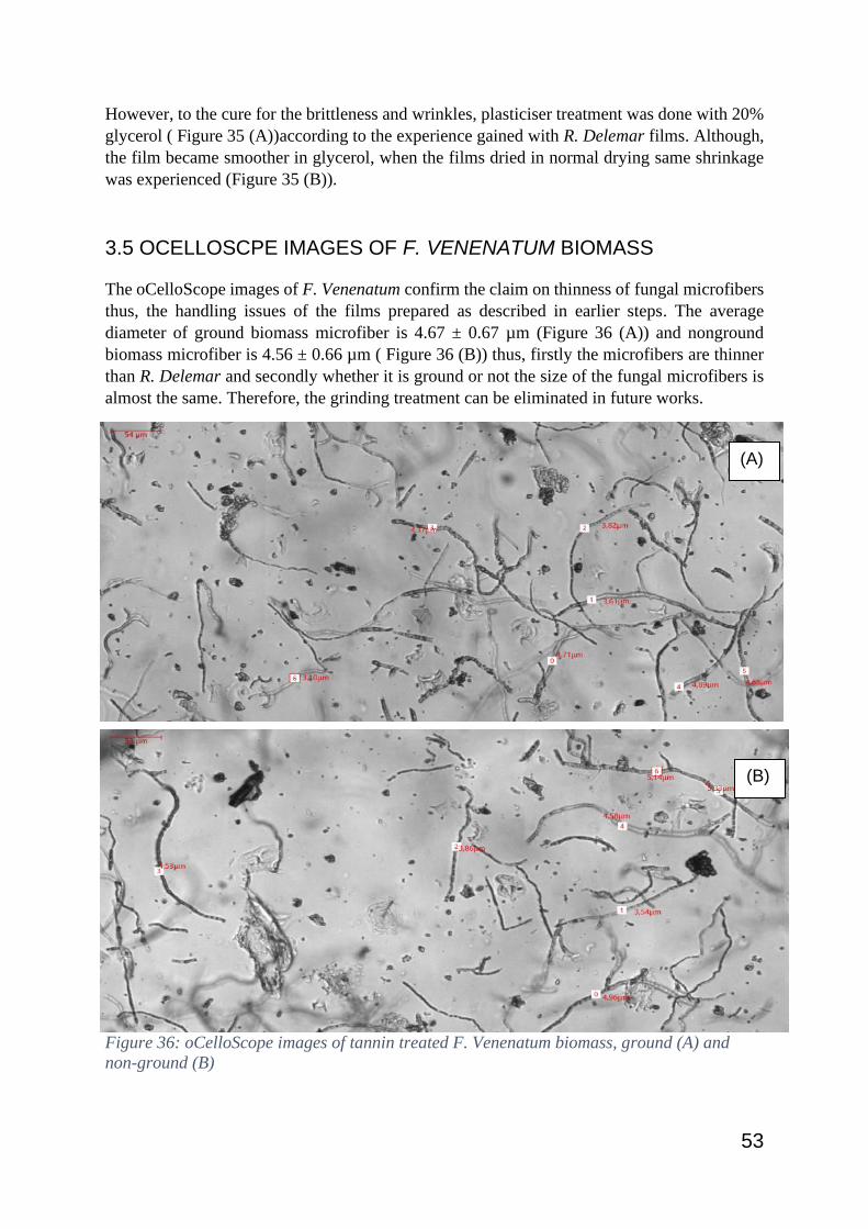
53
However, to the cure for the brittleness and wrinkles, plasticiser treatment was done with 20%
glycerol ( Figure 35 (A))according to the experience gained with R. Delemar films. Although,
the film became smoother in glycerol, when the films dried in normal drying same shrinkage
was experienced (Figure 35 (B)).
3.5 OCELLOSCPE IMAGES OF F. VENENATUM BIOMASS
The oCelloScope images of F. Venenatum confirm the claim on thinness of fungal microfibers
thus, the handling issues of the films prepared as described in earlier steps. The average
diameter of ground biomass microfiber is 4.67 ± 0.67 µm (Figure 36 (A)) and nonground
biomass microfiber is 4.56 ± 0.66 µm ( Figure 36 (B)) thus, firstly the microfibers are thinner
than R. Delemar and secondly whether it is ground or not the size of the fungal microfibers is
almost the same. Therefore, the grinding treatment can be eliminated in future works.
After considering all the issues such as, the brittleness, the handling hardness and the time
constrains it was decided not to continue making films with F. Venenatum biomass in this
project. Nonetheless, some future works regarding F. Venenatum biomass will be given in the
referred section.
Figure 36: oCelloScope images of tannin treated F. Venenatum biomass, ground (A) and
non-ground (B)
(A)
(B)

54
3.6 ATTEMPT TO MAKE A PRODUCT WITH THE FILMS
To check the flexibility and workability of the successful materials prepared using the fungal
biomass of R. Delemar first continues sheets were prepared (Figure 37 and 38). The films made
using both untreated and tannin treated biomass with both glycerol and binder post-treatments
(RD Neat + Glycerol + Binder and RD Tannin + Glycerol + Binder)
As expected, the biobased binder which used to enhance the properties has functioned as a
binder of one film to another in making a continues sheet using the circle patches. Working
with sewing was little harder since the films tend to tear with the forces created by the needle
or the thread. However, finally the stitching was completed without damaging the product.
Therefore, it is proven that the material is workable in this early laboratory scale thus with
further developments the future is bright.
Figure 37: Making a pouch using RD Tannin + Glycerol + Binder films
Figure 38: Making a coin wallet from RD Neat + Glycerol + Binder films

55
4. CONCLUSION
Production of a leather-like material in an emerging laboratory scale using fungal biomass of
Rhizopus Delemar grown on bread waste through an environmentally friendly and affable
process was successful. In this regard a wet-laid technique was applied to make films using
fungal microfibres by producing a non-woven mesh like material. Several hypotheses were
tested such as, using tannin to stabilise the fungal proteins to obtain a material like leather in
properties, using glycerol plasticising agent to reduce brittleness and enhance flexibility and
using binder to enhance the strength and water repellence.
Overall, films produced with untreated biomass and post-treated with glycerol (RD Neat +
Glycerol), the same films post-treated with glycerol and binder (RD Neat + Glycerol + Binder)
and films made with tannin treated biomass and post treated with both glycerol and binder (RD
Tannin + Glycerol + Binder) were selected as potential materials with tensile strengths of 7.74
± 0.55 MPa, 7.10 ± 0.60 MPa and 6.92 ± 0.51 MPa, respectively. Furthermore, the elongations
of the same materials were 4.82, 11.49 and 16.87, respectively. However, only untreated
biomass films with different treatments (RD Neat + Glycerol and RD Neat + Glycerol + Binder)
were able to categorise as hydrophobic, with the water contact angles above 90° even after 10
seconds. The tannin treated films were acting as hydrophilic with a good absorbance due to
the porosity of the surface.
On the other hand, film production using the biomass of F. Venenatum was not successful
mainly due to the hardness in handling and drying as a result of the thin nature of the fungal
fibres. The process should be further developed to make the film production successful
therefore the proposals are added in the future work.
Having said all the above, it is still understood that there is a huge gap between the laboratory
scale and industrial scale practices. There is a vast area to develop the produced material
towards leather substitute. Nonetheless, this process and the material produced has opened a
promising path towards sustainable leather alternatives in the future.

56
5. PROPOSED FUTURE WORK
Throughout the project some interesting hypotheses were experimented, and some became
successful while others were ineffective. However, there were more ideas to develop this
fungal functional material which will be listed in this section,
• The natural leather is a multi-layered material which has a gradient in tightness
throughout its cross-section (Meyer et al., 2021). During the project, several materials
were developed with different properties thus, different methodologies should be
designed to have layered materials, and it will be one step forward towards producing
a leather substitute.
• The growth of F.Venenatum was investigates to be higher at 26°C than 30°C which
was the temperature used earlier. However, a proper investigation is proposed with all
the temperatures between 25°C to 30°C to observe the growth.
• Since the thinness of F. Venenatum microfibres was a problem in both film making
and handling it is proposed to produce composites.
o Fungi – Natural fibres composites are proposed with fibres such as Cellulose
and Lyocell.
o Fungi – Fungi composites are also proposed by mixing fungal microfibers of
F. Venenatum and R. Delemar.
• Since bread waste contains particles which are unconsumed by the fungi, different
food wastes could be used as substrate and research on the properties of the material.
• Several testes are proposed to carry on the biodegradability, antimicrobial properties,
affinity to natural dyes etcetera, to test the feasibility of the material in industrial
scale.
• New methods to apply the binder is also proposed as it was identified as an unperfect
work.

57
REFERENCES
ABE, A., ODA, Y., ASANO, K. & SONE, T. 2007. Rhizopus delemar is the proper name for Rhizopus oryzae fumaric-malic acid producers. Mycologia, 99, 714-22.
ADAMCZYK, B., SIMON, J., KITUNEN, V., ADAMCZYK, S. & SMOLANDER, A. 2017. Tannins and Their Complex Interaction with Different Organic Nitrogen Compounds and Enzymes: Old Paradigms versus Recent Advances. ChemistryOpen, 6, 610-614.
AELENEI, N., POPA, M. I., NOVAC, O., LISA, G. & BALAITA, L. 2009. Tannic acid incorporation in chitosan-based microparticles and in vitro controlled release. Journal of Materials Science: Materials in Medicine, 20, 1095-1102.
ALDRED, E. M., BUCK, C. & VALL, K. 2009. Chapter 21 - Phenols. In: ALDRED, E. M., BUCK, C. & VALL, K. (eds.) Pharmacology. Edinburgh: Churchill Livingstone.
APPELS, F. V. W., VAN DEN BRANDHOF, J. G., DIJKSTERHUIS, J., DE KORT, G. W. & WÖSTEN, H. A. B. 2020. Fungal mycelium classified in different material families based on glycerol treatment. Communications Biology, 3, 334.
BARTNICKI-GARCIA, S. 1968. Cell Wall Chemistry, Morphogenesis, and Taxonomy of Fungi. Annual Review of Microbiology, 22, 87-108.
BONDLOLL, S. 2016. This Beer Is Made From Old Bread and it’s coming to the U.S. soon. [Online]. huffppost. Available: https://www.huffpost.com/entry/toast-ale-beer-from-bread_n_57ab2cd1e4b06e52746e76e1?guccounter=1&guce_referrer=aHR0cHM6Ly93d3cuYmluZy5jb20v&guce_referrer_sig=AQAAAAeE-Ahe-iQzMs7p2bhBHfg4WzxZyUgzd0RddZXr39vGI02O3UImYwtRxP5u4FhXD5MDvJ2qV2HtjyoRUXq3aflnlS0f-MQAQu5PKPMJIaKBPoED3JfbeRFe5Gv__LWLNxUlVx51WQkRKDydQ_LZRi2C__sPvnpC9QEbChwkfKXf [Accessed 09-16 2020].
BRANCOLI, P., LUNDIN, M., BOLTON, K. & ERIKSSON, M. 2019. Bread loss rates at the supplier-retailer interface–analysis of risk factors to support waste prevention measures. Resources, Conservation and Recycling, 147, 128-136.
BULE, M., KHAN, F., NISAR, M. F. & NIAZ, K. 2020. Tannins (hydrolysable tannins, condensed tannins, phlorotannins, flavono-ellagitannins).
CARRIS, L., LITTLE, C. & STILES, C. 2012. Introduction to Fungi. Plant Health Instructor. CHANEY, R. L. 1983. LEATHER MANUFACTURING WASTES. 285-295. CHOUDHARY, A. K. & BANSAL, P. 2017. 8 - Drape measurement technique using manikins with the
help of image analysis. In: NAYAK, R. & PADHYE, R. (eds.) Manikins for Textile Evaluation. Woodhead Publishing.
CHUNG, K.-T., WONG, T. Y., WEI, C.-I., HUANG, Y.-W. & LIN, Y. 1998. Tannins and Human Health: A Review. Critical Reviews in Food Science and Nutrition, 38, 421-464.
CONCA, J. 2015. Making Climate Change Fashionable - The Garment Industry Takes On Global Warming [Online]. Forbes. Available: https://www.forbes.com/sites/jamesconca/2015/12/03/making-climate-change-fashionable-the-garment-industry-takes-on-global-warming/#1750178f79e4 [Accessed 09-20 2020].
CORRADINI, P., GALLO, S. & GESUALDI., F. 2016. A TOUGH STORY OF LEATHER
A journey into the tanning industry via the Santa Croce District. COVINGTON, A. D. 2009. Tanning Chemistry: The Science of Leather, Royal Society of Chemistry. EDANA. What are non wovens? [Online]. www.edana.org. Available: https://www.edana.org/nw-
related-industry/what-are-nonwovens [Accessed 09-22 2020]. FAO 2011. Global food losses and food waste – Extent, causes and prevention. Rome: Food and
Agriculture Organisation of the United Nations.

58
FAO 2013. Food wastage footprint - Impacts on natural resources. Food and Agriculture Organisation of the United Nations.
FAO 2016. World statistical compendium for raw hides and skins, leather and leather footwear
1999-2015. Rome. FERREIRA, J. A., LENNARTSSON, P. R., EDEBO, L. & TAHERZADEH, M. J. 2013. Zygomycetes-based
biorefinery: Present status and future prospects. Bioresource Technology, 135, 523-532. FIBER2FASHION. 2012. Various Pollutants Released into Environment by Textile Industry [Online].
Available: https://www.fibre2fashion.com/industry-article/6262/various-pollutants-released-into-environment-by-textile-industry [Accessed 09-15 2020].
GARCÍA, C. & PRIETO, M. A. 2019. Bacterial cellulose as a potential bioleather substitute for the footwear industry. Microbial Biotechnology, 12, 582-585.
GMOSER, R., A. FERREIRA, J. & LENNARTSSON, P. 2019a. Post-treatment of Fungal Biomass to Enhance Pigment Production. Applied Biochemistry and Biotechnology.
GMOSER, R., FRISTEDT, R., LARSSON, K., UNDELAND, I., TAHERZADEH, M. J. & LENNARTSSON, P. R. 2020. From stale bread and brewers spent grain to a new food source using edible filamentous fungi. Bioengineered, 11, 582-598.
GMOSER, R., SINTCA, C. & LENNARTSSON, P. 2019b. Combining submerged and solid state fermentation to convert waste bread into protein and pigment using the edible filamentous fungus N. intermedia. Waste Management, 97, 63-70.
GORDON, C., THOMAS, S., GRIFFEN, A., ROBSON, G. D., TRINCI, A. P. J. & WIEBE, M. G. 2001. Combined use of growth rate correlated and growth rate independent promoters for recombinant glucoamylase production in Fusarium venenatum. FEMS Microbiology Letters, 194, 229-234.
HAGERMAN, A. 2012. Fifty Years of Polyphenol-Protein Complexes. Recent Advances in Polyphenol Research, Volume 3, 3, 71-97.
HASLAM, E. 2007. Vegetable tannins – Lessons of a phytochemical lifetime. Phytochemistry, 68, 2713-2721.
HOSSEINI, S. M., KHOSRAVI, K., MOHAMMADIFAR, M. A. & NIKOOPOUR¹, H. 2009. Production of Mycoprotein by Fusarium venenatum Growth on Modified Vogel Medium. Asian Journal oF Chemistry, 21, 4017-4022.
ISO-130 2005. ISO 139:2005
Textiles — Standard atmospheres for conditioning and testing. ISO-527-2 2012. Plastics — Determination of tensile properties. ISO-3376 2020. Leather — Physical and mechanical tests — Determination of tensile strength and
percentage elongation. JACOMETTI, V. 2019. Circular Economy and Waste in the Fashion Industry. Laws, 8, 27. JONES, M., BHAT, T., KANDARE, E., THOMAS, A., JOSEPH, P., DEKIWADIA, C., YUEN, R., JOHN, S., MA,
J. & WANG, C.-H. 2018. Thermal Degradation and Fire Properties of Fungal Mycelium and Mycelium - Biomass Composite Materials. Scientific Reports, 8, 17583.
JONES, M., GANDIA, A., JOHN, S. & BISMARCK, A. 2021. Leather-like material biofabrication using fungi. Nature Sustainability, 4, 9-16.
KHANBABAEE, K. & VAN REE, T. 2001. Tannins: Classification and Definition. Natural Product Reports, 18, 641-649.
KOCHHAR, S. L. 2016. Vegetable Tannins and Dyestuffs. In: KOCHHAR, S. L. (ed.) Economic Botany: A Comprehensive Study. 5 ed. Cambridge: Cambridge University Press.
KÖHNLEIN, M. 2020. Preparation of films and nonwoven composites from fungal microfibers grown in bread waste. Independent thesis Advanced level (degree of Master (Two Years)) Student thesis.
59
LINDSTRÖM, T., WÅGBERG, L. & LARSSON, T. 2005. On the nature of joint strength in paper - A review of dry and wet strength resins in paper manufacturing. Advances in Paper Science and Technology, 1.
LOHMAN, S. 2019. A Brief History of Bread
Bread, in all its various forms, is the most widely consumed food in the world. [Online]. www.histroy.com. Available: https://www.history.com/news/a-brief-history-of-bread [Accessed 09-16 2020].
MADIGAN, M. T. B., THOMAS D. 2014. Brock biology of microorganisms, Boston, [Mass.] ;, Pearson. MALIK, T. & ROY, P. K. 2012. Wet-Laid Non Wovens: An Overview. Textile Review. Gujarat: Athena
Information Solutions Pvt. Ltd. MAO, N. & RUSSELL, S. J. 2015. Chapter 13 - Fibre to Fabric: Nonwoven Fabrics. In: SINCLAIR, R. (ed.)
Textiles and Fashion. Woodhead Publishing. MARHENDRASWARI, M. D., MONDYLAKSITA, K., MILLATI, R., BUDHIJANTO, W., FERREIRA, J. A.,
NIKLASSON, C. & TAHERZADEH, M. J. 2020. Production of edible fungal (Rhizopus delemar CBS 145940) biomass from organosolv-pretreated oil palm empty fruit bunch (OPEFB) in submerged fermentation. IOP Conference Series: Materials Science and Engineering, 991, 012041.
MCLAUGHLIN, G. D., THEIS, E. R. & ORTHMANN, A. C. 1946. The Chemistry of Leather Manufacture The Journal of Physical Chemistry, 569-576.
MELIKOGLU, M., LIN, C. S. K. & WEBB, C. 2013. Stepwise optimisation of enzyme production in solid state fermentation of waste bread pieces. Food and Bioproducts Processing, 91, 638-646.
MELIKOGLU, M. & WEBB, C. 2013. Chapter 4 - Use of Waste Bread to Produce Fermentation Products. In: KOSSEVA, M. R. & WEBB, C. (eds.) Food Industry Wastes. San Diego: Academic Press.
MEYER, M., DIETRICH, S., SCHULZ, H. & MONDSCHEIN, A. 2021. Comparison of the Technical Performance of Leather, Artificial Leather, and Trendy Alternatives. Coatings, 11.
MONEY, N. P. 2016a. Chapter 1 - Fungal Diversity. Third Edition ed.: Elsevier Ltd. MONEY, N. P. 2016b. Chapter 3 - Spore Production, Discharge, and Dispersal. In: WATKINSON, S. C.,
BODDY, L. & MONEY, N. P. (eds.) The Fungi (Third Edition). Boston: Academic Press. MONTEFUSCO, A. S. F. 2005. The use of Nonwovens in air filtration. Filtration & Separation, 42, 30-
31. NAIR, R., THEIMYA, E.-H. & LENNARTSSON, P. 2017. Waste Bread Valorization Using Edible
Filamentous Fungi. NAM, S., SMITH, D. M. & DOU, Q. P. 2001. Tannic Acid Potently Inhibits Tumor Cell Proteasome
Activity, Increases p27 and Bax Expression, and Induces G<sub>1</sub> Arrest and Apoptosis. Cancer Epidemiology Biomarkers &amp; Prevention, 10, 1083.
PALOMINO, M., VEGA GUTIERREZ, S., VAN COURT, R. & CHEN, H.-L. 2020. Oil-Based Fungal Pigment from Scytalidium cuboideum as a Textile Dye. Journal of Fungi, 6, 53.
PARACHIN, N. S., HAHN-HÄGERDAL, B. & BETTIGA, M. 2011. 6.46 - A Microbial Perspective on Ethanolic Lignocellulose Fermentation. In: MOO-YOUNG, M. (ed.) Comprehensive Biotechnology (Second Edition). Burlington: Academic Press.
PRZYBYSZ, P., DUBOWIK, M., KUCNER, M. A., PRZYBYSZ, K. & PRZYBYSZ BUZAŁA, K. 2016. Contribution of Hydrogen Bonds to Paper Strength Properties. PloS one, 11, e0155809-e0155809.
QUILLERIET, A.-L. 2004. The Leather Book, New York, USA, Assouline Publishing. RAJENDRAN, P., MANICKAM, P., GUNASEKARAN, S., MURUGESAN, R. & THILAGAVATHI, G. 2013.
Natural dye production from Thermomyces sp fungi for textile application. Indian Journal of Fibre and Textile Research, 38, 276-279.
RILEY, A. 2012. 10 - Paper and paperboard packaging. In: EMBLEM, A. & EMBLEM, H. (eds.) Packaging Technology. Woodhead Publishing.

60
RUGGIERO, M. A., GORDON, D. P., ORRELL, T. M., BAILLY, N., BOURGOIN, T., BRUSCA, R. C., CAVALIER-SMITH, T., GUIRY, M. D. & KIRK, P. M. 2015. A Higher Level Classification of All Living Organisms. PLOS ONE, 10, e0119248.
SALMAN, A., TSROR, L., POMERANTZ, A., MOREH, R., MORDECHAI, S. & HULEIHEL, M. 2010. FTIR spectroscopy for detection and identification of fungal phytopathogenes. Spectroscopy, 24, 723489.
SHARMA, D., GUPTA, C., AGGARWAL, S. & NAGPAL, N. 2012. Pigment extraction from fungus for textile dyeing. Indian Journal of Fibre and Textile Research, 37, 68-73.
SIVAKESAVA, S., IRUDAYARAJ, J. & DEBROY, C. 2004. DIFFERENTIATION OF MICROORGANISMS BY FTIR-ATR AND NIR SPECTROSCOPY. Transactions of the ASABE, 47, 951-957.
SPATAFORA, J. W., CHANG, Y., BENNY, G. L., LAZARUS, K., SMITH, M. E., BERBEE, M. L., BONITO, G., CORRADI, N., GRIGORIEV, I., GRYGANSKYI, A., JAMES, T. Y., O’DONNELL, K., ROBERSON, R. W., TAYLOR, T. N., UEHLING, J., VILGALYS, R., WHITE, M. M. & STAJICH, J. E. 2016. A phylum-level phylogenetic classification of zygomycete fungi based on genome-scale data. Mycologia, 108, 1028-1046.
SUDHA, GUPTA, C. & AGGARWAL, S. 2014. Novel bio-colorants for textile application from fungi. Journal of the Textile Association, 74, 282-287.
SVAHN, S., GÖRANSSON, U., EL-SEEDI, H., BOHLIN, L., LARSSON, J., OLSEN, B. & CHRYSSANTHOU, E. 2012. Antimicrobial activity of filamentous fungi isolated from highly antibiotic-contaminated river sediment. Infection ecology & epidemiology, 2.
SVANSTRÖM, Å. Trehalose Metabolism and Stress Resistance in Aspergillus niger. 2013. UN 2015. The 2030 Agenda for Sustainable Development. WIEBE, M. 2002. Myco-protein from Fusarium venenatum: a well-established product for human
consumption. Applied Microbiology and Biotechnology, 58, 421-427. WRAP, W. 2009. Household food and drink waste in the UK. Report prepared by WRAP. Banbury, UK:
WRAP. ZHOU, B., JIN, X., LI, J., XU, W., LIU, S., LI, Y. & LI, B. 2014. Vacuum-assisted layer-by-layer electrospun
membranes: antibacterial and antioxidative applications. RSC Advances, 4, 54517-54524.

61
Street address: Allégatan 1 · Mailing address: 501 90 Borås · Phone: +46 33-435 40 00 · E-mail: [email protected] · Web: www.hb.se