development of novel membrane active lipidated peptidomimetics active against drug resistant...
TRANSCRIPT
Accepted Manuscript
Development of novel membrane active lipidated peptidomimetics activeagainst drug resistant clinical isolates
Sandeep Lohan, Arneesh kalanta, Praveen Sonkusre, Swaranjit Singh Cameotra,Gopal Singh Bisht
PII: S0968-0896(14)00557-4DOI: http://dx.doi.org/10.1016/j.bmc.2014.07.041Reference: BMC 11736
To appear in: Bioorganic & Medicinal Chemistry
Received Date: 30 May 2014Revised Date: 24 July 2014Accepted Date: 26 July 2014
Please cite this article as: Lohan, S., kalanta, A., Sonkusre, P., Cameotra, S.S., Bisht, G.S., Development of novelmembrane active lipidated peptidomimetics active against drug resistant clinical isolates, Bioorganic & MedicinalChemistry (2014), doi: http://dx.doi.org/10.1016/j.bmc.2014.07.041
This is a PDF file of an unedited manuscript that has been accepted for publication. As a service to our customerswe are providing this early version of the manuscript. The manuscript will undergo copyediting, typesetting, andreview of the resulting proof before it is published in its final form. Please note that during the production processerrors may be discovered which could affect the content, and all legal disclaimers that apply to the journal pertain.
1
Development of novel membrane active lipidated peptidomimetics active against drug
resistant clinical isolates
Sandeep Lohana, Arneesh kalantaa, Praveen Sonkusreb, Swaranjit Singh Cameotrab, Gopal
Singh Bishta*
aDepartment of Pharmacy, Jaypee University of Information Technology, Solan-173234,
India.
bEnvironmental Biotechnology & Microbial Biochemistry, Institute of Microbial Technology,
Chandigarh-160036, India.
*Corresponding Author: Dr. Gopal Singh Bisht
Department of Pharmacy, Jaypee University of Information Technology, Solan, India.
Email: [email protected]
Tel: 91-1792-239336
Fax: +91-1792-245362
2
Abstract:
A new series of small cationic lipidated peptidomimetics have been synthesized and found to
be highly active against several susceptible as well as drug resistant clinical isolates of
bacteria and fungi. All lipidated peptidomimetics do not cause significant lysis of human
erythrocytes (HC50 > 200 µg/mL). Calcein dye leakage experiment revealed membranolytic
effect of LPEP08 which was further confirmed by scanning electron microscopy (SEM). The
involvement of intracellular targets as an alternate mode of action was precluded by DNA
retardation assay. Additionally, LPEP08 exhibit high proteolytic stability and dose not elicit
resistance against drug resistant clinical isolate of S. aureus, even after 16 rounds of
passaging. These results demonstrate the potential of lipidated peptidomimetics as
biocompatible anti-infective therapeutics.
Keywords: peptidomimetics; antimicrobial peptides; lipopeptides; drug resistance;
membranolytic
3
1. Introduction
The dramatically increased frequency of infections caused by multi-drug resistant bacterial
and opportunistic fungal strains has driven research to expand the arsenal of anti-infective
agents. Recently, WHO has recognized the infection caused by multidrug-resistant pathogens
viz. methicillin resistant Staphylococcus aureus (MRSA), vancomycin resistant
Staphylococcus aureus (VRSA), drug resistant Pseudomonas aeruginosa and Klebsiella
pneumoniaeas major causes of morbidity and mortality [1, 2]. In addition, majority of the
invasive fungal infections caused by Aspergillus and Candida species emerged as major
threat to public health [3]. These trends have accentuate to develop new class of antibiotics
possessing novel mode of action as well as different cellular targets compared to
conventional antibiotics in order to decrease the possibility of resistance development.
Antimicrobial peptides (AMPs) are found in virtually all multicellular organisms and
functionally act as weapons to ward off pathogenic microbes in order to survive and thrive on
this planet [4, 5]. In general, AMPs are typically composed of 20-50 amino acid residues and
carrying a net positive charge (provided by Arg and Lys residues) with ≈50% hydrophobic
residues. The mode of action of AMPs is of particular interest, as it is thought to be non-
specific unlike conventional antibiotic drugs (usually directed against a specific cellular
receptor) [6, 7]. Mechanically majority of AMPs are bind and permeate cell membranes and
others have found to modulate the immune response or have targets within the cell. Taking
these findings together, AMPs display unique mode of action that could not deriving the
development of resistance [8, 9].
Lipopeptides constitute another class of native AMPs, which are produced non-ribosomally
in bacteria and fungi during cultivation on various carbon sources [10-12].Structurally, native
lipopeptides are complex molecules composed of aliphatic acid attached to the N-terminus of
cationic or anionic peptidic moiety [13]. The mode of lytic action of lipopeptides is via
4
perturbation of the cell membrane by unknown mechanisms which is similar to most of the
AMPs [14, 15]. Mechanistically electrostatic interaction between cationic lipopeptides and
negative membrane surface charge of bacteria is the initial step of their bactericidal activity.
On the other hand in the fungi lipopeptides bind to the negatively charged membrane
phosphatidylinositol (PI) and to the negatively charged terminal sialic acid moieties [16, 17].
Clinically used members of this novel class of antimicrobials includes daptomycin (active
only toward Gram-positive bacteria), polymyxin B (active only toward Gram-negative
bacteria), and echinocandins (β-1,3-D-glucan synthase inhibitors; active only toward fungi)
[18]. The major drawback associated with this class of antimicrobials is that the toxic dose is
close to the therapeutic dose [18].
It is well documented that conjugation of linear fatty acids to small cationic AMPs resulted
into enhancement of antimicrobial potential against Gram-negative and Gram-positive
bacteria [19-21]. In addition, chongsiriwatana et al. demonstrated that lipidation of small
peptoid sequences renders them more selective towards microbes without losing
antimicrobial activity [22]. The aliphatic tail region of lipopeptides was found to be essential
for their antimicrobial action as it may improves the hydrophobic interaction with
cytoplasmic membrane [23]. Thus, the development of small synthetic congeners which
mimics integral structural features may overcome some of the drawbacks associated with
current lipopeptide antibiotics.
Herein, we reported a new series of small cationic lipidated peptidomimetics synthesized by
incorporating 3-amino benzoic acid (3-ABA) as a turn motif between peptidic and aliphatic
tail region of the molecules. We confirmed the broad-spectrum antimicrobial potential and
low hemolytic action of lipidated peptidomimetics. For an initial investigation of bactericidal
mechanism of the lead molecules, calcein leakage experiments on model membranes were
performed and which was complemented by observing their effect on intact cells. Finally, the

5
proteolytic stability even in human blood plasma and inability of drug resistant S. aureus to
develop resistance aid in the future development of small lipidated peptidomimetics to treat
fatal infections.
2. Results and discussion
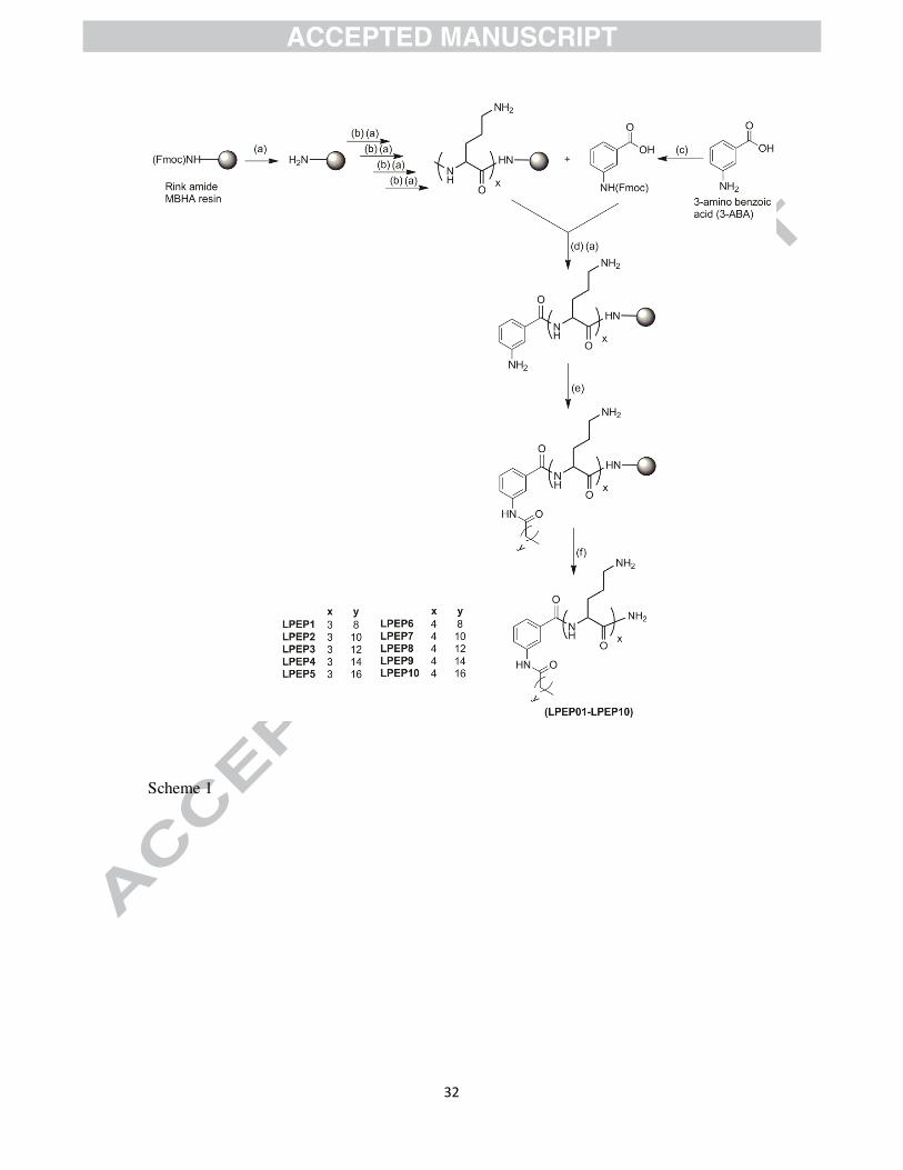
2. 1. Design and synthesis
In a drug development perspective, proteolytic enzyme susceptibility is one of the biggest
issues associated with the clinical applicability of peptide based therapeutics. Thus, with aim
to develop novel potent biocompatible anti-infective agents, we carried out the synthesis of
lipidated peptidomimetics using non-natural amino acid ornithine (Orn) as cationic charge
residue. To synthesize lipidated molecules, small peptide scaffold composed of 3 and 4 Orn
residues were acylated with fatty acid tail varying from 10 to 18 carbon atoms. It is well
known fact about AMPs that the antimicrobial activity is somewhat depends on their
secondary structure. For that reason, incorporation of any suitable moiety in the molecular
framework which provides a specific turn might boost up the antimicrobial action. The
incorporation of constrainedaromatic amino acid (3-ABA) in peptide sequence resulted into
the improvement of stability of folded conformation [24, 25] as well as antimicrobial
potential [26]. On the basis of these findings, we incorporated 3-ABA as a linker between
peptide and hydrophobic tail region (Scheme 1).
2.2. Antimicrobial activity
All synthesized lipidated peptidomimetics were screened against representative Gram-
positive and Gram-negative bacteria and fungi, including antibiotic resistant clinical isolates.
Lipidated peptidomimetics composed of 3 Orn residues and 10 carbon atoms long aliphatic
tail (LPEP01) was found to be almost inactive as antimicrobial. LPEP01 exhibits MIC > 50
µg/mL against all tested pathogens with an exception of having MIC = 31.5 µg/mL against S.
aureus MTCC 3160 (Table 1). A closer examination of the activity results revealed that
6
lipopeptides with aliphatic chain length ranging from 12 to 18 carbon atoms showed
improved antimicrobial activity. Among the lipidated peptidomimetics composed of 3 Orn
residues (LPEP01-LPEP05), highest activity was observed in case of lipopeptide having N-
terminus myristic acid (LPEP03) with MIC values of 2.5 µg/mL for E. coli and P. aeruginosa
and 3.1 µg/mL for S. aureus. In addition, LPEP03 showed good antibacterial activity against
antibiotic resistant clinical isolates of E. coli (MIC = 4 µg/mL) and S. aureus (MIC = 4.5
µg/mL). LPEP03 display moderate activity against B. subtilis with MIC = 15.5 µg/mL.
Further increase in the length of aliphatic tail resulted into decrease in antibacterial potential
(LPEP04 & LPEP05; Table 1). LPEP06, composed of 4 Orn residues and 10 carbon atoms
long aliphatic tail, displayed moderate activity (MIC values in the range of 17.5-35 µg/mL)
against all tested bacterial strains with an exception of having minimum killing effect against
B. subtilis (MIC = 100 µg/mL; Table 1). Similar impact of aliphatic chain length on
antibacterial potential was observed in case of lipidated peptidomimetics comprised of 4 Orn
residues as significant improvement in antibacterial activity was observed for lipidated
peptidomimetics bearing bulky aliphatic tail. LPEP08 exhibits maximum antibacterial
potential with MIC values of 1.5 µg/mL for E. coli and S. aureus and 2 µg/mL for P.
aeruginosa (Table 1). Further increment in aliphatic tail was not fruitful as somewhat
decrease in activity was observed in case of lipopeptides (LPEP09 & LPEP10) having 16 and
18 carbon atoms long aliphatic tail. It was interesting to note that all synthesized lipidated
peptidomimetics exhibit broad antibacterial activity spectrum with insignificant difference
between MIC values against susceptible pathogens and drug resistant clinical isolates.
Lipidated peptidomimetics with comparatively bulky aliphatic tail were found to be more
active towards fungal strains. LPEP10 has highest antifungal activity with MIC values of 1.5
µg/mL against C. albican and A. fumigatus, 2.5 µg/mL for A. niger, and 5.5 µg/mL for C.
neoformans. It was encouraging to observe similar antifungal potential of LPEP10 against

7
drug resistant clinical isolates of C. albican (MIC = 1.5 µg/mL) and A. fumigatus (MIC = 2
µg/mL; Table 2).
Analysing the antimicrobial activity results of lipidated peptidomimetics, we conclude that
activity is depends on the content of both cationic charge and hydrophobic bulk. Importantly,
in contrast to the most AMPs or natural lipopeptides that are active either against bacteria or
fungi alone [27], lipidated peptidomimetics reported here are highly potent against both
bacteria and fungi. The broad-spectrum antimicrobial potential of this library of compounds
reflected their candidature to develop as novel anti-infective agents.
2.3. Hemolytic activity
All synthesized lipidated peptidomimetics displayed batter selectivity towards microbial cells
(HC50 > 200 µg/mL) as summarized in Table 1. Lipidated peptidomimetics with bulky
aliphatic tail (16 &18 carbon atoms long aliphatic tail) showed higher affinity towards hRBC
as compared to the molecules bearing small fatty acid chain (10, 12, and 14 carbon atoms
long aliphatic tail). Thus, it seems that, as the length of aliphatic tail increases, the ability of
lipidated peptidomimetics to discriminate between anionic bacterial surface and zwitterionic
mammalian membrane decreases. These outcomes were in accordance with our earlier
findings [28]. Noticeably, most potent antibacterial lipidated peptidomimetic (LPEP08) has
significantly wider therapeutic index (SR = 333), which we defined as HC50/MICE.c and
HC50/MICS.a (Table 1).
2.4. Bactericidal kinetic assay
In contrast to the majority of the conventional antibiotics, AMPs are usually bactericidal,
rather than bacteriostatic [29]. To determine whether this ability is inherent to newly
synthesized lipidated peptidomimetics we performed time-kill assay by exposing E. coli and
S. aureus to various concentrations of LPEP08.The results clearly showed that LPEP08 was
bactericidal at 4 × MIC and 8 × MIC against both E. coli (Figure 1A) and S. aureus (Figure
8
1B). At lower concentrations (MIC and 2 × MIC) LPEP08 was able to inhibit the growth.
The results also demonstrated the rapid killing effect of LPEP08 at higher concentration
levels (4 × MIC and 8 × MIC), as nearly 5-log reduction in the growth of E. coli and S.
aureus was observed within 30 min of incubation (Figure 1A & 1B).
2.5. Biomembrane interaction study using model membranes
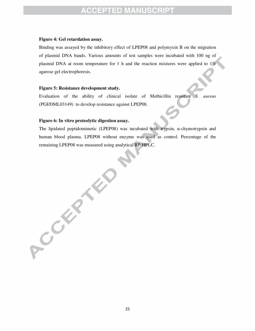
In the calcein leakage experiment, LPEP08 and LPEP10 at 4 µg/mL induced a rapid increase
in the fluorescence intensity, leading to a 58% and 44% dye release from bacterial membrane
mimicking liposomes, respectively. At 20 µg/mL, LPEP08 and LPEP10 caused 93% and
84% leakage, respectively. Whereas, LPEP06 at 4 µg/mL caused only 11% of dye leakage
from bacterial membrane mimicking liposomes and it was increased to nearly 67% at highest
used experimental concentration (20 µg/mL; Figure 2A). The better membrane interactions of
LPEP08 and LPEP10 were also in concord with our activity results. However, at 4 µg/mL
LPEP10 caused maximum dye leakage (57%) when incubated with fungal membrane
mimicking liposomes. In contrast, at 4 µg/mL LPEP06 and LPEP08 were able to induce 17%
and 29% leakage, respectively. At a concentration of 20 µg/mL the percentage of calcein
leakage reached 89% for LPEP10 and 77% and 45% dye leakage was observed for LPEP08
and LPEP06, respectively (Figure 2B). Thus, in agreement with earlier literature as well as
our activity and selectivity results these outcomes are indicative of the comparatively higher
affinity of bulky aliphatic tail conjugated lipidated peptidomimetic (LPEP10) toward
zwitterionic fungal membrane [17, 18, 28]. The outcomes of calcein dye leakage experiment
indicated that lipidated peptidomimetics damage bacterial and fungal cell membrane
mimicking liposomes in somewhat concentration dependant manner.
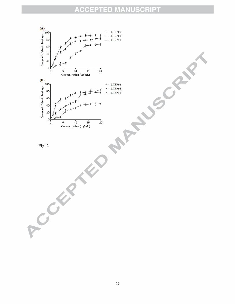
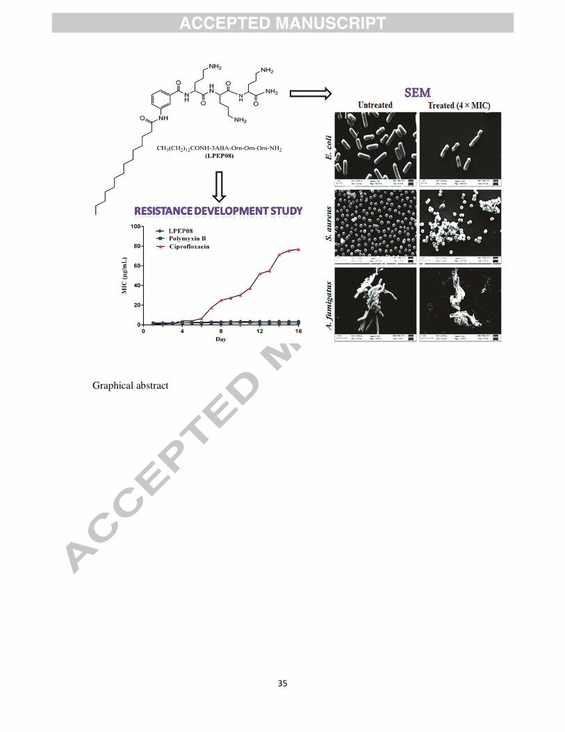
2.6. Surface disruption effect of lead lipopeptides in intact microbial cells
After knowing the membrane disruption effect of representative lipidated peptidomimetics
(LPEP08 & LPEP10), we further confirmed the mechanism of action by visualizing the effect
9
on intact bacterial (E. coli and S. aureus) and fungal (A. fumigatus) cells. SEM, a microscopic
technique has been used extensively for the elucidation of interaction of membrane active
peptides [30]. Morphological alterations caused by LPEP08 and LPEP10 when incubated
with bacterial and fungal cells respectively were observed under SEM. In SEM images we
visualized that untreated (control; Figure 3(A1-A3)) microbial cells exhibited bright and
smooth surface whereas treatment with lipidated peptidomimetics (LPEP08 and LPEP10)
similar to many other membrane active peptides such as melittin [31] resulted into surface
blebs and aggregation (Figure 3(B1-B3)). Therefore, SEM data suggested the membrane
perturbation effect of LPEP08 and LPEP10 on intact microbial cells which was analogues to
model microbial mimicking membranes (as evidenced by calcein dye leakage assay).
2.7. DNA binding assay
It is well documented that native lipopeptide antibiotic polymyxin B mainly active toward
Gram-negative bacterial strains [23]. The selective action of polymyxin B reflects the
possibility of their intracellular targets. These trends promoted us to determine the
involvement of any intracellular targets for antimicrobial action of lipidated peptidomimetics.
With this aim we compared the relative affinity of LPEP08 and polymyxin B to bind plasmid
DNA. In DNA binding assay, LPEP08 do not caused retardation in the movement of DNA
even at the highest used experimental concentration (12 µg/mL). On the other hand,
polymyxin B showed DNA binding at 12 µg/mL (Figure 4). These findings suggested a
predominant membrane disrupting mode of action for LPEP08, which is different from native
lipo-antibiotic polymyxin B.
2.8. Resistance development study
The efficacy of conventionally used antibiotics in treating infections caused by drug resistant
pathogens has been diminished as a result of pathogens ability to switch on to an alternate
metabolic pathway. It has already been reported that low propensity of resistance

10
development was observed in case of therapeutics that kill bacteria by targeting cell
membrane [32]. Therefore, the potential of drug resistant clinical isolate of S. aureus to
develop resistance was evaluated by serial passages of the bacterial cultures againstLPEP08.
Results in figure5confirmedthat bacterial pathogen was unable to develop resistance against
LPEP08 and native lipo-antibiotic polymyxin B when compared with ciprofloxacin as there
was insignificant change in the MIC after 16 passages.
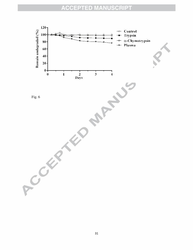
2.9. Evaluation of proteolytic stability
AMPs composed of genetically coded amino acids can be easily targeted by proteolytic
enzymes. Therefore, proteolytic degradation is one of the biggest hurdles in the development
of peptide based therapeutics. To investigate the proteolytic stability of our novel structural
framework, we examined degradation of LPEP08 by representative protease enzymes trypsin
and α-chymotrypsin. To further ensure the proteolytic stability, we tested LPEP08 in human
blood plasma also. The results from the stability experiment showed that no degradation was
observed for LPEP08 even after 4 days when incubated with α-chymotrypsin. It is well
documented that α-chymotrypsin cleaves the peptide bond on the C-terminal side of large
lipophilic amino acids (Phe, Trp) [33]. Consequently, the high stability of LPEP08 against α-
chymotrypsin might be due to the absence of any bulky hydrophobic residues in the structural
framework. Furthermore, LPEP08 was seemingly stable against proteolytic degradation by
trypsin, and after 4 days, 90% of the parent molecule was still intact. Mechanistically, trypsin
cleaves C-terminal to native cationic amino acids such as Lys and Arg [34]. Thus, the
incorporation of non-natural amino acid ornithine as cationic charged residues renders
LPEP08 immune toward tryptic degradation. It was further interesting to note that LPEP08
was found to be stable in plasma up to 24 h of incubation and after that it was slowly
degraded with approximately 76% of the peptidomimetic remaining after 4 days (Figure 6).
11
The results, thereby collectively demonstrated that the incorporation of ornithine could render
LPEP08 immune against proteolytic enzymes.
3. Conclusions
Overall, the present study adds to the repertoire of peptide based anti-infectives with lack of
mammalian toxicity and broad activity spectrum against several clinically relevant bacterial
and fungal strains. The pore-forming tendency of LPEP08 and LPEP10 in artificial
membrane along with weaker DNA-binding suggests their predominant membrane disrupting
mode of action. More importantly, LPEP08 was found immune to proteolytic degradation in
plasma and even drug resistant clinical isolate of S. aureus was unable to develop resistance.
In vivo efficacy and pharmacokinetic evaluation of lead molecules is currently under
progress. Therefore, taken together the advantageous features such as cell selectivity, broad-
spectrum antimicrobial properties, proteolytic stability, and most noticeably, no sign of
resistance development underscore the potential of LPEP08 for the development of novel
antimicrobial agent to treat infections caused by resistant pathogens in near future.
4. Experimental section
4.1. Materials
Rink amide MBHA resin and Fmoc-Protected ornithine (Fmoc-Orn(Boc)-OH)were
purchased from Novabiochem (Mumbai, India). Aromatic amino acid 3-amino benzoic acid
and Fmoc-Cl were obtained from Spectrochem (Mumbai, India). Fatty acids used for
acylation are caprylic acid, lauric acid, palmitic acid (Loba chemie), Myristic acid, and
stearic acid (Fluka). Other reagents used for solid phase synthesis of peptidomimetic
molecules included N-hydroxybenzotrizole (HOBt), N,N’-diisopropylcarbodiimide (DIC),
Piperidine, N,N-dimethylformamide (DMF) (Spectrochem, Mumbai, India),
dimethylsulphoxide (DMSO), dichloromethane (DCM), 1,2-Ethanedithiol (Merck, Mumbai,
India), and Trifluoro acetic acid (TFA; Loba chemie, Mumbai, India). Calcein, propidium

12
iodide (PI), 4’,6-diamidino-2-phenylindole (DAPI) and buffer material were purchased from
Sigma-Aldrich (India). All the solvents used for purification were of HPLC grade and
obtained from Merck (Mumbai, India). Buffers were prepared in double-distilled water.
4.2. General procedure for the Fmoc protection of 3-amino benzoic acid
To a solution of 3-amino benzoic acid (1.37 g, 10 mmol) in water (35 mL), was added
sodium hydrogen carbonate (2.52 g, 30 mmol), and the resulting mixture was cooled to 5°C
and it was slowly added with Fmoc-Cl (3.87 g, 15 mmol) as a solution in para-dioxane. The
resulting mixture was stirred at 0 °C for 1 h and allowed to warm to room temperature
overnight. Completion of reaction was monitored by precoated TLC plate. After the
completion of reaction water was added to the reaction mixture and the aqueous layer was
extracted with ethyl acetate. Then the organic layer was extracted twice with saturated
aqueous solution of sodium bicarbonate. The combined aqueous layers were acidified to a
pH of 2 with 10% HCl, and then extracted three times with ethyl acetate. The combined
organic layers were removed under reduced pressure to isolate title compound. The crude
material was used without any further purification.
4.2.1. Fmoc-3-ABA
1H NMR (400 MHz, DMSO d6) δ: 12.21 (s, 1H), 9.81 (s, 1H), 7.91 - 7.66 (m, 6H), 7.52 -
7.27 (m, 6H), 4.47 - 4.45 (d, J = 8Hz, 2H), 4.26 - 4.23 (t, J = 12Hz, 1H). MALDI-TOF: calcd
for C22H17NO4: 359.12, found 360.39.
4.3. General procedure for solid phase synthesis of lipidated peptidomimetics
Lipidated peptidomimetics were synthesized manually following standard Fmoc solid phase
protocols using Rink amide-4-methylbenzhydrylamine hydrochloride salt (MBHA) resin
(loading 0.79 mmol/g) as solid support [35]. Rink amide resin (100 mg) was washed in
CH2Cl2 (3 × 2 mL), which is followed by swelling in DMF (3 mL) for 25 min. The Fmoc
protecting group of resin was removed by treating with piperidine/DMF (20% v/v) mixture
13
for 10 min, followed by extensive washes with DMF (5 × 2 mL). Ornithine coupling was
performed by treatment of deprotected resin with Fmoc-Orn (Boc)-OH (0.14g; 4equiv) in the
presence of DIPC/HOBt in DMF. Each successive ornithine coupling step was followed by
Fmoc deprotection with piperidine/DMF (20% v/v) mixture. After coupling of 3 (LPEP1-
LPEP5) or 4 (LPEP6-LPEP10) ornithine residues, coupling of Fmoc protected 3-amino
benzoic acid was performed in the presence 2 equiv of DIPC/HOBt in DMF. Before acylation
of amino acid sequence with specific fatty acid fmoc deprotection was performed to generate
free amino group. Fatty acid acylation was performed by treatment of fatty acid (4equiv) in
the presence 2 equiv of DIPC/HOBt in DMF. After the desired sequences were assembled,
the peptidomimetic molecules were cleaved with a 5 mL solution of TFA/H2O/1,2-
Ethanedithiol (95:2.5:2.5) from solid support. The overview of the different synthetic steps
involved in the synthesis of lipopeptides is given in Scheme 1.
4.4. Purification
All crude peptidomimetics were analysed on a reversed-phase high performance liquid
chromatography (RP-HPLC) using a C18waters column (Spherisorb® , ODS2, 5µm, 4.6 mm ×
250mm) at room temperature. A linear gradient of 0.5-60% solvent B (0.05% TFA in
acetonitrile) in solvent A (0.05% TFA in water) over 35 min, followed by 60-0.5% solvent B
over 10 min was used at a flow rate of 0.5 mL/min. Preparative RP-HPLC was then
performed on a Waters column (Spherisorb® , ODS2, 5µm, 20 mm × 250 mm) using 0.5-60%
linear gradient of solvent B (0.05% TFA in acetonitrile) in solvent A (0.05% TFA in water)
over 35 min, followed by 60-0.5% solvent B over 10 min at a flow rate of 5 mL/min.
Electrospray mass spectroscopy (ESI-MS) was used to confirm their molecular weight.
Purified HPLC fractions were than lyophilized. See the supporting information for analytical
data (Mass spectra, 1H NMR, and HPLC chromatograms).
4.4.1. CH3(CH2)8CO-3-ABA-NH-Orn-Orn-Orn-NH2 (LPEP01)
14
1H NMR (400 MHz, DMSO-d6) δ 8.17 (s, 3H), 7.51 (d, J = 4.0 Hz, 1H), 7.36-7.27 (m, 3H),
7.12 (s, 2H), 4.22 (t, J = 20.0 Hz, 3H), 3.60 (s, 6H), 2.79-2.67 (m, 6H), 2.09 (t, J = 20.0 Hz,
2H), 1.72-1.42 (m, 12H), 1.22-1.11 (m, 16H), 0.82 (t, J = 20.0 Hz,3H). ESI-MS: calcd for
C32H56N8O5: 632.44, found 633.43; Purity (determined by RP-HPLC): 99.67%.
4.4.2. CH3(CH2)10CO-3-ABA-NH-Orn-Orn-Orn-NH2(LPEP02)
1H NMR (400 MHz, DMSO-d6) δ 8.26 (s, 3H), 7.81 (d, J = 4.0 Hz, 1H), 7.48-7.42 (m, 3H),
7.14 (s, 2H), 4.22 (t, J = 24.0 Hz, 3H), 3.45 (s, 6H), 2.76-2.63 (m, 6H), 2.11-2.03 (m, 2H),
1.68-1.43 (m, 13H), 1.27-1.15 (m, 19H), 0.82 (t, J = 24.0 Hz,3H). ESI-MS: calcd for
C34H60N8O5: 660.47, found 661.45; Purity (determined by RP-HPLC): 97.34%.
4.4.3. CH3(CH2)12CO-3-ABA-NH-Orn-Orn-Orn-NH2(LPEP03)
1H NMR (400 MHz, DMSO-d6) δ 8.47 (s, 3H), 7.62 (d, J = 4.0 Hz, 1H), 7.48-7.38 (m, 3H),
7.12 (s, 2H), 4.25 (t, J = 20.0 Hz, 3H), 3.64 (s, 6H), 2.88-2.73 (m, 7H), 2.09 (t, J = 12.0 Hz,
2H), 1.74-1.43 (m, 14H), 1.28-1.15 (m, 23H), 0.81 (t, J = 8.0 Hz,3H). ESI-MS: calcd for
C36H64N8O5: 688.50, found 689.48; Purity (determined by RP-HPLC): 97.65%.
4.4.4. CH3(CH2)14CO-3-ABA-NH-Orn-Orn-Orn-NH2(LPEP04)
1H NMR (400 MHz, DMSO-d6) δ 8.17 (s, 3H), 7.51 (d, J = 4.0 Hz, 1H), 7.36-7.27 (m, 3H),
7.12 (s, 2H), 4.24-4.17 (m, 3H), 3.51 (s, 6H), 2.81-2.77 (m, 6H), 2.10 (t, J = 28.0 Hz, 2H),
1.74-1.53 (m, 15H), 1.27-1.09 (m, 27H), 0.81 (t, J = 12.0 Hz, 3H). ESI-MS: calcd for
C38H68N8O5: 716.53, found 717.51; Purity (determined by RP-HPLC): 99.61%.
4.4.5. CH3(CH2)16CO-3-ABA-NH-Orn-Orn-Orn-NH2(LPEP05)
1H NMR (400 MHz, DMSO-d6) δ 8.29 (s, 3H), 7.79 (d, J = 4.0 Hz, 1H), 7.71-7.63 (m, 3H),
7.16 (s, 2H), 4.27-4.17 (m, 3H), 3.49 (s, 6H), 2.87-2.71 (m, 6H), 2.13 (t, J = 24.0 Hz, 2H),
1.78-1.46 (m, 16H), 1.31-1.11 (m, 30H), 0.82 (t, J = 12.0 Hz, 3H). ESI-MS: calcd for
C40H72N8O5: 744.56, found 745.53; Purity (determined by RP-HPLC): 99.62%.
4.4.6. CH3(CH2)8CO-3-ABA-NH-Orn-Orn-Orn-Orn-NH2(LPEP06)
15
1H NMR (400 MHz, DMSO-d6) δ 8.26 (s, 4H), 7.84 (d, J = 4.0 Hz, 1H), 7.63-7.58 (m, 3H),
7.14 (s, 2H), 4.27-4.18 (m, 4H), 3.46 (s, 8H), 2.76-2.67 (m, 8H), 2.15-2.05 (m, 2H), 1.73-
1.43 (m, 16H), 1.27-1.15 (m, 16H), 0.80 (t, J = 8.0 Hz, 3H). ESI-MS: calcd for C37H66N10O6:
746.52, found 747.48; Purity (determined by RP-HPLC): 98.77%.
4.4.7. CH3(CH2)10CO-3-ABA-NH-Orn-Orn-Orn-Orn-NH2(LPEP07)
1H NMR (400 MHz, DMSO-d6) δ 8.26 (s, 4H), 7.93 (d, J = 8.0 Hz, 1H), 7.48-7.43 (m, 3H),
7.13 (s, 2H), 4.27-418 (m, 4H), 3.47 (s, 8H), 2.82-2.66 (m, 8H), 2.16-2.03 (m, 2H), 1.76-1.41
(m, 17H), 1.29-1.09 (m, 19H), 0.82 (t, J = 12.0 Hz, 3H). ESI-MS: calcd for C39H70N10O6:
774.55, found 775.51; Purity (determined by RP-HPLC): 99.14%.
4.4.8. CH3(CH2)12CO-3-ABA-NH-Orn-Orn-Orn-Orn-NH2(LPEP08)
1H NMR (400 MHz, DMSO-d6) δ 8.28 (s, 4H), 7.87 (d, J = 8.0 Hz, 1H), 7.73-7.67 (m, 3H),
7.12 (s, 2H), 4.27-4.23 (m, 4H), 3.62 (s, 8H), 2.81-2.74 (m, 8H), 2.17-2.05 (m, 2H), 1.77-
1.46 (m, 17H), 1.27-1.12 (m, 22H), 0.79 (t, J = 12.0 Hz, 3H). ESI-MS: calcd for
C41H74N10O6: 802.58, found 803.53; Purity (determined by RP-HPLC): 99.06%.
4.4.9. CH3(CH2)14CO-3-ABA-NH-Orn-Orn-Orn-Orn-NH2(LPEP09)
1H NMR (400 MHz, DMSO-d6) δ 8.26 (s, 4H), 7.89 (d, J = 4.0 Hz, 1H), 7.48-7.43 (m, 3H),
7.12 (s, 2H), 4.27-4.19 (m, 4H), 3.44 (s, 8H), 2.79-2.73 (m, 8H), 2.13-2.04 (m, 2H), 1.76-
1.46 (m, 18H), 1.27-1.13 (m, 25H), 0.80 (t, J = 12.0 Hz, 3H). ESI-MS: calcd for
C43H78N10O6: 830.61, found 831.37; Purity (determined by RP-HPLC): 99.62%.
4.4.10. CH3(CH2)16CO-3-ABA-NH-Orn-Orn-Orn-Orn-NH2(LPEP10)
1H NMR (400 MHz, DMSO-d6) δ 8.26 (s, 4H), 7.84 (d, J = 4.0 Hz, 1H), 7.64-7.57 (m, 3H),
7.13 (s, 2H), 4.26-4.17 (m, 4H), 3.49 (s, 8H), 2.81-2.73 (m, 8H), 2.19-2.05 (m, 2H), 1.73-
1.46 (m, 19H), 1.26-1.11 (m, 28H), 0.81 (t, J = 12.0 Hz, 3H). ESI-MS: calcd for
C45H82N10O6: 858.64, found 859.61; Purity (determined by RP-HPLC): 99.68%.
4.5. Antibacterial activity
16
The antibacterial susceptibility testing of lipidated peptidomimetics was carried out by using
a modification of the Clinical Laboratory Standard Institute (CLSI) micro dilution broth assay
[36]. Briefly, the inoculums were prepared from mid-logarithmic phase bacterial cultures.
Each well of 96-well polypropylene microtiter plate (SIGMA) was inoculated with 90 µL of
bacterial suspension (105 CFU/mL) in Mueller-Hinton broth (HIMEDIA). Then 10 µL of
serial 2-fold diluted test samples in 0.001% acetic acid and 0.2% bovine serum albumin
(SIGMA) was added to the wells of microtiter plate. The microtiter plates were incubated
overnight with agitation at 37 °C and absorbance was read at 600 nm after 18 h. Cultures
(approximately 105 CFU/ mL) without test sample were used as positive control.
Uninoculated Mueller-Hinton broth was used as negative control. The tests were carried out
in triplicate. The minimum inhibitory concentration (MIC) is defined as the lowest
concentration of peptide that completely inhibits growth.
4.6. Antifungal Activity
The antifungal activity of the lipidated peptidomimetics was examined using the conditions
of the National Committee for Clinical Laboratory Standards (NCCLS) document M27-A.
The lipidated peptidomimetics were tested in sterile 96-well plates (BD Falcon Microtest
tissue culture plate) in a final volume of 200 µL as follows: 100 µL of a suspension
containing fungi at a concentration of 2.5 × 10³ cfu/mL in Roswell Park Memorial Institute
(RPMI) 1640 medium (with L-glutamine, without glucose and NaHCO3, buffered to pH 7.0
with 0.165 M MOPS) was added to 100 µL of water containing lipidated peptidomimetics in
serial 2-fold dilutions.. The fungi were incubated for 24 h (Aspergillus fumigatus and
Aspergillus niger) or 48-72 h (Candida albicans and Cryptococcus neoformans) at 37 °C in a
new brunswick scientific incubator shaker. Growth inhibition was determined by measuring
the absorbance at 600 nm in a microplate autoreader (Bio-tek Instruments). The antifungal
activity is expressed as the MIC, the concentration at which no growth was observed.
17
4.7. Hemolysis assay
Hemolytic activity measurements were performed on all synthesized lipidated
peptidomimetics. Freshly drawn human red blood cells (hRBC) were centrifuged for 15 min
at 1500 rpm to remove the buffy coat and washed three times with phosphate buffer saline
(35 mM phosphate buffer, 150 mM NaCl pH 7.2). 100 µL of the hRBC suspended 4% (v/v)
in PBS was plated into sterilized 96-well plates and then 100 µL of lipidated peptidomimetic
solutions (serial 2-fold dilution in PBS) were added to each well to reach a final volume of
200 µL on 96-well plates. The plates were incubated for 1 h at 37 °C without agitation and
centrifuged at 1000 g for 5 min. The supernatant (100 µL) in each well was transferred to
new 96-well plates. Hemolysis was monitored by measuring the absorbance of the released
hemoglobin at 440 nm using ELISA plate reader (Bio-Rad). Percent hemolysis was
calculated by the following formula:
Percentage hemolysis = 100[(A-A0)/(At-A0)]
Where, A represents absorbance of peptide sample at 440 nm and A0 and At represent zero
percent and 100% hemolysis determined in PBS and 1% Triton X-100, respectively.
4.8. Bactericidal kinetics
The bactericidal kinetics studies of lipidated peptidomimetics was performed on S. aureus
(MTCC 3160) and E. coli (MTCC 723). The bacterial cells were grown in MHB to mid-log
phase (2 × 10⁵ CFU/mL). The cells were either untreated (control) or treated to LPEP08 at
MIC, 2 × MIC, 4 × MIC, 8 × MIC and then incubated at 37 °C. Aliquots were removed at
different time interval (0, 15, 30, 60, 120, 240 min) and serial 10-fold dilutions were spread
on the Mueller-Hinton agar plate and incubated at 37 °C for 18 h to determine the viable
bacterial colony. To determine the log reduction of bacterial growth compared to untreated
control viable cell counts were plotted on a log scale graph (CFU/mL vs time; Figure 1).
4.9. Calcein dye leakage assay

18
In order to assess ability of the lipidated peptidomimetics (LPEP08 and LPEP10) to cause
leakage of liposomal content, calcein encapsulated bacterial (PE/PG; 7:3; w/w) fungal
PC/PE/PI/ergosterol (5:2.5:2.5:1, w/w), and hRBC (PC/cholesterol; 10:1; w/w) membrane
mimicking large unilamellar vesicles (LUVs) were used (See supplemental data). The
induced calcein dye leakage from the LUVs was monitored by measuring the fluorescence
intensity at an excitation wavelength of 490 nm and an emission wavelength of 520 nm on a
spectrofluorimeter. Lipidated peptidomimetics at various concentrations ranging from 1-20
µg/mL were incubated with liposome for 5 min before fluorescence measurement. For the
determination of 100% dye leakage 10 µL of 10% Triton X-100 in 10 mM Tris-HCl buffer
(150 mM NaCl, 0.1 mM EDTA) was added to dissolve the liposome. The percentage of dye
leakage caused by the peptides was calculated using the formula;
% Dye leakage = 100 [(F−F0)/ (Ft-F0)]
Where F is the fluorescence intensity achieved by the lipopeptides and F0 and Ft are
fluorescence intensities in buffer and with Triton X-100, respectively. All measurements
were made in triplicate.
4.10. Scanning electron microscopy (SEM)
Bacterial culture of E. coli (MTCC 0723) and S. aureus (MTCC 3160) were suspended at 1 ×
106 CFU/mL in 10 mM PBS, pH 7.4 supplemented with 100 mM NaCl, and incubated at 37
°C with LPEP08 at 4 × MIC. Intact cells of A. fumigatus (2.5 × 10⁷ CFU/mL) were incubated
with LPEP10 (4 × MIC) at 30°C for 30 min. Control were run in the absence of test samples.
After 30 min the cells were fixed with an equal volume of 4% glutaraldehyde in 0.2 MNa-
cacodylate buffer, pH 7.4, for 3 h at 4 °C followed by dehydration with graded series of
ethanol and dried the sample in HMDS (hexamethyl disilazane). Coating was done with gold
approximately 20 nm thicknesses and observed under scanning electron microscope (Leo 435
VP).
19
4.11. DNA-binding assay
Gel retardation experiments were performed for lead lipidated peptidomimetic (LPEP08) and
polymyxin B (as a standard) as described previously [37]. Briefly, 100 ng of the plasmid
DNA isolated from E. coli PGI/DML02292 (Supplemental data) was mixed with various
concentrations of LPEP08 in 20 µL of binding buffer (5% glycerol, 10 mMTris-HCl, pH 8.0,
1 mM EDTA, 1 mM dithiothreitol, 20 mM KCl, and 50 µg/mL bovine serum albumin).
Reaction mixtures were incubated at room temperature for 1 h. Subsequently, 4 µL of native
loading buffer was added (10% Ficoll 400, 10 mM Tris-HCl, pH 7.5, 50 mM EDTA, 0.25%
bromophenol blue, and 0.25% xylene cyanol), and a 20 µL aliquot subjected to 1% agarose
gel electrophoresis in 0.5 × Tris borate-EDTA buffer (45 mMTris-borate and 1 mMEDTA,
pH 8.0).
4.12. Resistance development study
We determine the potential of resistant clinical isolates of S. aureus to develop resistance
against LPEP08. Native lipopeptide antibiotic polymyxin B and conventionally used
fluoroquinolon antibiotic ciprofloxacin were also included in the study. The initial MIC
values of LPEP08, polymyxin B, and ciprofloxacin against S. aureus was obtained as
described above. Serial passage and MICs determination were performed in 96 well
microtiter plate containing test compound, each over a range of doubling dilution
concentrations. After the incubation period of 18 h the entire content of the triplicate wells
with concentration of test compound permitting visible growth were used to prepare the
bacterial dilution (approximately 2 × 10⁵ CFU/mL) for the successive exposure. The
experiment was repeated for 16 days.
4.13. Proteolytic digestion assay
The stability testing of lead peptidomimetics (LPEP08) against trypsin and α-chymotrypsin
were conducted using a modified version of earlier reported protocol [33]. Briefly, each test

20
sample was dissolved in a 0.1 M NH4HCO3 buffer (pH 8.2) to a final concentration of 1
mg/mL. The enzymes (trypsin and α-chymotrypsin) solutions were prepared by dissolving 1
mg of enzyme to 50 mL of 0.1 M NH4HCO3 buffer (pH 8.2). The test sample solution (150
µL), enzyme solution (150 µL), and 0.1 M NH4HCO3 buffer (1200 µL) were combined and
incubated at 37 ºC. Samples of 15 µL were collected at different time intervals, and 100 µL
10% (v/v) formic acid was added to stop the enzyme activity. For every test, a negative
control without enzyme was incubated to ensure that whether the degradation was due to the
enzyme or other factors. Quantitative analyses of remaining amount of the test samples were
performed by using RP-HPLC using a C18 waters column (Spherisorb®, ODS2, 5µm, 4.6 mm
× 250 mm) at room temperature. Solvents used in this system were: Solvent A, purified water
with 0.05% TFA, and solvent B, HPLC grade acetonitrile with 0.05% TFA. The gradient
chosen for separation started with an isocratic elution with 95% A and 5% B for 2 min, then a
linear gradient to 40% A and 60% B after 3 min. The gradient was increased linearly to 10%
A and 90% B after 10 min and was kept isocratic for 2 min. Flow speed was maintained at
0.2 mL/min for all set of experiments.
4.14. Plasma stability study
In vitro stability study of LPEP08 in human blood plasma was performed by using RP-HPLC
as described previously [38]. A stock solution of LPEP08 (1 µg/mL) was made by dissolving
in water. Freshly collected heparinized blood plasma (1 mL) was added with 50 µL of test
sample (LPEP08) stock solution and incubated at 37 °C. Dilution of the human plasma was
made in such a way that renders the proteolytic enzymes the limiting factor of the catalytic
reaction. After different time intervals 100 µL of the reaction solution was removed and
added to 200 µL of 95% ethanol for precipitation of plasma proteins. The cloudy reaction
sample is cooled at 4 ºC for 15 min and then centrifuge (18,000 g) for 2 min to pellet the
precipitated proteins. The clear supernatant was then analysed using RP-HPLC using a
21
C18waters column (Spherisorb®, ODS2, 5µm, 4.6 mm × 250 mm) at room temperature as
described above.
Acknowledgments
GSB thanks Department of science and Technology for financial support (SR/FT/CS-
61/2010). We acknowledge the Sophisticated Analytical Instrumentation Facility (SAIF),
Punjab University, Chandigarh, India for providing the NMR facility.
22
References:
1) Choi, S.; Isaacs, A.; Clements, D.; Liu, D.; Kim, H.; Scott, R. W.; Winkler, J. D.;
DeGrado, W. F. Proc. Natl Acad. U.S.A. 2009, 106, 6968.
2) Tew, G. N.; Scott, R. W.; Klein, M. L.; Degrado, W. F. Acc. Chem. Res. 2009, 43, 30.
3) Brakhage, A. A.Curr. Drug Targets 2005, 6, 875.
4) Zasloff, M. Nature 2002, 415, 389.
5) Mangoni, M.L.Cell Mol. Life Sci. 2011, 68, 2157.
6) Chongsiriwatana, N. P.; Patch, J. A.; Czyzewski, A. M.; Dohm, M. T.; Ivankin, A.;
Gidalevitz, D.; Zuckermann, R. N.; Barron, A.E. Proc. Natl. Acad. Sci. U.S.A. 2008,
105, 2794.
7) Shai, Y.Biochim. Biophys. Acta 1999, 1462, 55.
8) Fuertes, G.; Gimιnez, D.; Esteban-Martνn, S.; Sanchez-Muρoz, O.L.; Salgado, J. Eur.
Biophys. J. 2011, 40, 399.
9) Lohner, K. Gen. Physiol. Biophys. 2009, 28, 105.
10) Balkovec, J. M. Expert Opin. Invest. Drugs 1994, 3, 65.
11) De Lucca, A.J.; Walsh, T. J. Antimicrob. Agents Chemother. 1999, 43, 1.
12) Raaijmakers, J. M.; De Bruijn, I.; De Kock, M. J. Mol. Plant Microbe. Interact. 2006,
19, 699.
13) Jerala, R. Expert Opin. Invest. Drugs 2007, 16, 1159.
14) Straus, S.K.; Hancock, R. E. Biochim. Biophys. Acta 2006, 1758, 1215.
15) Epand, R. M. Biopolymers 1997, 43, 15.
16) Toniolo, C.; Crisma, M.; Formaggio, F.; Peggion, C.; Epand, R. F.; Epand, R. M. Cell.
Mol. Life Sci. 2001, 58, 1179.
17) Avrahami, D.; Shai, Y. Biochemistry 2003, 42, 14946.
18) Makovitzki, A.; Avrahami, D.; Shai, Y. Proc. Natl. Acad. Sci. U.S.A. 2006, 103, 15997.

23
19) Majerle, A.; Kidric, J.; Jerala, R. J. Antimicrob. Chemother. 2003, 51, 1159.
20) Mak, P.; Pohl, J.; Dubin, A.; Reed, M. S.; Bowers, S. E.; Fallon, M. T.; Shafer, W. M.
Int. J. Antimicrob. Agents 2003, 21, 13.
21) Lockwood, N. A.; Haseman, J. R.; Tirrell, M. V.; Mayo, K. H. Biochem. J. 2004, 378,
93.
22) Chongsiriwatana, N. P.; Miller, T. M.; Wetzler, M.; Vakulenko, S.; Karlsson, A. J.;
Palecek, S. P.; Mobashery, S.; Barron, A. E. Antimicrob. Agents Chemother. 2011, 55,
417.
23) Hu,Y.; Amin, M. N.; Padhee, S.; Wang, R. E.; Qiao, Q.; Bai, G.; Li, Y.; Mathew, A.;
Cao, C.; Cai, J. ACS Med. Chem. Lett. 2012, 3, 683.
24) Ramana Rao, M. H. V.; Kiran Kumar, S.; Kunwar, A. C. Tetrahedron Lett. 2003, 44,
7369.
25) Lengyel, G. A.; Eddinger, G. A.; Horne, W. S. Org. Lett. 2013, 15, 944.
26) Lundya, F. T.; Nelson, J.; Lockhart, D.; Greer, B.; Harriott, P.; Marley, J. J. Mol.
Immunol. 2008, 45, 190.
27) Lohan, S.; Bisht, G. S. Mini Rev. Med. Chem. 2013, 13, 1073.
28) Lohan, S.; Cameotra, S. S.; Bisht, G. S. Chem. Biol. Drug Des. 2013, 82, 557.
29) Niu,Y.; Wang, R. E.; Wu, H.; Cai, J.Future Med. Chem. 2012, 4, 1853.
30) Exposito, I. L.; Amigo, L.; Recio, I. Biochim. Biophys. Acta 2008, 1778, 2444.
31) Park, S. C.; Kim, J. Y.; Shin, S. O.; Jeong, C. Y.; Kim, M. H.; Shin, S. Y.; Cheong, G.
W.; Park, Y.; Hahm, K. S. Biophys. Res. Commun. 2006, 343, 222.
32) Wimley, W. C. ACS Chem. Biol. 2010, 5, 905.
33) Karstad, R.; Isaksen, G.; Brandsdal, B. O.; Svendsen, J. S.; Svenson, J. J. Med. Chem.
2010,53, 5558.

24
34) Svenson, J.; Stensen, W.; Brandsdal, B. O.; Haug, B. E.; Monrad, J.; Svendsen, J. S.
Biochemistry 2008, 47, 3777.
35) Merrifield, R. B. Science 1986, 232, 341.
36) Wikler, M.A.; Low, D. E.; Cockerill, F. R.; Sheehan, D. J.; Craig, W. A.; Tenover, F.
C.; Dudley, M. N. CLSI (formerly NCCLS) 2006 M7-A7.
37) Joshi, S.; Bisht, G. S.; Rawat, D. S. Kumar, A.; Kumar, R.; Maiti, S.; Pasha, S. Biochim.
Biophys. Acta 2010, 1798, 1864.
38) Haug, B. E.; Stensen, W.; Kalaaji, M.; Rekdal, Ø.; Svendsen, J. S. J. Med. Chem. 2008,
51, 4306.
Figure legends
Scheme 1. Overview of the solid phase synthesis of lipidated peptidomimetics.
Reagents and reaction conditions (a) Deprotection; 20% piperidine. (b) Coupling; Fmoc-
Orn(Boc)-OH, HOBt, DIPC, DMF. (c) Fmoc protection; Fmoc-Cl, NaHCO3 (d) Acylation;
Capric acid, Lauric acid, Myristic acid, Palmitic acid, and Stearic acid, HOBt, DIPC, DMF.
(e) Cleavage; TFA : 1, 2-Ethanedithiol : H2O (95:2.5:2.5).
Figure 1: Bactericidal kinetics.
Bactericidal kinetic study of LPEP08 against S. aureus (A) and E. Coli (B).The data obtained
are from two independent experiments performed in triplicate.
Figure 2: Calcein dye leakage effect of lipidated peptidomimetics.
LPEP06, LPEP08, and LPEP10 incubated with bacterial membrane mimicking LUVs (A)
and fungal membrane mimicking LUVs (B).The values are plotted as mean ± SD obtained
from two independent experiments.
Figure 3: Scanning electron micrographs.
E. coli untreated (A1); E. coli treated with LPEP08 at 4 × MIC (B1); S. aureus untreated
(A2); S. aureus treated with LPEP08 at 4 ×MIC (B2). A. fumigatus untreated (A3); A.
fumigatus treated with LPEP10 at 4 × MIC (B3).
25
Figure 4: Gel retardation assay.
Binding was assayed by the inhibitory effect of LPEP08 and polymyxin B on the migration
of plasmid DNA bands. Various amounts of test samples were incubated with 100 ng of
plasmid DNA at room temperature for 1 h and the reaction mixtures were applied to 1%
agarose gel electrophoresis.
Figure 5: Resistance development study.
Evaluation of the ability of clinical isolate of Methicillin resistant S. aureus
(PGI/DML03149) to develop resistance against LPEP08.
Figure 6: In vitro proteolytic digestion assay.
The lipidated peptidomimetic (LPEP08) was incubated with trypsin, α-chymotrypsin and
human blood plasma. LPEP08 without enzyme was used as control. Percentage of the
remaining LPEP08 was measured using analytical RP-HPLC.

26
Fig. 1
27
Fig. 2
28
Fig. 3

29
Fig. 4

30
Fig. 5
31
Fig. 6
32
Scheme 1

33
Table 1: Antibacterial activity and hemolytic activity of lipidated peptidomimetics.
aImipenem resistant clinical isolate of E. coli. bMethicillin resistant clinical isolate of S. aureus. cHC50 is the concentrations of lipidated peptidomimetic molecules at which 50% hemolysis was observed.
Code MIC (µg/mL) cHC50
(µg/mL) Theraputic index
E. coli
(MTCC 0723)
aE. coli
(PGI/DML02292) P. aeruginosa (MTCC 2295)
S. aureus
(MTCC 3160)
bS. aureus
(PGI/DML03149) B. subtilis
(MTCC 2763) HC50/MICE. c. HC50/MICS. a.
LPEP01 62.5 70 50 31.2 50 125 >1000 - -
LPEP02 12.5 10.5 4.5 6.25 12 100 900 72 144
LPEP03 2.25 4 2.5 3.1 4.5 15.5 750 333.33 241.93
LPEP04 3.5 4.5 5.5 5 5 20 450 128.57 90
LPEP05 6.25 7.5 10 12.5 10.5 25 250 40 20
LPEP06 17.5 25 25 20 35 100 1000 57.14 50
LPEP07 1.5 5.5 1.5 2.5 3.25 15.5 750 500 300
LPEP08 1.25 3.1 2 1.5 2 10 500 400 333.33
LPEP09 2 3.5 3.1 3 5 20 350 175 116.67
LPEP10 3.5 5 6.25 10 10.5 12.5 200 57.14 20
Ciprofloxacin 0.5 0.75 0.3 0.2 0.9 0.5 ND ND ND
Polymyxin B 0.3 0.2 0.15 1.25 1.5 1.5 ND ND ND

34
Table 2: Antifungal activity of lipidated peptidomimetics
Code MIC (µg/mL)
C. albican
(ATCC 24433)
aC. albican
(PGI/DML0109)
C. neoformans
(ATCC 2344) A. fumigatus
(ATCC 42203)
aA. fumigatus
(IGMC/LM1/0336)
A. niger
(ATCC 64028)
LPEP01 62.5 75 75 31.5 30 50
LPEP02 15.5 15 12.5 10 12.5 15
LPEP03 5.5 6.25 10 4.5 6.25 5
LPEP04 3 3.5 3.5 3.5 5 4.25
LPEP05 2.25 2 6.5 2 2.5 3.5
LPEP06 50 50 50 25 25 31.5
LPEP07 5.5 7.5 10.5 5 5.5 6.25
LPEP08 3.1 4 6.25 2.25 4 3
LPEP09 2 3.5 5 1.5 3.5 3.1
LPEP10 1.5 2 5.5 1.5 2 1.5
Amphotericin B 0.5 0.7 0.3 0.3 0.9 0.5 aFluconazole resistant clinical isolates
35
Graphical abstract