development of novel prime-boost strategies based on a tri-gene
TRANSCRIPT
Development of Novel Prime-Boost Strategies Based ona Tri-Gene Fusion Recombinant L. tarentolae Vaccineagainst Experimental Murine Visceral LeishmaniasisNoushin Saljoughian1, Tahereh Taheri1, Farnaz Zahedifard1, Yasaman Taslimi1, Fatemeh Doustdari1,
Azam Bolhassani1, Delaram Doroud2, Hiva Azizi3, Kazem Heidari4, Mohammad Vasei5, Nabiollah Namvar
Asl6, Barbara Papadopoulou3*, Sima Rafati1*
1 Molecular Immunology and Vaccine Research Laboratory, Pasteur Institute of Iran, Tehran, Iran, 2 Department of Quality Control, Research and Production Complex,
Pasteur Institute of Iran, Tehran, Iran, 3 Research Centre in Infectious Disease, CHUL Research Centre and Department of Microbiology, Infectious Disease and
Immunology, Laval University, Quebec, Canada, 4 Department of Epidemiology and Biostatistics, School of Public Health, Tehran University of Medical Sciences, Tehran,
Iran, 5 Department of Pathology, Shariati Hospital, Tehran University of Medical Sciences, Tehran, Iran, 6 Department of Laboratory of Animal Sciences, Pasteur Institute of
Iran, Tehran, Iran
Abstract
Visceral leishmaniasis (VL) is a vector-borne disease affecting humans and domestic animals that constitutes a serious publichealth problem in many countries. Although many antigens have been examined so far as protein- or DNA-based vaccines,none of them conferred complete long-term protection. The use of the lizard non-pathogenic to humans Leishmania (L.)tarentolae species as a live vaccine vector to deliver specific Leishmania antigens is a recent approach that needs to beexplored further. In this study, we evaluated the effectiveness of live vaccination in protecting BALB/c mice against L.infantum infection using prime-boost regimens, namely Live/Live and DNA/Live. As a live vaccine, we used recombinant L.tarentolae expressing the L. donovani A2 antigen along with cysteine proteinases (CPA and CPB without its unusual C-terminal extension (CPB-CTE)) as a tri-fusion gene. For DNA priming, the tri-fusion gene was encoded in pcDNA formulatedwith cationic solid lipid nanoparticles (cSLN) acting as an adjuvant. At different time points post-challenge, parasite burdenand histopathological changes as well as humoral and cellular immune responses were assessed. Our results showed thatimmunization with both prime-boost A2-CPA-CPB-CTE-recombinant L. tarentolae protects BALB/c mice against L. infantumchallenge. This protective immunity is associated with a Th1-type immune response due to high levels of IFN-c productionprior and after challenge and with lower levels of IL-10 production after challenge, leading to a significantly higher IFN-c/IL-10ratio compared to the control groups. Moreover, this immunization elicited high IgG1 and IgG2a humoral immune responses.Protection in mice was also correlated with a high nitric oxide production and low parasite burden. Altogether, these resultsindicate the promise of the A2-CPA-CPB-CTE-recombinant L. tarentolae as a safe live vaccine candidate against VL.
Citation: Saljoughian N, Taheri T, Zahedifard F, Taslimi Y, Doustdari F, et al. (2013) Development of Novel Prime-Boost Strategies Based on a Tri-Gene FusionRecombinant L. tarentolae Vaccine against Experimental Murine Visceral Leishmaniasis. PLoS Negl Trop Dis 7(4): e2174. doi:10.1371/journal.pntd.0002174
Editor: Eric Dumonteil, Universidad Autonoma de Yucatan, Mexico
Received September 19, 2012; Accepted March 8, 2013; Published April 18, 2013
Copyright: � 2013 Saljoughian et al. This is an open-access article distributed under the terms of the Creative Commons Attribution License, which permitsunrestricted use, distribution, and reproduction in any medium, provided the original author and source are credited.
Funding: This work was financially supported by grants from Pasteur Institute of Iran for N. Saljoughian’s PhD studentship. The funders had no role in studydesign, data collection and analysis, decision to publish, or preparation of the manuscript.
Competing Interests: The authors have declared that no competing interests exist.
* E-mail: [email protected] (BP); [email protected] (SR)
Introduction
Leishmaniasis is a vector-borne disease caused by different
Leishmania species that ranges from self-limiting cutaneous leish-
maniasis to fatal visceral leishmaniasis (VL) and is endemic in 88
tropical and subtropical countries [1]. VL is caused by members of
the L. donovani complex with a wide and growing prevalence and
incidence (http://www.who.int/en/) [2] and is considered as the
most severe form of leishmaniasis and is often fatal, if left untreated
[3,4]. L. infantum is responsible for VL in the Mediterranean basin,
extending to several Middle East and Asian countries [5]. VL has
emerged as an opportunistic infection in HIV-1 infected patients
in many parts of the world [6,7,8]. Currently, prophylactic or
therapeutic vaccines are not available and the control of the
disease depends exclusively on chemotherapy. However, drug-
resistant forms have developed from current chemotherapeutic
interventions [9,10]. Therefore, much attention has been given to
improve vaccination strategies. Although induction of lifelong
protection against reinfection in recovered individuals demon-
strates that a protective vaccine can be achieved, an effective
vaccine against human leishmaniasis has not been yet developed
[11]. Various vaccination strategies have been explored against
experimental leishmaniasis, with particular emphasis on their
efficacy against CL rather than VL [12,13]. First-generation anti-
leishmanial vaccines based on live parasites (leishmanization) are
the only successful intervention against leishmaniasis [14,15].
However, leishmanization was largely abandoned due to safety
issues. The development of second-generation vaccines for
Leishmania included recombinant proteins, polyproteins, DNA
vaccines, liposomal formulation, and dendritic cell vaccine
delivery systems [16,17,18]. Also, multicomponent vaccines have
been shown to protect against VL in experimental infection
PLOS Neglected Tropical Diseases | www.plosntds.org 1 April 2013 | Volume 7 | Issue 4 | e2174

systems [17,19,20,21,22,23]. Furthermore, it has been reported
that persistence of a small number of live parasites is essential for
maintaining durable immunity [24,25]. The only way to meet this
requirement is by using attenuated live vaccines. Attenuated
strains based either on long-term in vitro culturing [26] or culturing
under drug pressure [27] or on selection for temperature sensitivity
[28] and chemical mutagenesis [29] are not easily applicable to
human use because there is always a risk of reversion of the
organism to its virulent state. Alternatively, approaches based on
the genetic attenuation of Leishmania genes encoding virulence
factors or enzymes responsible for their synthesis and genes
essential for intracellular survival have been reported
[30,31,32,33,34,35,36]. Other approaches to develop live atten-
uated parasites as VL vaccines have utilized nonpathogenic
Leishmania species, an approach comparable to the use of BCG
as a vaccine against Mycobacterium tuberculosis infections.
Among different species of Leishmania, the lizard protozoan
parasite L. tarentolae has never been found associated with any form
of human leishmaniasis and is therefore considered nonpathogenic
[37]. Whilst L. tarentolae is capable of infecting mammalian cells
and transforming into amastigote-like forms, it is not however able
to persist long enough within macrophages [38,39]. The use of L.
tarentolae as a vaccine vector to deliver specific Leishmania antigens
mimicking live infection has also been explored. In a previous
study, the L. donovani amastigote-specific A2 antigen was expressed
in L. tarentolae, which lacks this protein [40,41] and used as a
vaccine strain in an experimental mouse model. Vaccination
protected susceptible mice against L. infantum challenge and was
associated with the production of high levels of IFN-c production
[42].
It has been reported that sera from either cured or active cases
of cutaneous and visceral leishmaniasis patients recognize the
recombinant cysteine proteinases CPA (rCPA) and CPB (rCPB) of
L. major and L. infantum [43,44] that are members of the papain
superfamily [45]. CPA (type II) and CPB (type I) are expressed at
higher levels in amastigotes [45] and stationary-phase promasti-
gotes [46]. An unusual C-terminal extension (CTE) of 110 amino
acids distinguishes CPB enzyme from the other CPs in the papain
superfamily [47,48]. Immunization with CTE also displayed both
type 1 and 2 immune signatures in experimental murine model of
L. infantum infection and therefore is not protective as a vaccine
candidate [49]. Furthermore, we have demonstrated that the
combination of CPA/CPB and CPA/CPB-CTE is more protective
against visceral and cutaneous leishmanial infections than the
individual forms [50,51,52,53,54]. Despite the proven antigenicity
and immunogenicity of these DNA vaccine candidates, their
largest drawback is the inefficient intracellular delivery of pDNA
causing low levels of gene expression, which in turn limits the
resulting immune responses [55]. In our previous studies, cationic
solid-lipid nanoparticles (cSLN) as an effective delivery system has
exhibited considerable low cytotoxicity, and it was able to protect
pDNA in a DNase I challenge assay [54,56].
Here, we use the A2-CPA-CPB-CTE tri-gene fusion as a DNA
vaccine formulated with cSLN and also a recombinant L. tarentolae
expressing the tri-gene fusion as a live vaccination strategy against
visceral leishmaniasis in two-modalities, namely DNA/Live and
Live/Live vaccination in BALB/c mice. We demonstrate that
prime-boost strategies harboring recombinant L. tarentolae-based
vaccines represent a promising immunization approach against
Leishmania infections.
Materials and Methods
ReagentsAll solutions were prepared using MilliQ ultrapure (Milli-
QSystem, Millipore, Molsheim, France) and apyrogenic water to
avoid surface-active impurities. Cetyl palmitate, Tween 80 and
cholesterol were purchased from Merck (Darmstadt, Germany).
G418, N-[1-(2,3-Dioleoyloxy) propyl]-N,N,Ntrimethylammonium
chloride (DOTAP), Sodium dodecyl sulfate (SDS) were purchased
from Sigma-Aldrich (Sigma, Deisenhofen, Germany). The mate-
rials applied for PCR, enzymatic digestion and agarose gel
electrophoresis were acquired from Roche Applied Sciences
(Mannheim, Germany). Cell culture reagents including M199
medium, HEPES, L-glutamine, adenosine, hemin, gentamicin,
RPMI and Schneider were sourced from Sigma (Darmstadt,
Germany) and Gibco (Gibco, Life Technologies GmbH, Karls-
ruhe, Germany), respectively. Fetal Calf Sera (FCS) was purchased
from (Gibco, Life Technologies GmbH, Karlsruhe, Germany).
DNA ConstructsThe A2 gene (with Kozak sequence) was digested from pUC57
vector (synthesized by Shine Gene Molecular Biotech, Inc) with
EcoRI and HindIII restriction sites. After sequence confirmation,
the A2 fragment was subcloned into the EcoRI and HindIII sites
of vector pGEM7zf(+) (Promega). The CPA fragment was
amplified from pGEM-CPA using Taq DNA Polymerase (Roche,
Germany) and the following primers: (forward, 59-
GTTAAGCTTCGCCCCCAGTGGTGT-39) including HindIII
restriction site (underlined); and (reverse, 59-TTTGCTAGCC-
TAGGCCGTTGTCGT-39) including NheI restriction site (un-
derlined). Then, the PCR-amplified CPA gene was cloned into
HindIII and NheI sites of pAT153 vector (Boca Scientific). The
CPB-CTE fragment was amplified from pGEM-CPB using Taq
DNA Polymerase (Roche, Germany) and the following primers:
(forward, 59-AATGCTAGCGATGCGGTGGACTGG-39) har-
boring NheI restriction site (underlined); and (reverse, 59-
ACTGGATCCCACATGCGCGGA-39) including BamHI re-
striction site (underlined). The PCR-amplified CPB-CTE gene
Author Summary
Visceral leishmaniasis (VL) is the most severe form ofleishmaniasis and has emerged as an opportunisticinfection in HIV-1 infected patients in many parts of theworld. Drug-resistant forms have developed so emergenceand increased the need for advanced preventive strate-gies. Using live avirulent organisms as a vaccine has beenproven to be more effective than other regimens. Thelizard protozoan parasite Leishmania tarentolae is consid-ered as nonpathogenic to humans. In our previous work, arecombinant L. tarentolae strain expressing the amasti-gote-specific L. donovani A2 antigen as a vaccine candi-date elicited protection against L. infantum challenge inmice. Furthermore, combinations of CPA/CPB cysteineproteinases were more protective against visceral andcutaneous Leishmania infections than the individual forms.Herein, we used DNA/Live and Live/Live prime-boostvaccination strategies against visceral leishmaniasis inBALB/c mice consisting of the A2-CPA-CPB-CTE tri-fusiongenes formulated with cationic solid lipid nanoparticlesand a recombinant L. tarentolae expressing the tri-fusion.Assessments of cytokine production, humoral responses,parasite burden and histopathological studies support thatthe recombinant L. tarentolae A2-CPA-CPB-CTE candidatevaccine elicits a protective response against visceralleishmaniasis in mice and represents an important stepforward in the development of new vaccine combinationsagainst Leishmania infections.
Recombinant Live Nonpathogenic Leishmania Vaccine
PLOS Neglected Tropical Diseases | www.plosntds.org 2 April 2013 | Volume 7 | Issue 4 | e2174

was cloned into the NheI and BamHI sites of pAT153 vector
downstream the CPA gene. Then CPA-CPB-CTE fusion gene was
digested with HindIII and BamHI restriction sites and cloned
downstream of the A2 gene in the pGEM7zf(+) vector. After
sequence confirmation, the A2-CPA-CPB-CTE fusion gene was
subcloned into the EcoRI and BamHI sites of vector pEGFP-N3
upstream of the GFP gene to generate pEGFP-A2-CPA-CPB-
CTE, and the correct insert orientation was confirmed by
restriction analysis. Then, the A2-CPA-CPB-CTE-GFP fragment
was subcloned into the BglII and NotI sites of vector pLEXSY-
NEO2 (EGE-233, Jena Bioscience, Germany) to generate
pLEXSY-A2-CPA-CPB-CTE-GFP. Also the A2-CPA-CPB-CTE
fragment was subcloned into the EcoRI and BamHI sites of vector
pcDNA3.1(2) (Invitrogen, Germany) to generate pcDNA-A2-
CPA-CPB-CTE as a DNA vaccine. pcDNA-A2-CPA-CPB-CTE
plasmid was purified by ion exchange chromatography with
Endofree Mega kit (QIAGEN, Germany).
Parasite Growth and TransfectionsThe L. tarentolae Tar II (ATCC 30267) strain was grown at
pH 7.2 and 26uC in M199 medium (Sigma) supplemented with
5% heat-inactivated fetal calf serum (FCS, Gibco), 40 mM
HEPES, 0.1 mM adenosine, 5 mg/ml hemin and 50 mg/ml
gentamicin. For transfection, 46107 log-phase parasites were
washed and re-suspended in 300 ml of electroporation buffer
(21 mM HEPES, 137 mM NaCl, 5 mM KCl, 0.7 mM
Na2HPO4, 6 mM glucose; pH 7.5) and mixed with 50 ml H2O
containing 15 mg of linearized pLEXSY-A2-CPA-CPB-CTE-GFP
with SwaI, stored on ice for 10 min, and electroporated (Bio-Rad
Gene Pulser Ecell, Germany) at 450 V and 500 mF as described
previously [57]. In brief, the electroporated promastigotes were
then incubated for 24 h in M199 10% medium at 26uC without
any drug (Neomycin or G418, Gibco, Germany), and plated on
solid media (2% of Noble agar and 2XM199 10% (vol/vol),
Sigma, Germany) containing 50 mg/ml of G418. The growth of
cells highly resistant to Neomycin was observed after 7–10 days.
Clones were selected on Noble agar plates and further propagated
in liquid M199 10% medium in the absence of G418. Expression
of EGFP in Leishmania promastigotes was evaluated by Epi-
fluorescent microscopy for up to 3 months (Nikon, E 200, ACT-1
software, Digital sight Camera, Japan).
Confirmation of the A2-CPA-CPB-CTE-GFP Fusion GeneExpression in Transgenic L. tarentolae
Integration of the expression cassette into the ssu locus was
confirmed by diagnostic PCR using genomic DNA of transgenic
strains as a template extracted by GF-1 Genomic DNA extraction
kit (Vivantis, Malaysia). We performed diagnostic PCR (annealing
temperature 60uC) with ssu forward primer F3001 (59-
GATCTGGTTGATTCTGCCAGTAG-39) and reverse primer
A1715 hybridizing within the 59UTR of the target gene (59-
TATTCGTTGTCAGATGGCGCAC-39) according to the
LEXSY Kit protocol (Jena bioscience, Germany). Primer pairs
including one primer hybridizing within the expression cassette
and one primer hybridizing to the ssu sequence not present in the
plasmid were used. Integration of the expression cassette into the
ssu locus yielded a 1 kb fragment that was not obtained in the
control reactions with the genomic DNA of L. tarentolae wild type.
Southern Blot AnalysisFor Southern blot analysis, 5 mg of transgenic L. tarentolae-GFP
and wild type L. tarentolae genomic DNA were digested with the
appropriate restriction enzymes (BglII/NotI). DNA was then
resolved on 0.7% agarose before being separated and transferred
onto membrane according to standard procedures [58]. The
membrane was then UV-crosslinked prehybridized with Church
mix buffer (7% SDS, 0.5 M NaPi, 1 mM EDTA, 1% BSA) for 1 to
2 h. For probe synthesis, 100 ng of the GFP ORF was used by
incorporating radiolabeled dCTP using Klenow enzyme. The
reaction was finished by addition of 1 ml EDTA (0.5 M) and
subsequently, the probe was purified by passing through a
Sephadex resin. The membrane was then washed once with
Wash buffer 1 (26 SSC, 0.5% SDS) at 25uC for 30 min, then 2
times at 65uC with Wash buffer 2 (16 SSC, 0.1% SDS) for
15 min. The membrane was then exposed for overnight and
developed by a Konicka Minolta developer.
RNA Extraction and Reverse-Transcription PCRRNA samples were extracted from promastigote forms using
RNeasy kit (Qiagen) and treated with RNase-free DNase for
30 min at 37uC to eliminate any remaining DNA. cDNA synthesis
was performed using the Qiagen Omniscript RT Kit from 1 mg of
RNA. To detect the A2, CPA, CPB-CTE and GFP cDNAs, PCR
reactions were carried out using specific primer pairs to amplify
each gene separately.
Fluorescence Microscopy and Flow Cytometry AnalysisPromastigote forms of L. tarentolae-A2-CPA-CPB-CTE-GFP
were examined for GFP expression by Epi-fluorescence micros-
copy. Promastigotes were centrifuged in 3000 rpm for 15 min
and after washing once with PBS, cells were re-suspended in PBS
and mounted on microscope slides. Expression of EGFP protein
was evaluated by Epi-fluorescent microscopy (Nikon, E 200,
ACT-1 software, Digital sight Camera, Japan). Also wild type (as
negative control) and GFP expressing promastigote forms were
analyzed for EGFP expression using flow cytometry. Parasites at
two different growth phases (logarithmic and stationary phases)
were centrifuged at 3000 rpm for 15 min, washed and then re-
suspended at 106 cell/ml in PBS and stored on ice. Cells were
analyzed on a FACS caliber flow cytometer (BD: Becton
Dickinson, Franklin Lakes, NJ) equipped with a 15 mV,
488 nm, air-cooled argon ion laser. 50,000 events were recorded
and EGFP expression in transgenic L. tarentolae was measured in
comparison with wild type (WT) parasites and L. tarentolae-GFP
expressing parasites.
Western Blot AnalysisPromastigote forms of L. tarentolae-A2-CPA-CPB-CTE-GFP were
harvested by centrifugation at 3000 rpm for 15 min and washed in
PBS. The pellets were immediately lysed in 26 SDS-PAGE
sample buffer (4.5 mM Tris-HCl, pH 6.8, 10%, v/v glycerol, 2%,
w/v SDS, 5%, v/v 2-mercaptoethanol, 0.05%, w/v bromophenol
blue) on ice and then boiled for 5 min. Samples were then loaded
on a 15% SDS-PAGE. The gels were transferred onto a
nitrocellulose membrane using a Bio-Rad wet blotting system
and incubated with blocking solution (PBS with 0.1% Tween 20
and 2.5% BSA) for 1 h. Washing was performed 3 times with
0.1% Tween 20 in PBS, and blots were incubated overnight with
previously prepared rabbit anti-CPB polyclonal antibodies [43,52]
as the first antibody at 1:50 dilution. The membranes were washed
three times and incubated for 90 min with peroxidase-conjugated
goat anti mouse IgG (1:5000, Sigma) assecondary antibodies.
Unbound secondary antibodies were removed by washing as
described above. Diaminobenzidine tetrahydrochloride (DAB,
Sigma) were used as the substrate to detect the desired bands of
the protein.
Recombinant Live Nonpathogenic Leishmania Vaccine
PLOS Neglected Tropical Diseases | www.plosntds.org 3 April 2013 | Volume 7 | Issue 4 | e2174
Mice InfectionsSix-week-old female BALB/c mice were obtained from the
breeding stock maintained at the Pasteur Institute of Iran. The L.
infantum strain MCAN/ES/98/LLM-877 was kindly provided by
WHO collaborating center for leishmaniasis, Servicio de Para-
sitologıa, Centro Nacional de Microbiologıa, Instituto de Salud
Carlos III, Madrid, Spain and kept virulent by continuous passage
in hamsters. Amastigotes were isolated from the spleen of infected
hamsters and cultured in NNN media in presence of 100 mg/ml of
gentamicin. Stationary-phase promastigotes were harvested after
5–6 days by centrifugation (2706g, 5 min, 4uC), washed three
times in PBS (8 mM Na2HPO4, 1.75 mM KH2PO4, 0.25 mM
KCl and 137 mM NaCl) and re-suspended at a concentration of
26108 parasites/ml. This preparation was frozen and thawed (F/
T) 10 times using liquid N2 and a 37uC water-bath and protein
concentration was determined by bicinchoninic acid reagent
(BCA, PIERCE, Chemical Co. Rochford III). For infection,
virulent promastigotes were harvested in the stationary phase,
washed in PBS and injected (107) by the lateral tail vein into
BALB/c mice.
Ethics StatementAll mouse experiments including maintenance, animals’ han-
dling program and blood sample collection were approved by
Institutional Animal Care and Research Advisory Committee of
Pasteur Institute of Iran (Education Office dated January, 2008),
based on the Specific National Ethical Guidelines for Biomedical
Research issued by the Research and Technology Deputy of
Ministry of Health and Medicinal Education (MOHME) of Iran
(issued in 2005).
Immunization Schedules and Parasite ChallengeTwo independent immunization experiments were carried out
in five groups of mice (n = 15 per treatment at each time point) and
all tests were done in duplicate or triplicate (number of mice per
group/time point n = 2–3). Results are shown as mean6S.E. of
measures obtained from 4–6 mice in different groups. Group 1
(DNA cSLN/Live) immunized with pcDNA-A2-CPA-CPB-CTE-
cSLN (50 mg of pcDNA-A2-CPA-CPB-CTE formulated by cSLN
nanoparticles as a chemical delivery as previously described [59]
as a prime and with 26107 recombinant L. tarentolae A2-CPA-
CPB-CTE as a boost; group 2 (L. tarentolae Live A2-CPA-CPB-CTE/
L. tarentolae Live A2-CPA-CPB-CTE) vaccinated with 26107
recombinant L. tarentolae-A2-CPA-CPB-CTE as prime and boost;
group 3 (PBS as a control); group 4 [(empty vector pcDNA-cSLN
(prime)/Live L. tarentolae wild type (boost) as a control)]; and group
5 (L. tarentolae Live/L. tarentolae Live) vaccinated with 26107L.
tarentolae wild type as prime and boost and used as a control. All
groups were immunized via footpad. Booster immunization was
carried out 3 weeks following the prime immunization. Three
weeks after the last immunization, all animals were challenged
with 107 stationary phase L. infantum promastigotes by lateral tail
vein.
Determination of Ag-Specific Antibody ResponsesSerum samples were analyzed by ELISA for specific antibodies
including IgG1 and IgG2a against either rA2, rCPs or Leishmania
F/T at two different time points: before challenge and 5 weeks
after challenge. Briefly, 96-well plates (Greiner) were coated with
rA2, rCPA and rCPB or L. tarentolae-A2-CPA-CPB-CTE or L.
infantum F/T, all at10 mg/ml, overnight at 4uC. Plates were
blocked with 100 ml of 1% BSA in PBS at 37uC for 2 h to prevent
nonspecific binding. Sera were added (with dilution of 1:100) and
incubated 2 h at 37uC. After three washes, Goat Anti-Mouse
IgG1-HPR (1:10,000, Southern Biotech, Canada) or Goat Anti-
Mouse IgG2a -HPR (1:10,000, Southern Biotech, Canada) were
added and incubated for 2 h at 37uC. After four washes, plates
were incubated for 30 min at 37uC with Peroxidase Substrate
System (KPL, ABTS) as substrate. Reactions were stopped with
1% SDS and the absorbance was measured at 405 nm.
Cytokine AssaysThree mice from each group were sacrificed before challenge
and also at 4 and 8 weeks after challenge and spleens were
homogenized. After red blood cell lysis using ACK lysis buffer
(0.15 M NH4Cl, 10 mM KHCO3 and 0.1 mM Na2EDTA),
splenocytes were washed and re-suspended in complete RPMI
medium (supplemented with 5% FCS, 1% l-glutamine, 1%
HEPES, 0.1% 2ME, 0.1% gentamicin). Cells were then seeded
at a density of 3.56106 cells/ml in the presence of rA2 (10 mg/ml),
rCPA (10 mg/ml) and rCPB (10 mg/ml), or L. infantum F/T
(25 mg/ml), or L. tarentolae-A2-CPA-CPB-CTE F/T (25 mg/ml), or
medium alone. Concanavalin A (Con A; 5 mg/ml) was also used in
all experiments as the positive control. Plates were incubated for
24 h for IL-2 measurement and 5 days for IFN-c and IL-10
measurement at 37uC in 5% CO2 humidified atmosphere. The
IL-2, IFN-c and IL-10 production in supernatants of splenocyte
cultures was measured by sandwich ELISA kits (R&D, Minneap-
olis, MN, USA), according to the manufacturer’s instructions. The
minimum detectable dose of mouse IFN-c, IL-10 and IL-2 is
typically less than 2, 4 and 7 pg/mL, respectively. All experiments
were run in duplicates.
Nitric Oxide AssayNitrite release was determined in 5-day stimulated splenocytes
supernatant at 8 weeks after challenge. In this case, as described in
Section 2.13, 100 ml of 5-day incubated culture splenocytes
supernatant was collected from each well and subsequently mixed
with an equal volume of Griess reagent [0.1 N (1-napthyl)
ethylenediamine dihydrochloride, 1% sulfanil amide in 5%
H3PO4] incubated 10 min at RT. Absorbance of the colored
complex was determined at 550 nm. The nitric oxide concentra-
tion of each corresponding sample was extrapolated from the
standard curve plotted with sodium nitrite serial dilution in culture
medium.
Determination of Parasite BurdenTwo mice from each group were sacrificed at 2, 4, 8 and 12
weeks after challenge and parasite burden was determined as
follows. A piece of spleen and liver was excised, weighed and then
homogenized with a tissue grinder in 2 ml of Schneider’s Drosophila
medium (Sigma, Germany) supplemented with 20% heat-inacti-
vated fetal calf serum and gentamicin (0.1%). Under sterile
conditions, serial dilutions ranging from 1 to 10220 were prepared
in wells of 96 well microtitration plates. After 7 and 14 days of
incubation at 26uC, plates were examined with an inverted
microscope at a magnification of 406. The presence or absence of
mobile promastigotes was recorded in each well. The final titer
was the last dilution for which the well contained at least one
motile parasite. The number of parasites per gram was calculated
in the following way: parasite burden = 2log10 (parasite dilution/
tissue weight) [60,61].
Furthermore, real time PCR was used to quantify parasite
burden in spleen and liver 4 weeks after challenge for vaccinated
groups (G1 and G2) and PBS control group (G3). Two mice from
each group were sacrificed and genomic DNA was extracted from
10 mg of spleen and 30 mg of liver tissues using DNeasy Blood &
Recombinant Live Nonpathogenic Leishmania Vaccine
PLOS Neglected Tropical Diseases | www.plosntds.org 4 April 2013 | Volume 7 | Issue 4 | e2174

Tissue kit (Qiagen). Two set of primers targeting a region of
kinetoplastid minicircle DNA of L. infantum named as RV1 and
RV2 (forward: 59-CTTTTCTGGTCCCGCGGGTAGG-39; re-
verse: 59-CCACCTGGCCTATTTTACACCA-39) were used
[62]. Absolute copy number of the target sequence was measured
using Applied Biosystem 7500 real time PCR system. L. infantum
genomic DNA was used in 10-fold dilutions corresponding to
26105 parasites and used in real time PCR to draw the standard
curve. For quantification of parasites in tissues, 300 ng of DNA
was subjected to the reaction containing 5 pmol of each forward
and reverse primers, 12.5 ml Qiagen QuantiFast SYBR Green
Master Mix in total volume of 25 ml. All reactions were
performed in duplicate. Conditions for PCR amplification were
as follows: 95uC for 10 min; 40 cycles consisting of 95uC for 15 s,
58uC for 30 s, and 72uC for 40 s. Specific amplification of the
target region was confirmed by gel electrophoresis of the PCR
products.
Histopathological StudiesLiver, spleen and bone marrow tissues of two animals from each
group at 4, 8, and 14 weeks after challenge were fixed by 10%
neutral-buffered formalin for 24 h, dehydrated by immersion in
increasing concentrations of ethanol (70%, 95%, and then 100%)
and then xylene was added before embedding in paraffin wax.
Four-micrometer-thick slides were prepared from paraffin blocks
and were stained with hematoxylin and eosin (H&E) method. The
slides were examined with an Olympus microscope (BX41) and
photos were prepared by a DP11 digital camera (Olympus). The
slides were reviewed by a pathologist who was not aware of the
original treatment of the groups. The parenchyma of the liver was
assessed for hepatocyte damage including fatty change, hydropic
changes, cholestasis, liver cell necrosis and regenerative changes.
The portals were assessed for degree of inflammation and interface
hepatitis. Both areas were evaluated for presence of granuloma,
neutrophils, plasma cells and lymphocytes as well as intracellular
parasites. The micro-architecture of spleen composed of lymphoid
follicles, splenic cord, and parafollicular area were evaluated.
Presence of granuloma, giant cells and neutrophils were also
assessed.
Statistical AnalysisStatistics were performed using Graph-Pad Prism 5.0 for
Windows (Graphpad Software Inc 2007, San Diego, Calif.,
USA) as well as SPSS version 18. All the data were analyzed
with one way ANOVA (Multiple-comparison Tukey post Hoc test)
and when required with a Student’s t-test. The correlation
between the ratio of IFN-c/IL-10 production and differences in
parasite burden at weeks 4 and 8 were calculated using Spearman
Correlation method (2 tailed). The area under the curve (AUC) of
parasite burden graphs was calculated using Graph-Pad Prism 5.0
software program. The results were considered statistically
significant when p,0.05.
Results
Generation of a Recombinant L. tarentolae StrainExpressing the A2-CPA-CPB-CTE-EGFP tri-Fusion Gene
Recombinant L. tarentolae stably expressing the L. donovani A2
antigen along with L. infantum cysteine proteinases CPA and CPB
without its unusual C-terminal extension (CPB-CTE) as a tri-fusion
gene (A2-CPA-CPB-CTE) together with the EGFP gene were
generated by introducing the linearized pLEXSY-A2-CPA-CPB-
CTE-EGFP vector into the 18S rRNA ssu locus of L. tarentolae (Fig.
S1A), as indicated in Materials and Methods. Specific targeting of
the expression cassette into the ssu locus was confirmed both by
genomic PCR (Fig. S1B) and by Southern blot analysis using an
EGFP-specific probe (Fig. S1C). Amplification of each gene from
cDNA resulted in a PCR product of the expected size, hence
confirming the expression of A2-CPA-CPB-CTE-EGFP by L.
tarentolae at the mRNA level (Fig. S1D). Expression of EGFP in
L. tarentolae-A2-CPA-CPB-CTE-EGFP and L. tarentolae-EGFP par-
asites was confirmed by fluorescence microscopy (Fig. 1A) and also
by fluorescence-activated cell sorting (FACS) analysis (Fig. 1B).
The percentage of L. tarentolae-A2-CPA-CPB-CTE-EGFP-express-
ing parasites was decreased in logarithmic phase (72%) as
compared to L. tarentolae-EGFP (93%) with decreasing in mean
fluorescent intensities (MFI, Fig. 1B) that may be due to the
addition of the tri-fusion genes before EGFP gene which possibly
alters translation of EGFP. The A2-CPA-CPB-CTE-EGFP expres-
sion was assessed by western blot analysis. As shown in Figure 1C,
an immunoreactive band of 102.56 kDa was detected in L.
tarentolae transgenic parasites using an anti-CPB antibody. Alto-
gether, these results are consistent both with a proper integration
Figure 1. Expression of the A2-CPA-CPB-CTE-EGFP tri-fusiongene by L.tarentolae. (A) Expression of EGFP by recombinant L.tarentolae-EGFP promastigotes (left) and L. tarentolae-A2-CPA-CPB-CTE-EGFP promastigotes (right) before and after glinting of fluorescence. (B)Percentage of the EGFP positive population in L. tarentolae transfectedwith either pLEXSY-EGFP (left) or pLEXSY-A2-CPA-CPB-CTE-EGFP (clone#5, right) as determined by flow cytometry. (C) Western blot analysisfor evaluating expression of the A2-CPA-CPB-CTE-EGFP fusion protein. A102.56 kDa band corresponding to the A2-CPA-CPB-CTE-EGFP proteinwas detected in the recombinant L. tar-A2-CPA-CPB-CTE-EGFP bywestern blotting using an anti-CPB antibody. No band was seen inlane 1 representing a negative control (L. tarentolae wild type).doi:10.1371/journal.pntd.0002174.g001
Recombinant Live Nonpathogenic Leishmania Vaccine
PLOS Neglected Tropical Diseases | www.plosntds.org 5 April 2013 | Volume 7 | Issue 4 | e2174

of the fused A2-CPA-CPB-CTE-EGFP gene into L. tarentolae and
with its constitutive expression.
Immunization with Live Recombinant L. tarentolae-A2-CPA-CPB-CTE Protects Mice against L. infantum InfectiousChallenge
We first examined whether immunization with the A2-CPA-
CPB-CTE recombinant L. tarentolae was protective against L.
infantum infectious challenge. For DNA vaccination with rA2-
CPA-CPB-CTE, the pcDNA-A2-CPA-CPB-CTE was formulated
into cationic lipid particles with nanometer range (,240–250 nm).
It was shown previously that this formulation facilitates the uptake
by dendritic cells and macrophages, thereby enhancing antigen
expression, processing and presentation and resulting in stronger
immune effects [59]. Five groups of mice were considered for
immunization with two subsequent repeats as described in
Materials and Methods. The results are shown as mean6S.E. of
measures obtained from these two independent experiments. The
degree of protection against infection was determined by weekly
measurement of the parasite burden in the spleen and the liver at
2, 4, 8 and 12 weeks post-challenge and by comparing the area
under the curves (AUC), as explained in the statistical analysis
section in Materials and Methods. As shown in Figure 2,
immunization using a DNA A2-CPA-CPB-CTE/Live L. tar A2-
CPA-CPB-CTE (G1) and L. tar A2-CPA-CPB-CTE/L. tar A2-CPA-
CPB-CTE (G2) prime-boosting regiments drastically (p,0.01)
reduced the infection levels in both the liver (Fig. 2A and 2B)
and the spleen (Fig. 2D and 2E) at 4 weeks after L. infantum
challenge in contrast to the control groups (G3 to G5, n = 4 at each
point/organ). The liver parasite load (Fig. 2A and 2B) of all control
groups increased early following infection, reaching its maximum
at 4 weeks after challenge to rapidly decline. However, the parasite
burden in the vaccinated groups G1 and G2 peaked with a 4 week
delay compared to the control groups. We observed a minor
increase in the parasite burden in the spleen of G1 and G2 groups
at week 8 but this level remained stable up to 12 weeks after
infection (Fig. 2D and 2E). In the liver, there was no significant
difference between all groups at 12 weeks post-infection which
resulted in the no significant difference in the liver AUC of DNA/
Live (G1) and Live/Live (G2) regimens with their related controls
G4 and G5, respectively (Fig. 2C). Carrion et al. demonstrated that
after intravenous injection of BALB/c mice with 103, 105 or 106
promastigotes of L. infantum, VL infection would be established but
the development of quantifiable immunohistological features like
parasite persistence were dependent on the inoculum size [63].
According to Carrion et al. herein control of the hepatic infection
in all groups did not result into complete clearance of the parasite
in the liver (at week 12 there were still few detectable parasites
present) due most likely to high inoculum (107). In the spleen, the
highest parasite burden in control groups (G3, G4 and G5) was
observed at 12 weeks after challenge and the organ remained
chronically infected (Fig. 2D and 2E). Interestingly, only the
vaccinated groups G1 and especially G2, which was immunized
with Live L. tar A2-CPA-CPB-CTE/Live L. tar A2-CPA-CPB-CTE
vaccine regimen, were able to control the infection as illustrated by
the significant differences observed at 12 weeks after challenge in
comparison to the controls (G1 with G4 and G2 with G3 and G5).
Thus, the G2 vaccination regimen protects against infection better
than G5 (Live L. tar/Live L. tar). Also, the spleen AUC of DNA/
Live (G1) and Live/Live (G2) parasites indicate significant
differences with the controls G4 and G5 (Fig. 2F). As shown by
real time PCR, the parasite load in G3 is significantly higher than
in the two vaccinated groups (G1 and G2) both in the liver and the
spleen at 4 weeks after challenge (Fig. S2A and S2B).
Immunization with Live Recombinant L. tarentolae-A2-CPA-CPB-CTE Induces a Mixed Production of IFN-c and IL-10
The above experiments demonstrate that immunization with
recombinant L. tarentolae expressing A2-CPA-CPB-CTE confers a
significant protection against L. infantum infection in mice. Given
that production of IFN-c is considered an important requirement
for protection against L. infantum and that the presence of a small
amount of IL-10 could promote the induction of type-1 immunity
[64], we determined the levels of IFN-c and IL-10 at different
times pre- and post-challenge in cultures of splenocytes isolated
from immunized and control mice in response to rA2-rCPA-rCPB,
F/T L. tar A2-CPA-CPB-CTE-EGFP and F/T L. infantum as recall
antigens (Fig. 3A–C). In addition, we analyzed the levels of IL-2
production before challenge and also at 4 and 8 weeks after
challenge as well as the concentration of nitrite levels (NO2) only 8
weeks after challenge in the spleen of all five groups following
stimulation with rA2-rCPA-rCPB, F/T L.tarA2-CPA-CPB-CTE-
EGFP and F/T L. infantum antigens (n = 6 at each mentioned
period Fig. 3D–E). As shown in Figure 3A, stimulation of
splenocytes isolated from vaccinated groups G1 and G2 prior
and after challenge with all three recall antigens (e.g. rA2-rCPA-
rCPB, L. tar A2-CPA-CPB-CTE-EGFP lysate and L. infantum lysate)
elicited a significantly higher IFN-c production than control
groups G3, G4 and G5. The production of IFN-c in response to
rA2-rCPA-rCPB antigens reached the highest level
(524616.4 pg/ml) at 4 weeks after challenge in G2 (Live/Live).
This was significantly (p,0.05) different from G1 (DNA/Live)
(404619.9 pg/ml). IFN-c levels were maintained high even 8
weeks post-challenge in both G1 (404.56101 pg/ml) and G2
(331627 pg/ml) (Fig. 3A). Stimulation with F/T L. tarA2-CPA-
CPB-CTE-GFP recall antigen elicited also high IFN-c production
in G1 (600622.8 pg/ml) before challenge but this was not
significantly different from G2 (Fig. 3A). IFN-c levels remained
high (although slightly less than before challenge) after 4 and 8
weeks post-challenge in G1 but to a lesser extent in G2 (Fig. 3A).
The production of IL-10 upon antigen stimulation at 4-week and
especially at 8 weeks after challenge was lower in G1 and G2
vaccinated groups in comparison to the control groups (G3 to G5)
(Fig. 3B). We further calculated the IFN-c to IL-10 ratio for each
vaccinated group as a clear indicator of successful immunization
[33] (Fig. 3C). Indeed, the Leishmania-specific IFN-c/IL-10 ratios
upon stimulation with rA2-rCPA-rCPB and L. tar A2-CPA-CPB-
CTE-GFP recall antigens were higher in G1 and G2 compared to
the control G3, G4 and G5 mice both at 4 and 8 weeks after
challenge. Moreover, a clear Spearman correlation was observed
between the ratio of IFN-c/IL-10 production and differences in
parasite burden in liver (2.729*(p = .017)) and spleen cells
(2.640*(p = .046)) stimulated with F/T L. infantum at 4th week
after challenge. This means that higher amount of IFN-c/IL-10
ratio resulted in lower parasite burden in both spleen and liver at
4th week after challenge. The IFN-c/IL-10 ratio was drawn
against the parasite burden in both spleen and liver at 4th week
after challenge with individual values of each group (five groups) in
liver and spleen as well as their mean, and the negative slops in
both graphs are in concordance with this fact (Fig. S3A and S3B).
IL-2 production that is important for lymphocyte proliferation
was higher in G1 and G2 than in control groups (Fig. 3D).
Significant differences were seen in the amount of IL-2 production
for G2 at all time points in response to different recall antigens.
Similar levels of cytokines were produced following exposure to
ConA in all groups (data not shown).
The highest amount of nitric oxide, which is essential for killing
parasites inside infected macrophages, was observed with the
Recombinant Live Nonpathogenic Leishmania Vaccine
PLOS Neglected Tropical Diseases | www.plosntds.org 6 April 2013 | Volume 7 | Issue 4 | e2174

Figure 2. Liver and splenic parasite burden in all groups following immunization and infectious challenge with L. infantum. Theparasite number in the liver (A, B) or spleen (D, E) was evaluated by Limiting Dilution Assay (LDA) at 2, 4, 8 and 12 weeks post-infection. The parasiteburden in the liver was compared between groups G1 and G3, G4 (controls) (A) and between G2 and G3, G5 (controls) (B). Areas contained under the
Recombinant Live Nonpathogenic Leishmania Vaccine
PLOS Neglected Tropical Diseases | www.plosntds.org 7 April 2013 | Volume 7 | Issue 4 | e2174

Live/Live modality vaccinated mice (G2) upon stimulation with
rA2-rCPA-rCPB, F/T L. tarA2-CPA-CPB-CTE-GFP and F/T L.
infantum antigens at 8 weeks after challenge (Fig. 3E).
Immunization with Live Recombinant L. tarentolae-A2-CPA-CPB-CTE Induces both IgG1 and IgG2a Responses
To compare IgG isotypes in different groups, all sera were
assayed by ELISA before (Fig. 4A–B) and 5 weeks after (Fig. 4C)
challenge. As shown in Figures 4A and 4C, both rA2-rCPA-rCPB
and F/T L. infantum specific IgG1 and IgG2a were higher in
vaccinated groups G1 and G2 compared to the control groups.
Although G1 shows higher amount of rA2-rCPA-rCPB specific
IgG2a, there were no significant differences with control groups.
Also, increased amount of F/T L. infantum specific IgG1 was seen
in G5 at 5 weeks after challenge. Interestingly, in G2 vaccinated
mice with Live L.tar A2-CPA-CPB-CTE-GFP/Live L.tar A2-CPA-
CPB-CTE-GFP, a higher amount of F/T L. tar A2-CPA-CPB-CTE-
GFP specific IgG1 and IgG2a was detected in comparison to G1
and G4 vaccinated with DNA/Live modality (Fig. 4B).
Histopathological Studies in the Vaccinated MiceTwo animals from all groups at each round of experiment were
sacrificed at specific time points (4, 8, and 14 weeks) after challenge
to collect liver, spleen and bone marrow samples for histopath-
ological analysis. In spleen, splenic cords were normal and
somewhat thickened in vaccinated groups but were thin and in
some cases were disappeared in other groups. Normal follicle
formation of the spleen was noted in G1 and G2 in 4th and 8th
week but the normal follicles were disappeared in the other groups
(G3, G4 and G5) (Fig. 5A). Absence of follicles and derangement
of splenic cords disfigured the total architecture of the spleen. In
total of 18 non-vaccinated mice (G3, G4 and G5) during 4, 8 and
14 weeks after challenge, the architecture was completely distorted
in 12, minimally changed in 3 and unchanged in 3 mice. However,
in vaccinated groups (G1 and G2, total of 12 mice), the
architecture was relatively unchanged in 9 and minimally changed
in 3 mice (Fig. 5A). Spleen granuloma formation was noted only in
3 mice of non-vaccinated cases in 4th week and not seen later. We
could not find parasites in the spleen in vaccinated groups (G1 and
G2) but they could be seen in nearly all non-vaccinated groups
(G3, G4 and G5, Fig. 5B).
In the liver, mononuclear cell infiltration in portals was more
prominent in non-vaccinated groups at week 14th. Plasma cells
were only seen in these groups. Interface hepatitis was not a
significant finding. Parasites in the portals were only seen in non-
vaccinated groups (G3 and G5) in 8th week (data not shown).
In all groups, lobular inflammation was seen at 4th week,
increased significantly in 8th week and decreased at week 14. The
severity of this inflammatory response at 4th week was higher in
G3 compared to the other groups (15–16/10 hpf (high power field)
v.s 2–4/10 hpf of vaccinated groups, p,0.05) (Fig. 6). Inflamma-
tory cell infiltration was seen in the liver parenchyma in groups 1,
2, 3, 4 and 5 at 4th week after challenge (Fig. S4). Portal granuloma
formation (either mature or immature) was seen as early as 8th
week in non vaccinated group, but in parenchyma of all groups
they were seen at this time and persisted until 14th week (Fig. 5C
and 5D). Parasite in the parenchyma was visualized only in one
out of 12 mice of vaccinated groups but it was seen in 11 out of 18
non-vaccinated mice. Occurrence of cholestasis was noted at 8th
week with no predilection to any group (data not shown). There
was no significant difference in neutrophil infiltration, fatty
changes, giant cell formation, and kupffer cell hyperplasia between
groups (data not shown). Liver cell necrosis was minimal and no
significant regenerative changes were noted (data not shown). No
parasites were found in bone marrow macrophages and no
significant changes in bone marrow of all examined mice were
observed (data not shown).
Discussion
Although preventive vaccines are recognized as the best and
most cost-effective protection measure against pathogens, no
effective vaccines are available to control leishmaniasis. Leishmania
vaccine development has been proven to be a difficult and
challenging task and is hampered by an inadequate knowledge of
disease pathogenesis, the complexity of immune responses needed
for protection, and the cost of vaccine development [16,65].
Immunization against experimental visceral leishmaniasis in
murine models has been reported to be more difficult to achieve
than for the cutaneous form. This may be due to the more
complex situation in immunopathology of murine infection with
VL species. In particular, the outcome of VL infection in mice
does not depend on the Th1 versus Th2 subset expansion [66,67].
Opposite to L. major murine model, this dichotomy seems
inadequate in human and murine forms of VL. Type 1 immune
responses are suppressed by IL-10 and TGF-b [68]. Based on
recent data about T reg expansion during infection, Foxp3
expressing CD4+CD25+ T cells were found responsible for TGF-band CD4+CD252Foxp32 T cells for IL-10 production [69]. In
some areas, the inoculation of infectious material isolated from
cutaneous lesions or cultured virulent promastigotes (a process
called leishmanization) in hidden parts of uninfected individuals
has been used to prevent further lesions [70]. These individuals
develop a strong immunity to reinfection, sometimes only after
unpleasant clinical episodes. Thus, there is a general consensus
among Leishmania vaccine researchers that parasite persistence and
exposure to complex antigens in the right context over time may
be important for effective protective response and could be
achieved by live attenuated parasites immunization. However,
reversion of these parasites to the virulent form restricts their use
[71]. Early attempts for the development of attenuated strains (e.g.
obtained through long-term in vitro culture, c-irradiation, selection
for temperature sensitivity, or chemical mutagenesis) did not elicit
a protective immunity [72]. On the other hand, manipulation of
the Leishmania genome through discovery of new genes involved in
parasite growth and survival revives the potential of a live
curves (AUC) obtained in A and B were compared between groups G1, G2 and their related controls groups G4 and G5, respectively (C). The parasiteburden in spleen was compared between groups G1 and G3, G4 (controls) (D) and between G2 and G3, G5 (controls) (E). Areas contained under thecurves (AUC) obtained in D and E were compared between groups G1, G2 and their related controls groups G4, G5, respectively (F). The number oftwo independent repeats is shown here as mean6S.E. of measures obtained from 4 mice of each group (see Materials and methods for more detailabout the groups). G1 [vaccinated with DNA A2-CPA-CPB-CTE-cSLN (prime) and Live L. tarentolae-A2-CPA-CPB-CTE (boost)]; G2 [vaccinated with Live L.tarentolae-A2-CPA-CPB-CTE (prime) and Live L. tarentolae-A2-CPA-CPB-CTE (boost)]; G3 (control PBS); G4 [(DNA vector alone (prime) and Live L.tarentolae wild type (boost)], and G5 [Live L. tarentolae wild type (prime) and Live L. tarentolae wild type (boost)]. Only significant differences areshown in the graphs. The asterisk sign (*) indicates the significant difference between G1 and G2 with their respective controls G4 and G5,respectively and the plus sign (+) indicates the significant difference between G1 and G2 with G3 control at the indicated time points as determinedby Student’s test (p,0.05 denoted as *, p,0.01 denoted as **and p,0.001 denoted as ***).doi:10.1371/journal.pntd.0002174.g002
Recombinant Live Nonpathogenic Leishmania Vaccine
PLOS Neglected Tropical Diseases | www.plosntds.org 8 April 2013 | Volume 7 | Issue 4 | e2174

Recombinant Live Nonpathogenic Leishmania Vaccine
PLOS Neglected Tropical Diseases | www.plosntds.org 9 April 2013 | Volume 7 | Issue 4 | e2174

attenuated parasite vaccine with reduced risk of reversion. In this
context, several successful attenuated lines of L. donovani, such as a
partial knockout of A2-A2rel gene clusters with attenuated
virulence in mice [73], a biopterin transporter knockout with
reduced infectivity and induced protection against challenge [35]
and a centrin (LdCen12/2) knockout with reduced parasite
survival in macrophages [74] have been generated. Using these
live attenuated Leishmania vaccines, which most closely mimic the
natural course of infection, could be very beneficial. On the other
hand, return back to virulence may occur, and there is an
increasing need to develop new safer live vaccine vectors that are
capable of enhancing antigen presentation and eliciting potent
immune responses without the risk of disease development in
humans. Consequently, a live nonpathogenic to humans parasitic
vector consisting of a lizard parasitic protozoan, L. tarentolae, has
been introduced by Breton et al. as a candidate vaccine against
visceral leishmaniasis [38]. We have demonstrated previously that
a recombinant L. tarentolae strain expressing the L. donovani A2 gene
elicited a strong protective immunity against virulent L. infantum
challenge [42]. We have also showed that vaccination with L.
infantum cysteine proteinases type I and II as part of a prime-boost
strategy induced a strong parasite-specific Th1 response and
conferred protection against parasite challenge [51].
As a follow-up of our previous work, the objective of this study
was to generate a recombinant L. tarentolae strain stably expressing
three candidate antigens as part of a tri-gene fusion, including the
L. donovani A2 and L. infantum CPA and CPB genes and to evaluate
its potential as a candidate live vaccine against VL using different
prime-boost vaccine modalities. Immunization of BALB/c mice
with recombinant L. tarentolae A2-CPA-CPB-CTE as a DNA/Live
or a Live/Live prime-boost regimen (groups G1 and G2) elicited a
significant protective immunity against a high dose virulent L.
infantum challenge. Moreover, mice immunized with these
modalities did control the parasite propagation both in the liver
and the spleen at 4 weeks but also 12 weeks after challenge. It is
worth mentioning that tissue parasite loads were measured with
the limiting dilution assay that is semi-quantitative and may give
inconsistent results due to medium and incubation conditions.
Hence, for more accurate measurements of parasite burden
considering both LDA and qPCR is highly recommended.
Therefore at week 4 after infectious challenge, we also confirmed
the quantity differences between vaccinated groups (G1 and G2)
and PBS control group (G3) by qPCR in both liver and spleen.
Interestingly, the obtained results for this specific time point are in
accordance with semi-quantitative LDA.
In our experimental system, immunization of BALB/c mice
with recombinant L. tarentolae A2-CPA-CPB-CTE revealed the
induction and secretion of IFN-c and IL-10, although IFN-c was
much higher than IL-10. Determining the main source of IL-10
production has some beneficial results for understanding mecha-
nisms of resistance and susceptibility to VL. Here, in immunized
G1 and G2 groups, we detected high IL-10 production, especially
at 8 weeks after challenge, upon stimulation with L. infantum lysate
as a recall antigen. The sources of IL-10 in human VL have not
been defined yet. IL-10 can be produced by several cell types such
as monocytes, macrophages, B cells and CD4+ T cells. It has been
described that several subpopulations of IL-10 producing CD4+ T
cells have the ability to inhibit the response of other T cells. One of
the important sources of IL-10 in the suppression of anti-
leishmanial immunity in human VL is T regulatory (T reg) cells
that arise from CD252Foxp32 T cells [75]. Also, there are some
CD3+ CD252 depleted cells that produce both IFN-c and IL-10
cytokines. Simultaneous production of IFN-c and IL-10 by human
T cell clones can be induced by IL-12 [76]. In addition, IL-10
Figure 3. Cytokine production by splenocytes in vaccinated and control mice. IFN-c (panel A), IL-10 (panel B), IFNc/IL-10 ratio (panel C), IL-2(panel D) and nitrite (NO2) (panel E) production after stimulation with rA2-rCPA-rCPB (column 1), F/T L. tarentolae A2-CPA-CPB-CTE-EGFP (column 2)and F/T L. infantum WT (column 3) as recall antigens. G1 to G5 groups are as indicated in Figure 2. Each bar represents a mean6S.E. in pg/ml (for allcytokines) or in mm for nitrite. The number of independent repeats was two and all tests were done in triplicates (number of mice per group/timepoint n = 3) and the results are pooled and shown as mean6S.E. of measures obtained from 6 mice in different groups. The asterisk indicates thesignificant difference between values at the indicated time points as determined by Student’s test (p,0.05 denoted as *, p,0.01 denoted as **,p,0.001 denoted as *** and n.s. denoted as non significant).doi:10.1371/journal.pntd.0002174.g003
Figure 4. Analysis of the specific humoral response induced in vaccinated and control mice before and after challenge. Mice werebled after two vaccinations before challenge and 5 weeks after challenge. Sera were obtained from individual mice from each group and pooled(n = 15). G1 to G5 groups are as indicated in Figure 2. Before challenge sera were tested for anti rA2-rCPA-rCPB (A) and F/T L. tarentolae A2-CPA-CPB-
CTE-EGFP (B) and after challenge sera were tested for anti F/T L. infantum (C) antibodies by an isotype-specific ELISA. The asterisk indicates thesignificant difference between values at the indicated time points as determined by Student’s test (p,0.05 denoted as *, p,0.01 denoted as **,p,0.001 denoted as *** and n.s. denoted as non significant).doi:10.1371/journal.pntd.0002174.g004
Recombinant Live Nonpathogenic Leishmania Vaccine
PLOS Neglected Tropical Diseases | www.plosntds.org 10 April 2013 | Volume 7 | Issue 4 | e2174

Figure 5. Liver and spleen histological sections stained with hematoxylin and eosin (H&E). (A) Splenic architecture of normal (non-immunized) mice in comparison with groups G1 to G5 as indicated in Figure 2 at 4th week post-infection with 107 L. infantum stationarypromastigotes. Disruption of the splenic architecture accompanied by lymphoid depletion could be seen in control mice infected with 107 L.
Recombinant Live Nonpathogenic Leishmania Vaccine
PLOS Neglected Tropical Diseases | www.plosntds.org 11 April 2013 | Volume 7 | Issue 4 | e2174
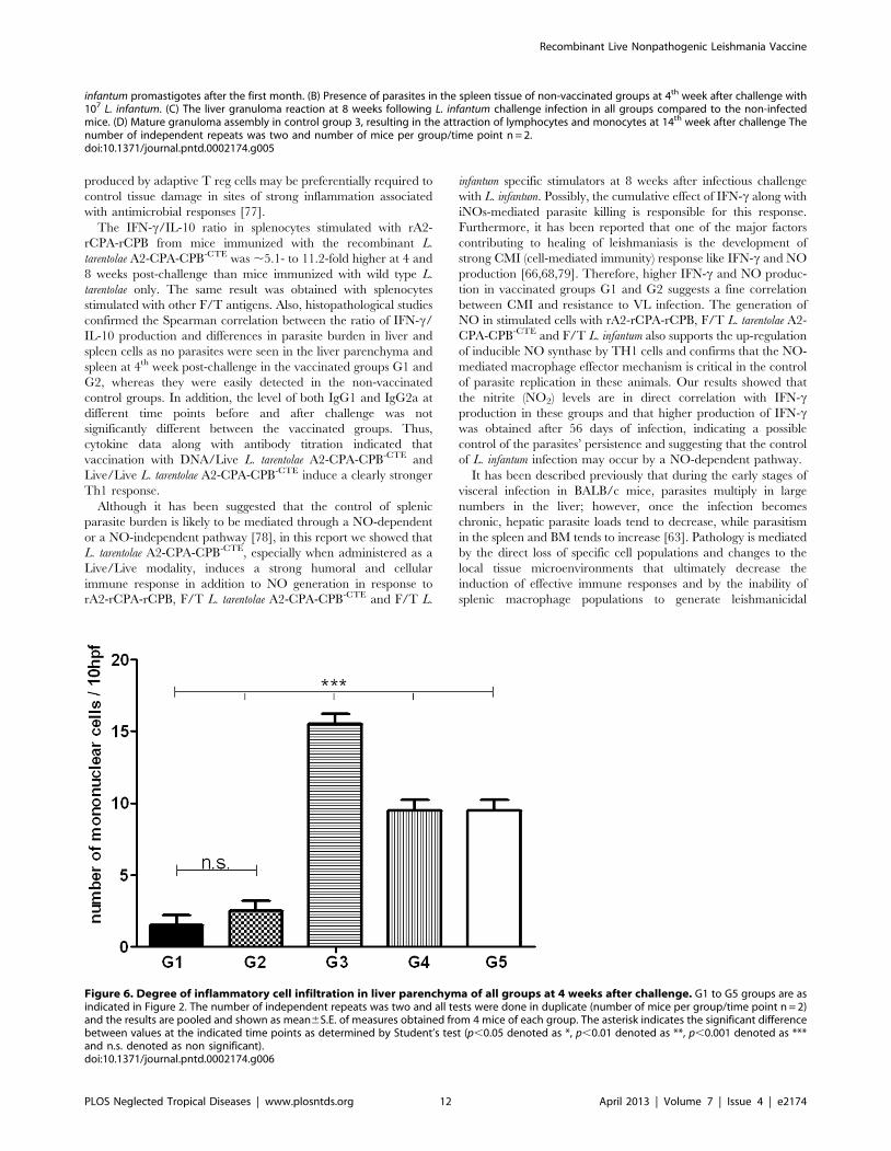
produced by adaptive T reg cells may be preferentially required to
control tissue damage in sites of strong inflammation associated
with antimicrobial responses [77].
The IFN-c/IL-10 ratio in splenocytes stimulated with rA2-
rCPA-rCPB from mice immunized with the recombinant L.
tarentolae A2-CPA-CPB-CTE was ,5.1- to 11.2-fold higher at 4 and
8 weeks post-challenge than mice immunized with wild type L.
tarentolae only. The same result was obtained with splenocytes
stimulated with other F/T antigens. Also, histopathological studies
confirmed the Spearman correlation between the ratio of IFN-c/
IL-10 production and differences in parasite burden in liver and
spleen cells as no parasites were seen in the liver parenchyma and
spleen at 4th week post-challenge in the vaccinated groups G1 and
G2, whereas they were easily detected in the non-vaccinated
control groups. In addition, the level of both IgG1 and IgG2a at
different time points before and after challenge was not
significantly different between the vaccinated groups. Thus,
cytokine data along with antibody titration indicated that
vaccination with DNA/Live L. tarentolae A2-CPA-CPB-CTE and
Live/Live L. tarentolae A2-CPA-CPB-CTE induce a clearly stronger
Th1 response.
Although it has been suggested that the control of splenic
parasite burden is likely to be mediated through a NO-dependent
or a NO-independent pathway [78], in this report we showed that
L. tarentolae A2-CPA-CPB-CTE, especially when administered as a
Live/Live modality, induces a strong humoral and cellular
immune response in addition to NO generation in response to
rA2-rCPA-rCPB, F/T L. tarentolae A2-CPA-CPB-CTE and F/T L.
infantum specific stimulators at 8 weeks after infectious challenge
with L. infantum. Possibly, the cumulative effect of IFN-c along with
iNOs-mediated parasite killing is responsible for this response.
Furthermore, it has been reported that one of the major factors
contributing to healing of leishmaniasis is the development of
strong CMI (cell-mediated immunity) response like IFN-c and NO
production [66,68,79]. Therefore, higher IFN-c and NO produc-
tion in vaccinated groups G1 and G2 suggests a fine correlation
between CMI and resistance to VL infection. The generation of
NO in stimulated cells with rA2-rCPA-rCPB, F/T L. tarentolae A2-
CPA-CPB-CTE and F/T L. infantum also supports the up-regulation
of inducible NO synthase by TH1 cells and confirms that the NO-
mediated macrophage effector mechanism is critical in the control
of parasite replication in these animals. Our results showed that
the nitrite (NO2) levels are in direct correlation with IFN-cproduction in these groups and that higher production of IFN-cwas obtained after 56 days of infection, indicating a possible
control of the parasites’ persistence and suggesting that the control
of L. infantum infection may occur by a NO-dependent pathway.
It has been described previously that during the early stages of
visceral infection in BALB/c mice, parasites multiply in large
numbers in the liver; however, once the infection becomes
chronic, hepatic parasite loads tend to decrease, while parasitism
in the spleen and BM tends to increase [63]. Pathology is mediated
by the direct loss of specific cell populations and changes to the
local tissue microenvironments that ultimately decrease the
induction of effective immune responses and by the inability of
splenic macrophage populations to generate leishmanicidal
infantum promastigotes after the first month. (B) Presence of parasites in the spleen tissue of non-vaccinated groups at 4th week after challenge with107 L. infantum. (C) The liver granuloma reaction at 8 weeks following L. infantum challenge infection in all groups compared to the non-infectedmice. (D) Mature granuloma assembly in control group 3, resulting in the attraction of lymphocytes and monocytes at 14th week after challenge Thenumber of independent repeats was two and number of mice per group/time point n = 2.doi:10.1371/journal.pntd.0002174.g005
Figure 6. Degree of inflammatory cell infiltration in liver parenchyma of all groups at 4 weeks after challenge. G1 to G5 groups are asindicated in Figure 2. The number of independent repeats was two and all tests were done in duplicate (number of mice per group/time point n = 2)and the results are pooled and shown as mean6S.E. of measures obtained from 4 mice of each group. The asterisk indicates the significant differencebetween values at the indicated time points as determined by Student’s test (p,0.05 denoted as *, p,0.01 denoted as **, p,0.001 denoted as ***and n.s. denoted as non significant).doi:10.1371/journal.pntd.0002174.g006
Recombinant Live Nonpathogenic Leishmania Vaccine
PLOS Neglected Tropical Diseases | www.plosntds.org 12 April 2013 | Volume 7 | Issue 4 | e2174
mechanisms or to recruit appropriate cells for eliminating the
parasite. Furthermore, the intensity of pathological changes in the
visceral organs of BALB/c mice can vary depending on the initial
inoculum size [63,80]. Pathological studies showed that after
infection with 107 L. infantum promastigotes, control groups
exhibited severe histopathological alterations in both the spleen
and liver at the peak of parasite burden. Once in the liver, the
development of cell-mediated immune responses is essential for the
clearance of L. infantum parasites. In contrast, the spleen ultimately
becomes the site of parasite persistence [63,81,82], suggesting that
it is more susceptible to L. infantum infection than the liver [83].
Interestingly, the leishmanicidal efficacy of hepatic granulomas is
dependent on their degree of maturation [63,84,85]. Among these
alterations, we detected the appearance of granulomas in different
maturation stages and giant cell granulomas in amastigotes in the
liver of all groups infected with L. infantum, which results in liver
parasite clearance. However, disruption of the splenic architecture
accompanied by lymphoid depletion only in control groups results
in spleen parasite persistence.
Overall, our results on cytokine production, humoral responses,
parasite burden and histopathological studies support that
immunization with the novel recombinant L. tarentolae A2-CPA-
CPB-CTE candidate vaccine protects mice against visceral leish-
maniasis when administered as a prime-boost modality more than
L. tarentolae [38] or L. tarentolae A2 [42]. The next step will be to
determine the long-term memory protection in mice or hamsters
and to evaluate the effectiveness of this promising live vaccine
against L. infantum in dogs as an important outbreed animal model
for VL research.
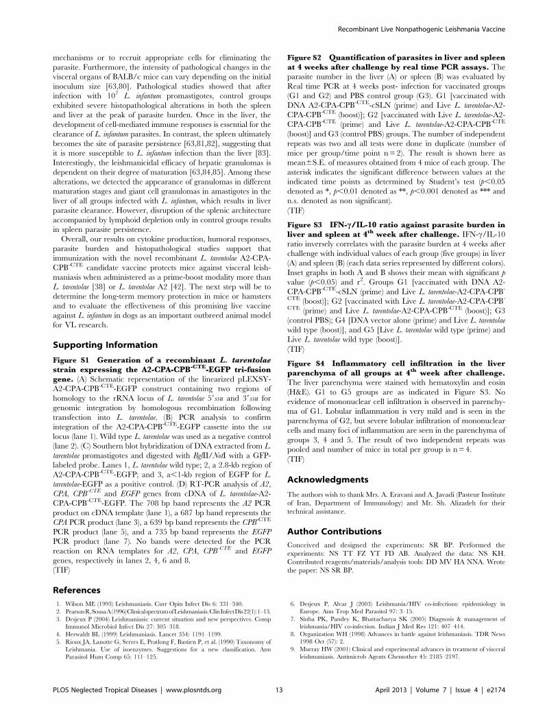
Supporting Information
Figure S1 Generation of a recombinant L. tarentolaestrain expressing the A2-CPA-CPB-CTE-EGFP tri-fusiongene. (A) Schematic representation of the linearized pLEXSY-
A2-CPA-CPB-CTE-EGFP construct containing two regions of
homology to the rRNA locus of L. tarentolae 59ssu and 39ssu for
genomic integration by homologous recombination following
transfection into L. tarentolae. (B) PCR analysis to confirm
integration of the A2-CPA-CPB-CTE-EGFP cassette into the ssu
locus (lane 1). Wild type L. tarentolae was used as a negative control
(lane 2). (C) Southern blot hybridization of DNA extracted from L.
tarentolae promastigotes and digested with BglII/NotI with a GFP-
labeled probe. Lanes 1, L. tarentolae wild type; 2, a 2.8-kb region of
A2-CPA-CPB-CTE-EGFP; and 3, a,1-kb region of EGFP for L.
tarentolae-EGFP as a positive control. (D) RT-PCR analysis of A2,
CPA, CPB-CTE and EGFP genes from cDNA of L. tarentolae-A2-
CPA-CPB-CTE-EGFP. The 708 bp band represents the A2 PCR
product on cDNA template (lane 1), a 687 bp band represents the
CPA PCR product (lane 3), a 639 bp band represents the CPB-CTE
PCR product (lane 5), and a 735 bp band represents the EGFP
PCR product (lane 7). No bands were detected for the PCR
reaction on RNA templates for A2, CPA, CPB-CTE and EGFP
genes, respectively in lanes 2, 4, 6 and 8.
(TIF)
Figure S2 Quantification of parasites in liver and spleenat 4 weeks after challenge by real time PCR assays. The
parasite number in the liver (A) or spleen (B) was evaluated by
Real time PCR at 4 weeks post- infection for vaccinated groups
(G1 and G2) and PBS control group (G3). G1 [vaccinated with
DNA A2-CPA-CPB-CTE-cSLN (prime) and Live L. tarentolae-A2-
CPA-CPB-CTE (boost)]; G2 [vaccinated with Live L. tarentolae-A2-
CPA-CPB-CTE (prime) and Live L. tarentolae-A2-CPA-CPB-CTE
(boost)] and G3 (control PBS) groups. The number of independent
repeats was two and all tests were done in duplicate (number of
mice per group/time point n = 2). The result is shown here as
mean6S.E. of measures obtained from 4 mice of each group. The
asterisk indicates the significant difference between values at the
indicated time points as determined by Student’s test (p,0.05
denoted as *, p,0.01 denoted as **, p,0.001 denoted as *** and
n.s. denoted as non significant).
(TIF)
Figure S3 IFN-c/IL-10 ratio against parasite burden inliver and spleen at 4th week after challenge. IFN-c/IL-10
ratio inversely correlates with the parasite burden at 4 weeks after
challenge with individual values of each group (five groups) in liver
(A) and spleen (B) (each data series represented by different colors).
Inset graphs in both A and B shows their mean with significant p
value (p,0.05) and r2. Groups G1 [vaccinated with DNA A2-
CPA-CPB-CTE-cSLN (prime) and Live L. tarentolae-A2-CPA-CPB-
CTE (boost)]; G2 [vaccinated with Live L. tarentolae-A2-CPA-CPB-
CTE (prime) and Live L. tarentolae-A2-CPA-CPB-CTE (boost)]; G3
(control PBS); G4 [DNA vector alone (prime) and Live L. tarentolae
wild type (boost)], and G5 [Live L. tarentolae wild type (prime) and
Live L. tarentolae wild type (boost)].
(TIF)
Figure S4 Inflammatory cell infiltration in the liverparenchyma of all groups at 4th week after challenge.The liver parenchyma were stained with hematoxylin and eosin
(H&E). G1 to G5 groups are as indicated in Figure S3. No
evidence of mononuclear cell infiltration is observed in parenchy-
ma of G1. Lobular inflammation is very mild and is seen in the
parenchyma of G2, but severe lobular infiltration of mononuclear
cells and many foci of inflammation are seen in the parenchyma of
groups 3, 4 and 5. The result of two independent repeats was
pooled and number of mice in total per group is n = 4.
(TIF)
Acknowledgments
The authors wish to thank Mrs. A. Eravani and A. Javadi (Pasteur Institute
of Iran, Department of Immunology) and Mr. Sh. Alizadeh for their
technical assistance.
Author Contributions
Conceived and designed the experiments: SR BP. Performed the
experiments: NS TT FZ YT FD AB. Analyzed the data: NS KH.
Contributed reagents/materials/analysis tools: DD MV HA NNA. Wrote
the paper: NS SR BP.
References
1. Wilson ME (1993) Leishmaniasis. Curr Opin Infect Dis 6: 331–340.
2. PearsonR,SousaA(1996)ClinicalspectrumofLeishmaniasis.ClinInfectDis22(1):1–13.
3. Desjeux P (2004) Leishmaniasis: current situation and new perspectives. Comp
Immunol Microbiol Infect Dis 27: 305–318.
4. Herwaldt BL (1999) Leishmaniasis. Lancet 354: 1191–1199.
5. Rioux JA, Lanotte G, Serres E, Pratlong F, Bastien P, et al. (1990) Taxonomy of
Leishmania. Use of isoenzymes. Suggestions for a new classification. Ann
Parasitol Hum Comp 65: 111–125.
6. Desjeux P, Alvar J (2003) Leishmania/HIV co-infections: epidemiology in
Europe. Ann Trop Med Parasitol 97: 3–15.
7. Sinha PK, Pandey K, Bhattacharya SK (2005) Diagnosis & management of
leishmania/HIV co-infection. Indian J Med Res 121: 407–414.
8. Organization WH (1998) Advances in battle against leishmaniasis. TDR News
1998 Oct (57): 2.
9. Murray HW (2001) Clinical and experimental advances in treatment of visceral
leishmaniasis. Antimicrob Agents Chemother 45: 2185–2197.
Recombinant Live Nonpathogenic Leishmania Vaccine
PLOS Neglected Tropical Diseases | www.plosntds.org 13 April 2013 | Volume 7 | Issue 4 | e2174

10. Sundar S, More D, Singh M, Singh V, Sharma S, et al. (2000) Failure ofpentavalent antimony in visceral leishmaniasis in India: report from the center of
the Indian epidemic. Clin Infect Dis 31: 1104–1107.
11. Modabber F (1995) Vaccines against leishmaniasis. Ann Trop Med Parasitol 89Suppl 1: 83–88.
12. Handman E (2001) Leishmaniasis: current status of vaccine development. Clin
Microbiol Rev 14: 229–243.
13. Sukumaran B, Madhubala R (2004) Leishmaniasis: current status of vaccinedevelopment. Curr Mol Med 4: 667–679.
14. Khamesipour A, Dowlati Y, Asilian A, Hashemi-Fesharki R, Javadi A, et al.(2005) Leishmanization: use of an old method for evaluation of candidate
vaccines against leishmaniasis. Vaccine 23: 3642–3648.
15. Nadim A, Javadian E, Tahvildar-Bidruni G, Ghorbani M (1983) Effectiveness ofleishmanization in the control of cutaneous leishmaniasis. Bull Soc Pathol Exot
Filiales 76: 377–383.
16. Evans KJ, Kedzierski L (2012) Development of Vaccines against VisceralLeishmaniasis. J Trop Med 2012: 892817.
17. Coler RN, Reed SG (2005) Second-generation vaccines against leishmaniasis.
Trends Parasitol 21: 244–249.
18. Piedrafita D, Xu D, Hunter D, Harrison RA, Liew FY (1999) Protective immune
responses induced by vaccination with an expression genomic library of
Leishmania major. J Immunol 163: 1467–1472.
19. Molano I, Alonso MG, Miron C, Redondo E, Requena JM, et al. (2003) A
Leishmania infantum multi-component antigenic protein mixed with live BCG
confers protection to dogs experimentally infected with L. infantum. VetImmunol Immunopathol 92: 1–13.
20. Coler RN, Goto Y, Bogatzki L, Raman V, Reed SG (2007) Leish-111f, a
recombinant polyprotein vaccine that protects against visceral Leishmaniasis byelicitation of CD4+ T cells. Infect Immun 75: 4648–4654.
21. Gradoni L, Foglia Manzillo V, Pagano A, Piantedosi D, De Luna R, et al. (2005)Failure of a multi-subunit recombinant leishmanial vaccine (MML) to protect
dogs from Leishmania infantum infection and to prevent disease progression in
infected animals. Vaccine 23: 5245–5251.
22. Bertholet S, Goto Y, Carter L, Bhatia A, Howard RF, et al. (2009) Optimized
subunit vaccine protects against experimental leishmaniasis. Vaccine 27: 7036–
7045.
23. Chakravarty J, Kumar S, Trivedi S, Rai VK, Singh A, et al. (2011) A clinical
trial to evaluate the safety and immunogenicity of the LEISH-F1+MPL-SE
vaccine for use in the prevention of visceral leishmaniasis. Vaccine 29: 3531–3537.
24. Belkaid Y, Piccirillo CA, Mendez S, Shevach EM, Sacks DL (2002)CD4+CD25+ regulatory T cells control Leishmania major persistence and
immunity. Nature 420: 502–507.
25. Uzonna JE, Wei G, Yurkowski D, Bretscher P (2001) Immune elimination ofLeishmania major in mice: implications for immune memory, vaccination, and
reactivation disease. J Immunol 167: 6967–6974.
26. Mitchell GF, Handman E, Spithill TW (1984) Vaccination against cutaneousleishmaniasis in mice using nonpathogenic cloned promastigotes of Leishmania
major and importance of route of injection. Aust J Exp Biol Med Sci 62 (Pt 2):
145–153.
27. Daneshvar H, Coombs GH, Hagan P, Phillips RS (2003) Leishmania mexicana
and Leishmania major: attenuation of wild-type parasites and vaccination withthe attenuated lines. J Infect Dis 187: 1662–1668.
28. Gorczynski RM (1985) Immunization of susceptible BALB/c mice against
Leishmania braziliensis. II. Use of temperature-sensitive avirulent clones ofparasite for vaccination purposes. Cell Immunol 94: 11–20.
29. Kimsey PB, Theodos CM, Mitchen TK, Turco SJ, Titus RG (1993) An
avirulent lipophosphoglycan-deficient Leishmania major clone induces CD4+ Tcells which protect susceptible BALB/c mice against infection with virulent L.
major. Infect Immun 61: 5205–5213.
30. Titus RG, Gueiros-Filho FJ, de Freitas LA, Beverley SM (1995) Development ofa safe live Leishmania vaccine line by gene replacement. Proc Natl Acad Sci U S A
92: 10267–10271.
31. Alexander J, Coombs GH, Mottram JC (1998) Leishmania mexicana cysteineproteinase-deficient mutants have attenuated virulence for mice and potentiate a
Th1 response. J Immunol 161: 6794–6801.
32. Hubel A, Krobitsch S, Horauf A, Clos J (1997) Leishmania major Hsp100 isrequired chiefly in the mammalian stage of the parasite. Mol Cell Biol 17: 5987–
5995.
33. Silvestre R, Cordeiro-Da-Silva A, Santarem N, Vergnes B, Sereno D, et al.(2007) SIR2-deficient Leishmania infantum induces a defined IFN-gamma/IL-
10 pattern that correlates with protection. J Immunol 179: 3161–3170.
34. Uzonna JE, Spath GF, Beverley SM, Scott P (2004) Vaccination withphosphoglycan-deficient Leishmania major protects highly susceptible mice
from virulent challenge without inducing a strong Th1 response. J Immunol 172:3793–3797.
35. Papadopoulou B, Roy G, Breton M, Kundig C, Dumas C, et al. (2002) Reduced
infectivity of a Leishmania donovani biopterin transporter genetic mutant and itsuse as an attenuated strain for vaccination. Infect Immun 70: 62–68.
36. Dumas C, Ouellette M, Tovar J, Cunningham ML, Fairlamb AH, et al. (1997)
Disruption of the trypanothione reductase gene of Leishmania decreases itsability to survive oxidative stress in macrophages. EMBO J 16: 2590–2598.
37. Raymond F, Boisvert S, Roy G, Ritt JF, Legare D, et al. (2012) Genome
sequencing of the lizard parasite Leishmania tarentolae reveals loss of genes
associated to the intracellular stage of human pathogenic species. Nucleic AcidsRes 40: 1131–1147.
38. Breton M, Tremblay MJ, Ouellette M, Papadopoulou B (2005) Live
nonpathogenic parasitic vector as a candidate vaccine against visceralleishmaniasis. Infect Immun 73: 6372–6382.
39. Wilson V, Southgate B (1978) Lizard leishmania. Biology of the Kinetoplastida.
San Diego, CA: Academic Press: 244–268.
40. Charest H, Matlashewski G (1994) Developmental gene expression inLeishmania donovani: differential cloning and analysis of an amastigote-stage-
specific gene. Mol Cell Biol 14: 2975–2984.
41. Azizi H, Hassani K, Taslimi Y, Najafabadi HS, Papadopoulou B, et al. (2009)
Searching for virulence factors in the non-pathogenic parasite to humans
Leishmania tarentolae. Parasitology 136: 723–735.
42. Mizbani A, Taheri T, Zahedifard F, Taslimi Y, Azizi H, et al. (2009)
Recombinant Leishmania tarentolae expressing the A2 virulence gene as a novel
candidate vaccine against visceral leishmaniasis. Vaccine 28: 53–62.
43. Rafati S, Salmanian AH, Hashemi K, Schaff C, Belli S, et al. (2001)
Identification of Leishmania major cysteine proteinases as targets of the immune
response in humans. Mol Biochem Parasitol 113: 35–43.
44. Rafati S, Nakhaee A, Taheri T, Ghashghaii A, Salmanian AH, et al. (2003)
Expression of cysteine proteinase type I and II of Leishmania infantum and theirrecognition by sera during canine and human visceral leishmaniasis. Exp
Parasitol 103: 143–151.
45. Coombs GH, Mottram JC (1997) Proteinases of trypanosomes and Leishmania:biology and control. London, UK: Oxford CAB International. pp. 177–179.
46. Robertson CD, Coombs GH, North MJ, Mottram JC (1996) Parasite cysteine
proteinases. Perspectives in Drug Discovery and Design. ESCOM SciencePublisher 6: 99.
47. Mottram JC, North MJ, Barry JD, Coombs GH (1989) A cysteine proteinase
cDNA from Trypanosoma brucei predicts an enzyme with an unusual C-terminal extension. FEBS Letters 258: 211–215.
48. Huete-Perez J, Engel J, Brinen L, Mottram JC, McKerrow J (1999) Protease
trafficking in two primitive eukaryotes is mediated by a prodomain protein motif.Journal of Biological Chemistry 274: 16249–16256.
49. Rafati S, Zahedifard F, Azari MK, Taslimi Y, Taheri T (2008) Leishmania
infantum: prime boost vaccination with C-terminal extension of cysteineproteinase type I displays both type 1 and 2 immune signatures in BALB/c
mice. Exp Parasitol 118: 393–401.
50. Rafati S, Nakhaee A, Taheri T, Taslimi Y, Darabi H, et al. (2005) Protectivevaccination against experimental canine visceral leishmaniasis using a combi-
nation of DNA and protein immunization with cysteine proteinases type I and IIof L. infantum. Vaccine 23: 3716–3725.
51. Rafati S, Zahedifard F, Nazgouee F (2006) Prime-boost vaccination using
cysteine proteinases type I and II of Leishmania infantum confers protectiveimmunity in murine visceral leishmaniasis. Vaccine 24: 2169–2175.
52. Rafati S, Salmanian AH, Taheri T, Vafa M, Fasel N (2001) A protective cocktail
vaccine against murine cutaneous leishmaniasis with DNA encoding cysteineproteinases of Leishmania major. Vaccine 19: 3369–3375.
53. Zadeh-Vakili A, Taheri T, Taslimi Y, Doustdari F, Salmanian AH, et al. (2004)
Immunization with the hybrid protein vaccine, consisting of Leishmania majorcysteine proteinases Type I (CPB) and Type II (CPA), partially protects against
leishmaniasis. Vaccine 22: 1930–1940.
54. Doroud D, Zahedifard F, Vatanara A, Taslimi Y, Vahabpour R, et al. (2011) C-
terminal domain deletion enhances the protective activity of cpa/cpb loaded
solid lipid nanoparticles against Leishmania major in BALB/c mice. PLoS NeglTrop Dis 5: e1236.
55. Greenland JR, Letvin NL (2007) Chemical adjuvants for plasmid DNA vaccines.
Vaccine 25: 3731–3741.
56. Doroud D, Vatanara A, Zahedifard F, Gholami E, Vahabpour R, et al. (2010)
Cationic solid lipid nanoparticles loaded by cysteine proteinase genes as a novel
anti-leishmaniasis DNA vaccine delivery system: characterization and in vitroevaluations. J Pharm Pharm Sci 13: 320–335.
57. Papadopoulou B, Roy G, Ouellette M (1992) A novel antifolate resistance geneon the amplified H circle of Leishmania. EMBO J 11: 3601–3608.
58. Sambrook J, Fritsch E, Maniatis T (1989) Molecular Cloning: A Laboratory
Manual. Cold Spring Harbor, NY: Cold Spring Harbor Press.
59. Doroud D, Zahedifard F, Vatanara A, Najafabadi AR, Taslimi Y, et al. (2011)Delivery of a cocktail DNA vaccine encoding cysteine proteinases type I, II and
III with solid lipid nanoparticles potentiate protective immunity againstLeishmania major infection. J Control Release 153: 154–162.
60. Viana da Costa A, Huerre M, Delacre M, Auriault C, Correia Costa JM, et al.
(2002) IL-10 leads to a higher parasite persistence in a resistant mouse model ofLeishmania major infection. Parasitol Int 51: 367–379.
61. Buffet PA, Sulahian A, Garin YJ, Nassar N, Derouin F (1995) Culture
microtitration: a sensitive method for quantifying Leishmania infantum in tissuesof infected mice. Antimicrob Agents Chemother 39: 2167–2168.
62. Lachaud L, Marchergui-Hammami S, Chabbert E, Dereure J, Dedet JP, et al.
(2002) Comparison of Six PCR Methods Using Peripheral Blood for Detectionof Canine Visceral Leishmaniasis. Journal of Clinical Microbiology 40: 210–215.
63. Carrion J, Nieto A, Iborra S, Iniesta V, Soto M, et al. (2006) Immunohistological
features of visceral leishmaniasis in BALB/c mice. Parasite Immunol 28: 173–183.
64. Alexander J, McFarlane E (2008) Can type-1 responses against intracellular
pathogens be T helper 2 cytokine dependent? Microbes Infect 10: 953–959.
Recombinant Live Nonpathogenic Leishmania Vaccine
PLOS Neglected Tropical Diseases | www.plosntds.org 14 April 2013 | Volume 7 | Issue 4 | e2174

65. Modabber F (2010) Leishmaniasis vaccines: past, present and future.
Int J Antimicrob Agents 36 Suppl 1: S58–61.
66. Kaye PM, Curry AJ, Blackwell JM (1991) Differential production of Th1- and
Th2-derived cytokines does not determine the genetically controlled or vaccine-
induced rate of cure in murine visceral leishmaniasis. J Immunol 146: 2763–
2770.
67. Alexander J, Bryson K (2005) T helper (h)1/Th2 and Leishmania: paradox
rather than paradigm. Immunol Lett 99: 17–23.
68. Wilson ME, Jeronimo SM, Pearson RD (2005) Immunopathogenesis of infection
with the visceralizing Leishmania species. Microb Pathog 38: 147–160.
69. Rodrigues OR, Marques C, Soares-Clemente M, Ferronha MH, Santos-Gomes
GM (2009) Identification of regulatory T cells during experimental Leishmania
infantum infection. Immunobiology 214: 101–111.
70. Handman E (1997) Leishmania vaccines: old and new. Parasitol Today 13: 236–
238.
71. Handman E (2001) Protective saliva: a novel approach to a Leishmania vaccine.
Trends Parasitol 17 513–514.
72. Streit JA, Recker TJ, Filho FG, Beverley SM, Wilson ME (2001) Protective
immunity against the protozoan Leishmania chagasi is induced by subclinical
cutaneous infection with virulent but not avirulent organisms. J Immunol 166:
1921–1929.
73. Zhang WW, Matlashewski G (2001) Characterization of the A2-A2rel gene
cluster in Leishmania donovani: involvement of A2 in visceralization during
infection. Mol Microbiol 39: 935–948.
74. Selvapandiyan A, Debrabant A, Duncan R, Muller J, Salotra P, et al. (2004)
Centrin gene disruption impairs stage-specific basal body duplication and cell
cycle progression in Leishmania. J Biol Chem 279: 25703–25710.
75. Nylen S, Maurya R, Eidsmo L, Manandhar KD, Sundar S, et al. (2007)
Splenic accumulation of IL-10 mRNA in T cells distinct from CD4+CD25+(Foxp3) regulatory T cells in human visceral leishmaniasis. J Exp Med 204:
805–817.
76. Gerosa F, Paganin C, Peritt D, Paiola F, Scupoli MT, et al. (1996) Interleukin-12
primes human CD4 and CD8 T cell clones for high production of bothinterferon-gamma and interleukin-10. J Exp Med 183: 2559–2569.
77. O’Garra A, Vieira PL, Vieira P, Goldfeld AE (2004) IL-10-producing and
naturally occurring CD4+ Tregs: limiting collateral damage. J Clin Invest 114:1372–1378.
78. Murray HW, Delph-Etienne S (2000) Roles of endogenous gamma interferonand macrophage microbicidal mechanisms in host response to chemotherapy in
experimental visceral leishmaniasis. Infect Immun 68: 288–293.
79. Haldar JP, Ghose S, Saha KC, Ghose AC (1983) Cell-mediated immuneresponse in Indian kala-azar and post-kala-azar dermal leishmaniasis. Infect
Immun 42: 702–707.80. Nieto A, Domınguez-Bernal G, Orden JA, Fuente RDL, Madrid-Elena N, et al.
(2011) Mechanisms of resistance and susceptibility to experimental visceralleishmaniosis: BALB/c mouse versus syrian hamster model. Veterinary
Research 42: 39.
81. Stanley AC, Engwerda CR (2007) Balancing immunity and pathology in visceralleishmaniasis. Immunol Cell Biol 85: 138–147.
82. Bankoti R, Stager S (2012) Differential Regulation of the Immune Response inthe Spleen and Liver of Mice Infected with Leishmania donovani. J Trop Med
2012: 639304.
83. Rolao N, Cortes S, Gomes-Pereira S, Campino L (2007) Leishmania infantum:mixed T-helper-1/T-helper-2 immune response in experimentally infected
BALB/c mice. Exp Parasitol 115: 270–276.84. Murray HW, Squires KE, Miralles CD, Stoeckle MY, Granger AM, et al. (1992)
Acquired resistance and granuloma formation in experimental visceralleishmaniasis. Differential T cell and lymphokine roles in initial versus
established immunity. J Immunol 148: 1858–1863.
85. Stager S, Alexander J, Carter KC, Brombacher F, Kaye PM (2003) Bothinterleukin-4 (IL-4) and IL-4 receptor alpha signaling contribute to the
development of hepatic granulomas with optimal antileishmanial activity. InfectImmun 71: 4804–4807.
Recombinant Live Nonpathogenic Leishmania Vaccine
PLOS Neglected Tropical Diseases | www.plosntds.org 15 April 2013 | Volume 7 | Issue 4 | e2174