development of pharyngeal apparatus and palate
TRANSCRIPT

Development of pharyngeal
apparatus and palate
By
Dr. Abdul Waheed Ansari
Chairperson &Prof. Anatomy,
RAKCODS. RAKMHSU.
12/18/2014 1

Learning outcomes for this developmental topic
• What are the components of pharyngeal apparatus?
• Enlist the derivatives of all pharyngeal arches
• Mention the derivatives of pharyngeal pouches
• What are the derivatives of pharyngeal clefts?
• Name the developmental sequences of hard and soft palate
• Explain the congenital forms of pharyngeal apparatus
12/18/2014 2

The pharyngeal apparatus are the bronchial apparatus
developing at the cranial end of embryo during 4th &5th
week of embryogenesis
• The result of the pharyngeal apparatus are the development of face, neck, organs like tongue, tonsil, thyroid, parathyroid, thymus, middle ear, muscles of face, muscles of mastication, laryngeal and pharyngeal muscles.
• The pharyngeal arches are bilateral/paired swellings that surround the foregut of the embryo from day 20 to day 35 of development .
• These arches are numbered 1,2,3,4, and 6.
12/18/2014 3
On approximately day 20 of development
• The first arch develops, followed by the second and the third.
• By the time that arches 4 and 6 develop, the first two arches are no longer distinctly visible externally.
• The cellular elements of the pharyngeal apparatus are formed by cells that are derived from ectoderm, endoderm, mesoderm and neural crest cells.
• Ectoderm lines the external surfaces of the pharyngeal arches.
• The ectodermally lined depressions between the pharyngeal arches are called pharyngeal grooves.
12/18/2014 4

Components of a pharyngeal arch
• Each arch has its own artery, nerve, cartilage rod “skeleton”, and a group of muscle cells.
• The first structure to develop in each arch is the aortic (pharyngeal) arch artery.
• The arteries, like the arches, develop in a rostro-caudal sequence.
• While the fourth aortic arch artery is forming, the arteries of the first and second arches are breaking up and disappear.
• The third arch artery becomes part of the common carotid, the fourth arch artery becomes the arch of the aorta, and the sixth becomes the pulmonary artery.
12/18/2014 5

Nerves and muscles of pharyngeal archesPharyngeal arch Cranial nerve Muscles
12/18/2014 6
1st Trigeminal (V) Muscles of mastication
2nd Facial (VII) Muscles of facial
expression
3rd Glossopharyngeal
(VIII)
Stylopharyngeus
4th
6th
Vagus (X)
External laryngeal
nerve
Recurrent laryngeal
nerve
Laryngeal musculature
Pharyngeal constrictors

Endoderm lines the internal surfaces of
the pharyngeal arches.
• The endodermally lined depressions between the pharyngeal arches are called pharyngeal pouches.
• They will form endocrine glands, tonsillar crypts and thymus.
• The tongue is formed by the endoderm of the floor of mouth by 2 lateral lingual swellings, median lingual swelling, cupola of His and hypobranchial eminence.
12/18/2014 7

12/18/2014 8

The embryonic composition of
pharyngeal arch
• The core is made up of mesenchyme covered externally by ectoderm and internally by endoderm.
• The first pharyngeal arch separates into two prominences, the maxillary and mandibular prominences.
• The maxillary prominence gives rise to the maxilla, zygomatic bone, and a portion of the vomer bone.
• The mandibular prominence forms the mandible and the squamous temporal bone.
• The second pharyngeal arch (hyoid arch) contributes, along with parts of the third and fourth arches, to the formation of the hyoid bone.
• The cartilages of larynx develops from 6th arch.12/18/2014 9

12/18/2014 10

Pharyngeal pouches are endodermal
• They protrude between the two succeeding arches from inside.
• The first pouch is between first and second arch. It give rise to middle ear cavity and auditory tube.
• The second pouch will form the tonsillar crypts.
• The third pouch develops into inferior parathyroid glands and thymus.
• The fourth pouch will form the superior parathyroid glands.
• The distal pharyngeal complex will form ultimo bronchial body- the parafollicular cells/”C” cells of thyroid gland.
• The thyroid gland perse arises from a diverticulum from the floor of oral cavity, thyroglossal duct. This duct descends inferiorly and settle down in neck.
12/18/2014 11

12/18/2014 12

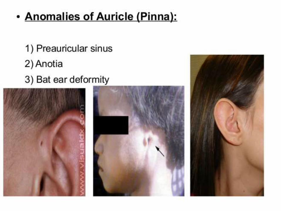
The derivative of first pharyngeal cleft
• It forms the external acoustic meatus.• The membrane so farmed between the cleft and
pouch remains as tympanic membrane.• There are very little elements of mesoderm
between first cleft and pouch.• The remaining pharyngeal cleft closes down. If
they persists, they form cervical cyst or sinus.• The external ear is formed around the brim of
first and second arches externally, by six ear hillocks, which later forms the external pinna.
12/18/2014 13

External ear development
12/18/2014 14
12/18/2014 15

Origin of the pharyngeal arch musculature
1st pharyngeal arches Mm. masseter, temporalis,
pterygoideus (12)
Mandibular nerve from the
trigeminal nerve
(HN V 3)
2nd pharyngeal arches Mimic musculature:
Mm. Stylohyoideus, digastricus
(venter posterior) and stapedius
Facial nerve
(HN VII)
3rd pharyngeal arches M. stylopharyngeus N. glossopharyngeus (HN IX)
4th pharyngeal arches Almost the entire pharynx and
larynx musculature
N. vagus and accessory
(HN X and XI)
5th pharyngeal arches and
cervical somites
M. sternocleidomastoideus and
trapezius
N. accessory
(HN XI)
12/18/2014 16

Development of palate
• The face develops from 5 prominences, frontonasal, right and left maxillary and right and left mandibular prominences.
• The maxillary prominences sends out a horizontal shelves, the palatine shelves from inside. This later fuses in the mid line separating the nasal cavity from the oral cavity.
• The posterior 1/3rd of palate is soft and the palatine muscles contribute to its formation. Failure of closure of palatine shelves in the midline can give rise to cleft palate or cleft lip.
• A bifid uvula is a minor degree of non-fusion of two palatine shelves.
12/18/2014 17

Development of upper lip
12/18/2014 18
Philtrum= medial nasal processes
Lateral part of upper lip= maxillary processes

The palate development
• Primary palate is formed fusion of medial nasal processes.
• The secondary palate is the outgrowth from the maxillary prominences medially inside and when tongue is growing from below the two halves of palatine shelves fuse together in the midline.
12/18/2014 19

Fusion of palatine shelves forming palate.
The four maxillary incisor teeth develop in pre-maxilla
12/18/2014 20
Incidences of cleft palate
• Roughly 1 out of every 900 children are born with a cleft palate.
• About 70% of these children will also have a cleft lip.
• The other one third will have only a cleft palate.
12/18/2014 21

Cleft hard and soft palate due to non fusion of palatine processes
with each other
12/18/2014 22

Cleft lip due to non fusion of frontonasal and maxillary processes
12/18/2014 23

The congenital forms of pharyngeal apparatus• The cervical cyst and fistulas are
congenital malformation when the 2nd pharyngeal arch fails to grow caudally over the 3rd and 4th arches.
• They usually occur in carotid triangle.
• Axial CT scan showing a right branchial cleft cyst
12/18/2014 24

Thyroglossal duct cysts
• Are remnants of the embryonic thyroglossal duct that may occur anywhere from the base of the tongue to the thyroid gland.
• The majority, however, are found at the level of the thyrohyoid membrane, under the deep cervical fascia.
• They are midline or just off the midline, and move up and down upon swallowing.
12/18/2014 25

Mandibulofacial Dysostosis (Treacher Collins Syndrome)
12/18/2014 26
This malformation due to disturbed development of neural crest cells, malformation of mandible and external ear.
Hypoplasia of first arch elements.

References
• http://quizlet.com/16788403/embryology-pharyngeal-apparatus-flash-cards/
• http://quizlet.com/6930680/gross-anatomy-development-of-the-pharyngeal-apparatus-the-head-and-neck-flash-cards
• http://www.columbia.edu/itc/hs/medical/humandev/2004/Chapt9-PharyngealArches.pdf
• http://www.ghorayeb.com/BranchialCleft.html
• http://www.youtube.com/watch?v=r18fexJ-UnE
• http://www.youtube.com/watch?v=rlA6ncGwwAU
• https://web.duke.edu/anatomy/embryology/craniofacial/craniofacial.html
• https://syllabus.med.unc.edu/courseware/embryo_images/unit-hednk/hednk_htms/hednk033.htm
12/18/2014 27