development of vaccine conjugates based on dengue …opus.bath.ac.uk/57424/1/ali_s_mphil.pdf ·...
TRANSCRIPT

Development of vaccine conjugates based
on Dengue virus using a staphylococcal
immune evasion protein
Ali Almansoor
A thesis submitted for the degree of Master of Philosophy
(University of Bath)
Department of Biology and Biochemistry
2017
i
COPYRIGHT
Attention is drawn to the fact that copyright of this thesis rests with the author. A
copy of this thesis has been supplied on condition that anyone who consults it is
understood to recognise that its copyright rests with the author and that they must
not copy it or use material from it except as permitted by law or with the consent of
the author.
If you wish to include copyright material belonging to others in your thesis, you are
advised to check with the copyright owner that they will give consent to the
inclusion and public availability online of any of their material in the thesis. If the
material is to be copied other than by photocopying or facsimile then the request
should be put to the publisher or the author in accordance with the copyright
declaration in the volume concerned. If, however, a facsimile or photocopy will be
included, then it is appropriate to write to the publisher alone for consent.
This thesis may be made available for consultation within the University Library and
may be photocopied or lent to other libraries for the purposes of consultation with
effect from...................(date)
Signed on behalf of the Faculty/School of Department of Biology and Biochemistry

ii
Table of contents 1 Introduction ......................................................................................................... 2
1.1 Current treatment ......................................................................................... 4 1.2 Innate immunity........................................................................................... 6 1.3 The complement system .............................................................................. 8 1.4 Complement C3d and Staphylococcal Sbi protein as an adjuvant strategy 13 1.5 Dengue non-structural protein 1 (NS1) as a vaccine antigen .................... 15 1.6 Outline of the project objectives ................................................................ 16
2 Materials and Methods ...................................................................................... 18 2.1 Epression and purification of recombinant Sbi-III-IV-NS1 ...................... 18
2.1.1 Cloning .................................................................................................. 18 2.1.2 Expression of Sbi-III-IV-NS1 using E. coli BL21 (DE3) ..................... 18 2.1.3 Purification ............................................................................................ 19
2.2 Isolation of the Sbi-III-IV-NS1 plasmid DNA .......................................... 19 2.3 Expression of Sbi-III-IV using Rosetta ™ 2(DE3) ................................... 20 2.4 Sodium Dodecyl Sulphate Polyacrylamide Gel Electrophoresis (SDS-PAGE) 21 2.5 Optimization of the expression and purification of inclusion bodies ........ 22
2.5.1 Optimization of the expression of recombinant Sbi-III-IV-NS1 ........... 22 2.5.2 Washing and Processing Protocol for Inclusion Bodies........................ 22 2.5.3 Inclusion Bodies – Optimizing solubilisation and refolding ................. 23 2.5.4 Purification using immobilized metal affinity chromatography ........... 24
2.6 Measurement of the protein concentration ................................................ 24 2.7 Complement activation assay assessment using the Wielisa kit ............... 24 2.8 Circular dichroism (CD) ............................................................................ 26 2.9 Western blotting ........................................................................................ 26
3 Results ............................................................................................................... 29 3.1 Expression and purification of recombinant Sbi-III-IV-NS1 .................... 29 3.2 Expression of Sbi-III-IV-NS1 into Rosetta ™ 2(DE3) ............................. 32 3.3 Optimisation of the purification of recombinant Sbi-III-IV-NS1 from inclusion bodies using Nickel column affinity chromatography ........................... 33 3.4 Complement activation assay assessment using the Wieslab kit .............. 37 3.5 Circular dichroism (CD) ............................................................................ 38 3.6 Western blotting ........................................................................................ 41
4 Discussion.......................................................................................................... 43
5 Summary, conclusions and future work ............................................................ 50
6 References ......................................................................................................... 53
7 Appendices ........................................................................................................ 60 7.1 Appendix 1 ................................................................................................ 60
7.1.1 Sbi-III-IV-NS1 conjugate. ..................................................................... 60 7.2 Appendix 2 ................................................................................................ 62 7.3 Appendix 3 ................................................................................................ 64
7.3.1 Buffers and solutions ............................................................................. 64 7.4 Appendix 4 ................................................................................................ 66

iii
7.4.1 Media ..................................................................................................... 66

iv
Table of figures Figure 1: Dengue virus genome diagram shows the capsid (C), envelope (E) and membrane (M) proteins and the other 7 are non-structural proteins ........................... 3 Figure 2: Schematic representation of the roles of innate and adaptive immunity in responding to infectious disease (30) .......................................................................... 7 Figure 3: Schematic representation of the complement pathways ............................ 10 Figure 4: Schematic representation of the domain structure of Sbi taken from (60) 13 Figure 5: diagrammatic representation of how C3d binding to a target antigen via CR2 ............................................................................................................................ 14 Figure 6: Expression trials for Sbi-III-IV-NS1 ......................................................... 30 Figure 7: Purification of Sbi-III-IV-NS1 on AKTA affinity chromatography at 25°C ................................................................................................................................... 30 Figure 8: Expression trials for Sbi-III-IV-NS1 ......................................................... 31 Figure 9: Purification of Sbi-III-IV-NS1 on AKTA ion affinity chromatography at 16°C ........................................................................................................................... 31 Figure 10: Expression of Sbi-III-IV-NS1 in Rosetta cells ........................................ 33 Figure 11: Screening of the expressed Sbi-III-IV-NS1 protein in different re-folding buffers ........................................................................................................................ 34 Figure 12: Purification of Sbi-III-IV-NS1 following incubation and refolding using buffer 4 ...................................................................................................................... 35 Figure 13: Purification of Sbi-III-IV-NS1 following incubation and refolding using buffer 8 ...................................................................................................................... 35 Figure 14: Purification of Sbi-III-IV-NS1 following incubation and refolding using buffer 12 .................................................................................................................... 36 Figure 15: Functional activity assessment of the Sbi-III-IV-NS1 fusion protein in complement activation............................................................................................... 37 Figure 16: Smoothed data of 3 scans of the CD spectra of the Sbi-III-IV-NS1 fusion protein in PBS............................................................................................................ 38 Figure 17: CD spectra of Sbi-III-IV-NS1 fusion protein in PBS .............................. 39 Figure 18: Smoothed data of 3 scans of the CD spectra of the Sbi-III-IV-NS1 fusion protein in PBS............................................................................................................ 40 Figure 19: Western blot of Sbi-III-IV-NS1 ............................................................... 41

v
Acknowledgments
I would like to express my appreciation to my supervisor professor Jean van den
Elsen for giving me the opportunity to be a member of his research group and for all
help which was given to me by him. I extend my thanks to Prof Sue Wonnacott for
her advice and patience. Special thanks to Dr Mareike Posner for her time, patience
and support and also thanks for Dr. Abhishek Upadhyay.
My gratitude also goes to Marjorie Gibbon, Chase, and Omar, there are not enough
words to describe their friendly behaviour.

vi
Abstract
Dengue viruses (DENV) are members of the Flavivirus family and are thought to
infect up to 400 million people per annum and have the potential to lead to large
epidemics with subsequent human and economic consequences. The 2 current
vaccines being developed are at the clinical trial stage and focus on the induction of
neutralising antibodies but there are at least 5 different serotypes of the virus and it
appears that induction of immunity to any one serotype will not subsequently
provide cross protection to the others. The purpose of this project is to develop an
immunisation strategy that will provide protection across all serotypes. A common
antigen expressed by all serotypes is designated NS1 which is a non-structural
protein expressed on the surface of infected cells. However, on its own NS1 is
poorly immunogenic so the intention here is to develop a vector system that
combines the expression of NS1 epitopes with those of a highly immunogenic
protein derived from Staphylococcus aureus the Staphylococcal immunoglobulin-
binding protein, Sbi. Sbi is composed of a number of separate domains each of
which has characteristic interactive properties with C3. Previous studies have shown
that Sbi N-terminal domain IV (Sbi-IV) binds to C3 and its proteolytic fragments,
however, the Sbi domains III and IV are essential for fluid phase consumption of C3
activating via the alternative complement pathway. In this project the domains III
and IV of Sbi have been cloned and expressed in E. coli and conjugated to NS1.
These can bind to complement C3, causing cleavage, and lead to the generation of
C3d which is able to crosslink binding between the NS1 antigen and complement
receptor 2 (CR2) present on phagocytic cells and B cells to greatly enhance a TH2
(antibody) mediated response and ultimately, the generation of memory TH2 cells to
provide expansion of B cells (plasma cells) producing DENV specific neutralising
antibodies.
vii
List of Abbreviations
AMP Ampicillin
APS Ammonium persulphate
IPTG Isopropyl- β-D-thiogalactoside
LB Luria broth
CBB Coomassie Brilliant Blue
CDS Coomassie de-staining solution
E.coli Escherichia coli
WIESLAB® Enzyme immunoassay for assessment of Complement
functional activity
CP Classical pathway of complement system
AP Alternative pathway of complement system
LP Lectin pathway of complement system
DENF Dengue Fever
DENV Dengue Virus
NS1 Non-structural protein of Dengue Virus
Sbi Staphylococcal immunoglobulin-binding protein
C3 Complement component 3
CR2 complement receptor 2
BL21 competent E. coli cells
RT Room temperature
CD Circular Dichroism
OD Optical density
DMSO Dimethyl sulfoxide
DMSO Dimethyl sulfoxide
DNA Deoxyribonucleic acid
mRNA Messenger RNA
SDS Sodium dodecyl sulphate
SDS-PAGE Sodium dodecyl sulphate polyacrylamide gel
electrophoresis
TBS Tris buffered saline
TEMED N,N,N’,N, tetramethylenediamine

viii
PCR Polymerase chain reaction
RNA Ribonucleic acid
ELISA Enzyme-linked immunosorbent assay
PRRs Pathogen recognition receptors
PAMPs Pathogen-associated molecular patterns
LTA Lipoteichoic acid
LPS Lipopolysaccaride
MBL Mannose Binding Lectin
MASPs Mannose binding lectin Associated Serine Protease
FD Factor D
FB Factor D
MAC Membrane Attack Complex
PNH Paroxysmal Nocturnal Hemoglobinuria
aHUS atypical Haemolytic Uremic Syndrome
AMD Age-related Macular Degeneration
FDC follicular dendritic cells
BCR B cell surface receptors
Abs Antibodies
DHF Dengue hemorrhagic fever
DSS Dengue shock syndrome
LB Luria-Bertani
DDT DL-Dithiothreitol
PBS Phosphate buffered saline
TBS Tris-buffered saline
PEG polyethylene glycol

Chapter one: Introduction
1
Chapter one: Introduction

Chapter one: Introduction
2
1 Introduction
Dengue is a viral disease transmitted and spread largely by the mosquito genus
Aedes aegypti (and to some extent A. Albopictus) which is easily recognised by the
silvery white pattern on its scales and has become endemic in many tropical areas of
Africa and Asia. The virus is largely spread by female mosquitoes when they prey
on large mammals and humans as they require a blood meal to produce eggs. The
virus is normally present in the salivary glands of the mosquito, so upon biting the
host the virus is transmitted and then propagates in the host (1, 2).
The mosquito – human - mosquito cycle of transmission leads to a typical cycle
being: infected mosquito bite leading to (after about 4 days) acute viremia in the
host which usually lasts for about 5 days and then the development of symptoms of
dengue which can last for a week. The mosquito vectors become infected when they
feed on humans during the four/five-day period of viraemia. The virus passes from
the mosquito intestinal tract to the salivary glands after an extrinsic incubation
period, a process that takes approximately 10 days and is most rapid at high ambient
temperatures (3). Mosquito bites after the extrinsic incubation period result in
infection, which might be promoted by mosquito salivary proteins (4).
Although the many cases of Dengue fever are misclassified or indeed under-reported
it is generally recognised that there are as many as 400 million Dengue infections
per annum of which about a quarter are shown as clinically relevant infections. From
these figures published by the World Health Organisation (WHO) it has been
estimated that up to half a million people require hospitalisation because of Dengue
fever per annum (5). Following infection there are no specific drug treatments
available and patient survival depends largely on maintaining patient homeostasis
through transfusion of fluids.
The Dengue virus is a positive sense single stranded RNA virus with 10 encoded
genes. The genome is translated as a single polypeptide which is subsequently
cleaved into the 10 encoded proteins. Of these 10, three are capsid (C), envelope (E)

Chapter one: Introduction
3
and membrane (M) proteins and the other 7 are non-structural proteins that are
important in viral replication and assembly (Figure 1)(6, 7).
Figure 1: Dengue virus genome diagram shows the capsid (C), envelope (E) and membrane (M) proteins and the other 7 are non-structural proteins
Over a period of time it has become clear that there are a number of different
serotypes of Dengue virus defined by variations in the structure of vial envelope
proteins and these have been defined as DEN-1, DEN-2, DEN-3 and DEN-4. The
serotype classification is largely based on E envelope gene sequence data (1485 bp)
defining the 4 distinct serotypes but advanced sequencing techniques are likely to
uncover even greater variations including the recently described 5 serotype (DENV-
5). DENV-2 is associated with most severe disease progression with a greater
propensity for transmissibility, increased virulence and faster replication (8, 9).
Recovery from infection by one serotype provides lifelong immunity against that
particular serotype. However, cross-immunity to the other serotypes after recovery is
only partial and temporary. Subsequent infections by other serotypes increases the
risk of developing severe dengue fever (10).
It has been proposed that the process of infection through dengue virus initial
exposure and uptake by dendritic cells in the skin proceeds via ICAM3-grabbing
non-integrin (DC-SIGN) receptors of immature dendritic cells (11) which then
migrate and mature in regional lymph nodes promoting further presentation of viral
epitopes and thus enhancement of disease progression through the development of
inflammation and the recruitment of mature T cells. This phenomenon involves the
development and maturation of both type 1 and type 2 immune responses and leads

Chapter one: Introduction
4
to the activation of monocytes and macrophages which can participate in so called
antibody-dependent enhancement (ADE) (12) (13). ADE is particularly enhanced in
the presence of phagocytes which naturally take up immune complexes (via Fc
receptors) formed from DENVs and non-neutralizing antibodies. There is a
suggestion that the non-neutralizing antibodies found in recovering patients result
from either previous heterotypic dengue infections or from low concentrations of
dengue antibodies of maternal origin found in infant sera (14)(. Thus the co-
existence of four DENV serotypes in a given population might allow the
augmentation of population based immune protection by the ADE phenomenon
(15).
1.1 Current treatment
The protocols used for dealing with severe dengue infection were developed as long
ago as the 1960’s following an outbreak affecting many children in Thailand (16)
and have been gradually refined since then. Accurate diagnosis is vitally important
in recognising the early signs of disease onset such as a rising haemocrit and falling
platelet levels as well as abdominal pain and vomiting (although these can also be
signs of non-dengue related disease) (17). ELISA and real-time PCR tests can also
aid accurate diagnosis but these are often impractical in real-life settings. In practice,
treatment is largely through management of fluids but in severe disease this is
difficult as fluid overload can be just as dangerous as too little fluids (18).
There have also been considerable efforts applied to the development of anti-dengue
drugs especially those aimed at the inhibition of viral entry which is mediated by the
dengue virus E protein and there has been some promising early results (19). Other
potential targets are the proteins NS3 and NS5 which have a pivotal role in cell
replication but these are very much still at the developmental rather than clinical
trial phase (20).
As current treatment regimes are often quite ineffective, vaccination is one of the
main approaches being undertaken to try and prevent infection in the human host.
However, this is complicated because of the large number of different serotypes of
the virus. It has been shown that immunisation with one of the 4 well recognised

Chapter one: Introduction
5
serotypes does not confer protection against the others and to compound the problem
it has been well described that following immunisation of one serotype, infection
with one of the other viral serotypes exacerbates disease progression which is
referred to as immune enhancement of the disease (10).
Vaccine design most usually involves inducing the production of ‘neutralising
antibodies’. Neutralising antibodies are an important aspect of preventing repeated
infection to microorganisms. They are usually produced towards the later stages of
an infection and are generally specific for the receptor ligands on the microorganism
preventing binding to targeted host tissues. In the case of DENV infections where up
to 4 or 5 separate and discrete subtypes have been shown to exist, it has been
suggested that the partial cross-reactivity of antibodies induced by an earlier
infection of one DENV subtype may not be of sufficient avidity to neutralise a
secondary infection with a discrete subtype and rather than give protection against
infection may provide a degree of opsinisation enhancing uptake of the virus into
antigen presenting cells (e.g. macrophages) leading to increased spread throughout
the body and enhanced viral replication – so called antibody-dependent
enhancement (ADE) (12, 21).
So currently there are no really effective vaccines against Dengue virus but a
number of approaches with the backing of WHO are being tested. The most
advanced trials are with attenuated forms of the virus normally given in
combinations of serotypes. As mentioned above, one of the main concerns about
inducing immunity to Dengue following immunisation is the problem of disease
enhancement and it has thought that by immunising simultaneously with multiple
sub-types of the virus this could be overcome. However, preliminary evidence
suggests that even this approach may not circumvent the problem of disease
enhancement caused by the production of neutralising antibodies produced with low
affinity and sub-optimal titres (22, 23).
As discussed earlier, current vaccine strategies are not effective because of the broad
range of serotypes existing in wild populations and, indeed, may actually lead to
greater pathogenesis because of disease enhancement. Thus targeting the non-
Chapter one: Introduction
6
structural and highly conserved protein for vaccine development could provide an
important new approach to this serious health problem.
1.2 Innate immunity
The immune system consists of 2 separate ‘arms’ - innate and adaptive immunity.
While adaptive immunity has the property of being highly specific and also
possesses ‘memory’ of previous infections it takes a long period to become
effective. Thus a sudden highly virulent infection can lead to death before adaptive
immunity is able to neutralise the pathogen by recruitment of T-lymphocytes, B-
lymphocytes and humoral components that include immunoglobulins and cytokines
(24, 25). This is why the innate immune response plays a vital role as the ‘first line’
of immune defence (Figure 2).
The innate immune system on the other hand is able to respond to pathogens and
recognise pathogenic organisms using pathogen recognition receptors (PRRs) which
are able to bind to microbial associated molecules or molecular ‘patterns’ present
only on micro-organisms (pathogen-associated molecular patterns (PAMPs), not
present in human tissues (26). Examples of PAMPs include Lipoteichoic acid (LTA)
of Gram-positive bacteria, Lipopolysaccaride (LPS) of Gram-negative bacteria,
mannan in yeasts, microbial glycolipids and RNA and DNA from viruses. The
critical element of the innate immune response is its ability to very quickly mount an
effective defence against viruses and bacteria. The importance of innate immunity to
protection from microbial infection is illustrated by the fact that there are only very
rare cases of people found with poorly effective innate immune responses (27-29).

Chapter one: Introduction
7
Figure 2: Schematic representation of the roles of innate and adaptive immunity in responding to infectious disease (30)

Chapter one: Introduction
8
1.3 The complement system
The complement system is a crucial arm of the innate immune defence. It consists of
more than 30 proteins in serum, tissue fluids and on cell surfaces (31). Hepatocytes
are considered to be the primary site for biosynthesis of complement proteins
although extra-hepatic sources of complement protein synthesis have also been
reported which include monocytes, macrophages, pulmonary epithelial cells,
osteoblasts, adipose tissue and myoblasts (32). There are three different pathways of
complement activation: namely the classical pathway (CP), the alternative pathway
(AP) and the lectin pathway (LP) (igure 3). Complement proteases exist in serum as
inactive zymogens that, upon activation, develop into a cascade of enzymatic
reactions involving the assembly of proteolytic complexes and the conversion of
complement-zymogens from their inactive state to an enzymatically active state
(30). Upon activation, all of the three pathways converge at the C3 cleavage stage
which is the most crucial step in complement activation.
The classical pathway is mainly initiated by the binding of C1 to antigen-antibody
complexes (33). Clusters of IgG1, IgG2, IgG3 and IgM molecules on antigen
surfaces lead to binding of the classical pathway recognition molecule C1q. This
binding then leads to activation of the associated serine protease C1r. Upon
activation, C1r cleaves and activates C1s which in turn cleaves C4 into C4a and
C4b. In a second cleavage step, active C1s cleaves C4-bound C2 into C2a and C2b.
C4b and C2a stay bound to the surface to form the classical pathway C3 convertase,
C4bC2a while the C4a and C2b are released into the fluid phase (34-36). In addition
to the antigen-antibody route of classical pathway activation, some microorganisms
and compounds have been found to directly initiate the activation of the classical
pathway including C-reactive protein, Gram-negative bacteria and viral envelopes
(37).
Another route of complement activation is the lectin pathway. This pathway is
initiated by the binding of lectin carbohydrate recognition molecules to pathogen

Chapter one: Introduction
9
surfaces. In the human, more than five different recognition molecules have been
identified so far, including Mannose Binding Lectin (MBL), ficolins (L-ficolin [also
known as ficolin-1], M-ficolin [also known as ficolin-2], H-ficolin [also known as
ficolin-1]) and Collectin-11 (CL-11). The pathway involves three serine proteases
i.e. MASP-1, MASP-2 and MASP-3 for Mannose binding lectin Associated Serine
Protease (38-40).
The binding of carbohydrate recognition molecules to their target leads to the
activation of its associated serine protease. Similarly to the classical pathway
molecule C1r, MASP-2 cleaves both C4 and C2 generating C4bC2a which is in
effect the lectin pathway equivalent of C3 convertase. MASP-1, however, has the
ability to cleave C2 but not C4 (41). The most recently discovered MASP, MASP-3,
is devoid of enzymatic activity against C2, C3 and C4 (40). However, recent
publications have claimed that MASP-1 and MASP-3 have the ability to cleave and
activate the alternative pathway effector enzyme factor D (42).
C3 convertase produced by either the classical pathway or the lectin pathway,
C4bC2a, cleaves its unique substrate, C3, into two unequally sized fragments, C3a
(9 kDa) and C3b (171 kDa). C3b binds to C3 convertase to form C5 convertase,
C4bC2a(C3b)n (35, 43).
The alternative complement pathway (AP) plays a crucial role in immune protection
and it has been suggested that approximately 80-90% of all complement activation
goes via the alternative pathway (44). As shown earlier, the key component of the
initiation of complement activation is by the cleavage of C3 to C3b and other
cleavage products and this can be initiated through the classical or lectin pathways
which in turn can activate the highly effective alternative pathway. In the alternative
pathway of complement activation, factor B (FB) binds to a C3b-bound activator
surface and undergoes conformational changes exposing a new cleavage site for
factor D (FD). FD cleaves C3bB to form the alternative pathway C3 convertase
(C3bBb) and the latter cleaves C3 into C3a and C3b (35, 43)

Chapter one: Introduction
10
However, there is another route by which C3 can be cleaved using spontaneous
hydrolysis of native C3 to produce C3(H2O). The thio-ester bond of native C3
undergoes hydrolysis and that lead to emerge new binding site of FB which allows
binding of FB to form the C3(H2O)B complex. In turn factor D cleaves factor B in
that complex to form C3(H2O)Bb which is a further route to the generation of AP
activation (34, 35).
Following C3 activation, a C5 convertase will be formed by the binding one or more
molecules of C3b to C3 convertase, C4BC2a(C3b)n or C3bBb(C3b)n, that will
activate complement C5 via cleaving it into C5a and C5b. The activated C5
stimulates the assembly of the terminal complement components C6, C7, C8 and C9
with the formation of the so-called Membrane Attack Complex (MAC). MAC has
the capacity to insert into target cell membranes with subsequent lead to lysis of
these susceptible target cells (45).
Figure 3: Schematic representation of the complement pathways
The 3 different complement pathways that have different initiating pathways which converge at the C3 cleavage stage which is the most crucial step in complement activation (46).
As a part of the innate immune system, complement protects the body against
invading pathogens via one or more of the following mechanisms; recruitment of
immune cells, opsonisation and lysis of pathogens via MAC formation (47).

Chapter one: Introduction
11
Complement activation leads to a constant release of pro-inflammatory signals,
including the anaphylatoxins C3a and C5a, which trigger phagocytosis. C3a and C5a
are strong chemoattractants that trigger activation of phagocytes (i.e. monocytes,
macrophages and neutrophils) to the site of infection to remove the opsonised cells
(48).
In spite of the important and crucial role of the complement system in innate
immune defence against infection, complement activation can have a detrimental
effect on self-cells and is thus tightly controlled by a number of feedback signals.
The contribution of the complement system to the pathophysiology and development
of several autoimmune diseases has been well documented and uncontrolled
complement activation can also lead to the development of inflammatory diseases
such as Paroxysmal Nocturnal Hemoglobinuria (PNH), atypical Haemolytic Uremic
Syndrome (aHUS), Age-related Macular Degeneration (AMD), and
membranoproliferative glomerulonephritis (49).
Viral infections have the ability to initiate the activation all 3 complement pathways.
For instance, C1q, C3b and C4b can bind directly to viron surfaces leading to
opsinisation and the recruitment of phagocytic cells and may also directly prevent
the interaction of viruses with their appropriate receptors preventing uptake (50). In
addition, C3 has been shown to directly inactivate human immunodeficiency virus
and the formation of multivalent complexes of viruses, antibodies and complement
serve to neutralise viral infectivity. The terminal components of the classical
complement pathway (C5a-C9) have also been shown to directly attack viral
envelopes leading to pore formation and osmotic lysis and loss of viral viability as
has been shown for alphaviruses, coronaviruses, herpesviruses, and retroviruses (51,
52).
During dengue hemorrhagic fever DHF, the complement cascade is also activated
and the levels of the complement activation products C3a and C5a correlate with the
severity of illness (53). Both soluble and membrane-associated NS1 have been
demonstrated to directly activate human complement and the levels of the terminal
complement complex (SC5b–9) and plasma NS1 concentration both correlate with
disease severity, suggesting a link between the virus, complement activation and the
Chapter one: Introduction
12
development of DHF and dengue shock syndrome (DSS) (54). Alternative
hypotheses of dengue pathogenesis include the suggestion that secondary T-cell
responses are blunted because stimulation of the T-cell memory response results in
the production of heterotypic CD4+ and CD8+ cells that have a diminished capacity
to neutralise the infection but, at the same time, release inflammatory cytokines that
contribute to disease severity (55); this leads to the development/maturation of
DENVs with an increased virulence causing more severe disease (56); and, finally,
there has been a suggestion that cross-reactivity between the NS1 protein and
components of human platelets and endothelial cells leads to the production of cross
reactive antibodies that damage these cells increasing the pathological consequences
(57).

Chapter one: Introduction
13
1.4 Complement C3d and Staphylococcal Sbi protein as an adjuvant
strategy
In the cleavage process which produces C3b from C3, another important fragment is
produced (C3d), which is the final degradation product of C3. As a natural and
powerful opsonin, C3d provides an excellent way of switching on Th2 activation.
C3d binds to complement receptor 2 (CR2) that is located on the surface of follicular
dendritic cells (FDC), B cells and some subsets of T cells. C3d stimulates antigen
presentation by FDCs and helps to maintain B cell memory which in turn can be
highly effective in initiating and perpetuating an immune response, thus acting as an
adjuvant for any linked antigen (58, 59).
Vaccine development can be focused through different effector pathways of the
immune response to establish immunological memory and classical approaches to
vaccine design try to induce the production of neutralising antibodies which are
effective against viral infections as these prevent entry of the virus (upon later
exposure) to its target cells. For this to be successful, it is necessary for the vaccine
to initiate a Th2 response which induces antibody mediated immunity. The
Staphylococcus aureus immune evasion protein Sbi is unique in its ability to interact
with components of both the adaptive and innate arms of the immune system. Sbi
has 4 domains – Sbi-I and Sbi-II bind IgG and Sbi-IV, on its own, specifically
inhibits the alternative pathway of complement activation but when linked to Sbi-III
induces a futile consumption of complement (Figure 4).
Figure 4: Schematic representation of the domain structure of Sbi taken from (60)

Chapter one: Introduction
14
Thus expression of a combination of Sbi domains III and IV has the potential to
recruit and activate complement through binding to the C3d component of
complement (60). Normally activation of B cells and the initiation of B cell clonal
proliferation requires cross-linking of the target antigen epitope with the appropriate
B cell surface receptors (BCR) but by combining the antigen with C3d, the
combined antigen can directly crosslink the BCR on any one B cell precursor with
CR2, greatly enhancing the potential activation of B cells even when these may have
a very low precursor frequency. In addition, enhanced expression of C3d can act as a
chemical attractant, again improving the activation of Th2 directed immunity (59)
(Figure 5).
Figure 5: diagrammatic representation of how C3d binding to a target antigen via CR2
This binding can greatly enhance stimulation of B cells and consequently enhance phagocytosis leading to enhanced antibody maturation and long-term memory (61)

Chapter one: Introduction
15
1.5 Dengue non-structural protein 1 (NS1) as a vaccine antigen
Given the proven difficulties in developing an effective vaccine against the different
serotypes of the Dengue virus, this project sets out a coherent approach which can
lead to the induction of neutralising antibodies caused by local complement
activation and opsonisation of the antigen by using the complement C3d fragment as
a natural adjuvant. Rather than trying to target the numerous antigens present on the
various serotypes the project sets out to target the non-structural protein 1 (NS1) of
the virus that is highly conserved and is present in all serotypes of DENV (62) thus
overcoming the problem of trying to induce a protective response to the numerous
subtypes. NS1 is a glycosylated 48kD protein that has been shown to be important
not only in viral replication but also seems to play a role in immune evasion (63,
64).
It has also been shown that NS1 is a major target of humoral immunity in patients
recovering from disease and is also believed to be involved in DENV pathogenesis.
Some authors have suggested the NS1 protein itself (as well as virons) when taken
up by endothelial cells in the liver may actually promote and enhance viral infection
of the liver cells so this evidence supports the concept that the non-structural viral
protein NS1 plays an important role in DENV disease pathogenesis (53, 65). NS1 is
also a useful diagnostic marker for the presence of dengue virus using enzyme-
linked immunosorbent assays (ELISAs) as it is secreted and circulates in the plasma
even at an early stage of viral infection. It has been shown that NS1 can directly
bind host components of complement which leads to inhibition of complement
activation in solution (66). The immune response to dengue virus (DENV) infection
generates high levels of antibodies (Abs) to NS1, particularly in cases of secondary
infection (67, 68). It has also been suggested that NS1 may contain epitopes that
mimic self-epitopes present on host molecules intensifying the pathogenic effects of
DENV infection as seen with DHF and DSS. Increasing evidence for the important
role of NS1 in the development of DENV pathogenesis thus enhances the case for
focusing attention on NS1 as a target for inducing protective immunity (54, 69, 70).
Chapter one: Introduction
16
1.6 Outline of the project objectives
The project therefore set out to express a pGEX-htb construct (combining the Sbi-
III-IV and NS1 genes) using the pET28a expression system in BL21 E. coli.
Possibly because of a lack of glycosylation in the E. coli expression system the
fusion protein was expressed in inclusion bodies because of poor solubility, so a
large part of the project was devoted to investigating the best method to yield a
soluble/functional protein which could be characterised by circular dichroism and
assessed for its functional activity by complement activation assays in vitro.
The key aims of the project were therefore to:
• Express the vector in E. coli and purify the expressed protein.
• Characterise the fusion protein biochemically (e.g. sequencing, circular
dichroism).
• Assess the function of the fusion protein in complement activation assays
and complement C3b deposition.
Chapter two: Materials and Methods
17
Chapter Two: Materials and Methods

Chapter two: Materials and Methods
18
2 Materials and Methods
2.1 Epression and purification of recombinant Sbi-III-IV-NS1
2.1.1 Cloning
The coding sequence of Sbi domains 3 and 4 was cloned into the pET28a
(Kanr)vector using the NheI and BamHI restriction sites and, in addition, the coding
sequence of Dengue virus NS1 was inserted using the BamHI and XhoI restriction
sites to make a plasmid construct that can be expressed in E. coli. The recombinant
Sbi-III-IV-NS1 carries a N-terminal His-tag for protein purification. (This work was
performed by Dr Gyles Cozier). See appendix 1 for the coding sequence.
2.1.2 Expression of Sbi-III-IV-NS1 using E. coli BL21 (DE3)
The E. coli BL21 (DE3) strain was used to express the Sbi-III-IV-NS1 fusion
protein. Bacteria containing the plasmid were grown overnight in LB medium
containing a final concentration of 1 mM of Kanamycin at 37°C. 15ml of the
overnight culture (primary culture) were added to 1L of LB medium containing 1
mM. Cells were grown at 37°C with shaking at 180 rpm. The expression of the Sbi-
III-IV-NS1 fusion protein was induced at different temperatures i.e. 12°C, 16°C and
25°C during the exponential growth phase (OD600=0.8) in the presence of 0.5mM
isopropyl thiogalactoside (IPTG). E. coli cells were then centrifuged at 8000g for 20
min at different time points including 16h, 12h and 8h after induction. The cell pellet
was resuspended in 20ml of HisA buffer (20mM Tris + 300mM NaCl + 20mM
imidazole). The re-suspended cells were sonicated on ice 6 times at 80% amplitude
for 30 seconds separated by 5 minute intervals and the resulting cell lysate was
centrifuged at 60,000g for 30min at 4°C.
Chapter two: Materials and Methods
19
2.1.3 Purification
The supernatant was collected and the recombinant protein purified by ion exchange
chromatography using HiTrapTM HP column (GE Healthcare). The HiTrapTM HP
chromatography column was operated by using the AKTA purifier system of GE
Healthcare which allows monitoring the progress of purification and measuring the
conductivity and UV/Vis absorbance at 280 nm for detecting the proteins. The
supernatant was loaded onto a 1ml Hitrap column (GE healthcare) using an AKTA
purifier (GE) with a flow rate of between 0.8ml/min and 1.5 ml/min. The loaded
column was washed with 5 column volumes of HisA buffer and the bound Sbi-III-
IV-NS1 fusion protein was eluted with HisB (20mM Tris, 300mM NaCl, 500mM
imidazole) elution buffer.
2.2 Isolation of the Sbi-III-IV-NS1 plasmid DNA
The E. coli BL21 (DE3) strain did not express the protein as a soluble protein. In
order to transform the Sbi-III-IV-NS1 plasmid into a different bacterial host as
another approach to express the required protein, the The pET28a (Kanr) vector and
Sbi-III-IV-NS1 plasmid DNA was isolated and purified using the GeneJET Plasmid
Miniprep Kit (Thermo- fisher). Following the manufactures instructions, briefly, 7
ml of the overnight culture was centrifuged for 10 minutes at 2000 g at 4 OC. The
supernatant was discarded and the pellet re-suspended with 250 µl of cell re-
suspension solution, mixed by vortex and transferred into a micro centrifuge tube
followed by adding 250 µl of cell lysis solution and the tubes were then kept for 2-3
minutes at room temperature (RT). At the end of the incubation, 10 µl of alkaline
protease solution was added and mixed and then the micro centrifuge tube was left
at RT for 3 minutes. In the next step, 350 µl of neutralisation solution was added and
mixed immediately. Samples were spun down at 13,000 g for 10 minutes. The clear
supernatant was transferred onto a spin column which has a filter (provided by the
kit) and centrifuged for 60 second. The flow through was discard and the column
was washed by adding 750 µl of washing buffer to the spin column and the column
was centrifuged at 13,000 g for 60 seconds. The flow through was discarded and the
column left in a safety cabinet for 15 minutes to remove the ethanol and the plasmid
Chapter two: Materials and Methods
20
DNA was eluted in clean tubes by adding 55-65 µl of Nuclease free water and
centrifuged at a maximum speed of 13,000 g for 60 seconds and the eluted DNA
was stored at -20oC.
2.3 Expression of Sbi-III-IV using Rosetta ™ 2(DE3)
The pET28a (Kanr) vector and of the Sbi-III-IV-NS1 plasmid were extracted from
the transformed cells using the GeneJET Plasmid kit. The Rosetta™ 2(DE3)
competent cells were recovered from -20°C and thawed on ice. 5μl of the extracted
plasmid were added to 10µl of the competent cells and mixed gently, then incubated
on ice for 20 minutes. After that, cells were heat shocked in a water bath at 42°C for
45 seconds, to allow uptake of the plasmid by bacterial cells, then transferred
immediately into ice for another 2 minutes then 900 µl of LB broth was added to the
tubes and incubated at 37°C for 1-2 hours with gentle shaking at 180 rpm. Two
different volumes; 50 µl and 100 µl were placed onto LB media plates containing 1
mM of kanamycin and chloramphenicol antibiotics (35µg/ml) and then incubated
overnight at 37°C. The next day, one colony was picked up using a sterile tip then
inoculated in a universal tube containing 10ml LB medium, and incubated at 37°C
for overnight with shaking at 180 rpm. For protein expression, the growth culture
was transferred to a flask containing 1 L of LB medium containing 1 mM of
kanamycin and 35 μg/ml chloramphenicol and the flask was incubated at 37°C with
shaking until the OD600=0.8. At this time, protein expression was induced by
addition of IPTG to a final concentration of 1mM. Bacteria were harvested after 4,
8, 12 and 18 hours of incubation by centrifugation at 4000 x g, 4ºC, for 20 minutes
and the cell pellets were re-suspended in PBS for washing. The cells were then
sonicated on ice for 6 times at 55% amplitude for 30 seconds separated by 5 minutes
and the cell lysate was centrifuged at 6000 x g for 30 minutes at 4ºC.

Chapter two: Materials and Methods
21
2.4 Sodium Dodecyl Sulphate Polyacrylamide Gel Electrophoresis
(SDS-PAGE)
In order to identify the molecular weight of the proteins, samples were resolved by
SDS-PAGE (NOVEX).40 µl of the purified protein was mixed with 10 µl of 6X
SDS loading dye containing 1% β-mercaptoethanol (reducing conditions) and the
mixture was heated at 90°C for few minutes. 25 µl of this mixture were loaded into
12% SDS gel wells. The gels were run at 150 volts for 40 minutes. The gels were
then stained with Coomassie Brilliant Blue.
In order to identify the molecular weight and visualize the protein bands, gels were
incubated with Coomassie brilliant blue R-250 (Serva)solution with gentle shaking
for 20 - 25 minutes and the gel was de-stained by using de-staining solution (30%
methanol, 10% acetic acid, 60% H2O).

Chapter two: Materials and Methods
22
2.5 Optimization of the expression and purification of inclusion
bodies
2.5.1 Optimization of the expression of recombinant Sbi-III-IV-NS1
To optimize the Sbi-III-IV-NS1 protein expression E. coli BL21 (DE3) strain was
used. Bacteria containing the plasmid were grown overnight at 37°C with shaking at
180 rpm in LB medium supplemented with 1 mM of kanamycin. 15ml of the
primary culture was added to 1L of LB medium containing 1 mM kanamycin. The
culture was incubated at 37°C until the OD600 reached 0.8. The Sbi-III-IV-NS1
expression was induced by adding IPTG to a final concentration of 1mM. After 4-5
hours of incubation, the bacteria were spun down at 8000 g for 20 minutes. The cell
pellet was washed once in 40 ml of PBS and the pellet was then kept at -80 ºC.
2.5.2 Washing and Processing Protocol for Inclusion Bodies
Following protein expression, four buffers were used to process the cell pellet
including: lysis buffer, wash buffer 1 (Triton X-100), wash buffer 2 (Urea) and Bug
Buster. Firstly, the pellet was recovered from -80 ºC and defrosted on ice. Forty
millilitres of lysis buffer (25mM Tris-HCL pH 8.0, 150 mM NaCl, 0.5 mg/ml
lysozyme, 1 mM EDTA) were used to dissolve the pellet and a tablet of protease
cocktail inhibitor (Roche) was added. The mixture was incubated for 30 minutes at
room temperature (RT) with shaking. A mixer buffer (5 mM MgCl2, 5 μg/ml
DNAse) was added to the solution and incubated for 15 minutes at RT with shaking
followed by sonication (amplitude at 60% 8 to 9 pulses, 30 seconds per pulse with 3
minute between) and then centrifugation at 20,000g for 20 minutes at 4ºC. The
lysate was collected and supernatant was discarded. The pellet was then re-
suspended in 40 ml of wash buffer 1 (25 mM Tris-HCL pH 8.0, 0.5 M NaCL, 0.5%
Triton X-100, 1mM EDTA. pH 8). Two sonication pulses were applied before
further purification followed by centrifugation as in the previous step. After
discarding the supernatant, wash buffer 2 was used (25mM Tris-HCL pH 8.0, 0.5 M
NaCl, 1 mg/ml sodium deoxycholate, 1 M Urea) followed by another round of
sonication and the mixture was then centrifuged. The pellet was washed with 40 ml

Chapter two: Materials and Methods
23
of 1:10 protein extraction master mix reagent (BugBuster) followed by sonication
and spun down. After this step the pellet was dissolved in 10 ml of 25 mM Tris-
HCL pH 8.0 buffer. Finally, the suspended protein was distributed into 1ml
Eppendorf tubes, centrifuged at 13,000 rpm for 10 minutes and the pellets were
stored at -80 ºC.
2.5.3 Inclusion Bodies – Optimizing solubilisation and refolding
After completing washing and purification of the inclusion bodies, samples were
dissolved in 50 ml of solubilisation buffer (25 mM Tris-HCL pH 7.5, 8 M Urea with
5 mM DDT at pH 8) and incubated in a water bath at 42ºC for 15 minutes.
Subsequently, the suspension was centrifuged to remove any remaining insoluble
cell debris. The supernatant was collected and the concentration was adjusted to 1
mg/ml in each experiment to be used for the refolding process as the manufacture
instructions.
The refolding process was started by screening the optimal refolding buffer using
the QuickFold™ Protein Refolding Kit (Athena) for the Sbi-III-IV-NS1 protein. The
kit contains 15 buffers which were screened for their ability to refold the protein. 4
buffers were then chosen to be used for refolding experiments (see Appendix 3 for
buffers).
Once the appropriate refolding buffers were determined, 50 ml of the solution
containing soluble protein was mixed slowly by drop method with the refolding
buffer at 4ºC overnight, using a 50 ml syringe with gentle stirring. The refolded
protein was then dialysed against dialysis buffer (25 mM Tris-HCL pH 7.4, 150 mM
NaCl) at 4ᵒC overnight.
Chapter two: Materials and Methods
24
2.5.4 Purification using immobilized metal affinity chromatography
To facilitate the purification process, the code for six consecutive histidine residues
was added to the N-terminus of the Sbi-III-IV-NS1 plasmid insert. The HisGravi
Trap column (GE Healthcare) provides a rapid purification of Histidine-tagged
proteins. To obtain the maximum purity of the recombinant protein, a solution
containing the refolded protein was run through a HisGravi Trap column (GE
Healthcare) allowing the solution to flow through the column by gravity. The
column was then washed with 20 ml washing buffer (PBS buffer pH 7.4, containing
20 mM imidazole). Finally, the protein was eluted using 5 ml elution buffer (PBS
buffer pH 7.4, containing in order 100mM, 200mM, 300mM and 500mM imidazole)
to define the optimal elution concentration of imidazole.
2.6 Measurement of the protein concentration
To measure the Sbi-III-IV-NS1 protein concentration, the Nano drop 2000c
spectrophotometer (Thermo Scientific) was used, using an extinction coefficient of
16.29M-1 cm-1.
2.7 Complement activation assay assessment using the Wielisa kit
The complement system functional activity of the recombinant Sbi-III-IV-NS1
protein was assessed using Wieslab complement system screening kit (Euro
Diagnostica, COMPL 300 RUO) in accordance with the enclosed manufacturer
instruction. The assay system itself uses the principles of ELISA to quantitate the
functional different pathways of complement activation in human serum by using
labelled specific monoclonal antibodies that can detect the presence of neo-epitopes
generated by complement activation. Microtitre plate wells are coated with specific
lectin, classical or alternative pathway activator i.e. the LPS coated wells for the
Chapter two: Materials and Methods
25
alternative activation pathway, IgM coated wells for the classical activation pathway
and mannan coated wells for the MBL activation pathway of complement.
In order to insure that only one pathway is activated in each assay, the diluents used
in this assay contain pathway-specific blocker. For example, the alternative pathway
diluent contains Ethylene-bis (oxyethylenenitrilo) tetraacetic acid (EGTA) that
chelates the calcium required for the classical and the lectin pathway activation. The
low serum concentration cannot activate the alternative pathway as it requires high
serum concentration leaving the classical and the MBL pathways intact and in order
to discriminate between them, the classical pathway is only activated on IgM coated
strips in presence of divalent ions, calcium and magnesium. The MBL pathway is
activated on mannan coated wells as the lectin pathway Mannan-Binding Lectin
(MBL) recognition molecule binds to mannan and activated the complement system
via the lectin pathway.
In order to investigate the possible roles of the recombinant Sbi-III-IV-NS1 protein
in complement activation pathways, the recombinant protein was mixed with normal
human serum to a final concentration of 0.5 mg/ml and incubated for 30 minutes at
30˚C. The mixture was then diluted 1:101 with the classical pathway and MBL
pathway diluents (low serum concentration in a buffer containing calcium and
magnesium), as provided with the kit, and 1:18 with the alternative pathway diluent
(high serum concentration in a buffer containing magnesium and EGTA). After that,
100 μL of the diluted serum were added to the corresponding plate wells and 100 μL
of the positive control (complement reference serum) and negative control sera
provided were added to other wells. Wells received buffer only were considered as
blank and plate incubated for one hour at 37°C. Plate was washed three times with
300 μL of washing solution followed by adding 100 μL of specific alkaline-
phosphatase labelled antibody to a new antigen expressed in C5b-9 complex were
added to each well and the plate was then incubated at room temperature for 30
minutes. After washing the plate 3 times, 100 μL of substrate solution was added to
each well followed by incubation for 30 minutes at room temperature. The
absorbance, correlated to complement activity, was measured at 405 nm using a
microplate reader. The blank (diluent) absorbance was subtracted from samples,
negative and positive control absorbance. The mean of pathway activated was

Chapter two: Materials and Methods
26
calculated from the formula [sample - negative control]/ [positive control - negative
control] x100.
2.8 Circular dichroism (CD)
Circular dichroism (CD) is a useful method for determining the secondary folded
structure of proteins and works by determining the asymmetry of molecules due to
differences in absorption between left- and right- handed circular polarised light
(71). In essence the CD signal between 190-250 nm and 250-350 nm can indicate
characteristics such as a-helices and disulphide bonds allowing prediction of
characteristics such as the presence of helices, b-sheet formation and coiling of the
protein. Here CD experiments were performed on an Applied Photophysics
Chiracsan instrument. The experiment was done in three scans at 20°C across a
wavelength range of 200-300 nm and the path length was set at 1 mm and scanning
intervals were 0.5 seconds. The protein sample concentration was 0.5 mg/ml in PBS.
2.9 Western blotting
Following expression of the pET28a plasmid containing the insert for the Sbi-III-IV-
NS1 fusion protein in E. coli BL21, the bacteria were lysed and following SDS-
PAGE and western blotting were performed to identify the Sbi-III-IV-NS1
recombinant protein. The SDS-PAGE separated proteins were transferred
electrophoretically onto a nitrocellulose membrane at 25V for 12 minutes in transfer
buffer. The membrane was then blocked with blocking buffer (5% skimmed milk
in TBS) at room temperature, followed by washing the membrane twice with
TBS/Tween-20 and the membrane was then incubated for 1h at RT with polyclonal
rabbit anti-Sbi antibody [kind gift from Prof Timothy Foster, Trinity College
Dublin] diluted 1:5000 in wash buffer. After that, the membrane was washed 3 times
for 10 minutes each and incubated with HRP conjugated goat anti-rabbit IgG
antibody (Thermo-Fisher, 815-968-0747) diluted 1:5000 in wash buffer for 1h. After
this, the membrane was extensively washed (3 times for 10 minutes each). The
Chapter two: Materials and Methods
27
antibody was probed by using Pierce ECL western blotting substrate (Thermo
Scientific). The last step was to expose the membrane to a light sensitive film before
being developed and photographed.

Chapter Three: Results
28
Chapter Three: Results

Chapter Three: Results
29
3 Results
3.1 Expression and purification of recombinant Sbi-III-IV-NS1
The purpose of the project was to define and optimise a method for obtaining
efficient expression of the hybrid protein Sbi-III-IV-NS1. The Sbi-III-IV-NS1
construct was expressed using the pET28a vector in E. coli BL21. This vector is
commonly used as it incorporates a sequence that includes a repetitive histidine
sequence (HisTag) to be incorporated at the N terminal of the transcribed
recombinant protein and thus allows purification using nickel column affinity
chromatography.
Sbi-III-IV-NS1 expression in E. Coli BL21 (DE3) was found to be present in
inclusion bodies in all the conditions that were tried including different temperatures
and different IPTG concentrations. It is quite common for the expression of
recombinant proteins in E. coli to be found in inclusion bodies due to aggregation of
the protein largely because prokaryotic cells do not glycosylate expressed proteins
and these then become aggregated and form inclusion bodies where the protein is
likely to be misfolded and inactive. Figure (6 A-B) shows SDS gel separation of the
transformed bacterial proteins indicating that most of the proteins present
accumulated as inclusion bodies.
The Sbi-III-IV-NS1 recombinant protein was purified by affinity chromatography
using a HisTrapTM HP column (GE Healthcare) which was operated using the
AKTA purifier system. The sample was loaded onto a 1ml Hitrap column using the
AKTA purifier (GE) and the loaded column was washed with 5 column volumes of
HisA buffer and the bound Sbi-III-IV-NS1 fusion protein was eluted with HisB
(20mM Tris, 300mM NaCl, 500mM imidazole) elution buffer. Figures 7 and 9 show
the purification of Sbi-III-IV-NS1 using the AKTA purifier then judged by SDS gel
(Figure 8).

Chapter Three: Results
30
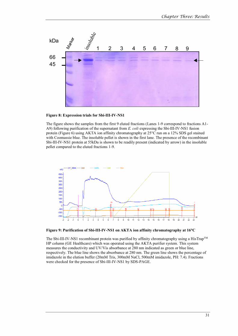
Figure 6: Expression trials for Sbi-III-IV-NS1
This figure shows a 12% SDS-gel stained with Coomassie blue of samples from E. coli expressing the Sbi-III-IV-NS1 fusion protein. The tracks include samples taken from the supernatant of un-induced cultures, supernatant from cultures induced with IPTG, bacterial extracts (after sonication) of the soluble fraction and extracts from the insoluble (bacterial pellet) fraction. The molecular weight ladder is shown in the left hand column. The transformed bacteria were cultured at both 25oC (A) and 16oC (B). In the induced cultures the presence of the recombinant Sbi-III-IV-NS1 protein at 55kDa.
Figure 7: Purification of Sbi-III-IV-NS1 on AKTA affinity chromatography at 25°C
The Sbi-III-IV-NS1 recombinant protein was purified by affinity chromatography using a HisTrapTM HP column (GE Healthcare) which was operated using the AKTA purifier system. This system measures the conductivity and UV/Vis absorbance at 280 nm indicated as green or blue line, respectively. The blue line shows the absorbance at 280 nm. The green line shows the percentage of imidazole in the elution buffer (20mM Tris, 300mM NaCl, 500 mM imidazole PH: 7.4). Fractions were checked for the presence of Sbi-III-IV-NS1 by SDS-PAGE.
Chapter Three: Results
31
kDa
6645
1 2 3 4 5 6 7 8 9insoluble
Mar
ker
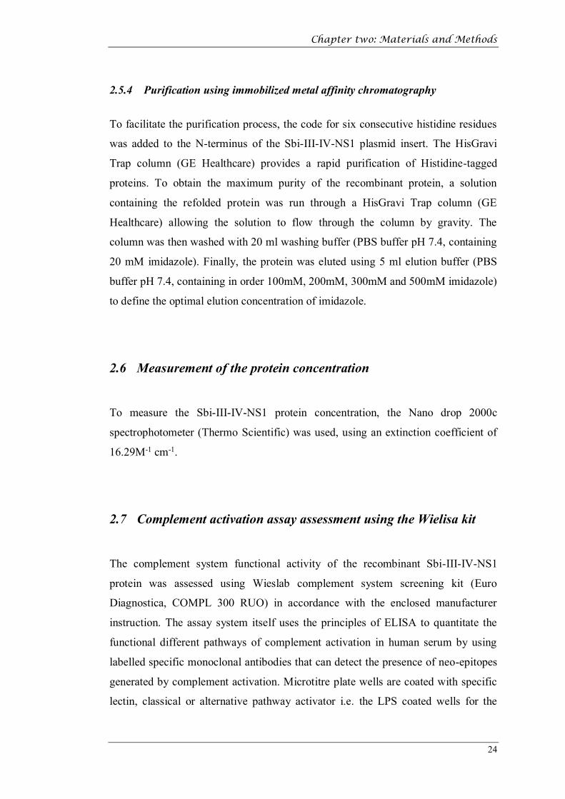
Figure 8: Expression trials for Sbi-III-IV-NS1
The figure shows the samples from the first 9 eluted fractions (Lanes 1-9 correspond to fractions A1-A9) following purification of the supernatant from E. coli expressing the Sbi-III-IV-NS1 fusion protein (Figure 6) using AKTA ion affinity chromatography at 25°C run on a 12% SDS gel stained with Coomassie blue. The insoluble pellet is shown in the first lane. The presence of the recombinant Sbi-III-IV-NS1 protein at 55kDa is shown to be readily present (indicated by arrow) in the insoluble pellet compared to the eluted fractions 1-9.
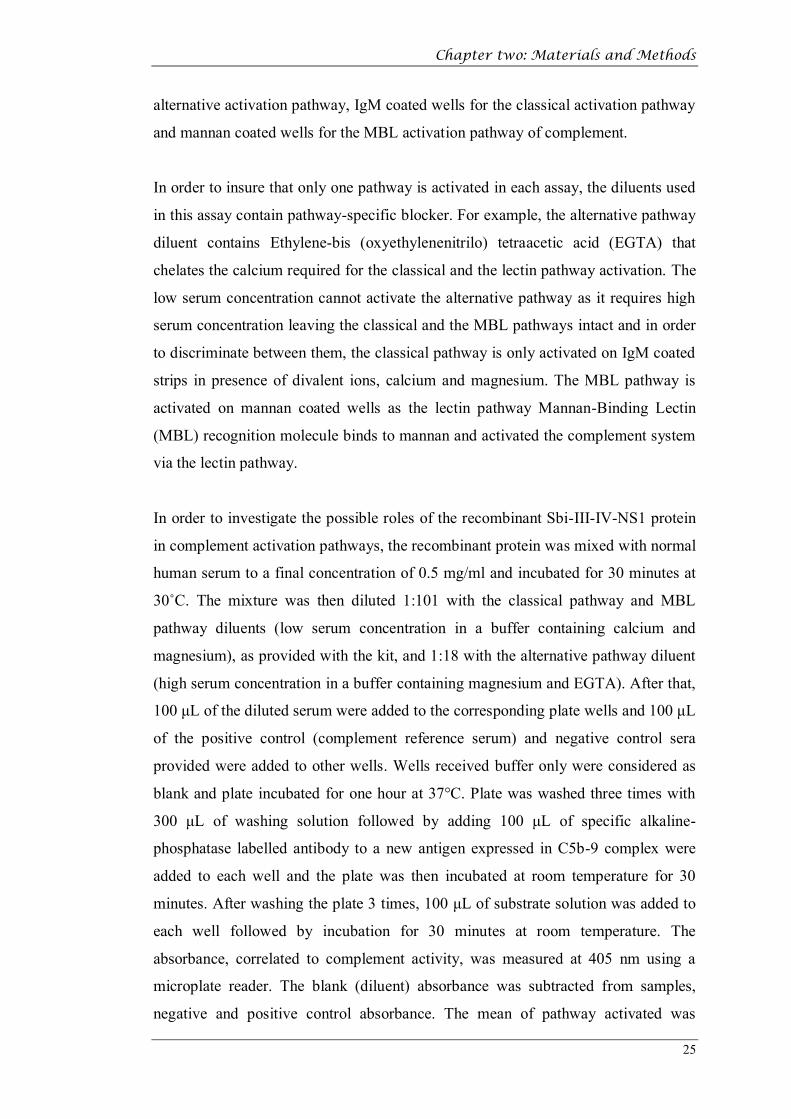
Figure 9: Purification of Sbi-III-IV-NS1 on AKTA ion affinity chromatography at 16°C
The Sbi-III-IV-NS1 recombinant protein was purified by affinity chromatography using a HisTrapTM HP column (GE Healthcare) which was operated using the AKTA purifier system. This system measures the conductivity and UV/Vis absorbance at 280 nm indicated as green or blue line, respectively. The blue line shows the absorbance at 280 nm. The green line shows the percentage of imidazole in the elution buffer (20mM Tris, 300mM NaCl, 500mM imidazole, PH: 7.4). Fractions were checked for the presence of Sbi-III-IV-NS1 by SDS-PAGE.
Chapter Three: Results
32
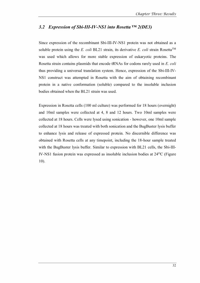
3.2 Expression of Sbi-III-IV-NS1 into Rosetta ™ 2(DE3)
Since expression of the recombinant Sbi-III-IV-NS1 protein was not obtained as a
soluble protein using the E. coli BL21 strain, its derivative E. coli strain RosettaTM
was used which allows for more stable expression of eukaryotic proteins. The
Rosetta strain contains plasmids that encode tRNAs for codons rarely used in E. coli
thus providing a universal translation system. Hence, expression of the Sbi-III-IV-
NS1 construct was attempted in Rosetta with the aim of obtaining recombinant
protein in a native conformation (soluble) compared to the insoluble inclusion
bodies obtained when the BL21 strain was used.
Expression in Rosetta cells (100 ml culture) was performed for 18 hours (overnight)
and 10ml samples were collected at 4, 8 and 12 hours. Two 10ml samples were
collected at 18 hours. Cells were lysed using sonication - however, one 10ml sample
collected at 18 hours was treated with both sonication and the BugBuster lysis buffer
to enhance lysis and release of expressed protein. No discernible difference was
obtained with Rosetta cells at any timepoint, including the 18-hour sample treated
with the BugBuster lysis buffer. Similar to expression with BL21 cells, the Sbi-III-
IV-NS1 fusion protein was expressed as insoluble inclusion bodies at 24OC (Figure
10).

Chapter Three: Results
33
Figure 10: Expression of Sbi-III-IV-NS1 in Rosetta cells
This figure shows samples from E. Coli Rosetta cells expressing the Sbi-III-IV-NS1 fusion protein. The tracks show samples taken from the overnight induction with IPTG at different time points (4, 8, 12 and 18 hours) (lanes 2, 3, 4 and 5) which were then lysed by sonication and then run on a 12% SDS-PAGE gel and stained with Coomassie blue. The sample shown in Lane 6 was lysed by both the BugBuster buffer and by sonication. Lane 7 shows the extract of the sonicated cell pellet at 18 hours and lane 1 shows the culture supernatant. The molecular weight ladder is indicated to the far left and the arrows show the expected position of the Sbi-III-IV-NS1 fusion protein (55kDa).
3.3 Optimisation of the purification of recombinant Sbi-III-IV-NS1
from inclusion bodies using Nickel column affinity
chromatography
Preliminary experiments had indicated that rather than being expressed as a soluble
protein the recombinant Sbi-III-IV-NS1 protein was insoluble and accumulated in
inclusion bodies within the E. coli cells. In order to optimise the purification the
protein was solublised by using 8M urea and incubated in a series of different
refolding buffers as mention in the methods section. Following HisGravi Trap
affinity column purification the recombinant Sbi-III-IV-NS1 protein was eluted with
PBS buffer containing 300 mM imidazole. The 15 eluted fractions from the different
refolding buffer incubations were analysed by SDS-PAGE as shown in figure 11.

Chapter Three: Results
34
Figure 11: Screening of the expressed Sbi-III-IV-NS1 protein in different re-folding buffers
Following transfection of E. coli with the plasmid containing the Sbi-III-IV-NS1 insert, inclusion bodies were purified and exposed to a series of different refolding buffers from the Athena Refolding Kit and run on a12% SDS-gel which was stained with Coomassie blue. The gel shows the expected band size of Sbi-III-IV-NS1 (55kDa) and buffers 4, 8, 11 and 12 were more likely to be the optimal refolding buffers.
On the basis of the results shown in Figure 11 buffers 4, 8 and 12 were chosen to be
used for refolding and further purification experiments of recombinant Sbi-III-IV-
NS1 using its N-terminal histidine tag by affinity chromatography with a HisGravi
Trap column (GE Healthcare) as shown in Figures 12, 13 and 14. These are the
results of 3 separate experiments designed to optimise the imidazole (100-500 mM)
concentration used for the elution from the HisGravi Trap column and the optimal
refolding buffer.

Chapter Three: Results
35
kDa
55
70
300 mM
500 mM
Figure 12: Purification of Sbi-III-IV-NS1 following incubation and refolding using buffer 4
A 12% SDS-gel was stained with Coomassie blue and shows bands of the elution fractions after purification with a HisGravi Trap column (GE Healthcare) using different elution concentrations of imidazole (300 and 500 mM). The gel shows a major band at the expected molecular weight of 55kDafollowing elution with 300mM imidazole.
100 m
MkDa
200 m
M
300 m
M
500 m
MFlo
w thr
ough
Wash
5570
Figure 13: Purification of Sbi-III-IV-NS1 following incubation and refolding using buffer 8
A 12% SDS-gel was stained with Coomassie blue and shows bands of the elution fractions after purification with the HisGravi Trap column (GE Healthcare) using different elution concentrations of imidazole (100-500 mM) to optimise the elution conditions. The gel shows a major band at the expected molecular weight of 55kDa following elution with imidazole. F=flow through, W=wash.

Chapter Three: Results
36
70
kDa
55
100 mM 300 mM
Figure 14: Purification of Sbi-III-IV-NS1 following incubation and refolding using buffer 12
A 12% SDS-gel was stained with Coomassie blue and shows bands of the elution fractions after purification with a HisGravi Trap column (GE Healthcare) using different elution concentrations of imidazole (100 and 300 mM). The gel shows a major band at the expected molecular weight of 55kDafollowing elution with 300mM imidazole.
Chapter Three: Results
37
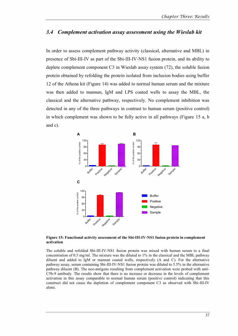
3.4 Complement activation assay assessment using the Wieslab kit
In order to assess complement pathway activity (classical, alternative and MBL) in
presence of Sbi-III-IV as part of the Sbi-III-IV-NS1 fusion protein, and its ability to
deplete complement component C3 in Wieslab assay system (72), the soluble fusion
protein obtained by refolding the protein isolated from inclusion bodies using buffer
12 of the Athena kit (Figure 14) was added to normal human serum and the mixture
was then added to mannan, IgM and LPS coated wells to assay the MBL, the
classical and the alternative pathway, respectively. No complement inhibition was
detected in any of the three pathways in contrast to human serum (positive control)
in which complement was shown to be fully active in all pathways (Figure 15 a, b
and c).
Buffer
Positiv
e
Negati
ve
Sample
0
30
60
90
120
% o
f the
pos
itve
cont
rol
Buffer
Positiv
e
Negati
ve
Sample
0
30
60
90
120
% o
f the
pos
itve
cont
rol
Buffer
Positiv
e
Negati
ve
Sample
0
30
60
90
120
% o
f the
pos
itve
cont
rol
Buffer
Positive
Negative
Sample
A B
C
Figure 15: Functional activity assessment of the Sbi-III-IV-NS1 fusion protein in complement activation
The soluble and refolded Sbi-III-IV-NS1 fusion protein was mixed with human serum to a final concentration of 0.5 mg/ml. The mixture was the diluted to 1% in the classical and the MBL pathway diluent and added to IgM or mannan coated wells, respectively (A and C). For the alternative pathway assay, serum containing Sbi-III-IV-NS1 fusion protein was diluted to 5.5% in the alternative pathway diluent (B). The neo-antigens resulting from complement activation were probed with anti-C5b-9 antibody. The results show that there is no increase or decrease in the levels of complement activation in this assay comparable to normal human serum (positive control) indicating that this construct did not cause the depletion of complement component C3 as observed with Sbi-III-IV alone.

Chapter Three: Results
38
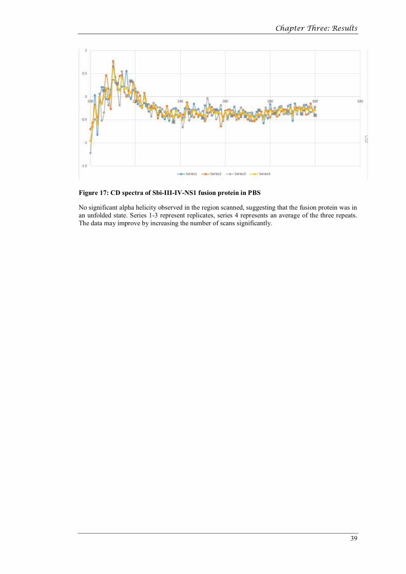
3.5 Circular dichroism (CD)
Since the Sbi-III-VI-NS1 contains an Sbi-IV domain, which is composed of three
alpha-helices (73), it was expected that some degree of helicty will be observed in
the CD data. The unstructured protein 1 (NS1) from Dengue virus is also known to
contain significant beta-sheet structure (74), which would be expected to give a
signal observable in the CD spectra in the region of 200-210 nm. The CD spectra
(Figures 16 & 18), however show no evidence of helical structure in this region
suggesting that the protein maybe present as a random coiled coil in solution
because it is unfolded. These results could be simply due to poor signal to noise ratio
in the region. The poor signal could be due to inaccuracy of measurement (protein
concentration) or inadequate number of scans performed on the sample.
Figure 16: Smoothed data of 3 scans of the CD spectra of the Sbi-III-IV-NS1 fusion protein in PBS
No peaks were observed in the region scanned, suggesting that the fusion protein was in an unfolded state though a poor signal to noise ratio makes it difficult to make this claim conclusively
Chapter Three: Results
39
Figure 17: CD spectra of Sbi-III-IV-NS1 fusion protein in PBS
No significant alpha helicity observed in the region scanned, suggesting that the fusion protein was in an unfolded state. Series 1-3 represent replicates, series 4 represents an average of the three repeats. The data may improve by increasing the number of scans significantly.

Chapter Three: Results
40
200 250 300 350-1.0
-0.5
0.0
0.5
wave length (nm)
Circ
ular
dic
hroi
sm (m
elde
g) SBI-NS1
200 250 300 350-0.8
-0.6
-0.4
-0.2
0.0
wave length (nm)
Circ
ular
dic
hroi
sm (m
elde
g)
buffer
Figure 18: Smoothed data of 3 scans of the CD spectra of the Sbi-III-IV-NS1 fusion protein in PBS
The buffer spectrum (lower panel) was subtracted to ensure accuracy of the data.

Chapter Three: Results
41
3.6 Western blotting
Western blotting was performed to confirm the identity of the Sbi-III-IV-NS1
recombinant protein seen at 55 kDa in the SDS-PAGE experiments. After SDS-
PAGE separated proteins were transferred electrophoretically onto a nitrocellulose
membrane at 25V for 12 minutes in transfer buffer. Buffer 12 was used for refolding
in this experiment. Figure 3-14 shows the Sbi-III-IV-NS1 recombinant protein on
SDS-PAGE with the expected band size of 55 kDa (A) and (B) shows the western
blot of the gel using polyclonal rabbit anti-Sbi antibody and probed with HRP
conjugated goat anti-rabbit IgG antibody. The eluted proteins were thus confirmed
as products of the recombinant plasmid.
kDa
5570
300 m
M
500 m
MA B
Figure 19: Western blot of Sbi-III-IV-NS1
A 12% SDS-gel was stained with Coomassie blue and shows bands of the elution fractions of refolding buffer 12, after purification with the HisGravi Trap column (GE Healthcare). The gel shows eluted fractions and shows a major band at the expected molecular weight of 55 kDa following elution with 300mM imidazole (A). Western blot analysis using polyclonal rabbit anti-Sbi antibody was used to confirm the identity of the eluted protein (B).

Chapter four: Discussion
42
Chapter four: Discussion

Chapter four: Discussion
43
4 Discussion
Dengue fever is a mosquito-borne tropical disease that affects millions of people,
which when left untreated, causes mortality in about 50% of cases (1, 2). Dengue
fever is caused by any of the 5 different serotypes of the dengue virus that have been
identified. Infection with a specific (DENV) serotype confers lifelong immunity
against that specific serotype but cross-protection against other serotypes lasts only a
few months (10). Previous studies have shown that vaccine-mediated localised
activation of complement factor C3 leads to improved pan-serotype immunological
memory and opsonisation by phagocytes (59). Thus, the ideal DENV vaccine would
contain a highly conserved immunogenic DENV protein conjugated to a
complement activator. Therefore, this study aimed to construct a fusion protein
comprising the highly-conserved DENV non-structural glycoprotein, NS1, and an
immune evasion factor of Staphylococcus aureus, Sbi, which interacts with Factor
H, C3b and C3b and C3d (75).
In order to achieve this aim the project set out to produce a recombinant protein that
could both induce an immune response to the conserved DENV non-structural
protein (NS1) and’ in tandem, induce effective complement activation which would
lead to highly efficient neutralisation and phagocytosis of infected cells and
neutralise the infectious pathway.
Using a previously cloned hybrid Sbi-III-IV-NS1 gene cloned into the expression
vector pET-28a, methods were optimised with E.coli BL21 and its derivative,
RosettaTM, to obtain sufficient quantities of the recombinant hybrid protein to initiate
vaccination strategies. However, although optimization of the expression conditions
in E. coli allowed abundant quantities of recombinant Sbi-III-IV-NS1 to be
produced (Figure 6) further experiments demonstrated that the expressed protein
was not soluble and was found in inclusion bodies within the bacteria. This is not an
uncommon finding in prokaryotic systems which do not allow appropriate
glycosylation of eukaryotic derived proteins. In addition, the induction of the
expression of the fusion protein with the prokaryotic promoter T7 and/or the absence
of the appropriate eukaryotic cellular machinery in E. coli BL21 (and its derivative,

Chapter four: Discussion
44
RosettaTM) required to maintain the fusion protein in its native state, may have led to
the expression of the denatured fusion protein in inclusion bodies (76).
In order to try and isolate the expressed recombinant Sbi-III-IV-NS1 from the
inclusion bodies, E. coli cells were first sonicated and the Sbi-III-IV-NS1
recombinant protein was first purified by affinity chromatography (Figures 7 and 9).
However, no peaks of eluted proteins were observed and elution fractions run on
SDS-PAGE showed no bands (Figure 8). This implied that no soluble proteins were
present in the extract and the recombinant protein was effectively trapped in
inclusion bodies, which has been described elsewhere (76).
Therefore, further experiments were performed in order to firstly release the
denatured Sbi-III-IV-NS1 recombinant protein and, secondly, to solubilise it and
allow the protein to refold into is native conformation.
Clearly stringent conditions were required to release and solubilise the denatured
protein so the E. coli BL2 cultured bacteria were lysed and sonicated and the extract
solubilised in 8M urea following which the protein concentration was adjusted to
1mg/ml and using the ‘drip technique’ was allowed to refold in a series of buffers
designed to promote protein refolding as mention in the methods section. Following
overnight incubation in the refolding buffer the Sbi-III-IV-NS1 recombinant protein
was isolated using HisGravi Trap affinity column purification and conditions for
optimal elution with imidazole was performed (Figures 12, 13 and 14).
Attempts to refold the protein from the inclusion bodies were not very successful
with the best results being obtained when polyethylene glycol (PEG) [i.e. in buffer
4] was excluded from the isolation buffers, which could have enhanced the activity
of Triton X-100, a non-ionic detergent that improves the solubility of proteins
(Figures 12). However, circular dichroism experiments on the purified and refolded
recombinant Sbi-III-IV-NS1 protein (Figure 16-18) were inconclusive. Circular
dichroism is a method which allows the determination of the secondary refolded
structure of the refolded recombinant Sbi-III-IV-NS1 protein but the Chirascan
failed to register any protein helical structural motifs as would be expected from the
published molecular models of Sbi-IV. In addition the non-structural protein 1
Chapter four: Discussion
45
(NS1) from Dengue virus is known to contain significant beta-sheet structure (74)
but again no signal was observed in the circular dichroism scan. This evidence
therefore suggests that the numerous attempts to purify and appropriately refold the
Sbi-III-IV-NS1 protein were ultimately unsuccessful.
While the clear advantage of using a mammalian expression system would be that
proteins would be glycosylated and therefore remain stable and be more likely to
remain in solution, it was thought that it might be preferable to use an E. coli
expression system because of the need to produce sufficiently large amounts of the
fusion protein that would allow testing in the in vitro assays that were established.
Converse to mammalian expression systems involving cell lines such as the human
embryonic kidney (HEK-293) and Chinese hamster ovary (CHO) cell lines, E. coli
systems offer rapid and high expression of recombinant proteins. Furthermore, use
of a mammalian expression system would have involved the generation of stable cell
lines that would express the fusion proteins; a time-consuming process that could be
complicated further by clonal variations and the need to keep the transgene stable
under selection pressure.
Since the expression of soluble recombinant protein in E. coli can be influenced by
different factors, such as temperature and IPTG concentration, in future studies, the
expression of Sbi-III-IV-NS1 fusion protein will be assessed at different
temperatures and lower (<1mM) IPTG concentrations in RosettaTM cells. It is
expected optimization of temperature and IPTG concentrations will increase yields
of the native Sbi-III-IV-NS1 fusion protein. Expression of the individual
components of the Sbi-III-IV-NS1 fusion protein separately, i.e. Sbi-III-IV by its
own and NS1 alone, may be productive as a reduction in the complexity of the
fusion protein may be responsible for its inability to fold correctly. Pooling of the
individual proteins (Sbi-III-IV and NS1) in subsequent assays would then be
attempted.
As NS1 is commonly secreted as an hexameric complex into the plasma of Dengue
fever patients (63, 77) oxidation-dependent disulphide bond formation between the
cysteine residues of the Sbi-III-IV-NS1 fusion protein (which possesses six potential
disulphide bonds), is likely to be somewhat ineffective in the reductive cytoplasmic
environment of E. coli (78). To counter the effect of reductases such as trxB (a

Chapter four: Discussion
46
thioredoxin reductase) present in the cytoplasm, an E. coli strain containing the trxB
mutation could be utilised. As the reductive activities of Thioredoxin-1 and -2
(encoded by trxA and trxC respectively) is dependent on their initial reduction by the
gene product of trxB, only non-reduced forms of the thioredoxins will be present in
the cytoplasm in a trxB mutant. These non-reduced thioredoxins can catalyse
disulphide bond formation (79) thereby possibly promoting the stability of the
recombinant Sbi-III-IV-NS1 fusion protein and its expression within the cytoplasm
in a soluble form. This method has been successfully used to express sufficient
amounts of cysteine-rich extracellular proteins such as the micronemal adhesive
protein from Toxoplasma gondii (80). Obtaining functional isoforms of the fusion
protein will therefore be tried using the E. coli trxB mutant expression system
(commercially available as Origami™ 2(DE3)pLysS) or by targeting the expressed
protein to the E. coli periplasm where disulphide bond-promoting enzymes abound
to optimise appropriate folding of the protein.
In addition to using a strain with reduced reductase activity, co-expression of the
Sbi-III-IV-NS1 protein with plasmids containing coding sequences for disulphide
oxidoreductase and isomerase, DsbA and DsbC respectively, may ensure stability of
the Sbi-III-IV-NS1 fusion protein as both DsbA and DsbC play crucial roles in the
stability and functional expression of proteins in vivo(81, 82). Alternatively, the Sbi-
III-IV-NS1 construct could be engineered to include a signal sequence that will
target the expressed protein to the periplasm where the Dsb family of proteins is
present.
In the Athena refolding kit experiments perhaps the optimal refolding was achieved
with buffer 4 as judged by SDS-PAGE (Figure 12) and this could be confirmed with
appropriate CD analysis. In future experiments detailed CD analysis of the Sbi-III-
IV-NS1 protein should be undertaken to see whether the protein is expressed in its
native confirmation. Further experiments might yield more definitive data if a higher
concentration of protein and/or an increased number of scans were used to improve
the signal to noise ratio.

Chapter four: Discussion
47
Alternatively, the production of the fusion protein could be performed in the
aforementioned mammalian expression systems (HEK-293 or CHO cell lines) or in
baculovirus-infected insect cell lines (83). Despite the limitations mentioned
previously, the key advantage of utilizing either insect or mammalian cell lines over
the E. coli system would be the production of soluble recombinant proteins more
likely to be expressed in their native conformation. These insect or mammalian
systems provide glycosylation and other post-translational modifications, which
could improve the solubility of the Sbi-III-IV-NS1 fusion protein. Exploiting the
baculovirus vector system in lepidopteron cells and larvae (84) could be more
productive since this system, termed MultiBac, affords the possibility of co-
expressing the Sbi-III-IV and NS1 subunits in separate bacmids to yield the
functional Sbi-III-IV-NS1 fusion protein complex. Following soluble protein
expression, purification of the Sbi-III-IV-NS1 fusion protein secreted into growth
media could be achieved using conventional monoclonal antibody-dependent
immune-purification techniques (62) identified several monoclonal antibodies that
bound to 3 highly-conserved epitopes of the NS1 protein. These antibodies, when
covalently-bound to Protein G-matrices via crosslinking agents, could be used to
isolate the soluble Sbi-III-IV-NS1 fusion protein from growth media.
Successful expression of the soluble Sbi-III-IV-NS1 fusion protein and its individual
components (whose expression will be pursued as described previously) will pave
the way for experiments in which the functional immunogenicity of the different
proteins, i.e. their ability to induce neutralising antibodies, will be compared in mice
(i.e. Sbi-III-IV-NS1 versus Sbi-III-IV and Sbi-III-IV-NS1 versus NS1).
In summary, an ideal vaccine for DENV will include antigenic components that are
highly conserved among all described serotypes of DENV and can induce long-term
immunological memory. The fusion protein described in this study satisfies vaccine
candidacy as it contains the highly-conserved DENV non-structural protein NS1 and
the Staphylococcal complement activator, Sbi. This fusion protein was successfully
expressed in this study but not in a soluble, biologically-active state. To obtain
functional isoforms of the fusion protein stability of the protein in the E. coli
expression system should be pursued by utilising an E. coli trxB mutant
(commercially available as Origami™ 2(DE3)pLysS) or by targeting the expressed

Chapter four: Discussion
48
protein to the E. coli periplasm where disulphide bond-promoting enzymes can
promote appropriate folding of the protein.

Chapter five: Summary and Conclusion
49
Chapter five: Summary and Conclusions

Chapter five: Summary and Conclusion
50
5 Summary, conclusions and future work
The purpose of the project was to develop a method for enhancing the development
of effective immunity against Dengue Fever. It has previously been shown that the
recruitment of C3d greatly enhances humoral immunity so therefore the aim of the
project was to directly associate Sbi from staphylococcus (which activates the
alternative complement pathway and indirectly recruits C3d) to NS1 a key target for
immunoprotection against DENV. The aim of the project has therefore to express
and produce sufficient quantities of Sbi-III-IV-NS1 for vaccine studies. However,
firstly a number of hurdles had to be overcome, notably determining the optimal
conditions for Sbi-III-IV-NS1 expression using different E. coli strains and different
culture conditions including, time, temperature and IPTG concentration. Secondly it
became clear that the Sbi-III-IV-NS1 protein was not present in soluble extracts of
the transformed E. coli BL2 bacteria and that the Sbi-III-IV-NS1 recombinant
protein was expressed in a denatured form in inclusion bodies. A number of methods
were then used to optimize the isolation and the appropriate refolding of the Sbi-III-
IV-NS1 recombinant protein which were assessed by SDS-PAGE and CD. Although
significant progress was made there was insufficient time to progress to the next
stage of the project which was to use the purified Sbi-III-IV-NS1 protein to assess
its ability to produce resistance to DENV (in mice) using the construct to recruit C3d
which should enhance humoral immunity to the virus and pave the way for future
vaccine based experiments.

Chapter five: Summary and Conclusion
51
Future directions Firstly, future studies could assess the expression of the Sbi-III-IV-NS1 fusion
protein at different temperatures and lower (<1mM) IPTG concentrations in
RosettaTM cells as well as trying to express the individual components Sbi-III-IV
and NS1 separately as the complexity of the fusion protein may contribute to its lack
of solubility and inability to fold appropriately.
Secondly, as refolding of this recombinant protein is dependent of the formation of a
number of disulphide bonds the effect of reductases such as trxB (a thioredoxin
reductase) present in the cytoplasm, an E. coli strain containing the trxB mutation
could be utilized. In addition, plasmids containing coding sequences for disulphide
oxidoreductase and isomerase may improve the stability of the Sbi-III-IV-NS1
fusion protein or, alternatively, the Sbi-III-IV-NS1 construct could be engineered to
include a signal sequence that will target the expressed protein to the periplasm
where the Dsb family of proteins is present.
Finally, if ssuccessful expression of a soluble form of the Sbi-III-IV-NS1 fusion
protein and its individual components can be achieved it will then be possible to
study their ability to induce neutralising antibodies in mouse model systems.

Chapter six: References
52
Chapter six: References
Chapter six: References
53
6 References
1. Gubler DJ. The changing epidemiology of yellow fever and dengue, 1900 to 2003: full circle? Comp Immunol Microbiol Infect Dis. 2004;27(5):319-30. 2. WHO. Dengue: Guidelines for Diagnosis, Treatment, Prevention and Control. 2009. 3. Watts DM, Burke DS, Harrison BA, Whitmire RE, Nisalak A. Effect of temperature on the vector efficiency of Aedes aegypti for dengue 2 virus. The American journal of tropical medicine and hygiene. 1987;36(1):143-52. Epub 1987/01/01. 4. Schneider BS, Soong L, Zeidner NS, Higgs S. Aedes aegypti salivary gland extracts modulate anti-viral and TH1/TH2 cytokine responses to sindbis virus infection. Viral immunology. 2004;17(4):565-73. Epub 2005/01/27. 5. Guzman MG, Halstead SB, Artsob H, Buchy P, Farrar J, Gubler DJ, et al. Dengue: a continuing global threat. Nature reviews Microbiology. 2010;8(12 Suppl):S7-16. 6. Wang E, Ni H, Xu R, Barrett AD, Watowich SJ, Gubler DJ, et al. Evolutionary relationships of endemic/epidemic and sylvatic dengue viruses. Journal of virology. 2000;74(7):3227-34. 7. Rodriguez-Roche R, Alvarez M, Gritsun T, Halstead S, Kouri G, Gould EA, et al. Virus evolution during a severe dengue epidemic in Cuba, 1997. Virology. 2005;334(2):154-9. 8. Holmes EC. Molecular epidemiology and evolution of emerging infectious diseases. Br Med Bull. 1998;54(3):533-43. 9. Normile D. Tropical medicine. Surprising new dengue virus throws a spanner in disease control efforts. Science. 2013;342(6157):415. 10. Thisyakorn U, Thisyakorn C. Latest developments and future directions in dengue vaccines. Ther Adv Vaccines. 2014;2(1):3-9. 11. Aintablian A, Hamby RI, Hoffman I, Hartstein ML, Wisoff BG. New Q waves after bypass grafting: correlations between graft patency, ventriculogram and surgical venting technique. Journal of electrocardiology. 1976;9(4):321-7. Epub 1976/01/01. 12. Halstead SB, O'Rourke EJ. Dengue viruses and mononuclear phagocytes. I. Infection enhancement by non-neutralizing antibody. The Journal of experimental medicine. 1977;146(1):201-17. Epub 1977/07/01. 13. Goncalvez AP, Engle RE, St Claire M, Purcell RH, Lai CJ. Monoclonal antibody-mediated enhancement of dengue virus infection in vitro and in vivo and strategies for prevention. Proceedings of the National Academy of Sciences of the United States of America. 2007;104(22):9422-7. Epub 2007/05/23. 14. Kliks SC, Nimmanitya S, Nisalak A, Burke DS. Evidence that maternal dengue antibodies are important in the development of dengue hemorrhagic fever in infants. The American journal of tropical medicine and hygiene. 1988;38(2):411-9. Epub 1988/03/01. 15. Cummings DA, Schwartz IB, Billings L, Shaw LB, Burke DS. Dynamic effects of antibody-dependent enhancement on the fitness of viruses. Proceedings of
Chapter six: References
54
the National Academy of Sciences of the United States of America. 2005;102(42):15259-64. Epub 2005/10/12. 16. Gan VC. Dengue: Moving from Current Standard of Care to State-of-the-Art Treatment. Curr Treat Options Infect Dis. 2014;6(3):208-26. 17. Dengue: Guidelines for Diagnosis, Treatment, Prevention and Control: New Edition. Geneva2009. 18. Wills BA, Nguyen MD, Ha TL, Dong TH, Tran TN, Le TT, et al. Comparison of three fluid solutions for resuscitation in dengue shock syndrome. The New England journal of medicine. 2005;353(9):877-89. 19. Wang QY, Patel SJ, Vangrevelinghe E, Xu HY, Rao R, Jaber D, et al. A small-molecule dengue virus entry inhibitor. Antimicrobial agents and chemotherapy. 2009;53(5):1823-31. 20. Tomlinson SM, Malmstrom RD, Watowich SJ. New approaches to structure-based discovery of dengue protease inhibitors. Infect Disord Drug Targets. 2009;9(3):327-43. 21. Diamond MS, Pierson TC, Fremont DH. The structural immunology of antibody protection against West Nile virus. Immunological reviews. 2008;225:212-25. Epub 2008/10/08. 22. Burton DR, Williamson RA, Parren PW. Antibody and virus: binding and neutralization. Virology. 2000;270(1):1-3. 23. Guirakhoo F, Kitchener S, Morrison D, Forrat R, McCarthy K, Nichols R, et al. Live attenuated chimeric yellow fever dengue type 2 (ChimeriVax-DEN2) vaccine: Phase I clinical trial for safety and immunogenicity: effect of yellow fever pre-immunity in induction of cross neutralizing antibody responses to all 4 dengue serotypes. Human vaccines. 2006;2(2):60-7. 24. Bonilla FA, Oettgen HC. Adaptive immunity. J Allergy Clin Immunol. 2010;125(2 Suppl 2):S33-40. 25. Janeway CA, Jr., Medzhitov R. Innate immune recognition. Annu Rev Immunol. 2002;20:197-216. 26. Medzhitov R. Recognition of microorganisms and activation of the immune response. Nature. 2007;449(7164):819-26. 27. Mogensen TH. Pathogen recognition and inflammatory signaling in innate immune defenses. Clinical microbiology reviews. 2009;22(2):240-73, Table of Contents. 28. Stetson DB, Medzhitov R. Recognition of cytosolic DNA activates an IRF3-dependent innate immune response. Immunity. 2006;24(1):93-103. 29. Rasmussen SB, Sorensen LN, Malmgaard L, Ank N, Baines JD, Chen ZJ, et al. Type I interferon production during herpes simplex virus infection is controlled by cell-type-specific viral recognition through Toll-like receptor 9, the mitochondrial antiviral signaling protein pathway, and novel recognition systems. Journal of virology. 2007;81(24):13315-24. 30. Schwaeble W, Dahl MR, Thiel S, Stover C, Jensenius JC. The mannan-binding lectin-associated serine proteases (MASPs) and MAp19: four components of the lectin pathway activation complex encoded by two genes. Immunobiology. 2002;205(4-5):455-66. Epub 2002/10/25. 31. Walport MJ. Complement. First of two parts. The New England journal of medicine. 2001;344(14):1058-66. Epub 2001/04/05. 32. Laufer J, Katz Y, Passwell JH. Extrahepatic synthesis of complement proteins in inflammation. Molecular immunology. 2001;38(2-3):221-9.

Chapter six: References
55
33. Manderson AP, Pickering MC, Botto M, Walport MJ, Parish CR. Continual low-level activation of the classical complement pathway. The Journal of experimental medicine. 2001;194(6):747-56. 34. Sim RB, Day AJ, Moffatt BE, Fontaine M. Complement factor I and cofactors in control of complement system convertase enzymes. Methods Enzymol. 1993;223:13-35. 35. Fujita T, Matsushita M, Endo Y. The lectin-complement pathway--its role in innate immunity and evolution. Immunological reviews. 2004;198:185-202. Epub 2004/06/18. 36. Schwaeble WJ, Lynch NJ, Clark JE, Marber M, Samani NJ, Ali YM, et al. Targeting of mannan-binding lectin-associated serine protease-2 confers protection from myocardial and gastrointestinal ischemia/reperfusion injury. Proc Natl Acad Sci U S A. 2011;108(18):7523-8. 37. Claus DR, Siegel J, Petras K, Osmand AP, Gewurz H. Interactions of C-reactive protein with the first component of human complement. J Immunol. 1977;119(1):187-92. Epub 1977/07/01. 38. Matsushita M, Fujita T. Activation of the classical complement pathway by mannose-binding protein in association with a novel C1s-like serine protease. The Journal of experimental medicine. 1992;176(6):1497-502. 39. Thiel S, Vorup-Jensen T, Stover CM, Schwaeble W, Laursen SB, Poulsen K, et al. A second serine protease associated with mannan-binding lectin that activates complement. Nature. 1997;386(6624):506-10. Epub 1997/04/03. 40. Dahl MR, Thiel S, Matsushita M, Fujita T, Willis AC, Christensen T, et al. MASP-3 and its association with distinct complexes of the mannan-binding lectin complement activation pathway. Immunity. 2001;15(1):127-35. Epub 2001/08/04. 41. Hajela K, Kojima M, Ambrus G, Wong KH, Moffatt BE, Ferluga J, et al. The biological functions of MBL-associated serine proteases (MASPs). Immunobiology. 2002;205(4-5):467-75. Epub 2002/10/25. 42. Takahashi M, Ishida Y, Iwaki D, Kanno K, Suzuki T, Endo Y, et al. Essential role of mannose-binding lectin-associated serine protease-1 in activation of the complement factor D. The Journal of experimental medicine. 2010;207(1):29-37. 43. Matsushita M, Endo Y, Fujita T. Cutting edge: complement-activating complex of ficolin and mannose-binding lectin-associated serine protease. Journal of immunology. 2000;164(5):2281-4. 44. Harboe M, Mollnes TE. The alternative complement pathway revisited. J Cell Mol Med. 2008;12(4):1074-84. 45. Unsworth DJ, Virgo PF, Lock RJ. Immunoglobulin E deficiency: a forgotten clue pointing to possible immunodeficiency? Ann Clin Biochem. 2011;48(Pt 5):459-61. 46. Endo Y, Matsushita M, Fujita T. The role of ficolins in the lectin pathway of innate immunity. The international journal of biochemistry & cell biology. 2011;43(5):705-12. Epub 2011/02/15. 47. Avirutnan P, Mehlhop E, Diamond MS. Complement and its role in protection and pathogenesis of flavivirus infections. Vaccine. 2008;26 Suppl 8:I100-7. 48. Ricklin D, Hajishengallis G, Yang K, Lambris JD. Complement: a key system for immune surveillance and homeostasis. Nature immunology. 2010;11(9):785-97. Epub 2010/08/20.
Chapter six: References
56
49. Ricklin D, Hajishengallis G, Yang K, Lambris JD. Complement: a key system for immune surveillance and homeostasis. Nature immunology. 2010;11(9):785-97. 50. Hannan J, Young K, Szakonyi G, Overduin MJ, Perkins SJ, Chen X, et al. Structure of complement receptor (CR) 2 and CR2-C3d complexes. Biochemical Society transactions. 2002;30(Pt 6):983-9. 51. Mochizuki Y, de Ming T, Hayashi T, Itoh M, Hotta H, Homma M. Protection of mice against Sendai virus pneumonia by non-neutralizing anti-F monoclonal antibodies. Microbiology and immunology. 1990;34(2):171-83. 52. Spear GT, Takefman DM, Sullivan BL, Landay AL, Jennings MB, Carlson JR. Anti-cellular antibodies in sera from vaccinated macaques can induce complement-mediated virolysis of human immunodeficiency virus and simian immunodeficiency virus. Virology. 1993;195(2):475-80. 53. Malasit P. Complement and dengue haemorrhagic fever/shock syndrome. The Southeast Asian journal of tropical medicine and public health. 1987;18(3):316-20. Epub 1987/09/01. 54. Avirutnan P, Punyadee N, Noisakran S, Komoltri C, Thiemmeca S, Auethavornanan K, et al. Vascular leakage in severe dengue virus infections: a potential role for the nonstructural viral protein NS1 and complement. The Journal of infectious diseases. 2006;193(8):1078-88. Epub 2006/03/18. 55. Mongkolsapaya J, Dejnirattisai W, Xu XN, Vasanawathana S, Tangthawornchaikul N, Chairunsri A, et al. Original antigenic sin and apoptosis in the pathogenesis of dengue hemorrhagic fever. Nature medicine. 2003;9(7):921-7. Epub 2003/06/17. 56. Rico-Hesse R. Microevolution and virulence of dengue viruses. Advances in virus research. 2003;59:315-41. Epub 2003/12/31. 57. Lin CF, Wan SW, Cheng HJ, Lei HY, Lin YS. Autoimmune pathogenesis in dengue virus infection. Viral immunology. 2006;19(2):127-32. Epub 2006/07/05. 58. Toapanta FR, Ross TM. Complement-mediated activation of the adaptive immune responses: role of C3d in linking the innate and adaptive immunity. Immunol Res. 2006;36(1-3):197-210. 59. van den Elsen JM, Isenman DE. A crystal structure of the complex between human complement receptor 2 and its ligand C3d. Science. 2011;332(6029):608-11. 60. Burman JD, Leung E, Atkins KL, O'Seaghdha MN, Lango L, Bernado P, et al. Interaction of human complement with Sbi, a staphylococcal immunoglobulin-binding protein: indications of a novel mechanism of complement evasion by Staphylococcus aureus. J Biol Chem. 2008;283(25):17579-93. 61. Dempsey PW, Allison ME, Akkaraju S, Goodnow CC, Fearon DT. C3d of complement as a molecular adjuvant: bridging innate and acquired immunity. Science. 1996;271(5247):348-50. 62. Masrinoul P, Diata MO, Pambudi S, Limkittikul K, Ikuta K, Kurosu T. Highly conserved region 141168 of the NS1 protein is a new common epitope region of dengue virus. Japanese journal of infectious diseases. 2011;64(2):109-15. Epub 2011/04/27. 63. Flamand M, Megret F, Mathieu M, Lepault J, Rey FA, Deubel V. Dengue virus type 1 nonstructural glycoprotein NS1 is secreted from mammalian cells as a soluble hexamer in a glycosylation-dependent fashion. Journal of virology. 1999;73(7):6104-10.

Chapter six: References
57
64. Muller DA, Young PR. The flavivirus NS1 protein: molecular and structural biology, immunology, role in pathogenesis and application as a diagnostic biomarker. Antiviral research. 2013;98(2):192-208. 65. Couvelard A, Marianneau P, Bedel C, Drouet MT, Vachon F, Henin D, et al. Report of a fatal case of dengue infection with hepatitis: demonstration of dengue antigens in hepatocytes and liver apoptosis. Human pathology. 1999;30(9):1106-10. Epub 1999/09/24. 66. Somnuke P, Hauhart RE, Atkinson JP, Diamond MS, Avirutnan P. N-linked glycosylation of dengue virus NS1 protein modulates secretion, cell-surface expression, hexamer stability, and interactions with human complement. Virology. 2011;413(2):253-64. 67. Dussart P, Petit L, Labeau B, Bremand L, Leduc A, Moua D, et al. Evaluation of two new commercial tests for the diagnosis of acute dengue virus infection using NS1 antigen detection in human serum. PLoS Negl Trop Dis. 2008;2(8):e280. 68. Thomas L, Verlaeten O, Cabie A, Kaidomar S, Moravie V, Martial J, et al. Influence of the dengue serotype, previous dengue infection, and plasma viral load on clinical presentation and outcome during a dengue-2 and dengue-4 co-epidemic. Am J Trop Med Hyg. 2008;78(6):990-8. 69. Young PR, Hilditch PA, Bletchly C, Halloran W. An antigen capture enzyme-linked immunosorbent assay reveals high levels of the dengue virus protein NS1 in the sera of infected patients. J Clin Microbiol. 2000;38(3):1053-7. 70. Omokoko MD, Pambudi S, Phanthanawiboon S, Masrinoul P, Setthapramote C, Sasaki T, et al. A highly conserved region between amino acids 221 and 266 of dengue virus non-structural protein 1 is a major epitope region in infected patients. Am J Trop Med Hyg. 2014;91(1):146-55. 71. Greenfield NJ. Using circular dichroism spectra to estimate protein secondary structure. Nature protocols. 2006;1(6):2876-90. 72. Seelen MA, Roos A, Wieslander J, Mollnes TE, Sjoholm AG, Wurzner R, et al. Functional analysis of the classical, alternative, and MBL pathways of the complement system: standardization and validation of a simple ELISA. Journal of immunological methods. 2005;296(1-2):187-98. 73. Clark EA, Crennell S, Upadhyay A, Zozulya AV, Mackay JD, Svergun DI, et al. A structural basis for Staphylococcal complement subversion: X-ray structure of the complement-binding domain of Staphylococcus aureus protein Sbi in complex with ligand C3d. Molecular immunology. 2011;48(4):452-62. 74. Akey DL, Brown WC, Dutta S, Konwerski J, Jose J, Jurkiw TJ, et al. Flavivirus NS1 structures reveal surfaces for associations with membranes and the immune system. Science. 2014;343(6173):881-5. 75. Haupt K, Reuter M, van den Elsen J, Burman J, Halbich S, Richter J, et al. The Staphylococcus aureus protein Sbi acts as a complement inhibitor and forms a tripartite complex with host complement Factor H and C3b. PLoS pathogens. 2008;4(12):e1000250. 76. Samuelson JC. Recent developments in difficult protein expression: a guide to E. coli strains, promoters, and relevant host mutations. Methods in molecular biology. 2011;705:195-209. 77. Alcon S, Talarmin A, Debruyne M, Falconar A, Deubel V, Flamand M. Enzyme-linked immunosorbent assay specific to Dengue virus type 1 nonstructural protein NS1 reveals circulation of the antigen in the blood during the acute phase of
Chapter six: References
58
disease in patients experiencing primary or secondary infections. J Clin Microbiol. 2002;40(2):376-81. 78. Derman AI, Prinz WA, Belin D, Beckwith J. Mutations that allow disulfide bond formation in the cytoplasm of Escherichia coli. Science. 1993;262(5140):1744-7. 79. Stewart M, Kent HM, McCoy AJ. The structure of the Q69L mutant of GDP-Ran shows a major conformational change in the switch II loop that accounts for its failure to bind nuclear transport factor 2 (NTF2). Journal of molecular biology. 1998;284(5):1517-27. 80. Saouros S, Blumenschein TM, Sawmynaden K, Marchant J, Koutroukides T, Liu B, et al. High-level bacterial expression and purification of apicomplexan micronemal proteins for structural studies. Protein Pept Lett. 2007;14(5):411-5. 81. Zapun A, Bardwell JC, Creighton TE. The reactive and destabilizing disulfide bond of DsbA, a protein required for protein disulfide bond formation in vivo. Biochemistry. 1993;32(19):5083-92. 82. Zhang S, Huang F, Zhao J, Wen L, Zhou F, Yang P. Determination of thiols in urinary sample by capillary-column liquid chromatography with amperometric detection at a carbon electrode. Talanta. 2002;58(3):451-8. 83. Kost TA, Condreay JP, Jarvis DL. Baculovirus as versatile vectors for protein expression in insect and mammalian cells. Nature biotechnology. 2005;23(5):567-75. Epub 2005/05/07. 84. Berger I, Fitzgerald DJ, Richmond TJ. Baculovirus expression system for heterologous multiprotein complexes. Nature biotechnology. 2004;22(12):1583-7. Epub 2004/11/30.

Chapter seven: Appendices
59
Chapter seven: Appendices

Chapter seven: Appendices
60
7 Appendices
7.1 Appendix 1
7.1.1 Sbi-III-IV-NS1 conjugate.
The coding sequence of Sbi domains 3 and 4 was cloned into the pET28a
(Kanr)vector using the NheI and BamHI restriction sites and, in addition, the coding
sequence of Dengue virus NS1 was inserted using the BamHI and XhoI restriction
sites to make a plasmid construct that could be expressed in E. coli. The
recombinant Sbi-III-IV-NS1 carried an N-terminal His-tag for protein purification.
(This work was performed by Dr Gyles Cozier). Sbi-III-IV is in red. NS1 is in blue.

Chapter seven: Appendices
61
(HisTag) (Thrombin) ATG GGC AGC AGC CAT CAT CAT CAT CAT CAC AGC AGC GGC CTG GTG CCG CGC GGC AGC CAT M G S S H H H H H H S S G L V P R G S H (NheI) ATG GCT AGC GAA CGT CAA AAT ATT GAA AAT GCG GAT AAA GCA ATT AAA GAT TTC CAA GAT M A S E R Q N I E N A D K A I K D F Q D AAC AAA GCA CCA CAC GAT AAA TCA GCA GCA TAT GAA GCT AAC TCA AAA TTA CCT AAA GAT N K A P H D K S A A Y E A N S K L P K D TTA CGC GAT AAA AAT AAC CGC TTT GTA GAA AAA GTT TCA ATT GAA AAA GCA ATC GTT CGT L R D K N N R F V E K V S I E K A I V R CAT GAT GAG CGT GTG AAA TCA GCA AAT GAT GCA ATC TCA AAA TTA AAT GAA AAA GAT TCA H D E R V K S A N D A I S K L N E K D S ATT GAA AAC AGA CGT TTA GCA CAA CGT GAA GTT AAC AAA GCA CCT ATG GAT GTA AAA GAG I E N R R L A Q R E V N K A P M D V K E CAT TTA CAG AAA CAA TTA GAC GCA TTA GTA GCT CAA AAA GAT GCT GAA AAG AAA GTG GCG H L Q K Q L D A L V A Q K D A E K K V A (BamHI) GGA TCC GAC AGT GGT TGC GTT GTG AGC TGG AAG AAC AAA GAA CTG AAA TGC GGC AAT GGG G S D S G C V V S W K N K E L K C G N G ATA TTT GTC ACA GAT AAC GTG CAT ACA TGG ACA GAA CAA TAC AAG TTC CAA CCA GAA TCC I F V T D N V H T W T E Q Y K F Q P E S CCT TCA AAA CTG GCT TCA GCT ATT CAG AAA GCT CAT GAA GAG GGC ATT TGT GGA ATC CGC P S K L A S A I Q K A H E E G I C G I R TCA GTA ACA AGA CTG GAA AAT CTT ATG TGG AAA CAG ATA ACA CCG GAA TTG AAC CAC ATC S V T R L E N L M W K Q I T P E L N H I CTA TCA GAA AAT GAA GTG AAG CTG ACT ATC ATG ACA GGA GAC ATT AAA GGA ATC ATG CAG L S E N E V K L T I M T G D I K G I M Q GTA GGA AAA CGA TCT CTG CAG CCC CAA CCC ACT GAG CTG AGG TAT TCA TGG AAA ACA TGG V G K R S L Q P Q P T E L R Y S W K T W GGT AAA GCA AAA ATG CTC TCC ACA GAA CTC CAC AAC CAG ACC TTC CTT ATT GAT GGT CCC G K A K M L S T E L H N Q T F L I D G P GAA ACA GCA GAA TGC CCC AAC ACA AAC AGA GCT TGG AAT TCA CTG GAA GTT GAG GAC TAT E T A E C P N T N R A W N S L E V E D Y GGC TTT GGA GTA TTC ACC ACC AAT ATA TGG CTA AAA TTG AGA GAG AAG CAG GAT GTA TTT G F G V F T T N I W L K L R E K Q D V F TGT GAC TCA AAA CTT ATG TCA GCG GCC ATA AAG GAC AAC AGA GCC GTC CAT GCT GAT ATG C D S K L M S A A I K D N R A V H A D M GGT TAT TGG ATA GAA AGT GCA CTC AAT GAC ACA TGG AAG ATA GAA AAA GCC TCT TTC ATT G Y W I E S A L N D T W K I E K A S F I GAA GTT AAA AGT TGC CAC TGG CCA AAG TCA CAC ACT CTT TGG AGC AAT GGA GTG CTA GAA E V K S C H W P K S H T L W S N G V L E AGT GAG ATG ATA ATT CCA AAG AAT CTC GCT GGA CCA GTG TCA CAA CAC AAT AAC AGA CCA S E M I I P K N L A G P V S Q H N N R P GGC TAT TAC ACA CAA ACA GCA GGA CCT TGG CAT CTA GGC AAA CTT GAG ATG GAC TTT GAT G Y Y T Q T A G P W H L G K L E M D F D TTC TGC AAA GGA ACT ACA GTG GTG GTG ACC GAG GAC TGT GGA AAC AGA GGA CCC TCT TTA F C K G T T V V V T E D C G N R G P S L AGA ACA ACC ACT GCC TCT GGA AAA CTT ATA ACG GAA TGG TGC TGT CGA TCT TGC ACA TTG R T T T A S G K L I T E W C C R S C T L CCA CCG TTA AGA TAC AGA GGT GAG GAT GGA TGC TGG TAT GGG ATG GAA ATT AGA CCA TTG P P L R Y R G E D G C W Y G M E I R P L AAA GAG AAA GAA GAA AAT CTG GTC AAC TCC TTG GTC ACA GCC STOP K E K E E N L V N S L V T A

Chapter seven: Appendices
62
7.2 Appendix 2
The purification of expressed Sbi-III-IV-NS1 by AKTA affinity chromatography
using different conditions.
ALISBI34NS123032015:10_UV1_280nm ALISBI34NS123032015:10_Conc ALISBI34NS123032015:10_Fractions ALISBI34NS123032015:10_Inject ALISBI34NS123032015:10_Logbook
-1000
0
1000
2000
3000
mAU
0 20 40 60 80 100 120 140 mlF2 X1 WasteA1 A2 A3 A4 A5 A6 A7 A8 A9 A10 A11 A12 B1 B2 B3 B4 B5 B6
Purification of Sbi-III-IV-NS1 by AKTA affinity chromatography at 12oC
Purification of Sbi-III-IV-NS1 by AKTA affinity chromatography at 16oC

Chapter seven: Appendices
63
Purification of Sbi-III-IV-NS1 by AKTA affinity chromatography at 20oC
Purification of Sbi-III-IV-NS1 by AKTA affinity chromatography at 25oC

Chapter seven: Appendices
64
7.3 Appendix 3
7.3.1 Buffers and solutions
7.3.1.1 Protocol to process Inclusion body pellets
Solubilisation buffer (25 mM Tris-HCL pH 7.5, 8 M Urea with 5 mM DDT at
pH 8) 50 ml
1 Lysis buffer (lysosome)
(25mM Tris-HCL pH 8.0, 150 mM NaCl, 0.5
mg/ml lysozyme, 1 mM EDTA) 50 ML
2 Wash buffer (Triton)
(25 mM Tris-HCL pH 8.0, 0.5 M NaCL, 0.5%
Triton X-100, 1 mM EDTA) 50 ML
3 Wash buffer (Urea)
(25mM Tris-HCL pH 8.0, 0.5 M NaCl, 1
mg/ml Sodium deoxycholic, 1 M Urea) 50 ML
7.3.1.2 Refolding Buffers from the Athena kit
Buffer 4 50 mM MES pH 6.0, 240 mM NaCl, 10 mM KCl, 2 mM MgCl2, 2
mM CaCl2, 0.5 M arginine, 0.5% Triton X-100, 1 mM GSH, 0.1
mM GSSH
Buffer 6 50 mM MES pH 6.0, 240 mM NaCl, 10 mM KCl, 1 mM EDTA,
0.5 M arginine, 0.4 M sucrose, 0.5% Triton X-100, 0.05%
polyethylene glycol 3550, 1 mM GSH, 0.1 mM GSSH
Buffer 8 50 mM Tris-Cl pH 8.5, 9.6 mM NaCl, 0.4 mM KCl, 2 mM MgCl2,
2 mM CaCl2, 0.4 M sucrose, 0.5% Triton X-100, 0.05%
polyethylene glycol 3550, 1 mM GSH, 0.1 mM GSSH
Buffer 12 50 mM Tris-Cl pH 8.5, 240 mM NaCl, 10 mM KCl, 1 mM EDTA,

Chapter seven: Appendices
65
0.05% polyethylene glycol 3550, 1 mM GSH, 0.1 mM GSSH
7.3.1.3 Akta Purification buffers
His-A
(washing)
20mM Tris + 300mM NaCl +20mM imidazole pH 7.4
His-B
(elution)
20mM Tris + 300mM NaCl +500mM imidazole pH 7.4
7.3.1.4 TBS buffer
7.3.1.5 SDS-PAGE
7.3.1.5.1 Separating gel 12%
Tris (ph8) 1.5625 ml
Acrylamide 1.867 ml
Water 2.77 ml
TEMED 2.8125 ul
APS 10% 31.25 ul
7.3.1.5.2 Staking gel
0.5 M Tris (pH6.8) 0.7813 ml

Chapter seven: Appendices
66
Acrylamide 0.3125 ml
Water 2 ml
TEMED 3.125 ul
APS 10% 15.625 ul
7.4 Appendix 4
7.4.1 Media
7.4.1.1 Luria broth
10 gm Bacto Tryptone
5 gm Yeast extract
10 gm NaCl
Total volume 1000 ml
7.4.1.2 Stock Solution Preparation of 1 M IPTG
2.38 gm IPTG
5 gm Yeast extract
Filter 0.22 μ syringe filter
Total final volume 10 ml